Orthotic Management of The Problematic Scoliosis* · PDF file · 2015-11-02Orthotic...
10
Orthotic Management of The Problematic Scoliosis* by Siegfried W. Paul, C.P.O.** A study of 135 patients under treatment with the Milwaukee Or- thosis conducted two years ago re- vealed excellent results which are to be expected for the properly treated case. Missing in my pres- entation as well as in most known publications was a discussion of the Problematic Case, i.e., those pa- tients which require the fullest at- tention of all of the members of a "Scoliosis Team." The Scoliosis Clinic at Newington Children's Hospital has seen a most dramatic increase in its patient load. A previous study was based on 135 patients, we now would have to discuss over 600 patients in a simi- lar paper. Today, I shall discuss some of these Problematic Cases and our concepts of treatment at Newington. The initial diagnosis of Scoliosis will place the patient in one of three major categories. The idiopathic, paralytic, and juvenile scoliosis. Problematic cure patterns are most frequently seen in the paralytic and juvenile groups. Problematic idiopathic curves are basically related to the following circumstances: A rigid lumbar curve, severe pelvic obliquity, the, high thoracic lordosis observed in conjunction with a narrow A-P di- ameter of the chest and the high thoracic curve with rotation of vertebral bodies. Paralytic scoliosis results in the most severe structural changes which are secondary to cerebral palsy, polio, and lower motor neu- ron disturbances. The congenital and juvenile scoliosis cases demand our special attention since problems primarily due to scoliosis easily shadow the spinal problem. Scoliosis is fre- quently seen secondary to neurofi- bromatosis, amyotonia congenita, Wernig Hoffmans disease, osteo- genesis imperfecta, arthrogryposis, hemivetebra, Marphans syndrome, and Ehlers-Danlos syndrome. *This Paper was prepared for and presented at the 1970 Assembly of Interbor in Turin, Italy. **Director, Orthotic and Prosthetic Department, Newington Children's Hos- pital, Newington, Connecticut.
Transcript of Orthotic Management of The Problematic Scoliosis* · PDF file · 2015-11-02Orthotic...

Orthotic Management of The Problematic Scoliosis*
by Siegfried W. Paul, C.P.O.**
A study of 135 patients under treatment with the Milwaukee Orthosis conducted two years ago revealed excellent results which are to be expected for the properly treated case. Missing in my presentation as well as in most known publications was a discussion of the Problematic Case, i.e., those patients which require the fullest attention of all of the members of a "Scoliosis Team."
The Scoliosis Clinic at Newington Children's Hospital has seen a most dramatic increase in its patient load. A previous study was based on 135 patients, we now would have to discuss over 600 patients in a similar paper.
Today, I shall discuss some of these Problematic Cases and our concepts of treatment at Newington.
The initial diagnosis of Scoliosis will place the patient in one of three
major categories. The idiopathic, paralytic, and juvenile scoliosis. Problematic cure patterns are most frequently seen in the paralytic and juvenile groups.
Problematic idiopathic curves are basically related to the following circumstances: A rigid lumbar curve, severe pelvic obliquity, the, high thoracic lordosis observed in conjunction with a narrow A-P diameter of the chest and the high thoracic curve with rotation of vertebral bodies.
Paralytic scoliosis results in the most severe structural changes which are secondary to cerebral palsy, polio, and lower motor neuron disturbances.
The congenital and juvenile scoliosis cases demand our special attention since problems primarily due to scoliosis easily shadow the spinal problem. Scoliosis is frequently seen secondary to neurofibromatosis, amyotonia congenita, Wernig Hoffmans disease, osteogenesis imperfecta, arthrogryposis, hemivetebra, Marphans syndrome, and Ehlers-Danlos syndrome.
*This Paper was prepared for and presented at the 1970 Assembly of Interbor in Turin, Italy.
**Director, Orthotic and Prosthetic Department, Newington Children's Hospital, Newington, Connecticut.

Fig. 1 Functional Principle of Spring Loaded Lumbar Pad.
Fig. 2 Spring Loaded Lumbar Pad Applied to Patient.
Our basic approaches to these problems can be summarized as follows:
The idiopathic curve is ordinarily treated according to concepts outlined by Drs. Schmid, Blount, and Moe. Although the purpose of this presentation is not to discuss the Milwaukee routine, I would like to show the functional Milwaukee orthosis as applied at Newington.
As you will recall, careful placement of the pressure pads will result in a three-point pressure system aided by the distraction from pelvic and head support. The problematic curve will require precise arrangement of this pressure system.
The rigid lumbar curve should be first treated with a localizer cast in an attempt to loosen the structure of the curve.
The Milwaukee orthosis with a pelvic section fitted tight on the side of the curve and loose on the opposite side with a functional lumbar pad and pressure coordinating components will be most effective in stabalizing and balancing of the curves.
The Orthotist can do little for the patient with a severe pelvic ob-
liquity. A level pelvis is mandatory for the proper fit of the Milwaukee orthosis and a shoe lift will frequently establish this prerequisite.
The high thoracic lordosis in conjunction with a narrow chest is treated with anterior pressure pads in addition to the routine pressure system. Exercises intended to expand the chest are also helpful.
High thoracic primary curves with severe rotation respond to a shoulder flange.
This shoulder ring flange will actively derotate the veterbral bodies as well as control the high thoracic curve. Over-correction is possible at this level and application of an L-shaped pad replacing the ring flange will be indicated.
The L-shaped pad excludes the shoulder girdle but includes the scapula, exerting greater lateral force than the regular thoracic pad.
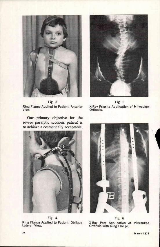
Fig. 3 Ring Flange Applied to Patient, Anterior View.
Fig. 4 Ring Flange Applied to Patient, Oblique Lateral View.
Fig. 5 X-Ray Prior to Application of Milwaukee Orthosis.
Fig. 6 X-Ray Post Application of Milwaukee Orthosis with Ring Flange.
Our primary objective for the severe paralytic scoliosis patient is to achieve a cosmetically acceptable,

Fig. 7 Patient Post-Polio after Treatment with Localizer Casts for Two Years (treatment of choice prior to introduction of Milwaukee Orthosis).
Fig. 8 Same Patient Posterior View Bending (Note sharp razoring).
Fig. 9 X-Ray of This Patient in His Second Orthosis.
functional correction and to maintain this correction. Severe paralytic curves will not stabilize with bony maturity and spinal fusions will be performed once bony maturity has been reached.
THE FOLLOWING CASES WILL DEMONSTRATE
THIS APPROACH
Case No. 1: This is a 17-year-old boy who was first seen in 1957. He is a post-polio patient.
His severe curve was treated in localizers for two years. The first Milwaukee orthosis was applied in 1959. A spinal arthrodesis was performed in November 1967 and the orthosis continued till June 1969. This patient was fitted with three orthoses during this 10-year period. His curve was improved and today's clinical examination demonstrates a cosmetically acceptable result.
The next patient is a twenty-year-old female who was diagnosed as amyotonia congenita with secondary scoliosis. This patient is a good example for a case where an error in judgement led us to do too little too late. The patient was ambulatory till age seven at which time the scoliosis became decompensated. Early holding of the curve was attempted with corsets, followed by

Fig. 10 X-Ray of This Patient Post Arthrodesis.
Fig. 11 Initial X-Ray of This Patient Diagnosed as Amyotonia Congenita.
Fig. 12 X-Ray of Patient in Corset.
Fig. 13 Posterior View of Patient Ten Years Post Initial Visit.
an early version of the Milwaukee orthosis. A Harrington instrumentation was utilized two years ago. She
is now a wheel chair patient who is gainfully employed.
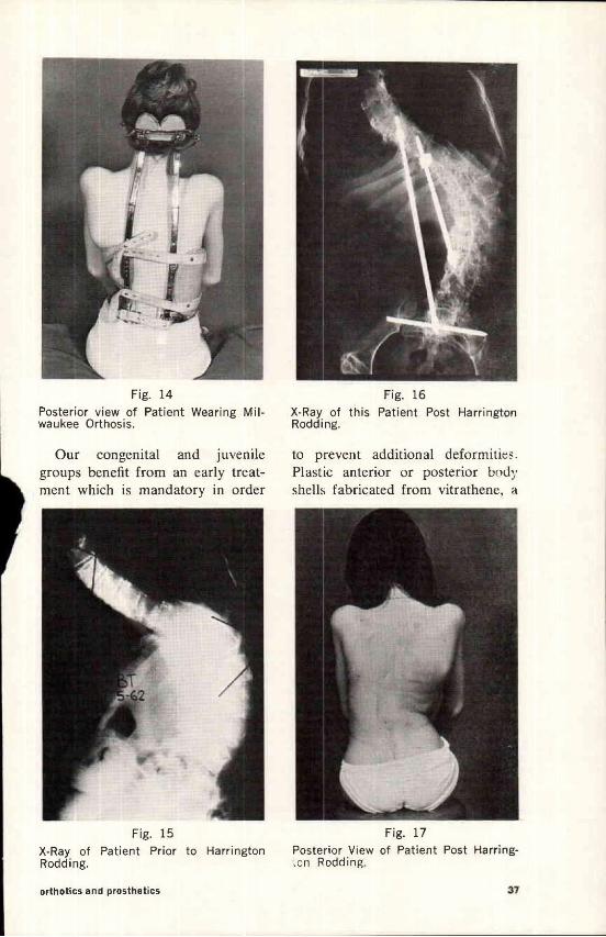
Fig. 14 Posterior view of Patient Wearing Milwaukee Orthosis.
Fig. 15 X-Ray of Patient Prior to Harrington Rodding.
Fig. 16 X-Ray of this Patient Post Harrington Rodding.
Fig. 17 Posterior View of Patient Post Harringion Rodding.
Our congenital and juvenile groups benefit from an early treatment which is mandatory in order
to prevent additional deformities. Plastic anterior or posterior body shells fabricated from vitrathene, a

Fig. 18 Myelomeningocele Patient Wearing Anterior Body Shell, Supine View.
Fig. 19 Patient Wearing a Newington Infantile Scoliosis Splint, Anterior View.
Fig. 20 Newington Infantile Scoliosis Splint.
Fig. 21 Anterior View of Patient Afflicted with Marphans Syndrome.
polythene plastic, have proven to be most effective. The lower motor
neuron disturbed patient will go into extensive orthotic management to become ambulatory.
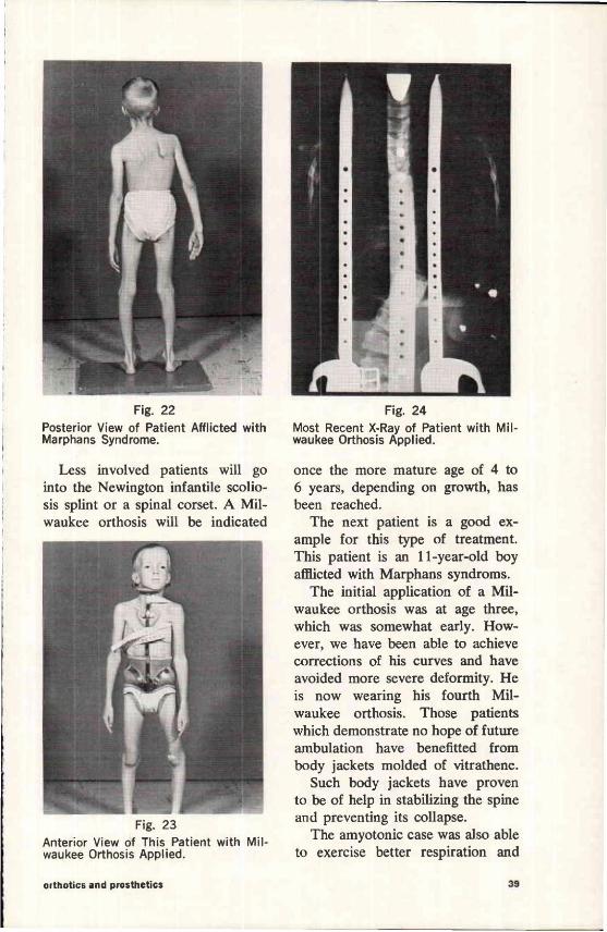
Fig. 22 Posterior View of Patient Afflicted with Marphans Syndrome.
Fig. 23 Anterior View of This Patient with Milwaukee Orthosis Applied.
Fig. 24 Most Recent X-Ray of Patient with Milwaukee Orthosis Applied.
Less involved patients will go into the Newington infantile scoliosis splint or a spinal corset. A Milwaukee orthosis will be indicated
once tne more mature age of 4 to 6 years, depending on growth, has been reached.
The next patient is a good example for this type of treatment. This patient is an 11-year-old boy afflicted with Marphans syndroms.
The initial application of a Milwaukee orthosis was at age three, which was somewhat early. However, we have been able to achieve corrections of his curves and have avoided more severe deformity. He is now wearing his fourth Milwaukee orthosis. Those patients which demonstrate no hope of future ambulation have benefitted from body jackets molded of vitrathene.
Such body jackets have proven to be of help in stabilizing the spine and preventing its collapse.
The amyotonic case was also able to exercise better respiration and

Fig. 25 Laterial View of Myelomeningocele Patient in Body Jacket.
Fig. 26 X-Ray of This Patient Without Body Jacket.
Fig. 27 X-Ray of This Patient With Body Jacket Applied.
Fig. 28 Reclining Seating Device for Patient with Werdnig Hoffmans Disease.
the retarded child was easier to manage by its custodians.
The same applies to the patients
with Werdnig Hoffmans disease, like this patient in a reclining seating device with lateral support.

CONCLUSION The examples presented here can
only highlight the many problems posed by scoliosis in its multitude of variations. Only the sharing of
knowledge obtained by all of us will help to advance our current status of Orthotic Technology in the treatment of scoliosis.
ACKNOWLEDGEMENTS: This paper could not have been
presented without the assistance and cooperation of the staff of the Newington Children's Hospital.
My special appreciation is extended to Dr. James Hardy, Scolio
sis Clinic Chief, Dr. Walter Bonne, Orthopedic Resident, the members of the Photography Department, Medical Records, the Radiology Department, and the Staff of my Department.