ORTHOPANTOMOGRAPH OP300• clearly seen on panoramic images Sometimes landmarks are not visible in...
11
A platform for changing needs. ORTHOPANTOMOGRAPH ® OP300 OP300 Digital panoramic imaging system Digital cephalometric imaging system Digital CB3D imaging system 2D 3D
Transcript of ORTHOPANTOMOGRAPH OP300• clearly seen on panoramic images Sometimes landmarks are not visible in...

1
A platform for changing needs.
ORTHOPANTOMOGRAPH® OP300
OP3
00
Dig
ital p
anor
amic
imag
ing
syste
m
Dig
ital c
epha
lom
etric
imag
ing
syste
m
Dig
ital C
B3D
imag
ing
syste
m
2D
3D

2 3
1946 Professor Y.V. Paatero publishes his first paper on Panoramic Tomography.
1951 “Pantomography” equipment is presented.
1961 The first dental panoramic X-ray, ORTHOPANTOMOGRAPH® OP1, is developed.
1964 Commercialization of the ORTHOPANTOMOGRAPH® units begins with models OP2 and OP3.
1978 ORTHOPANTOMOGRAPH® becomes the leading name within dental panoramic imaging with models
OP5/OC5, OP6 and OP10/OC10.
1992 New innovations, such as the lifting cassette head and linear tomography, are introduced along with the
OP100 product family.
1999 Direct digital ORTHOPANTOMOGRAPH® OP100 product family is introduced.
2006 New ORTHOPANTOMOGRAPH® product family OP200 is launched.
2007 Volumetric Tomography (VT) is developed to maximize the performance of an
ORTHOPANTOMOGRAPH® unit.
2009 A new member to the ORTHOPANTOMOGRAPH® product family – OP30 – is launched.
2011 ORTHOPANTOMOGRAPH® OP300, the most comprehensive 3-in-1 platform is launched to celebrate
50 years of ORTHOPANTOMOGRAPH® success.
2013 Introduction of the improved 3D image quality, new metal artifact reduction (MAR) tool and endo mode
for ORTHOPANTOMOGRAPH® OP300 3D images.
2013 New revision of ORTHOPANTOMOGRAPH® OP30 is launched.
Leading the way through the decades
Choose your own ORTHOPANTOMOGRAPH® OP30 OP200 OP300
Standard panoramic •
Advanced panoramic • •
TMJ imaging • • •
Volumetric Tomography •
Cone Beam 3D •
Cephalometric • •
ContentsA platform for your changing needs ... 5
Gold standard image quality ... 6
Confident diagnostics ... 8
Complete versatility ... 10
Clinical example images ... 12
ART OF IMAGING FOR IMPLANTOLOGY ... 15
ART OF IMAGING FOR ORTHODONTICS ... 16
ART OF IMAGING FOR ENDODONTICS ... 17
Dimensions ... 18
Technical specifications ... 19

4 5
ORTHOPANTOMOGRAPH® OP300 is the most comprehensive 3-in-1 platform
designed for today and tomorrow. OP300 combines an advanced panoramic
imaging system with either cephalometric or cone beam 3D or a combination of
both, giving you a truly adaptable platform for different imaging applications and
dental specialties. In addition, all of these options can be upgraded in the field
after the initial purchase.
AA platform for changing needs
Over 50 years of experience in panoramic imagingORTHOPANTOMOGRAPH®, introduced over 50 years ago, was a revolutionary groundbreaker and pacesetter for dental panoramic X-ray imaging. Today, ORTHOPANTOMOGRAPH® is regarded as the leading name in the panoramic X-ray world and often used even as a synonym for panoramic X-ray units.

6 7
GGold standard image quality
Image quality is a result of many elements, such as the carefully planned features, chosen
technology and sufficient technical characteristics of the system, along with proper patient
positioning. ORTHOPANTOMOGRAPH® OP300 combines all these for your benefit and
provides you with a perfect image – every single time. ORTHOPANTOMOGRAPH® OP300
masters the details.
Stable and open patient positioningA rigid 5-point positioning system, including forehead support, chin rest and bite fork, eliminates patient movement. The open design allows easy viewing and the positioning of the patient from either the left or right side.
Unsurpassed cephalometric resultsThe OP300 scanning cephalometric option offers unsurpassed visibility of tracing key reference points for orthodontic treatment planning. In addition, dose optimization is carried out by an adjustable scanning area and Automatic Facial Contour. AFC increases soft tissue visibility while decreasing patient dose.
Complete usabilityThe large 10” touchscreen with easy-to-use interface enables professional usage from the very beginning. The clear and user friendly structure of controls allows fast and effortless workflow for all imaging modalities.
Versatile software toolsCLINIVIEW™ software offers professional tools for processing and viewing digital X-ray images. Open architecture and DICOM® format images enable easy connectivity for 3D viewing and planning software.
Latest sensor technologyThe OP300 utilizes the latest in CMOS sensor technology. CMOS sensors provide a larger dynamic range combined with 14-bit image data and increased signal-to-noise ratio. The result is an intensely sharp image with reduction of unwanted under- and overexposures.
Metal artifact reduction (MAR) toolThe metal artifact reduction (MAR) tool offers the possibility of utilizing metal artifact reduction in 3D images. This improves diagnostic capabilities in cases where metallic artifacts from radio-opaque objects can be expected, for example:
• Endodontic analysis of teeth with root canal fillings
• Implant cases

8 9
CConfident diagnostics
Choose the best of five different layers – or automatically select just one layer for use as a conventional panoramic unit.
Multilayer pan increases the thickness of the focal area compared to traditional panoramic imaging: this decreases patient positioning errors and aids in difficult malocclusion cases.
Multilayer panThe OP300 multilayer panoramic option provides five panoramic images with only one scan. This enables forgiving patient positioning and reduces possible retake exposures. Multilayer images are achieved in the same scanning time and dose as the traditional panoramic scan.
6 cm
6 cm
4 cm
8 cm
SMARTVIEW™A two-dimensional scout image can be taken before the 3D examination to adjust the target position visually from GUI screen. This guarantees precise positioning and eliminates risk of retake exposures.
Select freely and fine tune FOV position from GUI.
SMARTVIEW™ takes a two- dimensional scout of the selected area.
Two available fields-of-view with 3D option: • 6 x 4 cm – a small FOV optimized for local
diagnostics like single implant planning, 3rd molar extractions and endodontic procedures, keeping the patient dose at a substantially reduced level.
• 6 x 8 cm – FOV covering complete dental arch for multiple implant placement and operations using surgical guides.
High resolution
Standard resolution
Two available resolutionsBoth FOV sizes are high enough to easily cover the jaw bone and occlusion level. For both FOV sizes it is possible to choose between two resolutions: • Standard scan takes only 10 seconds
with exposure time only 2.3 seconds with optimized patient dose
• High resolution scan offers extremely sharp images for more detailed diagnosis
Multilayer panoramic feature enables sharp images even in difficult malocclusion cases.
For 6 x 4cm FOV it is also possible to select endo mode for accurate diagnostic tasks:• 85 µm voxel size with MAR tool specially
designed for endodontic applications

10 11
CComplete versatility
UpgradeabilityOP300 is designed and built as an expandable platform where both 3D and cephalometric options are field upgradeable. The cephalometric arm can be ordered for left- or right-side configuration, which is adaptable and can be changed in the field. In panoramic positioning, side is also adaptable for left or right configuration to ensure optimal system performance and ease of use. OP300 can truly grow with your practice and be tailored to users’ preferences.
CephalometricsFull range of projections: lateral ceph, AP/PA, obliques.
1. Fully adjustable scanning area ensures that by exposing only the required region, patient safety is greatly increased.
2. Automatic facial contour (AFC) decreases exposure factors in the facial soft tissue region to provide improved visibility of soft tissue tracing points in addition to a reduction in patient dose.
PanoramicsA full range of panoramic imaging programs covers multiple modalities from everyday procedures to even more specialized imaging procedures. Automatic collimation for adults and pediatrics optimizes patient dose for increased patient safety.
3DTwo fields-of-view combined with integrated motorized chin rest enable free FOV positioning within maxillofacial area. This ensures multiple different modalities from 3D TMJ analysis into implant planning with drilling guides.
100% dose, typical lateral full scan
52 – 38% dose with standard lateral ceph
Area of lowered AFC dose
Area of lowered AFC dose
Only 43 – 32% dose with pediatric ceph
12 13
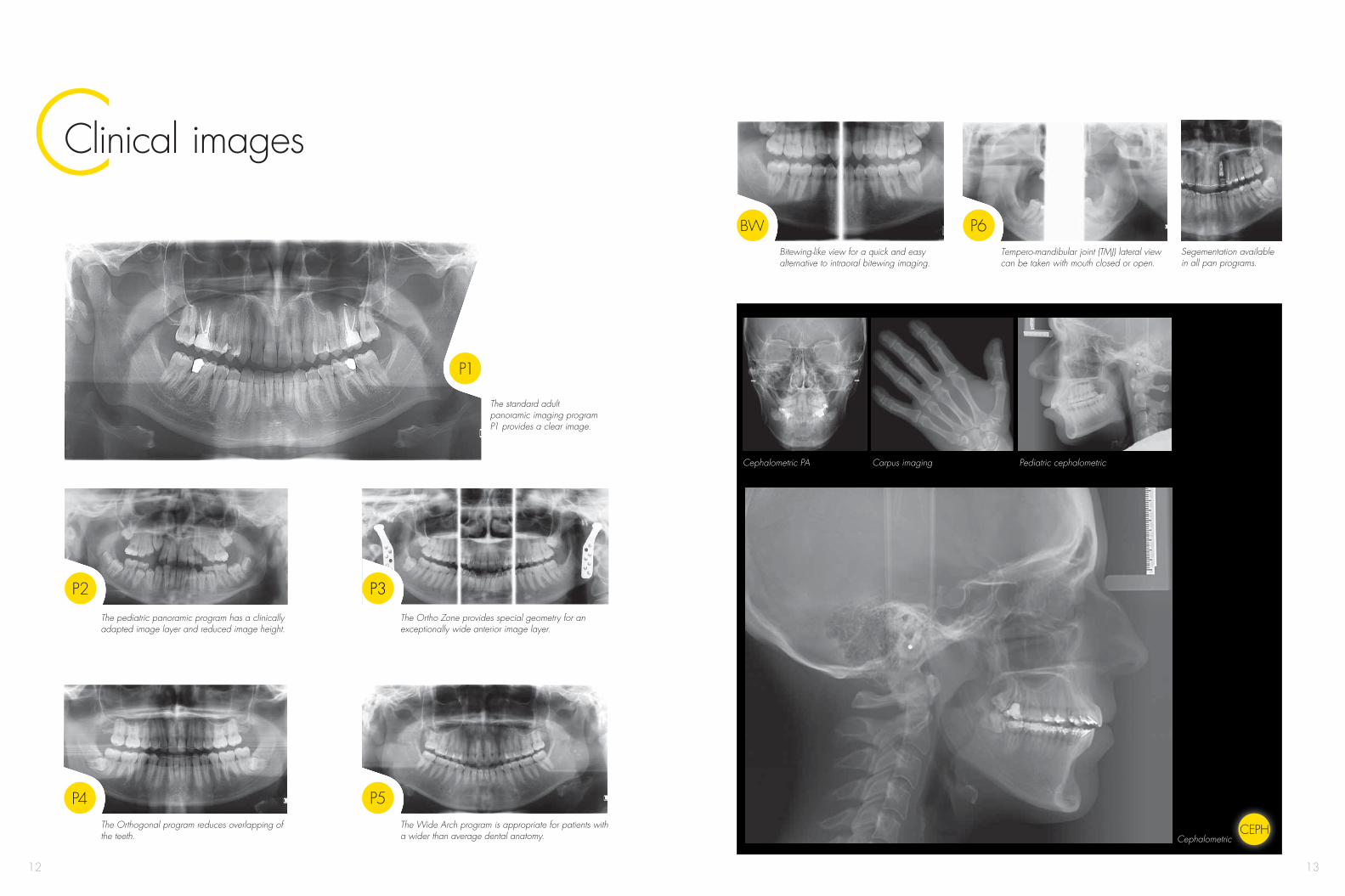
CClinical images
The pediatric panoramic program has a clinically adapted image layer and reduced image height.
The Orthogonal program reduces overlapping of the teeth.
The Ortho Zone provides special geometry for an exceptionally wide anterior image layer.
The Wide Arch program is appropriate for patients with a wider than average dental anatomy.
Tempero-mandibular joint (TMJ) lateral view can be taken with mouth closed or open.
Carpus imaging
Bitewing-like view for a quick and easy alternative to intraoral bitewing imaging.
Cephalometric PA Pediatric cephalometric
The standard adult panoramic imaging program P1 provides a clear image.
Segementation available in all pan programs.
Cephalometric

14 15
IArt of imaging for Implantology
DENTAL IMPLANTOLOGY
Learn more www.op300.com/implantology
Diagnostic challenges• To evaluate sufficient bone structure and
quality, such as undercut and intrabony pathology, to avoid complications during surgery
• To evaluate sensitive anatomic structures like nerve canal, neighboring adjacent teeth and sinuses
• To conduct diagnostic, surgical and prosthodontical planning at once to avoid the need to refer the patient to another specialist
Solution• The OP300 gold standard image quality with
multilayer panoramic imaging feature and cone beam 3D imaging option takes implant dentistry to a new level
• Precise patient positioning with SMARTVIEW™ scout enables needed structures to be present at imaging area
• The OP300 implant concept combines aesthetically driven virtual implant planning and custom-made surgical templates with precise depth and angle control for your benefit
Two FOVs in 3D with versatile software cover a wide range of examinations.
Clinical images

16 17
EO and for EndodonticsArt of imaging for Orthodontics
ENDODONTICSORTHODONTICS
Learn more www.op300.com/endodonticsLearn more www.op300.com/orthodontics
Diagnostic challenge• Pan images are unclear for patients with malocclusion• Sometimes landmarks are not visible in ceph images• Orthodontic treatment often requires multiple images
for diagnosis and follow-up• The effect of impacted teeth, hyperdontia and
possible resorption on treatment plan is difficult to estimate with 2D images
Solution• The OP300 multilayer panoramic feature enables
sharp images even in difficult malocclusion cases• The dose-controlled Automatic Facial Contour (AFC)
guarantees excellent visibility for cephalometric tracing points and soft tissues
• Adjustable lateral ceph field of view is especially suitable for pediatric patients and treatment follow-up
• The additional small field-of-view CBCT with precise and free 3D positioning can be used for impacted teeth localization and other special cases
Diagnostic challenge• Roots and periodontal ligament not always
clearly seen on panoramic images • Root and rootcanal morphology is often
difficult to evaluate based on only 2D (periapical or panoramic) images
• Root canal fillings most often cause artifacts to the images and decreases the accuracy of diagnosing root fractures
Solution• With multilayer panoramic feature the sharp
layer can be easily adjusted to present roots and periodontal ligaments
• 6 x 4 cm (H x W) field of view 3D with accurate positioning improves diagnostics of atypical morphologies
• 3D program dedicated for endodontics with high resolution and MAR (Metal artifact reduction) tool helps in seeing small details surrounding radio-opaque objects

18 19
2007 mm (79.0”)
TD Technical specifications
Technical specifications
generator high frequency DC, 75–150 kHz
focal spot 0.5 mm IEC 336
tube voltage 57 – 90 kV
tube current 4 – 16 mA
minimum total filtration 3.2 mm AI
Panoramic Cephalometric
image detector CMOS image detector CMOS
sensor pixel size 100 µm sensor pixel size 100 µm
image pixel size 100 µm image pixel size 100 µm
scan time 8.6 – 16.1 s scan time 6.5 s – 20 s
image field height 151 mm image field width 160 mm – 270 mm
3D
image detector CMOS
image voxel size 85 µm – 300 µm
scan time 10 – 20 s
exposure time 2.34 s – 12.5 s, pulsed X-ray
image volume sizes (H x W) 61 mm x 41 mm, 61 mm x 78 mm
DICOM® support yes
DICOM® is the registered trademark of the National Electrical Manufacturers Association for its standards publications relating to digital communications of medical information.
Minimum system requirements for acquisition computer
processor 2.5 GHz dual core, or better
memory 3 GB RAM or more
hard disk 500 GB or more
expansion slot PCI Express x16, full length
network Gigabit Ethernet, 1000Base-T
power supply 500 watt minimum
operating system Windows 7 or Windows Vista (32 or 64-bit)
Please refer to CLINIVIEW™ Installation manual for full software specifications and requirements or contact your local dealer.
Dimensions
1610
– 24
10 m
m (6
3.4
– 94.
9”)
1300
mm
(51.
1”)
Chi
n su
ppor
t 898
– 17
58 m
m (3
5.5
– 69.
2”)
1385
– 14
25 m
m (5
4.5
– 56.
1”)
965 mm (38”)
min. 550 mm (21.7”) recommended
min. 550 mm (21.7”) recommended
Weight
panoramic 200 kg
with cephalometric option 250 kg
Height
min. room height 2050 – 2450 mm
Easy wheelchair accessibility.
20
© 2013 Instrumentarium Dental
207131-4 English
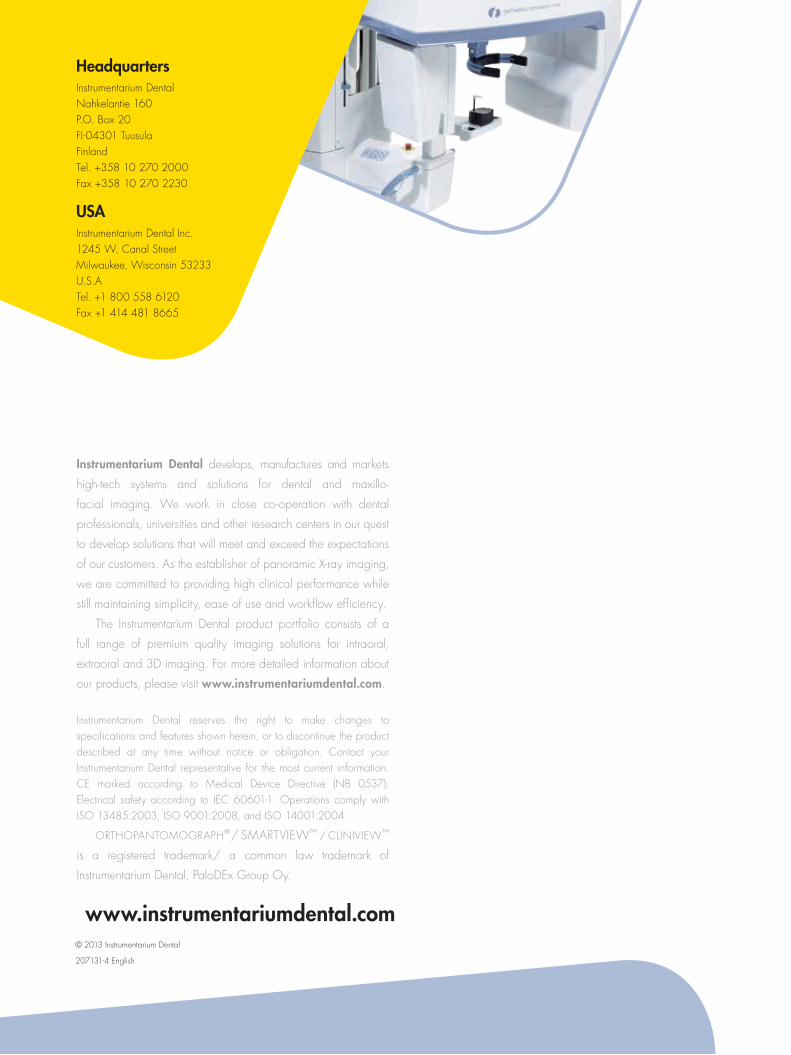
Instrumentarium Dental develops, manufactures and markets
high-tech systems and solutions for dental and maxillo-facial imaging. We work in close co-operation with dental
professionals, universities and other research centers in our quest
to develop solutions that will meet and exceed the expectations
of our customers. As the establisher of panoramic X-ray imaging,
we are committed to providing high clinical performance while
still maintaining simplicity, ease of use and workflow efficiency.
The Instrumentarium Dental product portfolio consists of a
full range of premium quality imaging solutions for intraoral,
extraoral and 3D imaging. For more detailed information about
our products, please visit www.instrumentariumdental.com.
Instrumentarium Dental reserves the right to make changes to specifications and features shown herein, or to discontinue the product described at any time without notice or obligation. Contact your Instrumentarium Dental representative for the most current information. CE marked according to Medical Device Directive (NB 0537). Electrical safety according to IEC 60601-1. Operations comply with ISO 13485:2003, ISO 9001:2008, and ISO 14001:2004.
ORTHOPANTOMOGRAPH® / SMARTVIEW™ / CLINIVIEW™
is a registered trademark/ a common law trademark of
Instrumentarium Dental, PaloDEx Group Oy.
www.instrumentariumdental.com
HeadquartersInstrumentarium DentalNahkelantie 160P.O. Box 20FI-04301 Tuusula FinlandTel. +358 10 270 2000 Fax +358 10 270 2230
USAInstrumentarium Dental Inc. 1245 W. Canal Street Milwaukee, Wisconsin 53233 U.S.ATel. +1 800 558 6120Fax +1 414 481 8665