![Practice For May: Cell Ultrastructure [114 marks]blogs.4j.lane.edu/.../2018/02/Cell-Ultrastructure-Test-1.pdfPractice For May: Cell Ultrastructure [114 marks]1. Which structure found](https://static.fdocuments.net/doc/165x107/5eda4db5b3745412b5711d9c/practice-for-may-cell-ultrastructure-114-marksblogs4jlaneedu201802cell-ultrastructure-test-1pdf.jpg)
ORIGINAL ARTICLE Ultrastructure and Molecular … and Molecular Phylogenetic Position of Heteronema...
14
ORIGINAL ARTICLE Ultrastructure and Molecular Phylogenetic Position of Heteronema scaphurum: A Eukaryovorous Euglenid with a Cytoproct Susana A. Breglia 1 , Naoji Yubuki & Brian S. Leander Canadian Institute for Advanced Research, Program in Integrated Microbial Biodiversity, Departments of Zoology and Botany, University of British Columbia, 6270 University Boulevard, Vancouver, BC, V6T 1Z4, Canada Keywords Dinema; euglenida; euglenozoa; feeding apparatus; Peranema. Correspondence B. Leander, Canadian Institute for Advanced Research, Program in Integrated Microbial Biodiversity, Departments of Zoology and Botany, University of British Columbia, 6270 University Boulevard, Vancouver, BC V6T 1Z4, Canada Telephone number: 604 822 2474; FAX number: 604 822 6089; e-mail: [email protected] Received: 19 May 2012; revised 12 September 2012; accepted September 13, 2012. doi:10.1111/jeu.12014 ABSTRACT Euglenids comprise a distinct clade of flagellates with diverse modes of nutri- tion, including phagotrophy, osmotrophy and phototrophy. Much of the previous research on euglenids has focused on phototrophic species because of their ecological abundance and significance as indicators for the health of aquatic ecosystems. Although largely understudied, phagotrophic species probably rep- resent the majority of euglenid diversity. Phagotrophic euglenids tend to be either bacterivorous or eukaryovorous and use an elaborate feeding apparatus for capturing prey cells. We characterized the ultrastructure and molecular phy- logenetic position of Heteronema scaphurum, a eukaryovorous euglenid col- lected in freshwater. This species was equipped with a distinct cytoproct through which waste products were eliminated in the form of faecal pellets; a cytoproct has not been reported in any other member of the Euglenida. Hetero- nema scaphurum also had a novel predatory mode of feeding. The euglenid ensnared and corralled several green algal prey cells (i.e. Chlamydomonas) with hook-like flagella covered in mucous before engulfing the bundle of prey cells whole. Molecular phylogenetic analyses inferred from small subunit rDNA sequences placed this species with other eukaryovorous euglenids, which was consistent with ultrastructural features associated with the feeding apparatus, flagellar apparatus, extrusomes, and pellicle. THE Euglenida is a diverse group of flagellates unified by both ultrastructural and molecular features. Euglenids share a distinctive pellicle that is formed by the plasma membrane, interlocking proteinaceous strips, subtending microtubules, and cisternae of endoplasmic reticulum (Esson and Leander 2006; Leander and Farmer 2000; Lean- der et al. 2007). Different modes of nutrition in the group (phagotrophy, osmotrophy and phototrophy) correlate with the number of strips and the degree of plasticity in the pel- licle (Leander and Farmer 2000, 2001a; Leander et al. 2001a). Bacterivorous species tend to have rigid cells with few (less than 16) longitudinally arranged strips, whereas eukaryovorous species tend to have a larger number (16 or more) of helically arranged strips on the cell surface; eukaryovorous species also tend to distort their cell shape in a manner that is characteristic of euglenids called “euglenoid movement” or “metaboly” (Leander et al. 2001b; Triemer and Farmer 1991; Triemer et al. 2006). This ability, coupled with the presence of a well-developed feeding apparatus consisting of microtubule-based rods and vanes, allows eukaryovorous species (e.g. Peranema, Dinema, and Urceolus) to capture and engulf large prey cells, such as microalgae. The engulfment of a green algal prey cell led to a secondary endosymbiotic event, which gave rise to a distinct clade of phototrophic euglenids (Gibbs 1978, 1981; Leander 2004; Leander et al. 2007; Rogers et al. 2007; Yamaguchi et al. 2012). Phagotrophic euglenids typically have two emergent fla- gella that are used to glide along substrates: one oriented straight in front of the cell (the anterior or dorsal flagel- lum) and the other that curves backwards and trails behind the cell (the posterior or ventral flagellum). One of the more common phagotrophic genera is Heteronema, which is found in both freshwater and marine environ- ments; about 43 species have been described so far (Schroeckh et al. 2003). Heteronema is morphologically very similar to Peranema under the light microscope; both lineages are metabolic, capable of gliding, possess conspicuous feeding rods within the anterior end of the cell, and have an anterior flagellum that is longer and thicker than the posterior flagellum (Lee et al. 2005). However, unlike Peranema, the posterior flagellum of © 2013 The Author(s) Journal of Eukaryotic Microbiology © 2013 International Society of Protistologists Journal of Eukaryotic Microbiology 2013, 60, 107–120 107 Journal of Eukaryotic Microbiology ISSN 1066-5234 Published by the International Society of Protistologists Eukaryotic Microbiology The Journal of
Transcript of ORIGINAL ARTICLE Ultrastructure and Molecular … and Molecular Phylogenetic Position of Heteronema...

ORIGINAL ARTICLE
Ultrastructure and Molecular Phylogenetic Positionof Heteronema scaphurum: A Eukaryovorous Euglenidwith a CytoproctSusana A. Breglia1, Naoji Yubuki & Brian S. Leander
Canadian Institute for Advanced Research, Program in Integrated Microbial Biodiversity, Departments of Zoology and Botany, University of
British Columbia, 6270 University Boulevard, Vancouver, BC, V6T 1Z4, Canada
Keywords
Dinema; euglenida; euglenozoa; feeding
apparatus; Peranema.
Correspondence
B. Leander, Canadian Institute for Advanced
Research, Program in Integrated Microbial
Biodiversity, Departments of Zoology and
Botany, University of British Columbia, 6270
University Boulevard, Vancouver, BC V6T
1Z4, Canada
Telephone number: 604 822 2474; FAX
number: 604 822 6089;
e-mail: [email protected]
Received: 19 May 2012; revised 12
September 2012; accepted September 13,
2012.
doi:10.1111/jeu.12014
ABSTRACT
Euglenids comprise a distinct clade of flagellates with diverse modes of nutri-
tion, including phagotrophy, osmotrophy and phototrophy. Much of the previous
research on euglenids has focused on phototrophic species because of their
ecological abundance and significance as indicators for the health of aquatic
ecosystems. Although largely understudied, phagotrophic species probably rep-
resent the majority of euglenid diversity. Phagotrophic euglenids tend to be
either bacterivorous or eukaryovorous and use an elaborate feeding apparatus
for capturing prey cells. We characterized the ultrastructure and molecular phy-
logenetic position of Heteronema scaphurum, a eukaryovorous euglenid col-
lected in freshwater. This species was equipped with a distinct cytoproct
through which waste products were eliminated in the form of faecal pellets; a
cytoproct has not been reported in any other member of the Euglenida. Hetero-
nema scaphurum also had a novel predatory mode of feeding. The euglenid
ensnared and corralled several green algal prey cells (i.e. Chlamydomonas) with
hook-like flagella covered in mucous before engulfing the bundle of prey cells
whole. Molecular phylogenetic analyses inferred from small subunit rDNA
sequences placed this species with other eukaryovorous euglenids, which was
consistent with ultrastructural features associated with the feeding apparatus,
flagellar apparatus, extrusomes, and pellicle.
THE Euglenida is a diverse group of flagellates unified by
both ultrastructural and molecular features. Euglenids
share a distinctive pellicle that is formed by the plasma
membrane, interlocking proteinaceous strips, subtending
microtubules, and cisternae of endoplasmic reticulum
(Esson and Leander 2006; Leander and Farmer 2000; Lean-
der et al. 2007). Different modes of nutrition in the group
(phagotrophy, osmotrophy and phototrophy) correlate with
the number of strips and the degree of plasticity in the pel-
licle (Leander and Farmer 2000, 2001a; Leander et al.
2001a). Bacterivorous species tend to have rigid cells with
few (less than 16) longitudinally arranged strips, whereas
eukaryovorous species tend to have a larger number (16 or
more) of helically arranged strips on the cell surface;
eukaryovorous species also tend to distort their cell shape
in a manner that is characteristic of euglenids called
“euglenoid movement” or “metaboly” (Leander et al.
2001b; Triemer and Farmer 1991; Triemer et al. 2006). This
ability, coupled with the presence of a well-developed
feeding apparatus consisting of microtubule-based rods
and vanes, allows eukaryovorous species (e.g. Peranema,
Dinema, and Urceolus) to capture and engulf large prey
cells, such as microalgae. The engulfment of a green algal
prey cell led to a secondary endosymbiotic event, which
gave rise to a distinct clade of phototrophic euglenids
(Gibbs 1978, 1981; Leander 2004; Leander et al. 2007;
Rogers et al. 2007; Yamaguchi et al. 2012).
Phagotrophic euglenids typically have two emergent fla-
gella that are used to glide along substrates: one oriented
straight in front of the cell (the anterior or dorsal flagel-
lum) and the other that curves backwards and trails
behind the cell (the posterior or ventral flagellum). One of
the more common phagotrophic genera is Heteronema,
which is found in both freshwater and marine environ-
ments; about 43 species have been described so far
(Schroeckh et al. 2003). Heteronema is morphologically
very similar to Peranema under the light microscope;
both lineages are metabolic, capable of gliding, possess
conspicuous feeding rods within the anterior end of the
cell, and have an anterior flagellum that is longer and
thicker than the posterior flagellum (Lee et al. 2005).
However, unlike Peranema, the posterior flagellum of
© 2013 The Author(s) Journal of Eukaryotic Microbiology © 2013 International Society of Protistologists
Journal of Eukaryotic Microbiology 2013, 60, 107–120 107
Journal of Eukaryotic Microbiology ISSN 1066-5234
Published bythe International Society of ProtistologistsEukaryotic Microbiology
The Journal of

Heteronema does not stick closely to the ventral side of
the cell. To date, members of this genus have only been
studied with light microscopy. Here, we characterize the
eukaryovorous euglenid Heteronema scaphurum using
not only light microscopy and video analysis, but also
scanning electron microscopy, transmission electron
microscopy, and molecular phylogenetic analysis of SSU
rDNA sequences. Our study demonstrated that this spe-
cies has a distinct and novel feeding behaviour involving
hook-like flagella and a mucilaginous web that captures
(green algal) prey before engulfing the cells whole. Het-
eronema scaphurum also eliminates remnant prey mate-
rial as faecal pellets through a novel cytoproct. The
discovery of these novel features in this species expands
our knowledge of euglenid diversity, especially with
regard to phagotrophic species, and provides improved
context for understanding the eukaryovorous origins of
phototrophic euglenids.
MATERIALS AND METHODS
Cell isolation and cultivation
A sample of freshwater sediments from a pond in Illinois,
USA, was collected during the Spring of 2007, and a cul-
ture was established at room temperature in a 0.01%
Knop medium (Saito et al. 2003) using Chlamydomonas as
prey cells.
Light microscopy and video analysis
Differential interference contrast (DIC) light micrographs
(LMs) were taken using a Zeiss Axioplan 2 imaging micro-
scope (Carl Zeiss Microscopy GmbH, Jena, Germany) and
a Leica DC500 digital chilled CCD camera (Leica Microsys-
tems Digital Imaging, Cambridge, U.K.). Cells were fixed
with 1% (v/v) glutaraldehyde for high magnification obser-
vations. Digital videos were taken with a PixeLink Mega-
pixel colour digital camera (LixeLink, Ottawa, ON, Canada)
connected to a Zeiss Axiovert 200 inverted microscope
(Carl Zeiss Microscopy GmbH).
Electron microscopy
Cells were fixed for SEM using a 4% osmium tetroxide
vapour protocol described previously (Leander and Farmer
2000). The cells were then transferred onto a 10 lm poly-
carbonate membrane filter, dehydrated with a graded eth-
anol series, and critical point dried with CO2 using a
Tousimis Critical Point Dryer. The filter was then mounted
on an aluminium stub, sputter coated with gold/palladium
using a Cressington 208HR High Resolution Sputter
Coater (Cressington Scientific Instruments Ltd., Watford,
England), and observed with a Hitachi S-4700 field emis-
sion scanning electron microscope (Hitachi High Techno-
logies Corp., Tokyo, Japan).
Cells were fixed for transmission electron microscopy
(TEM) using 2% glutaraldehyde in 0.1 M sodium cacody-
late buffer (SCB). The fixed cells were washed in 0.1 M
SCB three times, and postfixed in 1% (w/v) osmium
tetroxide in 0.2 M SCB, in ice, for 1 h. The cells were
then dehydrated through a graded series of ethanol and
100% acetone, and infiltrated with a graded series of
acetone-Epon 812 resin mixtures and 100% Epon 812
resin. Ultra-thin sections were collected on copper Form-
var-coated slot grids, stained with 2% (w/v) uranyl acetate
and lead citrate (Reynolds 1963), and observed using a
Hitachi H7600 electron microscope.
Brightness and contrast of all micrographs were
adjusted, as well as labelled and assembled into plates,
using Photoshop CS4.
DNA extraction and PCR amplification
Total genomic DNA was extracted using the MasterPure
Complete DNA and RNA purification Kit (Epicentre, Maddison,
WI; Catalogue number MC85200) from ~ 30 cells, following
the procedures provided by the manufacturer. Polymerase
chain reactions (PCR) of 18S rDNA were performed using
PuRe Taq Ready-To-Go PCR beads kit (GE Healthcare,
Buckinghamshire, U.K.), and the following primers: 5′-TGCGCTACCTGGTTGATCC-3′ and 5′-AACGGAATYAACCAGACARAT-3′. Amplified DNA fragments were purified from
agarose gels using UltraClean 15 DNA Purification Kit (MO
Bio, Carlsbad, CA), and subsequently cloned into the TOPO
TA Cloning Kit (Invitrogen, Carlsbad, CA). Three clones were
sequenced with the ABI Big-Dye reaction mix using the vector
primers and three internal primers: Dinema18s1R (5′-GGACTACGACGGTATCTGATCAT-3′), 475EugF (5′- AAGTCTGGTGCCAGCAGCYGC-3′) and DinemaSSU620F (5′- GCAA
GACAGCTGTGCGATAGCAA-3′). The new sequence was
screened with BLAST, identified by molecular phylogenetic
analyses, and submitted to the GenBank database
(JN566139).
Multiple sequence alignment and molecularphylogenetic analyses
We obtained a 2,860 bp sequence that corresponded to
positions 380–1687 of Euglena gracilis. The new 18S
rDNA sequence was analysed within the context of a 39-
taxon alignment consisting of taxa representing the Eugle-
nozoa (636 unambiguously aligned sites). Ambiguously
aligned positions and gaps were excluded.
Molecular phylogenetic relationships were inferred using
maximum likelihood (ML) and Bayesian inference (BI)
methods with PhyML v2.4.5 (Guindon and Gascuel 2003)
and MrBayes v3.1.1 (Ronquist and Huelsenbeck 2003),
respectively, with the graphical interface TOPALi v2.5
(Milne et al. 2009). The models used for ML and BI to
generate phylogenetic trees were chosen using Model
selection in TOPALi v2.5 (Milne et al. 2009). In both
cases, the nucleotide dataset was analysed using a gen-
eral-time-reversible (GTR) model of base substitutions,
plus a gamma correction (gamma value = 0.663) with
eight substitution rate categories and a proportion of
invariable sites = 0.165 (GTR + I + G). ML bootstrap analy-
sis of 100 replicates was performed with the same param-
© 2013 The Author(s) Journal of Eukaryotic Microbiology © 2013 International Society of Protistologists
Journal of Eukaryotic Microbiology 2013, 60, 107–120108
Ultrastructure and Phylogeny of Heteronema (Euglenida) Breglia et al.

eters described above. For the BI, the program MrBayes
was set to operate with four Monte-Carlo-Markov chains
(MCMC) (default temperature = 0.2). A total of 1,000,000
generations was calculated with trees sampled every 50
generations, resulting in 20,000 trees saved. A 10% burn-
in was applied, resulting in 18,000 trees used (i.e. 2,000
sampled trees were discarded).
RESULTS
General morphology
The cells were spindle-shaped with a tapering posterior
end, 45–70 lm long and approximately 30 lm wide
(n = 250). A vestibular opening was located at the ante-
rior end of the cell, which led to a flagellar pocket (FP)
and to a feeding pocket (Fe) (Fig. 1, 2). The posterior
end of the cell contained a distinct concavity, or “cyto-
proct”, through which material was periodically released
(Fig. 3–7, 11–13). Two heterodynamic flagella emerged
from the vestibular opening (Fig. 1–5, 8). The dorsal
(anterior) flagellum (DF) was approximately the same
length as the cell body (~ 60 lm long) and extended
straight forward while gliding; the ventral (posterior) fla-
gellum (VF) was slightly shorter, about 0.7 times the
cell length, and trailed freely beneath the cell. Both fla-
gella were adorned with hairs (Fig. 8–10). The cells
were capable of euglenoid movement and glided
smoothly, with the anterior flagellum extended in front
of the cell and probing the substrate, and with the ven-
Fig. 1–6. Light micrographs showing fixed cells of Heteronema scaphurum. 1. Side view showing dorsal and ventral flagella (DF and VF respec-
tively), flagellar pocket (FP) and feeding apparatus (arrowhead). Scale bar = 10 lm. 2. High magnification view showing feeding apparatus (arrow-
head). Scale bar = 5 lm. 3. Side view showing the nucleus with endosomes (arrow) and cytoproct (arrowhead). 4. Cell in dorsal view showing
the feeding pocket (Fe), engulfed prey Chlamydomonas (arrows), and cytoproct (arrowhead). 5. Dorsal-posterior view showing the dorsal flagellum
(DF) and the ventral flagellum (VF), and the feeding apparatus (arrowhead). Scale bars 3–5 = 10 lm. 6. Detail of Fig. 5 showing the cytoproct, and
material being excreted (arrowhead). Scale bar = 5 lm.
© 2013 The Author(s) Journal of Eukaryotic Microbiology © 2013 International Society of Protistologists
Journal of Eukaryotic Microbiology 2013, 60, 107–120 109
Breglia et al. Ultrastructure and Phylogeny of Heteronema (Euglenida)

Fig. 7–14. Scanning electron micrographs of Heteronema scaphurum. 7. Posterior view of a cell showing the cytoproct (arrow). Scale
bar = 10 lm. 8. Lateral view of the cell showing the dorsal and ventral flagella with mastigonemes (DF and VF respectively). Scale bar = 5 lm. 9.
Ventral view of the anterior tip of a cell showing two prey cells (Chlamydomonas, arrowhead) that have been captured by both flagella of the eu-
glenid. Scale bar = 10 lm. 10. Anterior view of a cell showing a mucus web (arrow) surrounding the prey cells. Scale bar = 10 lm. 11. Posterior
tip of a cell showing pellicle strips extending into the cytoproct (arrowhead). Scale bar = 1 lm. 12. Posterior tip of a cell showing material being
secreted from the cytoproct. Scale bar = 5 lm. 13. Posterior view of a cell showing a faecal pellet (arrow) being secreted through the cytoproct.
Scale bar = 10 lm. 14. Posterior view of a cell dividing along its antero-posterior axis. The arrow shows the thinner nascent pellicle strips. Scale
bar = 10 lm.
© 2013 The Author(s) Journal of Eukaryotic Microbiology © 2013 International Society of Protistologists
Journal of Eukaryotic Microbiology 2013, 60, 107–120110
Ultrastructure and Phylogeny of Heteronema (Euglenida) Breglia et al.

tral flagellum bent backwards beneath the cell. The cell
divided from anterior to posterior along the longitudinal
axis (Fig. 14).
Feeding behaviour
Heteronema scaphurum devoured Chlamydomonas. Dur-
ing this process, both flagella participated in the capture of
prey cells; the dorsal flagellum formed an arc, hooked sev-
eral prey cells, and pushed them towards the vestibular
opening (Fig. 15–20). The ventral flagellum participated in
the capture and manipulation of the prey cells by trapping
the cells within the arc formed by the dorsal flagellum. The
cell also secreted a sticky substance that functioned to
envelope the prey within a web of mucus (Fig. 9, 10).
Once the prey cells were moved against the vestibular
opening, the anterior end of the cell expanded as the fla-
gella continued to push the prey inwards (Fig. 16–20).Then the microtubular rods that separate the flagellar and
the feeding pockets pushed the flagella to one side, pre-
venting the passage of food into the flagellar pocket and,
at the same time, enlarging the feeding cavity, as the prey
cells were ingested whole. During this process, H. scaphu-
rum also distorted its shape, presumably to generate
forces required to facilitate the engulfment of the prey
(Fig. 19, 20). The entire process of feeding, from prey
capture to engulfment, took approximately one minute to
complete.
We also observed material that was released through
the posterior cytoproct during the feeding process (Fig. 6,
12, 13). The cells were able to discharge either mucilagi-
nous material (Fig. 12) or solid faecal pellets around 3 lmin diameter (Fig. 13). The faecal pellets accumulated and
were easily observed in the culture dishes.
Cell surface
Heteronema scaphurum had 28 pellicle strips (n = 100)
that ran along the antero-posterior axis of the cell and
formed a helically arranged pattern on the cell surface
(Fig. 7–14). All of the strips were approximately the same
width; there was no ventral groove or “flagellar strip” con-
taining the ventral flagellum. All of the strips extended into
the anterior vestibulum and the posterior cytoproct; there
were no indications of strip reduction on either end of the
cell (Fig. 7, 9–14). The number of strips doubled prior to
cytokinesis, whereby new thinner strips emerged
between the parental thicker strips (Fig. 14). The
transverse ultrastructure of the pellicle consisted of the
plasma membrane, relatively thick (100 nm wide)
Fig. 15–20. Time series of video microscope images of Heteronema scaphurum feeding on the green alga Chlamydomonas. The dorsal flagellum
(arrow) captures and guides the prey (arrowhead) towards the vestibular opening. Once in contact with the prey, the vestibular opening of the cell
expands, the flagella thrust the prey cells inwards, and the whole prey cells are engulfed.
© 2013 The Author(s) Journal of Eukaryotic Microbiology © 2013 International Society of Protistologists
Journal of Eukaryotic Microbiology 2013, 60, 107–120 111
Breglia et al. Ultrastructure and Phylogeny of Heteronema (Euglenida)
© 2013 The Author(s) Journal of Eukaryotic Microbiology © 2013 International Society of Protistologists
Journal of Eukaryotic Microbiology 2013, 60, 107–120112
Ultrastructure and Phylogeny of Heteronema (Euglenida) Breglia et al.

S-shaped proteinaceous strips, a discontinuous row of
microtubules beneath the heel of each strip, and cisternae
of endoplasmic reticulum (ER) (Fig. 21). The number of
microtubules underlining the strips was typically 10, but
varied from three to more than 18. The strips had an
uneven thickness, becoming thinner towards the zone of
articulation with the next strip. The arch terminated with a
conspicuous overhang that connected to the hook of the
adjacent strip by three bridges in the articulation zone (see
Leander and Farmer 2001b for definitions of terminology).
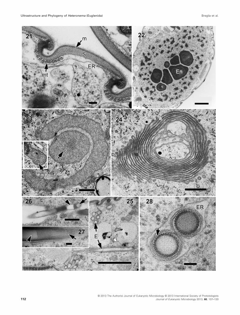
Cytoplasmic organelles
Light and transmission electron micrographs showed a
single nucleus with permanently condensed chromosomes
and several conspicuous, centrally located, endosomes
(Fig. 3, 22). The mitochondria had discoidal cristae
(Fig. 23), and robust Golgi bodies were formed of many
concentric cisternae (Fig. 24). Tubular extrusomes were
positioned immediately beneath the articulation zones
between S-shaped pellicle strips and throughout the cyto-
plasm, sometimes forming batteries of parallel units
(Fig. 25). The resting tubular extrusomes were approxi-
mately 2.7 lm long and 0.2 lm wide (Fig. 25–27). The ex-
trusomes were circular in cross-section, with a darkly
stained outer region surrounding a granular core (Fig. 26–28) and had helically arranged surface striations. The core
contained an anterior (clear) “cup” followed by a granular
dark band about 200 nm long and a lighter (apparently hol-
low) core (Fig. 26–28).
Feeding apparatus
The vestibular opening (V) at the anterior end of the cell led
to the flagellar pocket (Fe), as well as to the feeding pocket
(FP). The dorsal wall of the vestibulum was reinforced by
dense fibrous material (Fig. 29). The feeding apparatus
itself consisted of four structures: a dorsal rod (DR), a ven-
tral rod (VR), an accessory rod (AR), and four “vanes” (i.e.
a single row of microtubules affixed to a membrane). It
was positioned between the feeding pocket and the ante-
rior region of the flagellar pocket, separating them (Fig. 33,
34), and extended beyond the flagellar pocket for approxi-
mately one-third of the cell length (Fig. 31–33). The acces-
sory rod interacted with the ventral rod and the wall of the
feeding pocket. A group of microtubules lined the dorsal
side of the feeding pocket and supported a cluster of hairs
(“tomentum”) that extended into it (Fig. 29, 30, 33). The
rods were formed of interlinked microtubules embedded
in an amorphous matrix (Fig. 35–37). At the posterior end,
these were entirely formed of microtubules embedded in a
thin homogeneous matrix (Fig. 35); at the anterior end, the
rods contained microtubules within a heterogeneous
matrix that formed a conspicuous peripheral ring in trans-
verse sections (Fig.29–31, 37).The accessory rod was composed of only a few micro-
tubules embedded in a dense amorphous matrix (Fig. 38,
39). It also had a lamellar projection that extended towards
the anterior end of the cell (Fig. 39). At the most anterior
level of the feeding apparatus, the lamellar projection of
the accessory rod connected to a microtubule-lined ventral
lamella (VL) that extended inwards from the wall of the
vestibulum (Fig. 29, 30, 33). The ventral lamella arched
over the feeding pocket and joined the dorsal lamella (DL)
(Fig. 33). Near the anterior end of the cell, both the dorsal
and ventral rods were connected to one another. Near the
posterior end of the cell, the rods were also connected,
forming one microtubular bundle that was compartmental-
ized by the four vanes (Fig. 35). A series of more anterior
sections through the rods demonstrate the detachment of
the vanes from the rods (Fig. 36, 37). A striated fibre (SF)
lined the dorsal-right side of the feeding pocket (Fig. 33,
34, 45). A row of microtubules was positioned on both
sides of this fibre: four microtubules to the right, and eight
to more than eighteen microtubules on the left.
Flagellar apparatus
Details of the flagellar apparatus structures are shown in
Fig. 40–46. The flagellar pocket merged with the feeding
pocket near the anterior end of the cell, forming the ves-
tibulum. Two heterodynamic flagella emerged from the
base of the flagellar pocket, which was reinforced by elec-
tron-dense material near the vestibular opening (Fig. 43).
Each flagellum had axonemes with the typical 9 + 2
arrangement of microtubules (Fig. 42). Near the transition
zone, which appeared swollen, the two central microtu-
bules were absent, showing a 9 + 0 arrangement
(Fig. 40). Both flagella also contained paraxonemal rods
(PR) (Fig. 40–42) and conspicuous flagellar hairs (mastigo-
nemes) (Fig. 42). The paraxonemal rods in the dorsal
flagellum had a whorled disposition in transverse section;
the paraxonemal rods in the ventral flagellum had a lattice
of parallel fibres in transverse section (Fig. 41, 42).
A striated system of hairs connected the two flagella near
the vestibular opening (Fig. 42).
Like in other euglenids, the flagellar apparatus consisted
of two basal bodies and three microtubular roots. The fla-
Fig. 21–28. Transmission electron micrographs of Heteronema scaphurum. 21. Cross-section through the pellicle showing the plasma membrane
(m), thick proteinaceous strips (S), microtubules (mt), and cisternae of endoplasmic reticulum (ER). Scale bar = 100 nm. 22. Micrograph showing
the nucleus containing profiles of several conspicuous endosomes (En) and permanently condensed chromosomes. Scale bar = 2 lm. 23. Mito-
chondria with discoidal cristae (arrows). Scale bar = 500 nm. 24. Golgi bodies. Scale bar = 500 nm. 25. Longitudinal and cross-section views of
tubular extrusomes (E). Scale bar = 2 lm. 26. Longitudinal section of an extrusome showing an anterior clear “cup” (arrowhead) with operculum
(arrow). The region next to the cup, of approximately 200 nm in length, appears granular and darker than the rest of the tube. Scale
bar = 500 nm. 27. Detail of extrusome in longitudinal view, showing a striated outer region (arrow) and a clear, hollow core (arrowhead). Scale
bar = 100 nm. 28. Cross-section of extrusomes at different levels showing a dark and helically striated outer region (arrow), and a granular core.
Scale bar = 100 nm.
© 2013 The Author(s) Journal of Eukaryotic Microbiology © 2013 International Society of Protistologists
Journal of Eukaryotic Microbiology 2013, 60, 107–120 113
Breglia et al. Ultrastructure and Phylogeny of Heteronema (Euglenida)

© 2013 The Author(s) Journal of Eukaryotic Microbiology © 2013 International Society of Protistologists
Journal of Eukaryotic Microbiology 2013, 60, 107–120114
Ultrastructure and Phylogeny of Heteronema (Euglenida) Breglia et al.

Fig. 29–34. Transmission electron micrographs showing the feeding apparatus in Heteronema scaphurum at different levels along the longitudinal
axis of the cell. 29. Cross-section through the anterior end of the cell showing part of the feeding pocket (Fe), the dorsal rod (DR) and the acces-
sory rod (AR). The accessory rod has a lamellar expansion that connects with a microtubule-lined ventral lamella (VL) extending from the wall of
the feeding pocket. Microtubules line the dorsal side of the feeding pocket (arrows). A cluster of hairs or “tomentum” (arrowhead) extends from
the dorsal microtubules and into the feeding pocket. FM = fibrous material lining the anterior pocket of the cell; SF = striated fibre. Scale
bar = 1 lm. 30. Transversal section showing the enlarged vestibulum (V) formed by the merging of the flagellar pocket and the feeding pocket. A
cluster of hairs (arrowhead) still demarcates the deeper separation of the flagellar region and the feeding pocket (Fe). DF = dorsal flagellum. Scale
bar = 1 lm. 31. Cross-section at the anterior end of the cell. The region of the vestibulum that is continuous with the flagellar pocket is reinforced
by fibrous material (FM) and a striated fibre (SF). Both dorsal and ventral rods (DR and VR respectively) are visible, as well as the vanes of the
feeding apparatus (arrows). VF = ventral flagellum. Scale bar = 1 lm. 32. Oblique section showing the position of the dorsal and ventral rods (DR
and VR respectively) relative to the feeding pocket (Fe) and the flagellar pocket (FP). DF = dorsal flagellum; DL = dorsal lamella. Scale bar = 2 lm.
33. Semi-longitudinal section of the flagellar pocket (FP) and the feeding pocket (Fe). The ventral lamella (VL) arches over the feeding pocket and
connects to the dorsal lamella (DL). The arrowhead shows the cluster of hairs lining the feeding pocket. AR = accessory rod; DF = dorsal flagel-
lum; SF = striated fibre; VF = ventral flagellum; VR = ventral rod. Scale bar = 2 lm. 34. Separation of the feeding pocket (Fe) from the flagellar
pocket (FP). AR = accessory rod; DR = dorsal rod; SR = striated fibre; VR = ventral rod. Scale bars = 1 lm.
Fig. 35–39. Transmission electron micrographs showing the feeding apparatus in Heteronema scaphurum. 35–37. Non-consecutive serial cross-
sections through the rods. Scale bars = 500 nm. 35. A posterior section through the rods showing that they are connected, forming a single
structure entirely formed by microtubules embedded in a homogeneous matrix. Deep grooves in the microtubular bundle are lined by four vanes
(arrows). 36. A more anterior section through the rods showing separate bundles of microtubules embedded in a more heterogeneous matrix.
There are still grooves in the microtubular bundles associated with the four vanes. 37. An anterior section through the rods showing the detach-
ment of the vanes (arrows) from the rods. 38. Cross-section showing the relationship of the flagellar pocket (FP) and the feeding apparatus. The
rods are separated from the flagellar pocket and the vanes. Arrowhead: striated fibre; AR = accessory rod; DF = dorsal flagellum; DR = dorsal
rod; VF = ventral flagellum; VR = ventral rod. 39. Detail of the connection (arrow) between the ventral rod (VR) and the accessory rod (AR). The
arrowhead shows a striated fibre extending towards the pellicle. The double arrowhead shows the ventral lamella. Scale bars in 38–39 = 1 lm.
© 2013 The Author(s) Journal of Eukaryotic Microbiology © 2013 International Society of Protistologists
Journal of Eukaryotic Microbiology 2013, 60, 107–120 115
Breglia et al. Ultrastructure and Phylogeny of Heteronema (Euglenida)

© 2013 The Author(s) Journal of Eukaryotic Microbiology © 2013 International Society of Protistologists
Journal of Eukaryotic Microbiology 2013, 60, 107–120116
Ultrastructure and Phylogeny of Heteronema (Euglenida) Breglia et al.

gellar apparatus is shown in Fig. 44–46 from a posterior to
anterior view. The dorsal root (dr) originated from the dor-
sal basal body, and the ventral root (vr) and intermediate
root (ir) originated from the ventral basal body. The dorsal
root consisted of microtubules that extended towards the
anterior end of the cell and initially supported the dorsal
side of the flagellar pocket (Fig. 44, 45). The number of
microtubules increased as they extended anteriorly along
the flagellar pocket and ultimately became the microtu-
bules that subtend the pellicle strips (Fig. 43, 45, 46). The
intermediate root, initially formed by four microtubules,
was located between the dorsal and the ventral roots, and
supported the left side of the flagellar pocket (Fig. 44–46).The number of microtubules in this root increased towards
the anterior end of the flagellar pocket, joining the dorsal
root in a single microtubular band (dr + ir) that lined the
dorsal-left side of the flagellar pocket (Fig. 46). The ventral
root originated from the ventral basal body and initially
consisted of four microtubules (Fig. 44, 45). Towards the
anterior of the cell, the number of these microtubules
increased, and eventually reinforced the ventral side of
the flagellar pocket and the feeding apparatus (Fig. 46).
Molecular phylogenetic position
The 18S rDNA sequence (2,860 bp) of H. scaphurum con-
tained a number of insertions. Two of them were of con-
siderable length: the first one (at position 142 of the
sequence) was 567 bp long, and the second (at position
2,122 of the sequence) was 253 bp long. ML and Bayes-
ian analyses of the 39-taxon alignment resulted in identical
tree topologies that showed the new isolate clustering
within a clade of euglenids consisting of eukaryovorous,
primary osmotrophic, and phototrophic species (Fig. 47).
The phototrophic and primary osmotrophic euglenids
formed two well-supported subclades (ML boostrap
value = 83% and Bayesian posterior probability = 1.00 for
primary osmotrophs, and ML boostrap value = 99% and
Bayesian posterior probability = 1.00 for phototrophic
species). The eukaryovorous euglenids Peranema, Aniso-
nema, Dinema, and H. scaphurum, however, did not form
a distinct clade and instead formed a paraphyletic group.
DISCUSSION
Heteronema scaphurum had all the ultrastructural charac-
teristics distinctive of euglenozoans: a tripartite flagellar
root system, flagella with heteromorphic paraxial rods,
tubular extrusomes, and mitochondria with discoidal (pad-
dle-shaped) cristae (Simpson 1997; Willey et al. 1988).
Heteronema scaphurum also had a pellicle with proteina-
ceous strips, the best synapomorphy for euglenids, and a
complex feeding apparatus consisting of rods and vanes
that was typical of other eukaryovorous species (e.g. Pera-
nema and Dinema) (Triemer and Farmer 1991). In agree-
ment with these morphological attributes, our
phylogenetic analyses of 18S rDNA sequences robustly
placed H. scaphurum as a member of the Euglenida and,
more specifically, as part of a polytomy formed by all eu-
karyovorous, primary osmotrophic, and phototrophic eugle-
nids (to the exclusion of the bacterivorous in the analysis).
Heteronema scaphurum was originally described by
Skuja in 1934 and later reported in Australian freshwater
sites (Schroeckh et al. 2003). These descriptions, how-
ever, were solely based on light micrographs. Skuja
reported cells with a size range of 78–85 lm, and a diam-
eter of 40–46 lm for H. scaphurum, whereas the cells in
Australia were shorter (62–75 lm long), more within the
range of our isolate (45–70 lm in length and 30 lm in
diameter). Both Skuja and Schroeckh et al. described “a
characteristic dimple at the posterior end of the cell”
(Schroeckh et al. 2003), which we now know is the pos-
terior cytoproct in our isolate. No other data from previous
reports (e.g. scanning and transmission electron micros-
copy or molecular markers) are available, making a more
detailed comparison with our isolate impossible. The fea-
tures observed in our light micrographs, on the other
hand, seem to be in accordance with the previous descrip-
tions of this species, persuading us to name our isolate
H. scaphurum. However, the genus Heteronema has
Fig. 40–46. Transmission electron micrographs of the flagellar apparatus of Heteronema scaphurum. 40. Cross-section through the swollen
flagellar transition zone, showing a 9 + 0 arrangement of microtubules (arrow). PR = paraxial rod. Scale bar = 100 nm. 41. Oblique section
through the dorsal and ventral flagella (DF and VF respectively) showing the paraxial rods (arrows) and mastigonemes (arrowheads). Scale
bar = 500 nm. 42. Cross-section of the flagella (DF = dorsal flagellum; VF = ventral flagellum) showing the 9 + 2 arrangement of axonemal micro-
tubules and the paraxial rods (arrows). The paraxial rod in the DF has a whorled disposition, and the paraxial rod in the VF is composed of a lat-
tice-like pattern of fibres. A striated fibril (arrowhead) connects both flagella near the posterior end of the flagellar pocket. Scale bar = 100 nm.
43. Cross-section through the flagellar pocket at the anterior level showing the dorsal flagellum (DF), the ventral flagellum (VF), and pellicle strips
(arrows) extending into the flagellar pocket, which is lined by fibrous material (FM). Scale bar = 500 nm. 44. Cross-section through the flagellar
pocket at the level of the flagellar transition zones showing the three microtubular roots: the dorsal flagellar root (dr) is associated with the dorsal
basal body; the ventral and intermediate roots (vr and ir respectively) are associated with the ventral basal body. DR = dorsal rod; VR = ventral
rod. Scale bar = 500 nm. 45. Cross-section through the middle part of the flagellar pocket. The dorsal flagellar root (dr) is formed by microtubules
that extend along the dorsal side of the flagellar pocket. The intermediate flagellar root (ir) is positioned between the dorsal and the ventral roots,
supporting the left side of the flagellar pocket. The ventral root (vr) initially consists of four microtubules, supporting the ventral side of the pocket.
A striated fibre (SF) reinforces the dorsal side of the pocket. Rows of microtubules lie on both sides of SF: four microtubules on the left (arrow-
head) and sixteen linked microtubules (LMt) on the right side of striated fibre. DR = dorsal rod; VR = ventral rod. Scale bar = 1 lm. 46. Cross-sec-
tion through the anterior region of the flagellar pocket showing the fusion of the dorsal and intermediate roots (dr + ir). DR = dorsal rod;
VR = ventral rod. Scale bar = 2 lm.
© 2013 The Author(s) Journal of Eukaryotic Microbiology © 2013 International Society of Protistologists
Journal of Eukaryotic Microbiology 2013, 60, 107–120 117
Breglia et al. Ultrastructure and Phylogeny of Heteronema (Euglenida)

© 2013 The Author(s) Journal of Eukaryotic Microbiology © 2013 International Society of Protistologists
Journal of Eukaryotic Microbiology 2013, 60, 107–120118
Ultrastructure and Phylogeny of Heteronema (Euglenida) Breglia et al.

unclear generic limits, largely due to a continuum of varia-
tion with other genera, such as Metanema and Dinema
(Larsen and Patterson 1991), coupled with descriptions
based solely on light microscopy (Al-Qassab et al. 2002;
Lee et al. 2005; Schroeckh et al. 2003). The more compre-
hensive description of this species reported here using
SEM and TEM allows us to demarcate the ultrastructural
features of Heteronema species more precisely. More-
over, we provide the first molecular data (SSU rDNA
sequence) for this genus, which will contribute to a better
understanding of euglenid species boundaries.
Pellicle
The eukaryovorous euglenids described so far are capable
of euglenoid movement and have helically arranged, deli-
cate pellicle strips that range in total number between 20
and 56 (Leander et al. 2007). Euglenids with less than
about 16 strips tend to be rigid and are either bacterivor-
ous, osmotrophic, or phototrophic. Heteronema scaphu-
rum has a plastic pellicle formed by 28 helically arranged
strips without posterior strip reduction, which is consistent
with the range of features present in other eukaryovores.
In contrast to other eukaryovores (Peranema, Dinema, and
Urceolus), the pellicle strips of H. scaphurum were robust
in transverse section (100 nm thick) and had distinct over-
hangs in the articulation zones (Leander and Farmer 2000;
Leander et al. 2001b). Unlike Peranema trichophorum and
Dinema sulcatum, H. scaphurum did not possess a dis-
tinctly shaped “flagellar strip” that holds the ventral (pos-
terior or recurrent) flagellum on the ventral side of the cell
during gliding motility (Leander et al. 2001a).
Feeding apparatus
Triemer and Farmer (1991) described four types of feeding
apparatuses in euglenids (Types I–IV). Some bacterivores
have relatively simple feeding structures consisting of a
pocket lined by a row of microtubules (e.g. Type I appara-
tus in Petalomonas), whereas others have a robust and
well-developed feeding apparatuses consisting of rods and
vanes (e.g. Type II and IV in Ploeotia and Entosiphon
respectively) (Linton and Triemer 1999; Triemer and
Farmer 1991; Triemer and Fritz 1987). Eukaryovorous eu-
glenids also have a complex feeding system consisting of
a cytostome, four vanes, and two rods formed by varying
amounts of supporting microtubules and amorphous
matrix (e.g. Type III apparatus in Dinema and Peranema)
(Triemer and Farmer 1991). The feeding apparatus in H.
scaphurum conforms to Type III in this scheme and is
most similar to the apparatus found in Peranema tricho-
phorum (Nisbet 1974); the main difference is that the base
of the flagellar pocket is more expanded in H. scaphurum.
Moreover, the rods in P. trichophorum are capable of
projecting out from the cell to pierce the prey cell during
myzocytosis (Triemer 1997). According to Nisbet (1974),
the contraction of longitudinal lamellae attached to the
rods is responsible for moving the rods forward. This
behaviour was not observed in H. scaphurum.
Both H. scaphurum and P. trichophorum use their two
flagella like “arms” to manipulate prey and initiate phago-
cytosis (Triemer 1997). Heteronema scaphurum also
secretes a mucilaginous web through the vestibular open-
ing, a novel feature to capture and secure the prey cells
(usually several at a time). In both species, the rods move
the flagella to one side, preventing the passage of food
into the flagellar pocket. The widening of the vestibular
opening observed in H. scaphurum during phagocytosis
also occurs in P. trichophorum when it ingests whole
cells, although to a much lesser degree; H. scaphurum
enlarges its anterior end to several times its cell diameter.
This has also been observed in other kinds of eukaryotes,
such as raptorial ciliates. Dileptus lamella, for instance,
enlarges its cytostome to a width even wider than that of
the prey; Didinium nasutum has a fibrous ring that encir-
cles the base of its proboscis and stretches the cell body
to accommodate the prey (Verni and Gualtieri 1997). In H.
scaphurum, the thickenings and the striated fibre around
the flagellar pocket might have a similar function. Also,
the dorsal and ventral lamellae in H. scaphurum might play
a role in the expansion of the vestibular opening by pulling
from the opposite side of the flagellar pocket.
Faecal pellets in single-celled eukaryotes
After feeding, H. scaphurum secretes waste material
through the cytoproct, often in the form of faecal pellets.
Faecal pellets are found in both marine and fresh-water
environments and can vary in size and shape, ranging from
minipellets (measuring between 3 lm and 50 lm in diame-
ter) to larger pellets of more than 50 lm (Gowing and Silver
1985). The presence of faecal pellets in sediment samples
is usually attributed to meiofunal metazoans, dinoflagellates
(Buck and Newton 1995; Buck et al. 1990), ciliates (Stoec-
ker 1984), and radiolarians (Gowing and Silver 1985). This is
the first report of any member of the Euglenozoa discharg-
ing faecal pellets through a distinct cytoproct.
Ultrastructural identity and phylogenetic position
The SSU rDNA sequence of H. scaphurum had an unre-
solved position among the eukayrovorous species of eugle-
nids. Its ultrastructural characters are consistent with a
predator lifestyle (e.g. a feeding apparatus with short rods
located in the anterior end of the cell, similar to that of P.
trichophorum). Shorter rods presumably allow for more
flexibility in the posterior two-thirds of the cell which, cou-
pled with a growing number of pellicle strips, would give
eukaryovorous euglenids the elasticity to ingest bundles of
large prey cells. However, H. scaphurum has fewer pellicle
Fig. 47. Maximum likelihood tree, inferred from 39 small subunit (SSU) rDNA sequences showing the molecular phylogenetic position of
Heteronema scaphurum within euglenid using diplonemid and kinetoplastids as outgroups. The numbers above the stems are Bayesian posterior
probabilities over 0.95; ML bootstraps greater than 50% are shown below the stems.
© 2013 The Author(s) Journal of Eukaryotic Microbiology © 2013 International Society of Protistologists
Journal of Eukaryotic Microbiology 2013, 60, 107–120 119
Breglia et al. Ultrastructure and Phylogeny of Heteronema (Euglenida)

strips than P. trichophorum (28 vs. 56), as well as less cell
plasticity. The set of morphological characters in H. scaphu-
rum is somewhat intermediate between bacterivorous
(with fewer pellicle strips but longer rods) and eukaryovores
(with more pellicle strips and short rods). Also, H. scaphu-
rum shows novel characteristics such as a cytoproct from
which small pellets are expelled, and a feeding behaviour
that reflects the mechanism by which eukaryovorous
ancestors sequestered green algae and ultimately gave
rise to phototrophic euglenids (Yamaguchi et al. 2012).
ACKNOWLEDGMENTS
This research was supported by grants from the Tula Foun-
dation (Centre for Microbial Diversity and Evolution),
National Science and Engineering Research Council of Can-
ada (NSERC 283091-09), and the Canadian Institute for
Advanced Research, Program in Integrated Microbial Biodi-
versity. We would like to thank Dan Halloway for providing
water samples that contained Heteronema scaphurum.
LITERATURE CITED
Al-Qassab, S., Lee, W. J., Murray, S., Simpson, A. G. B. & Patterson,
D. J. 2002. Flagellates from stromatolites and surrounding sedi-
ments in Shark Bay, Western Australia. Acta Protozool., 41:91–144.Buck, K. R., Bolt, P. A. & Garrison, D. L. 1990. Phagotrophy and
fecal pellet production by an athecate dinoflagellate in Antarctic
sea ice. Mar. Ecol. Prog. Ser., 60:75–84.Buck, K. R. & Newton, J. 1995. Fecal pellet flux in Dabob Bay
during a diatom bloom: contribution of microzooplankton. Lim-
nol. Oceanogr., 40:306–315.Esson, H. J. & Leander, B. S. 2006. A model for the morphogenesis
of strip reduction patterns in phototrophic euglenids: evidence
for heterochrony in pellicle evolution. Evol. Dev., 8:378–388.Gibbs, S. P. 1978. The chloroplasts of Euglena may have evolved
from symbiotic green algae. Can. J. Bot., 56:2883–2889.Gibbs, S. P. 1981. The chloroplasts of some algal groups may
have evolved from endosymbiotic eukaryotic algae. Ann. N. Y.
Acad. Sci., 361:193–208.Gowing, M. M. & Silver, M. W. 1985. Minipellets: a new and abun-
dant size class of marine fecal pellets. J. Mar. Res., 43:395–418.Guindon, S. & Gascuel, O. 2003. A simple, fast, and accurate
algorithm to estimate large phylogenies by maximum likelihood.
Syst. Biol., 52:696–704.Larsen, J. & Patterson, D. J. 1991. The diversity of heterotrophic
euglenids. In: Patterson, D. J. & Larsen, J. (eds.), The Biology
of Free-Living Heterotrophic Flagellates. Systematics Associa-
tion Special Volume. Claredon Press, Oxford. p. 205–217.Leander, B. S. & Farmer, M. A. 2000. Comparative morphology of
the euglenid pellicle. I. Patterns of strips and pores. J. Eukaryot.
Microbiol., 47:469–479.Leander, B. S. & Farmer, M. A. 2001a. Evolution of Phacus
(Euglenophyceae) as inferred from pellicle morphology and SSU
rDNA. J. Phycol., 37:143–159.Leander, B. S. & Farmer, M. A. 2001b. Comparative morphology
of the euglenid pellicle. II. Diversity of strip substructure.
J. Eukaryot. Microbiol., 48:202–217.Leander, B. S., Triemer, R. E. & Farmer, M. A. 2001a. Character evo-
lution in heterotrophic euglenids. Eur. J. Protistol., 37:337–356.Leander, B. S., Witek, R. P. & Farmer, M. A. 2001b. Trends in the
evolution of the euglenid pellicle. Evolution, 55:2215–2235.
Leander, B. S. 2004. Did trypanosomatid parasites have photosyn-
thetic ancestors? Trends Microbiol., 12:251–258.Leander, B. S., Esson, H. J. & Breglia, S. A. 2007. Macroevolution
of complex cytoskeletal systems in euglenids. BioEssays,
29:987–1000.Lee, W. J., Simpson, A. G. B. & Patterson, D. J. 2005. Free-living
heterotrophic flagellates from freshwater sites in Tasmania
(Australia), a field survey. Acta Protozool., 44:321–350.Linton, E. W. & Triemer, R. E. 1999. Reconstruction of the feeding
apparatus in Ploeotia costata (Euglenophyta) and its relationship
to other euglenoid feeding apparatuses. J. Phycol., 35:313–324.Milne, I., Lindner, D., Bayer, M., Husmeier, D., McGuire, G., Mar-
shall, D. F. & Wright, F. 2009. TOPALi v2: a rich graphical inter-
face for evolutionary analyses of multiple alignments on HPC
clusters and multi-core desktops. Bioinformatics, 25:126–127.Nisbet, B. 1974. An ultrastructural study of the feeding apparatus
of Peranema trichophorum. J. Protozool., 21:39–48.Reynolds, E. S. 1963. The use of lead citrate at high pH as an elec-
tron-opaque stain in electron microscopy. J. Cell Biol., 17:208–212.Rogers, M. B., Gilson, P. R., Su, V., McFadden, G. I. & Keeling,
P. J. 2007. The complete chloroplast genome of the chlorarach-
niophyte Bigelowiella natans: evidence for independent origins
of chlorarachniophyte and euglenid secondary endosymbionts.
Mol. Biol. Evol., 24:54–62.Ronquist, F. & Huelsenbeck, J. P. 2003. MrBayes 3: Bayesian
phylogenetic inference under mixed models. Bioinformatics,
19:1572–1574.Saito, A., Suetomo, Y., Arikawa, M., Omura, G., Khan, S. M. M.
K., Kakuta, S., Suzaki, E., Kataoka, K. & Suzaki, T. 2003. Gliding
movement in Peranema trichophorum is powered by flagellar
surface motility. Cell Motil. Cytoskeleton, 55:244–253.Schroeckh, S., Lee, W. J. & Patterson, D. J. 2003. Free-living het-
erotrophic euglenids from freshwater sites in mainland Austra-
lia. Hydrobiologia, 493:131–166.Simpson, A. G. B. 1997. The identity and composition of the Eug-
lenozoa. Arch. Protistenkd., 148:318–327.Stoecker, D. K. 1984. Particle production by planktonic ciliates.
Limnol. Oceanogr., 29:930–940.Triemer, R. E. & Fritz, L. 1987. Structure and operation of the
feeding apparatus in a colorless euglenoid, Entosiphon sulca-
tum. J. Protozool., 34:39–47.Triemer, R. E. & Farmer, M. A. 1991. The ultrastructural organization of
the heterotrophic euglenids and its evolutionary implications. In: Patt-
erson, D. J. & Larsen, J. (eds.), The Biology of Free-Living Heterotro-
phic Flagellates. The Systematics Association, Oxford. p. 185–204.Triemer, R. E. 1997. Feeding in Peranema trichophorum revisited
(Euglenophyta). J. Phycol., 33:649–654.Triemer, R. E., Linton, E., Shin,W., Nudelman, A., Monfils, A., Bennett,
M. & Brosnan, S. 2006. Phylogeny of the Euglenales based upon
combined SSU and LSU rDNA sequence comparisons and descrip-
tion of Discoplastis gen. nov. (Euglenophyta). J. Phycol., 42:731–740.Verni, F. & Gualtieri, P. 1997. Feeding behaviour in ciliated
protists. Micron, 28:487–504.Willey, R. L., Walne, P. L. & Kivic, P. A. 1988. Phagotrophy and the
origins of the euglenoid flagellates. Crit. Rev. Plant Sci., 7:303–340.Yamaguchi, A., Yubuki, N. & Leander, B. S. 2012. Morphostasis
in a novel eukaryote illuminates the evolutionary transition from
phagotrophy to phototrophy: description of Rapaza viridis n.
gen. et sp. (Euglenozoa, Euglenida). BMC Evol. Biol., 12:29.
1 Present address: Centre for Comparative Genomics and Evolu-
tionary Bioinformatics, Department of Biochemistry and Molecular
Biology, Dalhousie University, 5850 College Street, Halifax, NS,
B3H 4R2, Canada
© 2013 The Author(s) Journal of Eukaryotic Microbiology © 2013 International Society of Protistologists
Journal of Eukaryotic Microbiology 2013, 60, 107–120120
Ultrastructure and Phylogeny of Heteronema (Euglenida) Breglia et al.