Original Article Progression of endometrial hyperplasia… · Endometrial hyperplasia and the 2014...
9
Int J Clin Exp Pathol 2016;9(2):1617-1625 www.ijcep.com /ISSN:1936-2625/IJCEP0018252 Original Article Progression of endometrial hyperplasia: a revisit under the 2014 WHO classifications Parker C Wilson, Natalia Buza, Pei Hui Department of Pathology, Yale School of Medicine, New Haven, CT 06510, USA Received October 21, 2015; Accepted December 22, 2015; Epub February 1, 2016; Published February 15, 2016 Abstract: 273 patients diagnosed with endometrial hyperplasia from 1999-2001 at our institution were classified using the 1994 and 2014 WHO criteria. By 1994 criteria: 189 patients had simple hyperplasia (SH), 8 had simple hyperplasia with atypia (SHA), 23 had complex hyperplasia (CH), and 53 had complex hyperplasia with atypia (CHA). By 2014 criteria: 212 patients had benign endometrial hyperplasia (BEH) and 61 patients had atypical endometrial hyperplasia/endometrioid intraepithelial neoplasia (AEH/EIN). The primary endpoint was development of endome- trioid carcinoma. With a median follow-up of 5.2 years, progression to endometrioid carcinoma occurred in 1.6% of patients with SH, 25% with SHA, 4.3% with CH, 24.5% with CHA, 1.9% with BEH and 24.5% with AEH. In patients that did not undergo hysterectomy for at least 1-year, 20% with AEH and 2.6% with BEH progressed to carcinoma. Patients with CHA had a shorter endometrioid carcinoma free survival time (EFS) than patients with SH (P < 0.0001) but was not different from SHA (P = 0.78) or CH (P = 0.02) after correction for multiple comparisons. Patients with SHA had a shorter EFS than patients with SH (P < 0.0001) but was not different from CH (P = 0.12). Patients with SH did not have a shorter EFS than patients with CH (P = 0.30). Median EFS was shorter in patients with AEH (10.9 years) than patients with BEH (16.3 years, HR = 0.08, 95% CI: 0.008 to 0.08, P < 0.0001). While confirming the diagnostic validity of both schemes, our data support the emphasis on cytological atypia by the two-tiered 2014 WHO classification. Keywords: Endometrial hyperplasia, progression, WHO classification Introduction Atypical endometrial hyperplasia is a pre-neo- plastic condition that proceeds to the mo- st common uterine malignancy, endometrioid adenocarcinoma (Type 1 endometrial cancer, 70-80% of endometrial cancer) [1]. While established criteria using combined evaluation of cytological and architectural abnormalities for histological classification of endometrial hyperplasia have been used since the mid 80’s, recent investigations have resulted in signifi- cant discussion of the overall clinical relevance for each subcategory of endometrial hyperpla- sia [2, 3]. Moreover, the evolving concept of endometrial intraepithelial neoplasia or EIN has enhanced our understanding of the molec- ular basis for the development of endometrioid adenocarcinoma [4]. In 2014, the World Health Organization (WHO) introduced the updated classification scheme for endometrial hyperplasia that has 2 cate- gories: benign endometrial hyperplasia (BEH), and atypical endometrial hyperplasia (AEH)/ endometrioid intraepithelial neoplasia (EIN) [5]. The defining feature of the 2014 classification scheme is the presence (AEH/EIN) or absence (BEH) of cytological atypia superimposed on a background of endometrial hyperplasia. The previous 1994 WHO classification scheme, pri- marily based on Kurman’s seminal study in 1985, uses both architectural and cytological abnormalities to classify the endometrial hyper- plasia into 4 categories: simple hyperplasia (SH), simple hyperplasia with atypia (SHA), com- plex hyperplasia (CH), and complex hyperplasia with atypia (CHA), which assessed both cyto- logical atypia and architectural complexity [2]. In light of the new WHO classification, it becomes necessary to compare the two schemes in a large study cohort with a long- term clinical follow-up. In this retrospective study, the prognostic values of the 1994 and
Transcript of Original Article Progression of endometrial hyperplasia… · Endometrial hyperplasia and the 2014...

Int J Clin Exp Pathol 2016;9(2):1617-1625www.ijcep.com /ISSN:1936-2625/IJCEP0018252
Original Article Progression of endometrial hyperplasia: a revisit under the 2014 WHO classifications
Parker C Wilson, Natalia Buza, Pei Hui
Department of Pathology, Yale School of Medicine, New Haven, CT 06510, USA
Received October 21, 2015; Accepted December 22, 2015; Epub February 1, 2016; Published February 15, 2016
Abstract: 273 patients diagnosed with endometrial hyperplasia from 1999-2001 at our institution were classified using the 1994 and 2014 WHO criteria. By 1994 criteria: 189 patients had simple hyperplasia (SH), 8 had simple hyperplasia with atypia (SHA), 23 had complex hyperplasia (CH), and 53 had complex hyperplasia with atypia (CHA). By 2014 criteria: 212 patients had benign endometrial hyperplasia (BEH) and 61 patients had atypical endometrial hyperplasia/endometrioid intraepithelial neoplasia (AEH/EIN). The primary endpoint was development of endome-trioid carcinoma. With a median follow-up of 5.2 years, progression to endometrioid carcinoma occurred in 1.6% of patients with SH, 25% with SHA, 4.3% with CH, 24.5% with CHA, 1.9% with BEH and 24.5% with AEH. In patients that did not undergo hysterectomy for at least 1-year, 20% with AEH and 2.6% with BEH progressed to carcinoma. Patients with CHA had a shorter endometrioid carcinoma free survival time (EFS) than patients with SH (P < 0.0001) but was not different from SHA (P = 0.78) or CH (P = 0.02) after correction for multiple comparisons. Patients with SHA had a shorter EFS than patients with SH (P < 0.0001) but was not different from CH (P = 0.12). Patients with SH did not have a shorter EFS than patients with CH (P = 0.30). Median EFS was shorter in patients with AEH (10.9 years) than patients with BEH (16.3 years, HR = 0.08, 95% CI: 0.008 to 0.08, P < 0.0001). While confirming the diagnostic validity of both schemes, our data support the emphasis on cytological atypia by the two-tiered 2014 WHO classification.
Keywords: Endometrial hyperplasia, progression, WHO classification
Introduction
Atypical endometrial hyperplasia is a pre-neo-plastic condition that proceeds to the mo- st common uterine malignancy, endometrioid adenocarcinoma (Type 1 endometrial cancer, 70-80% of endometrial cancer) [1]. While established criteria using combined evaluation of cytological and architectural abnormalities for histological classification of endometrial hyperplasia have been used since the mid 80’s, recent investigations have resulted in signifi-cant discussion of the overall clinical relevance for each subcategory of endometrial hyperpla-sia [2, 3]. Moreover, the evolving concept of endometrial intraepithelial neoplasia or EIN has enhanced our understanding of the molec-ular basis for the development of endometrioid adenocarcinoma [4].
In 2014, the World Health Organization (WHO) introduced the updated classification scheme
for endometrial hyperplasia that has 2 cate- gories: benign endometrial hyperplasia (BEH), and atypical endometrial hyperplasia (AEH)/endometrioid intraepithelial neoplasia (EIN) [5]. The defining feature of the 2014 classification scheme is the presence (AEH/EIN) or absence (BEH) of cytological atypia superimposed on a background of endometrial hyperplasia. The previous 1994 WHO classification scheme, pri-marily based on Kurman’s seminal study in 1985, uses both architectural and cytological abnormalities to classify the endometrial hyper-plasia into 4 categories: simple hyperplasia (SH), simple hyperplasia with atypia (SHA), com-plex hyperplasia (CH), and complex hyperplasia with atypia (CHA), which assessed both cyto-logical atypia and architectural complexity [2]. In light of the new WHO classification, it becomes necessary to compare the two schemes in a large study cohort with a long-term clinical follow-up. In this retrospective study, the prognostic values of the 1994 and

Endometrial hyperplasia and the 2014 WHO criteria
1618 Int J Clin Exp Pathol 2016;9(2):1617-1625
2014 classifications of endometrial hyperpla-sia were compared in 273 patients consecu-tively diagnosed with endometrial hyperplasia at a single tertiary medical center.
Materials and methods
Study cohort
This retrospective cohort study included pa- tients diagnosed with endometrial hyperplasia consecutively accessioned at our institution from 1999-2001 by endometrial biopsy or curettage. Eligible specimens were reviewed by three authors and categorized using the 1994 and 2014 WHO classifications for endometrial hyperplasia. All available subsequent endome-trial biopsies and hysterectomy specimens were also reviewed. A total of 273 cases were eventually included in the study. The number of slides of each curettage ranged 1 to 9 with a
median of 3. Follow-up endometrial sampling was available in 227 cases including curettage in 203 patients, and hysterectomy in 87 pa- tients. Slides of the hysterectomy were reviewed in 34 cases, including all cases that had atypi-cal endometrial hyperplasia or endometrioid carcinoma.
Study design
This retrospective study was designed to as- sess the differences in disease progression and endometrioid carcinoma-free survival be- tween women with endometrial hyperplasia diagnosed using the 1994 WHO four category scheme and 2014 WHO two tiered classi- fication.
Follow-up evaluation
All patients with at least 2 endometrial sampl- es were assessed for regression, persistence,
Figure 1. Representative histological presentations of endometrial lesions. A. Benign endometrial hyperplasia (BEH) by 2014 WHO classification/simple hyperplasia without atypia by 1994 WHO criteria. B. Atypical endometrial hyper-plasia/endometrioid intraepithelial neoplasia (AEH/EIN) by 2014 WHO classification/complex atypical hyperplasia by 1994 WHO criteria. C. Cytological/nuclear atypia in AEH/EIN. D. Well differentiated endometrioid adenocarci-noma, FIGO architectural grade 1.

Endometrial hyperplasia and the 2014 WHO criteria
1619 Int J Clin Exp Pathol 2016;9(2):1617-1625
development of atypia, and progression to car-cinoma. Regression in patients initially diag-nosed with BEH or AEH was defined as either: 1) normal phase endometrium in premenopau-al women and inactive or atrophic endometri-um in postmenopausal women on the last available follow-up biopsy or hysterectomy specimen, OR 2) patients who became clinically asymptomatic and were followed for at least 1 year. Persistence in patients initially diagnosed with BEH was defined as patients that had BEH, endometrial polyps, or disordered proliferative endometrium on last follow-up biopsy or BEH on a hysterectomy specimen. The development of atypia was defined as patients who were ini-tially diagnosed with BEH that progressed to AEH on the last available biopsy or hysterecto-my specimen. Progression to carcinoma was defined as patients that were initially diagnosed with BEH or AEH that were diagnosed with endometrioid carcinoma on any subsequent biopsy or hysterectomy specimen. The time to development of endometrioid carcinoma was the time from initial diagnosis of endometrial
hyperplasia to the first diagnosis of endometri-oid carcinoma by either endometrial biopsy or hysterectomy. All patients with only 1 endome-trial sample were considered as having no available follow-up (see above, the second cri-terion of regression).
Statistical analysis
Chi-square test or Fisher’s exact test for cate-gorical variables were used to compare the dif-ferences between patient groups categorized by 1994 and 2014 WHO endometrial hyperpla-sia criteria. Kaplan-Meier estimates were used to compute the survival functions among patient groups and were compared using log-rank tests. Endometrioid carcinoma free sur-vival (EFS) was defined as the time from initial endometrial hyperplasia diagnosis to endome-trioid carcinoma diagnosis by either curettage or hysterectomy. Only patients with follow-up endometrial sampling were included in the analysis. Patients were censored at the time of the time of their last curettage or hysterectomy. A value of P < 0.01 after adjustment for multi-
Table 1. Characteristics of patients eventually diagnosed with endometrioid carcinoma (N = 19)Initial Diagnosis of Hyperplasia
Age at Initial Diagnosis
Age at Carcinoma Diagnosis
Endometrioid Carcinoma Free Survival Time*
FIGO Architectural and Nuclear Grade
Carcinoma Diagnosed on Hysterectomy
SH/BEH 45 49 3 y 9 mo 17 d F1N1 NO56 72 16 y 3 mo 24 d F1N1 YES70 77 6 y 11 mo 29 d F2N2 NO
CH/BEH 64 64 24 d F1N2 NOSHA/AEH 55 59 3 y 8 mo 19 d F1N2 NO
72 72 1 mo 2 d F1N2 NOCAH/AEH 48 48 8 mo 18 d F1N1 YES
50 50 2 mo 18 d F1N1 YES51 51 1 mo 18 d F1N2 YES54 54 1 mo 3 d F1N1 YES54 55 4 mo 16 d F1N2 NO56 56 3 mo 17 d F1N1 NO56 56 2 mo 25 d F1N1 YES59 61 2 y 3 mo 21 d F1N2 NO60 60 1 mo 9 d F1N2 NO62 62 4 mo 2 d F1N1 NO69 80 10 y 11 mo 11 d F1N2 YES70 74 4 y 5 mo 24 d F1N1 NO78 78 1 mo 3 d F1N1 NO
*Endometrioid carcinoma-free survival time is the duration between the initial diagnosis and the first carcinoma diagnosis by either curettage or hysterectomy. SH-simple hyperplasia; CH-complex hyperplasia; SHA-simple hyperplasia with atypia; CAH-complex hy-perplasia with atypia; BEH-benign endometrial hyperplasia and AEH-atypical endometrial hyperplasia/endometrioid intraepithelial neoplasia.

Endometrial hyperplasia and the 2014 WHO criteria
1620 Int J Clin Exp Pathol 2016;9(2):1617-1625
ple comparisons was consid-ered statistically significant. The Cox proportional hazards model was used to adjust for age at initial presentation. Analyses were performed us- ing GraphPad Prism 6 for Mac OS X (GraphPad Software, Inc.) and XLSTAT for Mac OSX (Addinsoft, Inc).
Histological criteria
Simple hyperplasia is defined as diffuse abnormal glandular proliferation with balanced gland/stroma ratio without cytological atypia (Figure 1). The glands are abnormal in shape and size with out-po- uching and branching. Com- plex hyperplasia shows dimin-ished stromal component with more pronounced glan-dular abnormality than simple hyperplasia, and has no cyto-logical atypia. Atypical hyper-plasia (simple or complex) is defined by the presence of nuclear atypia: loss of polari-ty, nuclear rounding, enlarge-ment, pleomorphism, abnor-mal chromatin patterns and prominent nucleoli. Benign endometrial hyperplasia is defined by the 2014 WHO classification as “an exagger-ated proliferation of endome-trial glands of irregular size
Table 2. Patient characteristics and endometrioid carcinoma free survival by 1994 and 2014 WHO classifications/Kaplan-Meier analysis
Index Diagnosis No. of Patients
Median Follow-Up
(years)
Develop Endometrioid Carcinoma (n)
HR [95% CI] Mean Age (years) TH (n)
1994 SH 189 6.86 3 (1.5%) 1 51.4±10.3 42 (22.2%)1994 CH 23 8.21 1 (4.3%) 2.9 [0.2 to 147.4]* 54.3±13.2 10 (43.4%)1994 SHA 8 3.79 2 (25%) 24.1 [ND]** 52.0±11.8 3 (37.5%)1994 CHA 53 0.88 13 (24.5%) 79.4 [21.8 to 288.6]** 56.2±9.8 32 (60.3%)2014 BEH 212 6.98 4 (1.8%) 1 51.7±10.6 52 (24.5%)2014 AEH/EIN 61 0.88 15 (24.5%) 39.3 [12.3 to 126.2]# 55.8±9.9 35 (57.3%)Abbreviations: TH-total hysterectomy, EFS-endometrioid carcinoma free survival. SH-simple hyperplasia; CH-complex hyperpla-sia; SHA-simple hyperplasia with atypia; CAH-complex hyperplasia with atypia; BEH-benign endometrial hyperplasia and AEH/EIN-atypical endometrial hyperplasia/endometrioid intraepithelial neoplasia. *= P < 0.05 vs. CHA, **P < 0.001 vs. SH, #= P < 0.0001 vs. BEH. ND = data points had insufficient events to estimate median EFS.
Figure 2. Endometrioid carcinoma free survival by 1994 and 2014 WHO clas-sifications. Patients were categorized by their initial endometrial hyperplasia diagnosis using the 1994 (A) and 2014 WHO (B) criteria and endometrioid carcinoma free survival (EFS) was analyzed using Kaplan-Meier analysis. EFS is defined as the time from initial endometrial hyperplasia diagnosis to endo-metrioid carcinoma diagnosis by either curettage or hysterectomy. Patients were censored at the time of the time of their last curettage or hysterectomy.

Endometrial hyperplasia and the 2014 WHO criteria
1621 Int J Clin Exp Pathol 2016;9(2):1617-1625
and shape, with an associated increase in the gland to stroma ratio compared with prolifera-tive endometrium, but without significant cyto-logical atypia”. Atypical endometrial hyperpla-sia (AEH)/Endometrioid intraepithelial neopla-sia (EIN) is defined as “cytological atypia super-imposed on an endometrial hyperplasia”. The separation of well-differentiated endometrioid carcinoma from AEH/EIN is based on the pres-ence of stromal invasion characterized by one or more of the following of at least 5 mm in size: confluent glandular or cribriform pattern, com-plete replacement of intervening endometrial stroma by foamy macrophages; infiltrative growth with myofibroblastic or desmoplastic stromal reaction, or complex papillary architec-ture (villoglandular pattern).
Results
The study cohort consisted of 273 cases con-secutively identified in a two-year period. According to the 1994 criteria: 189 patients were categorized as simple hyperplasia (SH), 8 as simple hyperplasia with atypia (SHA), 23 as complex hyperplasia (CH), and 53 as complex hyperplasia with atypia (CHA). According to the 2014 criteria: 212 patients were categorized as benign endometrial hyperplasia (BEH), and 61 patients as atypical endometrial hyperpla-sia (AEH)/endometrioid intraepithelial neopla-sia (EIN). The median follow-up time was 6.9 years for patients initially diagnosed with benign endometrial hyperplasia and 325 days for patients with atypical endometrial hyperpla-sia. Overall, 19 patients (4 of 212 cases of
BEH, 1.9% and 15 of 61 cases of AEH/EIN, 24.6%) eventually developed endometrioid adenocarcinoma and all underwent staging hysterectomy (Tables 1 and 2). All carcinomas except one case in the study cohort were well-differentiated FIGO architectural grade 1 endo-metrioid adenocarcinoma. All of the carcino-mas that developed in these patients were con-fined to the endomyometrium and no patients had extrauterine or lymph node metastases.
Simple hyperplasia (189 patients)
The age at the time of initial diagnosis ranged from 24 to 98 years (mean = 51.4±10.3). The median number of procedures was 2 and the median follow-up time was 6.8 years (range = 0-21.6). Twenty-five patients had had at least 1 prior endometrial biopsy and 42 patients had no subsequent sampling. Forty-two patients had a hysterectomy. At the conclusion of the study: 64 patients had persistent endometrial hyperplasia, 98 patients had regressed, 7 patients had developed atypical endometrial hyperplasia, 3 patients had developed endo-metrioid carcinoma, and 1 patient had a hyster-ectomy for leiomyosarcoma.
Complex hyperplasia (23 patients)
The age at the time of initial diagnosis ranged from 25 to 77 years (mean = 54.3±13.2). The median number of procedures was 2 and the median follow-up time was 8.2 years (range = 65 days-14.4 years). Three patients had had at least 1 prior endometrial biopsy and 1 patient had no subsequent sampling. Ten patients had
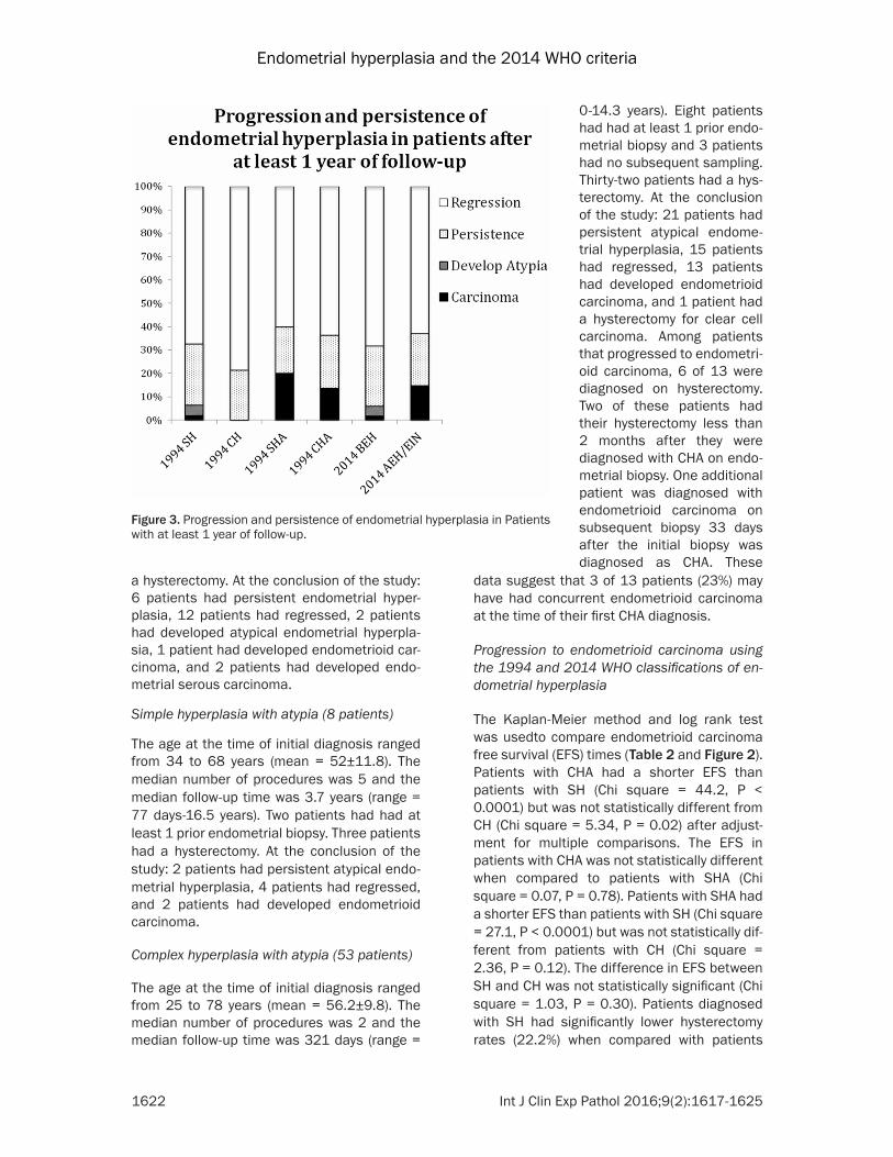
Table 3. Progression and persistence of endometrial hyperplasia in patients after at least 1 year of follow-up (n = 194)*Index Diagnosis
Progress to Carcinoma (n)
Develop Atypia (n)
Persistent Hyperplasia (n)
Regression (n)
Total Cases (n)
1994 SH 3 7 40 103 1531994 CH 0 0 3 11 141994 SHA 1 0 1 3 51994 CHA 3 0 5 14 222014 BEH 3 7 43 114 1672014 AEH/EIN 4 0 6 17 27Abbreviations: SH-simple hyperplasia; CH-complex hyperplasia; SHA-simple hyperplasia with atypia; CAH-complex hyperplasia with atypia; BEH-benign endometrial hyperplasia and AEH/EIN-atypical endometrial hyperplasia/endometrioid intraepithelial neoplasia. *Patients were categorized by their initial endometrial hyperplasia diagnosis using the 1994 and 2014 WHO criteria and had at least 1 year of follow-up (re-biopsied at least 1 year after their initial diagnosis or patients that were clinically asymptomatic and followed by PAP smear). Patients that underwent hysterectomy less than 1 year after their initial diagnosis were excluded.
Endometrial hyperplasia and the 2014 WHO criteria
1622 Int J Clin Exp Pathol 2016;9(2):1617-1625
a hysterectomy. At the conclusion of the study: 6 patients had persistent endometrial hyper-plasia, 12 patients had regressed, 2 patients had developed atypical endometrial hyperpla-sia, 1 patient had developed endometrioid car-cinoma, and 2 patients had developed endo-metrial serous carcinoma.
Simple hyperplasia with atypia (8 patients)
The age at the time of initial diagnosis ranged from 34 to 68 years (mean = 52±11.8). The median number of procedures was 5 and the median follow-up time was 3.7 years (range = 77 days-16.5 years). Two patients had had at least 1 prior endometrial biopsy. Three patients had a hysterectomy. At the conclusion of the study: 2 patients had persistent atypical endo-metrial hyperplasia, 4 patients had regressed, and 2 patients had developed endometrioid carcinoma.
Complex hyperplasia with atypia (53 patients)
The age at the time of initial diagnosis ranged from 25 to 78 years (mean = 56.2±9.8). The median number of procedures was 2 and the median follow-up time was 321 days (range =
data suggest that 3 of 13 patients (23%) may have had concurrent endometrioid carcinoma at the time of their first CHA diagnosis.
Progression to endometrioid carcinoma using the 1994 and 2014 WHO classifications of en-dometrial hyperplasia
The Kaplan-Meier method and log rank test was usedto compare endometrioid carcinoma free survival (EFS) times (Table 2 and Figure 2). Patients with CHA had a shorter EFS than patients with SH (Chi square = 44.2, P < 0.0001) but was not statistically different from CH (Chi square = 5.34, P = 0.02) after adjust-ment for multiple comparisons. The EFS in patients with CHA was not statistically different when compared to patients with SHA (Chi square = 0.07, P = 0.78). Patients with SHA had a shorter EFS than patients with SH (Chi square = 27.1, P < 0.0001) but was not statistically dif-ferent from patients with CH (Chi square = 2.36, P = 0.12). The difference in EFS between SH and CH was not statistically significant (Chi square = 1.03, P = 0.30). Patients diagnosed with SH had significantly lower hysterectomy rates (22.2%) when compared with patients
Figure 3. Progression and persistence of endometrial hyperplasia in Patients with at least 1 year of follow-up.
0-14.3 years). Eight patients had had at least 1 prior endo-metrial biopsy and 3 patients had no subsequent sampling. Thirty-two patients had a hys-terectomy. At the conclusion of the study: 21 patients had persistent atypical endome-trial hyperplasia, 15 patients had regressed, 13 patients had developed endometrioid carcinoma, and 1 patient had a hysterectomy for clear cell carcinoma. Among patients that progressed to endometri-oid carcinoma, 6 of 13 were diagnosed on hysterectomy. Two of these patients had their hysterectomy less than 2 months after they were diagnosed with CHA on endo-metrial biopsy. One additional patient was diagnosed with endometrioid carcinoma on subsequent biopsy 33 days after the initial biopsy was diagnosed as CHA. These

Endometrial hyperplasia and the 2014 WHO criteria
1623 Int J Clin Exp Pathol 2016;9(2):1617-1625
diagnosed with CH (43.4%, P < 0.04), and CHA (61.3%, P < 0.0001).
Patients with AEH developed EC had a shorter EFS than patients with BEH (Chi square = 31.1, P < 0.0001). The median EFS was 5957 days in patients diagnosed with BEH and 3998 days in patients diagnosed with AEH. Patients diag-nosed with AEH were 98% more likely to prog-ress to EC (HR = 0.02; 95% CI, 0.007 to 0.08, P < 0.0001). Age at initial presentation was a sig-nificant predictor of endometrioid carcinoma free survival (HR = 1.065; 95% CI, 1.012 to 1.12, P = 0.015) and the hazard ratios were adjusted using Cox regression. In multivariate analysis, patients diagnosed with AEH were 94% more likely to progress to EC (HR = 0.06; 95% CI, 0.02 to 0.19, P < 0.0001).
Endometrial hyperplasia progression to carci-noma, regression, and development of atypia among patients with at least 1 year of follow-up
194 patients had at least 1 year of follow-up and did not undergo hysterectomy in the first year after their initial diagnosis (Table 3 and Figure 3). Of the 153 patients initially diag-nosed with SH: 3 developed carcinoma, 7 pro-gressed to atypical hyperplasia, 40 had persis-tent endometrial hyperplasia, and 103 regre- ssed. Of the 14 patients initially diagnosed with CH: 3 had persistent endometrial hyperplasia, and 11 regressed. Of the 5 patients with simple hyperplasia with atypia: 1 progressed to carci-noma, 1 had persistent atypical endometrial hyperplasia, and 3 regressed. Of the 22 pa- tients with complex hyperplasia with atypia: 3 progressed to carcinoma, 5 had persistent atypical endometrial hyperplasia, and 14 re- gressed. A significantly higher proportion of patients initially diagnosed with AEH (4/27, 14.8%) developed endometrioid carcinoma when compared to patients diagnosed with BEH (3/167, 1.7%, P = 0.008).
Discussion
The overall findings in this study confirm the validity of both 1994 and 2014 WHO classifica-tions in predicting the clinical outcome of endo-metrial hyperplasia, comparable with Kurman’s original study [1]. Long-term follow-up of 273 patients diagnosed with endometrial hyperpla-
sia shows that patients initially diagnosed with atypical endometrial hyperplasia/endometrioid intraepithelial neoplasia (AEH/EIN) are 94% (HR = 0.06, 95% CI: 0.02 to 0.19, P < 0.0001) more likely to progress to endometrioid carci-noma than patients initially diagnosed with benign endometrial hyperplasia (BEH). The median endometrioid carcinoma-free survival time was 16.3 years among patients diagnosed with BEH and 10.9 years among patients diag-nosed with AEH. Kaplan-Meier analysis for endometrioid carcinoma-free survival among the 1994 WHO classification categories reveals that patients initially diagnosed with simple hyperplasia (SH) are not more likely to progress to carcinoma than patients diagnosed with complex hyperplasia (CH, P = 0.30). Similarly, patients initially diagnosed with complex hyper-plasia with atypia were not more likely to prog-ress to carcinoma than patients diagnosed with simple hyperplasia with atypia (SHA, P = 0.78). These findings suggest that the pres-ence of glandular architectural complexity is not an independent prognostic factor in the progression to endometrioid carcinoma. Fur- thermore, patients initially diagnosed with com-plex hyperplasia with atypia (CHA) progressed to endometrioid carcinoma more rapidly than patients initially diagnosed with SH (P < 0.0001) or CH (P = 0.02). Although the comparison between CHA and CH did not reach statistical significance when adjusted for multiple com-parisons (P < 0.01), the number of patients pro-gressing to endometrioid carcinoma was higher in the CHA group (13/53 = 24.5%) group when compared with the CH group (1/23 = 4%).
Using the 2014 WHO classification, a signifi-cant proportion of patients initially diagnosed with atypical endometrial hyperplasia/endome-trioid intraepithelial neoplasia will have concur-rent endometrioid carcinoma on their hysterec-tomy specimen if the procedure is performed within 2 months [6-10]. Among the 15 patients initially diagnosed with AEH/EIN that pro-gressed to endometrioid carcinoma, 3 of 15 patients were diagnosed on their hysterectomy specimen within 2 months of their initial endo-metrial biopsy (Table 1). Two additional patients were diagnosed with endometrioid carcinoma on subsequent biopsy. These data suggest that approximately 33.3% (5/15) of patients diag-nosed with atypical endometrial hyperplasia by biopsy may have concurrent endometrioid carcinoma.
Endometrial hyperplasia and the 2014 WHO criteria
1624 Int J Clin Exp Pathol 2016;9(2):1617-1625
As previously reported in other studies, all car-cinomas except one case in our cohort were well-differentiated FIGO grade 1 endometrioid adenocarcinoma [2, 11-23]. The median follow-up time was 6.9 years in patients initially diag-nosed with benign endometrial hyperplasia and 325 days in patients with atypical endometrial hyperplasia. All of the carcinomas that devel-oped in these patients were confined to the endomyometrium and no patients had lymph node metastases. When progression to endo-metrioid carcinoma was assessed in the sub-group of patients with at least 1 year of follow-up (Table 3), 4 of 27 patients initially diagnosed with atypical endometrial hyperplasia (14.8%) and 3 of 167 patients diagnosed with benign endometrial hyperplasia progressed to carci-noma (3/167 = 1.7%, P = 0.008).
The regression rates for patients that had at least 1 year of follow up was not statistically different among the 1994 categories (SH: 103/153 = 67.3%, CH: 11/14 = 78.5%, SHA: 3/5 = 60%, CHA: 14/22 = 63.6%, P = 0.78). Similarly, patients diagnosed with BEH (114/ 167 = 68.2%) did not have statistically differ-ent regression rates compared to AEH (17/27 = 62.9%, P = 0.66).
While our data confirm the diagnostic validity of both 1994 and 2014 WHO classifications in predicting progression of endometrial hyperpla-sia to endometrioid carcinoma, the simplified two-tiered 2014 WHO classification based on cytological atypia alone is sufficient in guiding clinical management of this disease. Women with benign endometrial hyperplasia (BEH) have a very low risk of progression to carcino-ma and can be managed conservatively. Wo- men with atypical endometrial hyperplasia (AEH) have a higher rate of progression to endo-metrioid carcinoma, which is almost always a well-differentiated one. The clinical manage-ment of patients with AEH is dependent on patient age and the desire to preserve fertility.
Disclosure of conflict of interest
None.
Address correspondence to: Dr. Parker C Wilson, Department of Pathology, Yale University, School of Medicine, 310 Cedar St. LH108 PO 208023, New Haven, CT 06510, USA. Tel: 843-817-9593; Fax: 203-785-7303; E-mail: [email protected]
References
[1] Setiawan VW, Yang HP, Pike MC, McCann SE, Yu H, Xiang YB, Wolk A, Wentzensen N, Weiss NS, Webb PM, van den Brandt PA, van de Vijver K, Thompson PJ, Strom BL, Spurdle AB, Soslow RA, Shu XO, Schairer C, Sacerdote C, Rohan TE, Robien K, Risch HA, Ricceri F, Rebbeck TR, Rastogi R, Prescott J, Polidoro S, Park Y, Olson SH, Moysich KB, Miller AB, McCullough ML, Matsuno RK, Magliocco AM, Lurie G, Lu L, Lissowska J, Liang X, Lacey JV Jr, Kolonel LN, Henderson BE, Hankinson SE, Hakansson N, Goodman MT, Gaudet MM, Garcia-Closas M, Friedenreich CM, Freudenheim JL, Doherty J, De Vivo I, Courneya KS, Cook LS, Chen C, Cerhan JR, Cai H, Brinton LA, Bernstein L, Anderson KE, Anton-Culver H, Schouten LJ and Horn-Ross PL. Type I and II endometrial can-cers: have they different risk factors? J Clin Oncol 2013; 31: 2607-2618.
[2] Kurman RJ, Kaminski PF and Norris HJ. The behavior of endometrial hyperplasia. A long-term study of “untreated” hyperplasia in 170 patients. Cancer 1985; 56: 403-412.
[3] Owings RA and Quick CM. Endometrial intraep-ithelial neoplasia. Arch Pathol Lab Med 2014; 138: 484-491.
[4] Baak JP, Mutter GL, Robboy S, van Diest PJ, Uyterlinde AM, Orbo A, Palazzo J, Fiane B, Lovslett K, Burger C, Voorhorst F and Verheijen RH. The molecular genetics and morphometry-based endometrial intraepithelial neoplasia classification system predicts disease progres-sion in endometrial hyperplasia more accu-rately than the 1994 World Health Organization classification system. Cancer 2005; 103: 2304-2312.
[5] Kurman RJ CM, Herrington CS, et al. WHO Classification of Tumours of Female Repro- ductive Organs. Lyon: International Agency for Research on Cancer; 2014.
[6] Chen YL, Cheng WF, Lin MC, Huang CY, Hsieh CY and Chen CA. Concurrent endometrial car-cinoma in patients with a curettage diagnosis of endometrial hyperplasia. J Formos Med Assoc 2009; 108: 502-507.
[7] Hahn HS, Chun YK, Kwon YI, Kim TJ, Lee KH, Shim JU, Mok JE and Lim KT. Concurrent endo-metrial carcinoma following hysterectomy for atypical endometrial hyperplasia. Eur J Obstet Gynecol Reprod Biol 2010; 150: 80-83.
[8] Giede KC, Yen TW, Chibbar R and Pierson RA. Significance of concurrent endometrial cancer in women with a preoperative diagnosis of atypical endometrial hyperplasia. J Obstet Gy- naecol Can 2008; 30: 896-901.
[9] Bilgin T, Ozuysal S, Ozan H and Atakan T. Coexisting endometrial cancer in patients with
Endometrial hyperplasia and the 2014 WHO criteria
1625 Int J Clin Exp Pathol 2016;9(2):1617-1625
a preoperative diagnosis of atypical endome-trial hyperplasia. J Obstet Gynaecol Res 2004; 30: 205-209.
[10] Merisio C, Berretta R, De Ioris A, Pultrone DC, Rolla M, Giordano G, Tateo S and Melpignano M. Endometrial cancer in patients with preop-erative diagnosis of atypical endometrial hy-perplasia. Eur J Obstet Gynecol Reprod Biol 2005; 122: 107-111.
[11] Gusberg SB and Kaplan AL. Precursors of Corpus Cancer. Iv. Adenomatous Hyperplasia as Stage O Carcinoma of the Endometrium. Am J Obstet Gynecol 1963; 87: 662-678.
[12] Tavassoli F and Kraus FT. Endometrial lesions in uteri resected for atypical endometrial hy-perplasia. Am J Clin Pathol 1978; 70: 770-779.
[13] Baak JP, Wisse-Brekelmans EC, Fleege JC, van der Putten HW and Bezemer PD. Assessment of the risk on endometrial cancer in hyperpla-sia, by means of morphological and morpho-metrical features. Pathol Res Pract 1992; 188: 856-859.
[14] Hunter JE, Tritz DE, Howell MG, DePriest PD, Gallion HH, Andrews SJ, Buckley SB, Kryscio RJ and van Nagell JR Jr. The prognostic and thera-peutic implications of cytologic atypia in pa-tients with endometrial hyperplasia. Gynecol Oncol 1994; 55: 66-71.
[15] Jensen HH, Hussain SF, Pedersen PH and Andreasson B. Atypical endometrial hyperp- lasia. Prognosis and course. Ugeskr Laeger 2000; 162: 666-669.
[16] Horn LC, Schnurrbusch U, Bilek K, Hentschel B and Einenkel J. Risk of progression in complex and atypical endometrial hyperplasia: clinico-pathologic analysis in cases with and without progestogen treatment. Int J Gynecol Cancer 2004; 14: 348-353.
[17] Shutter J and Wright TC Jr. Prevalence of un-derlying adenocarcinoma in women with atypi-cal endometrial hyperplasia. Int J Gynecol Pathol 2005; 24: 313-318.
[18] Mittal K, Sebenik M, Irwin C, Yan Z, Popiolek D, Curtin J and Palazzo J. Presence of endometri-al adenocarcinoma in situ in complex atypical endometrial hyperplasia is associated with in-creased incidence of endometrial carcinoma in subsequent hysterectomy. Mod Pathol 2009; 22: 37-42.
[19] Janicek MF and Rosenshein NB. Invasive en-dometrial cancer in uteri resected for atypical endometrial hyperplasia. Gynecol Oncol 1994; 52: 373-378.
[20] Widra EA, Dunton CJ, McHugh M and Palazzo JP. Endometrial hyperplasia and the risk of car-cinoma. Int J Gynecol Cancer 1995; 5: 233-235.
[21] Kimura T, Kamiura S, Komoto T, Seino H, Tenma K, Ohta Y, Yamamoto T and Saji F. Clinical over- and under-estimation in patients who underwent hysterectomy for atypical en-dometrial hyperplasia diagnosed by endome-trial biopsy: the predictive value of clinical pa-rameters and diagnostic imaging. Eur J Obstet Gynecol Reprod Biol 2003; 108: 213-216.
[22] Garuti G, Mirra M and Luerti M. Hysteroscopic view in atypical endometrial hyperplasias: A correlation with pathologic findings on hyster-ectomy specimens. J Minim Invasive Gynecol 2006; 13: 325-330.
[23] Reed SD, Newton KM, Garcia RL, Allison KH, Voigt LF, Jordan CD, Epplein M, Swisher E, Upson K, Ehrlich KJ and Weiss NS. Complex hyperplasia with and without atypia: clinical outcomes and implications of progestin thera-py. Obstet Gynecol 2010; 116: 365-373.

![Endometrium presentation - Dr Wright[1] · Endometrial Hyperplasia Simple hyperplasia Complex hyperplasia (adenomatous) Simple atypical hyperplasia ... Progression of Hyperplasia](https://static.fdocuments.net/doc/165x107/5b8a421e7f8b9a50388bc13d/endometrium-presentation-dr-wright1-endometrial-hyperplasia-simple-hyperplasia.jpg)