
Original Article Increased transforming growth factor-β1 ...ijppp.org/files/IJPPP1008005.pdf ·...
12
Introduction Transforming growth factor-β1 (TGF-β1) is an injury-related cytokine that exerts multifaceted functions in the developing and mature brain. Specifically, it is involved in the regulation of neuronal survival [1,2], migration [3,4], astro- cyte development and differentiation [5], and cerebral gene expression [6]. In the central nervous system (CNS), the expression of TGF-β1 increases acutely after various brain injuries including ischemia, trauma and encephalitis, or chronically during the process of neurodegen- erative illnesses [7,8,9]. The collected data sug- gest that TGF-β1 is important in orchestrating brain’s response to injury and/or aging. How- ever, the role of TGF-β1 in the regulation of cen- tral synaptic transmission remains unknown. The major excitatory neurotransmitter in the mammalian CNS is glutamate, which produces fast synaptic signalling through ionotropic gluta- mate receptors (iGluRs), including α-amino-3- hydroxy-5-methyl-4-isoxazoleproprionic acid (AMPA) receptor and N-methyl-D-aspartate (NMDA) receptor. A variety of brain diseases are associated with alterations in the function and/ or changes in the expression of iGluR com- plexes at glutamatergic synapses. For example, dysregulation of NMDA receptor activity is in- volved in neurological disorders such as Alz- heimer’s disease and schizophrenia [10]. Exces- sive activation of certain AMPA receptors con- tributes to both acute CNS injuries such as brain ischemia [11] and chronic neurodegenera- tive diseases such as amyotrophic lateral scle- rosis [12]. Inflammatory mediators contribute to the modu- lation of iGluRs in the cortex and hippocampus. For instance, tumor necrosis factor (TNF)-α [13,14] and interleukin (IL)-1β [15] modulate hippocampal AMPA receptor trafficking and/or expression. In particular, TGF-β1 modulates NMDA receptor mediated excitotoxicity [16,17]. We therefore hypothesized that TGF-β1 may regulate the expression of iGluRs in the hippo- campus. In the present study we examined the expression levels of iGluRs in the hippocampus Int J Physiol Pathophysiol Pharmacol 2011;3(1):9-20 www.ijppp.org /ISSN:1944-8171/IJPPP1008005 Original Article Increased transforming growth factor-β1 modulates glutamate receptor expression in the hippocampus James J. Bae 2,3,6 , Yun-Yan Xiang 1,2 , Alonso Martinez-Canabal ,3,4,5 , Paul W. Frankland 3,4,5 , Burton B. Yang 6 , Wei-Yang Lu 1,2 1 Molecular Brain Research Group, Robarts Research Institute, University of Western Ontario, London, Ontario, Can- ada; 2 Department of Anesthesia, 3 Department of Physiology, 4 Institute of Medical Science, University of Toronto; 5 Program in Neurosciences and Mental Health, Hospital for Sick Children; 6 Sunnybrook Health Sciences Centre, To- ronto, Ontario, Canada Received August 9, 2010; Accepted September 12, 2010; Epub September 15, 2010; Published January 1, 2011. Abstract: Transforming growth factor-beta 1 (TGF-β1) is an inflammation-related cytokine. Its expression in the brain increases under conditions of neurodegenerative diseases and injuries. Previous studies have shown that genomic alterations of TGF-β1 expression in the brain cause neurodegenerative changes in aged mice. The present study re- vealed that increased production of TGF-β1 in transgenic mice resulted in gliosis at young ages. In addition, the in- creased TGF-β1 augmented the expression of some key subunits of α-amino-3-hydroxy-5-methyl-4-isoxazoleproprionic acid (AMPA) and N-methyl-D-aspartate (NMDA) receptors in the hippocampus. Treatment of cultured hippocampal neurons with TGF-β1 facilitated neurite outgrowth and enhanced glutamate-evoked currents. Together, these data suggest that increased TGF-β1 alters ionotropic glutamate receptor expression and function in the hippocampus. Keywords: Cytokine, astrocytes, microglia, glutamate receptor, neurodegeneration
Transcript of Original Article Increased transforming growth factor-β1 ...ijppp.org/files/IJPPP1008005.pdf ·...
Introduction Transforming growth factor-β1 (TGF-β1) is an injury-related cytokine that exerts multifaceted functions in the developing and mature brain. Specifically, it is involved in the regulation of neuronal survival [1,2], migration [3,4], astro-cyte development and differentiation [5], and cerebral gene expression [6]. In the central nervous system (CNS), the expression of TGF-β1 increases acutely after various brain injuries including ischemia, trauma and encephalitis, or chronically during the process of neurodegen-erative illnesses [7,8,9]. The collected data sug-gest that TGF-β1 is important in orchestrating brain’s response to injury and/or aging. How-ever, the role of TGF-β1 in the regulation of cen-tral synaptic transmission remains unknown. The major excitatory neurotransmitter in the mammalian CNS is glutamate, which produces fast synaptic signalling through ionotropic gluta-mate receptors (iGluRs), including α-amino-3-hydroxy-5-methyl-4-isoxazoleproprionic acid (AMPA) receptor and N-methyl-D-aspartate
(NMDA) receptor. A variety of brain diseases are associated with alterations in the function and/or changes in the expression of iGluR com-plexes at glutamatergic synapses. For example, dysregulation of NMDA receptor activity is in-volved in neurological disorders such as Alz-heimer’s disease and schizophrenia [10]. Exces-sive activation of certain AMPA receptors con-tributes to both acute CNS injuries such as brain ischemia [11] and chronic neurodegenera-tive diseases such as amyotrophic lateral scle-rosis [12]. Inflammatory mediators contribute to the modu-lation of iGluRs in the cortex and hippocampus. For instance, tumor necrosis factor (TNF)-α [13,14] and interleukin (IL)-1β [15] modulate hippocampal AMPA receptor trafficking and/or expression. In particular, TGF-β1 modulates NMDA receptor mediated excitotoxicity [16,17]. We therefore hypothesized that TGF-β1 may regulate the expression of iGluRs in the hippo-campus. In the present study we examined the expression levels of iGluRs in the hippocampus
Int J Physiol Pathophysiol Pharmacol 2011;3(1):9-20 www.ijppp.org /ISSN:1944-8171/IJPPP1008005
Original Article Increased transforming growth factor-β1 modulates glutamate receptor expression in the hippocampus James J. Bae2,3,6, Yun-Yan Xiang1,2, Alonso Martinez-Canabal,3,4,5, Paul W. Frankland3,4,5, Burton B. Yang6, Wei-Yang Lu1,2
1Molecular Brain Research Group, Robarts Research Institute, University of Western Ontario, London, Ontario, Can-ada; 2Department of Anesthesia, 3Department of Physiology, 4Institute of Medical Science, University of Toronto; 5Program in Neurosciences and Mental Health, Hospital for Sick Children; 6Sunnybrook Health Sciences Centre, To-ronto, Ontario, Canada Received August 9, 2010; Accepted September 12, 2010; Epub September 15, 2010; Published January 1, 2011. Abstract: Transforming growth factor-beta 1 (TGF-β1) is an inflammation-related cytokine. Its expression in the brain increases under conditions of neurodegenerative diseases and injuries. Previous studies have shown that genomic alterations of TGF-β1 expression in the brain cause neurodegenerative changes in aged mice. The present study re-vealed that increased production of TGF-β1 in transgenic mice resulted in gliosis at young ages. In addition, the in-creased TGF-β1 augmented the expression of some key subunits of α-amino-3-hydroxy-5-methyl-4-isoxazoleproprionic acid (AMPA) and N-methyl-D-aspartate (NMDA) receptors in the hippocampus. Treatment of cultured hippocampal neurons with TGF-β1 facilitated neurite outgrowth and enhanced glutamate-evoked currents. Together, these data suggest that increased TGF-β1 alters ionotropic glutamate receptor expression and function in the hippocampus. Keywords: Cytokine, astrocytes, microglia, glutamate receptor, neurodegeneration
Excessive TGF-β1 increases iGluR expression
10 Int J Physiol Pathophysiol Pharmacol 2011;3(1):9-20
of mice overexpressing TGF-β1. Our studies re-vealed that chronically increased TGF-β1 causes an upregulation of AMPA and NMDA receptor subunits in the hippocampus. In addition, appli-cation of TGF-β1 effectively increased glutamate-evoked currents in cultured hippocampal neu-rons. Materials and methods Transgenic mice The TGF-β1 transgenic mice (line T64 [18], hereafter called TGF-β1 mice) used in the pre-sent study were the 2-month-old heterozygous. The line T64 was engineered to constitutively express active form of porcine TGF-β1 using a GFAP promoter [2,18,19,20,21]. Non-transgenic litter-matched wild type (WT) mice served as controls. In accordance with the ani-mal use protocol approved by the Animal Care Committee of the Hospital for Sick Children, mice were genotyped by means of PCR assay of the tail tissue. TGF-β1 mice and their WT litter-mates were anesthetised using halothane and then decapitated. The brains of some mice were quickly removed and the neocortex and hippo-campi were isolated, frozen and stored at -80°C. Some brains were fixed in 4% paraformaldy-hyde (PFA). Western Blot The general procedure of Western blotting was carried out as previously described [22]. Briefly, the cortical and hippocampal tissues were ho-mogenized in RIPA buffer containing 1% Non-idet P-40, 0.1% Sodium dodecyl sulphate, 0.5% Deoxycholic acid and protease inhibitors. Pro-tein concentrations were assayed by the Brad-ford method (Bio-Rad Laboratories, Hercules, CA, USA). Protein samples (30–60 µg) were fractionated on SDS-polyacrylamide gel electrophoresis and transferred to nitrocellulose membranes (Bio-Rad Laboratories). Membranes were blocked with 5% skim milk powder in TBS-T (0.1% Tween 20, 1mM Tris-HCl pH 8.0 and 150mM NaCl) or 5% bovine serum albumin (BSA) in TBS-T and were incubated at 4oC overnight in primary anti-bodies. The commercially available primary anti-bodies used included anti-glial fibrillary acidic protein (GFAP, 1:50,000, Abcam Inc., Cam-bridge, MA, USA), anti-GluR2 (1:200, Santa Cruz
Biotechnology, Santa Cruz, CA, USA), anti-GluR4 (1:200, Santa Cruz Biotechnology), anti-NMDAR1 (1:500, BD Biosciences, Bedford, MA, USA), anti-NMDAR2A/B (0.3 µg/mL, Millipore, Billerica, MA, USA), anti-porcine TGF-β1 (1:2,000, Cell Sciences, Inc., Canton, MA, USA), anti-β-actin (1:5,000, Sigma, St. Louis, MO, USA), and horseradish peroxidase (HRP)-conjugated Glyceraldehyde-3-phosphate dehy-drogenase (GAPDH) (1:5000, Abcam Inc.). β-actin or GAPDH were used as loading controls. After three washes in TBS-T, blots were incu-bated with appropriate HRP-conjugated secon-dary antibodies for 2 hours and proteins of in-terest were detected with ECL Western Blotting Detection Reagent (GE Healthcare, Bucking-hamshire, UK). The blotting films were scanned using GeneSnap (Syngene, Synoptics Ltd., Cam-bridge, UK) acquisition software and band den-sities were quantified using the GeneTool (Syngene, Synoptics Ltd.) program. The assay was repeated at least 2-3 times for each anti-body tested. Values of the band density were normalized to the level of respective β-actin or GAPDH. Cell Cultures Time-pregnant Wistar rat (Charles River, St. Con-stant, QC, Canada) was anesthetised using isofluorane and sacrificed by cervical disloca-tion. Rat fetus at embryonic day 18 (E18) were taken out. For primary culture of astrocytes, the neocortex was quickly dissected from the brain of rat fetus and mechanically triturated. The dissociated neocorticial cells were cultured in Dulbecco’s MEM (DMEM) (Invitrogen, Carlsbad, CA, USA) supplemented with 10% fetal bovine serum (FBS). Until 75% confluency, the cells were stripped from the dish and re-plated on poly-D-lysine (Sigma) coated glass coverslips. More than 95% of the cells grown on the cover-slips were astrocytes. The general procedure for culturing hippocampal neurons was the same as previously described [22]. Briefly, the hippo-campi were isolated from the fetus’ brain and put in plating medium, consisting of B27-supplemented Neurobasal medium (Invitrogen, Carlsbad, CA), with 0.5 mM L-glutamine, 25 μM glutamic acid, 0.5 mM sodium pyruvate, and 0.5% FBS. The hippocampal tissues were gently and mechanically triturated without enzymatic treatment. Dissociated neurons were seeded on poly-D-lysine coated 35 mm Nunc culture dish (Fisher Scientific, Pittsburgh, PA, USA), or
Excessive TGF-β1 increases iGluR expression
11 Int J Physiol Pathophysiol Pharmacol 2011;3(1):9-20
on top of cultured astrocytes that grew on glass coverslips. After overnight incubation (37oC, 95% air and 5% CO2), two-thirds of the medium were replaced with maintenance medium made up of Neurobasal medium, 0.5 mM L-glutamine and 2% B-27 (1:50) supplement. The cultured neurons together with astrocytes were used for immunostaining on 7th day in vitro (DIV). The hippocampal cells that were not co-cultured with astrocytes were grown over 12 days and the medium was replaced every 3-4 days. Under such culture conditions, more than 96% of dis-sociated hippocampal cells left in the dish were neurons with few astrocytes [22]. These hippo-campal neurons were treated with fresh TGF-β1 (4 ng/ml, R&D Systems, Minneapolis, MN, USA) on the first day of culture and were treated con-tinuously by adding TGF-β1 to the cultured me-dium every 3 days when changing the culture medium. Immunostaining and Confocal Microscopy Immunocytochemical procedure was performed as previously described [22]. For immunohisto-chemistry, fixed brain specimens were cut using a 1000 Plus Vibratome (Pelco 102, Ted Pella Inc., Redding, CA, USA) into 40-µm free-floating sections. Tissue sections were permeabilized in 0.25% Triton X-100, blocked in 10% normal serum in 0.25% Triton X-100 for 2 hours, washed with Phosphate-Buffered Saline (PBS), and then incubated with primary antibodies overnight. After three washes with PBS, Cy3- or FITC-conjugated secondary antibodies (Jackson ImmunoResearch Laboratories, Inc., West Grove, PA, USA) were added for incubation at room temperature for 2 hours. When necessary, the process was repeated for double labelling of another primary antibody. We repeated the im-munohistochemical assays of each protein in brain tissue sections of 2–4 mice. Primary anti-bodies used in immunostaining included anti-CD11b (1:150, AbD Serotec, Oxford, UK), anti-GFAP Cy3 conjugate (1:1,000, Sigma-Aldrich), and microtubule-associated protein 2 (MAP2, 1:400, Millipore). Confocal images of stained brain tissues were captured using a 40x lens in a Zeiss Axiovert 200 inverted microscope at-tached to Zeiss LSM510 scanning unit (Carl Zeiss, Göttingen, Germany). The visual field was randomly moved to a site of the cell-culture or tissue section. Digital images were analyzed using ImageJ (NIH) software.
Whole-cell Patch Clamp Recording Whole-cell patch clamp recordings were per-formed in cultured hippocampal neurons on the 11th and/or 12th DIV unless otherwise specified. All electrophysiological measurements were performed at room temperature (~24oC) using a MultiClamp 200B amplifier (Axon Instruments, Foster City, CA, USA). During recordings, the cultured cells were bathed with an extracellular solution (ECS) composed of (in mM): 154 NaCl, 2 CaCl2, 5 KCl, 5 HEPES, 10 Glucose, 0.001 tetrodotoxin (Alomone Labs Ltd., Jerusalem, Israel), with pH 7.4 and osmolarity ~315 mOsm. Cells in control and TGF-β1 treated dishes were randomly selected under microscope. The resis-tance of patch electrode was 3-4 MΩ when filled with intracellular solution (ICS) containing (in mM): 130 CsCl, 2 MgCl2, 1 CaCl2, 11 EGTA, 10 HEPES, 4 MgATP, with pH 7.3 and osmolarity ~315 mOsm. After whole-cell configuration, the recorded cell was voltage-clamped at -60 mV, and glutamate (100 μM) was focally applied to the cell by means of computer-controlled multi-barrel perfusion system (Warner Instruments, Hamden, CT, USA). The electrical signal was acquired via the Digidata 1322 acquisition sys-tem and saved in a computer using pCLAMP program (Axon Instruments) for off-line analysis using Clampfit program (Axon Instruments). Statistical Analyses Statistical analyses were performed with Sig-maplot software (SPSS, Chicago, IL, USA). All data were examined using Student’s t-tests and presented as mean ± standard error of mean. The statistical significance was considered at the p < 0.05. Results Confirmation of increased expression of TGF-β1 in the neocortex and hippocampus of TGF-β1 transgenic mice It was previously reported that both the trans-genically-expressed porcine TGF-β1 mRNA and the endogenousely expressed TGF-β1 mRNA are increased in the brain of TGF-β1 mice [18]. To confirm the PCR genotyping results, we per-formed Western blot assays of the hippocampal and neocortical homogenates of TGF-β1 mice and WT littermates using rabbit anti-porcine TGF-β1. The blotting results demonstrated the pres-
Excessive TGF-β1 increases iGluR expression
12 Int J Physiol Pathophysiol Pharmacol 2011;3(1):9-20
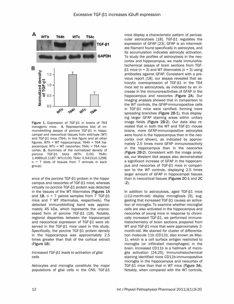
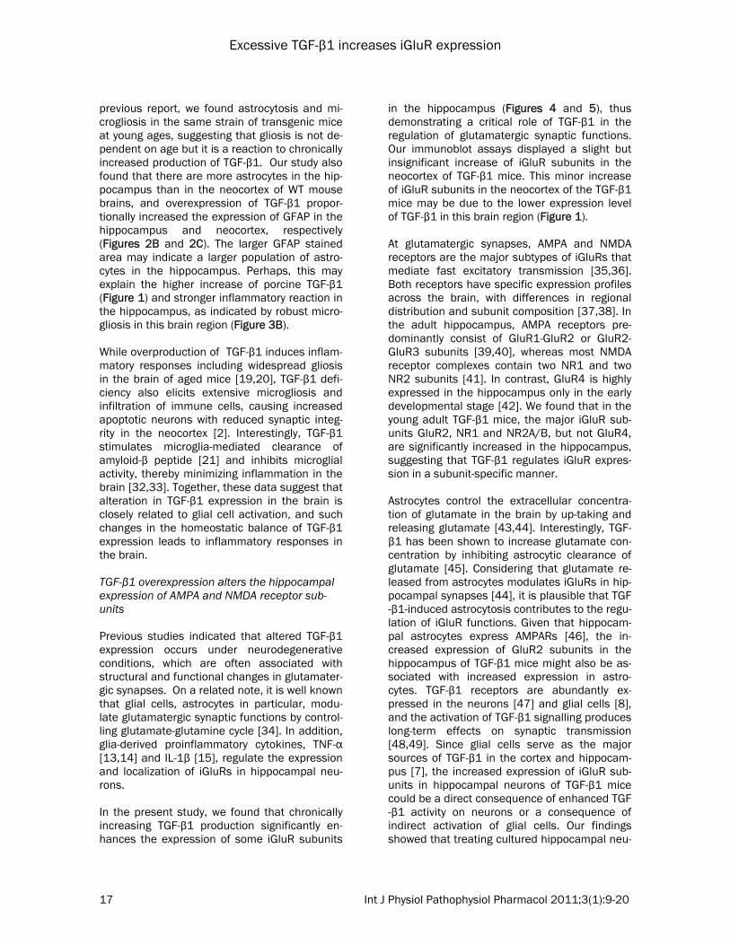
ence of the porcine TGF-β1 protein in the hippo-campus and neocortex of TGF-β1 mice, whereas virtually no porcine TGF-β1 protein was detected in the tissues of the WT littermates (Figures 1A and 1B, n = 7 paired samples from 7 TGF-β1 mice and 7 WT littermates, respectively). The detected immunoblotting band was approxi-mately 45 kDa, which represents the unproc-essed form of porcine TGF-β1 [18]. Notably, regional disparities between the hippocampal and neocortical expression of TGF-β1 were ob-served in the TGF-β1 mice used in this study. Specifically, the porcine TGF-β1 protein density in the hippocampus was approximately 2.5 times greater than that of the cortical extract (Figure 1B). Increased TGF-β1 leads to activation of glial cells Astrocytes and microglia constitute the major populations of glial cells in the CNS. TGF-β1
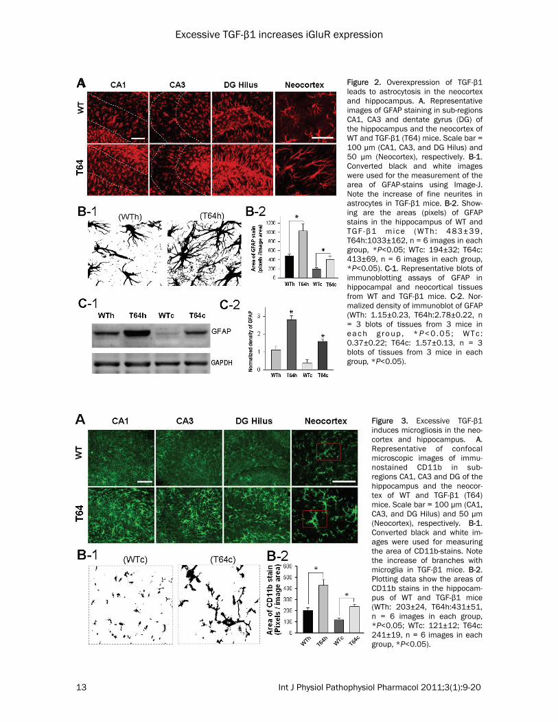
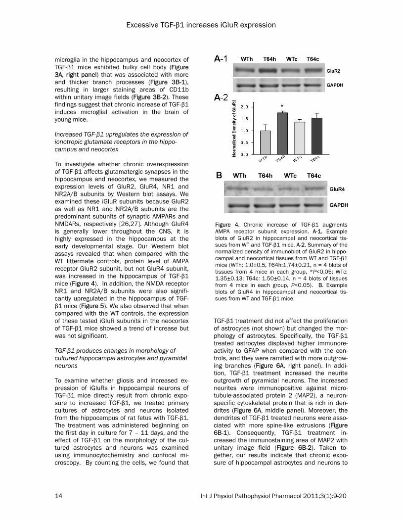
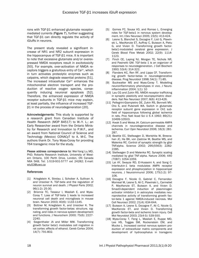
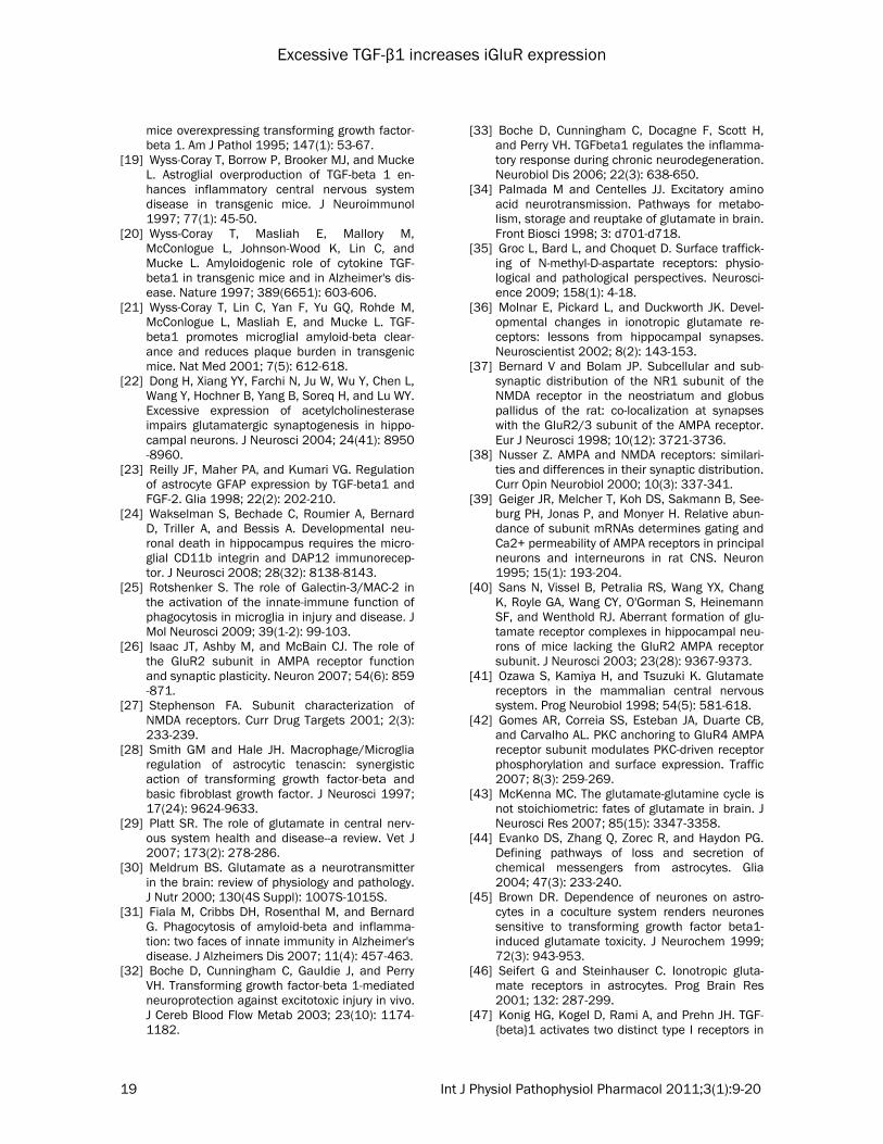
mice display a characteristic pattern of perivas-cular astrocytosis [18]. TGF-β1 regulates the expression of GFAP [23]. GFAP is an intermedi-ate filament found specifically in astrocytes, and its accumulation indicates astrocyte activation. To study the profiles of astrocytosis in the neo-cortex and hippocampus, we made immunohis-tochemical assays of brain sections from TGF-β1 mice (n = 3) and WT littermates (n = 3) using antibodies against GFAP. Consistent with a pre-vious report [18], our assays revealed that as-trocytic overexpression of TGF-β1 in the T64 mice led to astrocytosis, as indicated by an in-crease in the immunoreactivities of GFAP in the hippocampus and neocortex (Figure 2A). Our imaging analysis showed that in comparison to the WT controls, the GFAP-immunopositive cells in TGF-β1 mice were ramified, forming more spreading branches (Figure 2B-1), thus display-ing larger GFAP staining areas within unitary image fields (Figure 2B-2). Our data also re-vealed that in both the WT and TGF-β1 mouse brains, more GFAP-immunopositive astrocytes were found in the hippocampus than in the neo-cortex (not shown), as indicated by approxi-mately 2.5 times more GFAP immunoreactivity in the hippocampus than in the neocortex (Figure 2B-2). Consistent with the image analy-sis, our Western blot assays also demonstrated a significant increase of GFAP in the hippocam-pus and neocortex of TGF-β1 mice in compari-son to the WT controls, displaying 2.5 times larger amount of GFAP in hippocampal tissues than in neocortical tissues (Figures 2C-1 and 2C-2). In addition to astrocytosis, aged TGF-β1 mice (≥12-month-old) display microgliosis [9], sug-gesting that increased TGF-β1 causes an activa-tion of microglia. To examine whether microglial cells are also activated in the hippocampus and neocortex of young mice in response to chroni-cally increased TGF-β1, we performed immuno-histochemistry of brain sections prepared from WT and TGF-β1 mice that were approximately 2-month-old. We stained for cluster of differentia-tion molecule 11b (CD11b, also known as Mac-1), which is a cell surface antigen restricted to microglia (or infiltrated macrophages) in the brain. Increased CD11b is a hallmark of micro-glia activation [24,25]. Immunohistochemical staining identified more CD11b-immunopositive microglia in the hippocampus and neocortex of TGF-β1 mice than that in WT mice (Figure 3A). Notably, when compared with the WT controls,
Figure 1. Expression of TGF-β1 in brains of T64 transgenic mice. A. Representative blot of im-munoblotting assays of porcine TGF-β1 in hippo-campal and neocortical tissues from wild-type (WT) and TGF-β1 mice (T64). In this figure and all other figures, WTh = WT hippocampus; T64h = T64 hip-pocampus; WTc = WT neocortex; T64c = T64 neo-cortex. B. Summary of the normalized density of porcine TGF-β1 blots (WTh: 0.00; T64h: 1.4989±0.1187; WTc:0.00; T64c: 0.5415±0.1298; n = 7 blots of tissues from 7 animals in each group).
Excessive TGF-β1 increases iGluR expression
13 Int J Physiol Pathophysiol Pharmacol 2011;3(1):9-20
Figure 2. Overexpression of TGF-β1 leads to astrocytosis in the neocortex and hippocampus. A. Representative images of GFAP staining in sub-regions CA1, CA3 and dentate gyrus (DG) of the hippocampus and the neocortex of WT and TGF-β1 (T64) mice. Scale bar = 100 µm (CA1, CA3, and DG Hilus) and 50 µm (Neocortex), respectively. B-1. Converted black and white images were used for the measurement of the area of GFAP-stains using Image-J. Note the increase of fine neurites in astrocytes in TGF-β1 mice. B-2. Show-ing are the areas (pixels) of GFAP stains in the hippocampus of WT and TGF-β1 mice (WTh: 483±39, T64h:1033±162, n = 6 images in each group, *P<0.05; WTc: 194±32; T64c: 413±69, n = 6 images in each group, *P<0.05). C-1. Representative blots of immunoblotting assays of GFAP in hippocampal and neocortical tissues from WT and TGF-β1 mice. C-2. Nor-malized density of immunoblot of GFAP (WTh: 1.15±0.23, T64h:2.78±0.22, n = 3 blots of tissues from 3 mice in e a c h g r o u p , * P< 0 . 0 5 ; W T c : 0.37±0.22; T64c: 1.57±0.13, n = 3 blots of tissues from 3 mice in each group, *P<0.05).
Figure 3. Excessive TGF-β1 induces microgliosis in the neo-cortex and hippocampus. A. Representative of confocal microscopic images of immu-nostained CD11b in sub-regions CA1, CA3 and DG of the hippocampus and the neocor-tex of WT and TGF-β1 (T64) mice. Scale bar = 100 µm (CA1, CA3, and DG Hilus) and 50 µm (Neocortex), respectively. B-1. Converted black and white im-ages were used for measuring the area of CD11b-stains. Note the increase of branches with microglia in TGF-β1 mice. B-2. Plotting data show the areas of CD11b stains in the hippocam-pus of WT and TGF-β1 mice (WTh: 203±24, T64h:431±51, n = 6 images in each group, *P<0.05; WTc: 121±12; T64c: 241±19, n = 6 images in each group, *P<0.05).
Excessive TGF-β1 increases iGluR expression
14 Int J Physiol Pathophysiol Pharmacol 2011;3(1):9-20
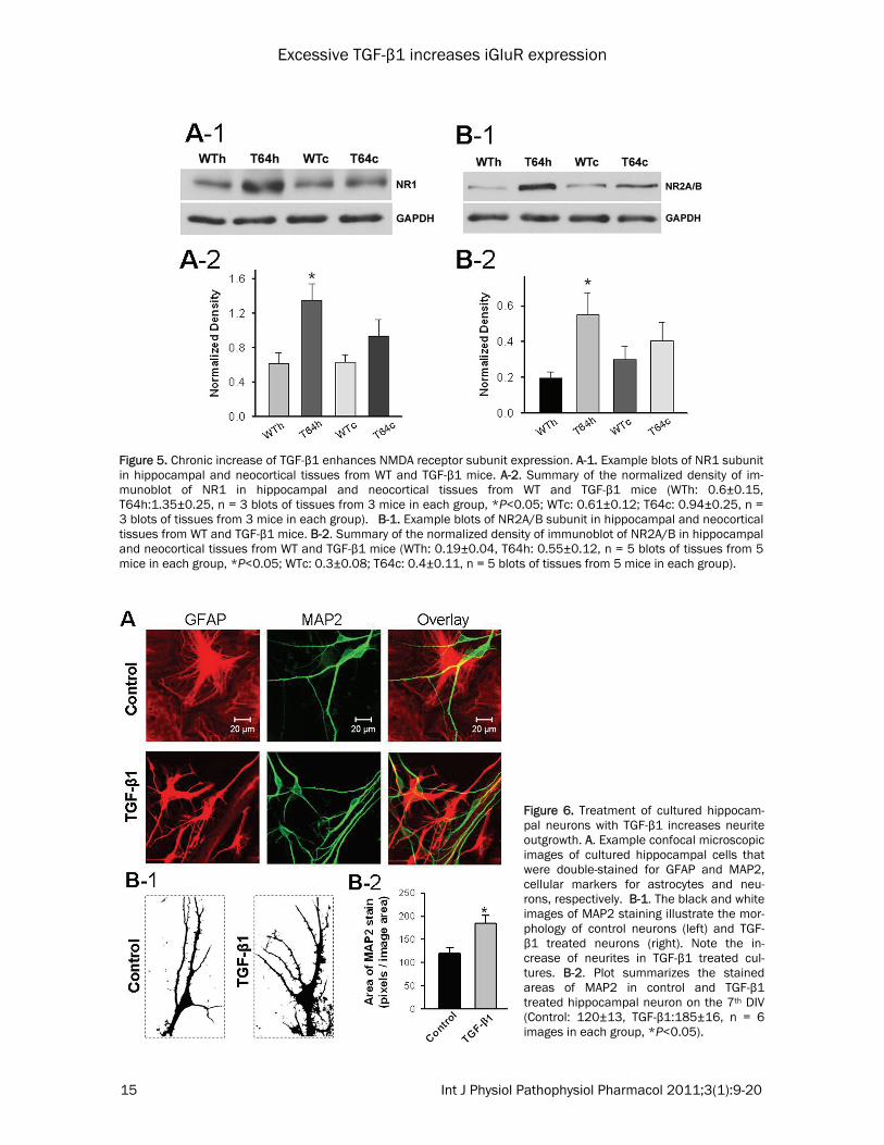
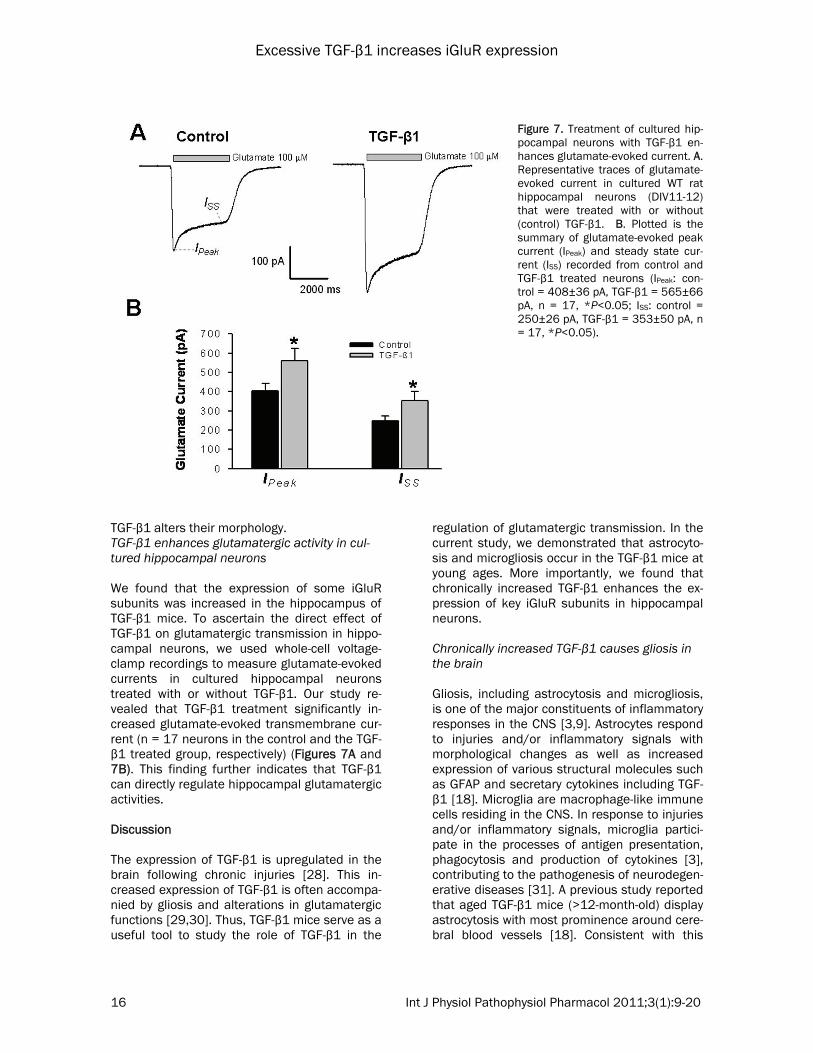
microglia in the hippocampus and neocortex of TGF-β1 mice exhibited bulky cell body (Figure 3A, right panel) that was associated with more and thicker branch processes (Figure 3B-1), resulting in larger staining areas of CD11b within unitary image fields (Figure 3B-2). These findings suggest that chronic increase of TGF-β1 induces microglial activation in the brain of young mice. Increased TGF-β1 upregulates the expression of ionotropic glutamate receptors in the hippo-campus and neocortex To investigate whether chronic overexpression of TGF-β1 affects glutamatergic synapses in the hippocampus and neocortex, we measured the expression levels of GluR2, GluR4, NR1 and NR2A/B subunits by Western blot assays. We examined these iGluR subunits because GluR2 as well as NR1 and NR2A/B subunits are the predominant subunits of synaptic AMPARs and NMDARs, respectively [26,27]. Although GluR4 is generally lower throughout the CNS, it is highly expressed in the hippocampus at the early developmental stage. Our Western blot assays revealed that when compared with the WT littermate controls, protein level of AMPA receptor GluR2 subunit, but not GluR4 subunit, was increased in the hippocampus of TGF-β1 mice (Figure 4). In addition, the NMDA receptor NR1 and NR2A/B subunits were also signifi-cantly upregulated in the hippocampus of TGF-β1 mice (Figure 5). We also observed that when compared with the WT controls, the expression of these tested iGluR subunits in the neocortex of TGF-β1 mice showed a trend of increase but was not significant. TGF-β1 produces changes in morphology of cultured hippocampal astrocytes and pyramidal neurons To examine whether gliosis and increased ex-pression of iGluRs in hippocampal neurons of TGF-β1 mice directly result from chronic expo-sure to increased TGF-β1, we treated primary cultures of astrocytes and neurons isolated from the hippocampus of rat fetus with TGF-β1. The treatment was administered beginning on the first day in culture for 7 – 11 days, and the effect of TGF-β1 on the morphology of the cul-tured astrocytes and neurons was examined using immunocytochemistry and confocal mi-croscopy. By counting the cells, we found that
TGF-β1 treatment did not affect the proliferation of astrocytes (not shown) but changed the mor-phology of astrocytes. Specifically, the TGF-β1 treated astrocytes displayed higher immunore-activity to GFAP when compared with the con-trols, and they were ramified with more outgrow-ing branches (Figure 6A, right panel). In addi-tion, TGF-β1 treatment increased the neurite outgrowth of pyramidal neurons. The increased neurites were immunopositive against micro-tubule-associated protein 2 (MAP2), a neuron-specific cytoskeletal protein that is rich in den-drites (Figure 6A, middle panel). Moreover, the dendrites of TGF-β1 treated neurons were asso-ciated with more spine-like extrusions (Figure 6B-1). Consequently, TGF-β1 treatment in-creased the immunostaining area of MAP2 with unitary image field (Figure 6B-2). Taken to-gether, our results indicate that chronic expo-sure of hippocampal astrocytes and neurons to
Figure 4. Chronic increase of TGF-β1 augments AMPA receptor subunit expression. A-1. Example blots of GluR2 in hippocampal and neocortical tis-sues from WT and TGF-β1 mice. A-2. Summary of the normalized density of immunoblot of GluR2 in hippo-campal and neocortical tissues from WT and TGF-β1 mice (WTh: 1.0±0.5, T64h:1.74±0.21, n = 4 blots of tissues from 4 mice in each group, *P<0.05; WTc: 1.35±0.13; T64c: 1.50±0.14, n = 4 blots of tissues from 4 mice in each group, P<0.05). B. Example blots of GluR4 in hippocampal and neocortical tis-sues from WT and TGF-β1 mice.
Excessive TGF-β1 increases iGluR expression
15 Int J Physiol Pathophysiol Pharmacol 2011;3(1):9-20
Figure 5. Chronic increase of TGF-β1 enhances NMDA receptor subunit expression. A-1. Example blots of NR1 subunit in hippocampal and neocortical tissues from WT and TGF-β1 mice. A-2. Summary of the normalized density of im-munoblot of NR1 in hippocampal and neocortical tissues from WT and TGF-β1 mice (WTh: 0.6±0.15, T64h:1.35±0.25, n = 3 blots of tissues from 3 mice in each group, *P<0.05; WTc: 0.61±0.12; T64c: 0.94±0.25, n = 3 blots of tissues from 3 mice in each group). B-1. Example blots of NR2A/B subunit in hippocampal and neocortical tissues from WT and TGF-β1 mice. B-2. Summary of the normalized density of immunoblot of NR2A/B in hippocampal and neocortical tissues from WT and TGF-β1 mice (WTh: 0.19±0.04, T64h: 0.55±0.12, n = 5 blots of tissues from 5 mice in each group, *P<0.05; WTc: 0.3±0.08; T64c: 0.4±0.11, n = 5 blots of tissues from 5 mice in each group).
Figure 6. Treatment of cultured hippocam-pal neurons with TGF-β1 increases neurite outgrowth. A. Example confocal microscopic images of cultured hippocampal cells that were double-stained for GFAP and MAP2, cellular markers for astrocytes and neu-rons, respectively. B-1. The black and white images of MAP2 staining illustrate the mor-phology of control neurons (left) and TGF-β1 treated neurons (right). Note the in-crease of neurites in TGF-β1 treated cul-tures. B-2. Plot summarizes the stained areas of MAP2 in control and TGF-β1 treated hippocampal neuron on the 7th DIV (Control: 120±13, TGF-β1:185±16, n = 6 images in each group, *P<0.05).
Excessive TGF-β1 increases iGluR expression
16 Int J Physiol Pathophysiol Pharmacol 2011;3(1):9-20
TGF-β1 alters their morphology. TGF-β1 enhances glutamatergic activity in cul-tured hippocampal neurons We found that the expression of some iGluR subunits was increased in the hippocampus of TGF-β1 mice. To ascertain the direct effect of TGF-β1 on glutamatergic transmission in hippo-campal neurons, we used whole-cell voltage-clamp recordings to measure glutamate-evoked currents in cultured hippocampal neurons treated with or without TGF-β1. Our study re-vealed that TGF-β1 treatment significantly in-creased glutamate-evoked transmembrane cur-rent (n = 17 neurons in the control and the TGF-β1 treated group, respectively) (Figures 7A and 7B). This finding further indicates that TGF-β1 can directly regulate hippocampal glutamatergic activities. Discussion The expression of TGF-β1 is upregulated in the brain following chronic injuries [28]. This in-creased expression of TGF-β1 is often accompa-nied by gliosis and alterations in glutamatergic functions [29,30]. Thus, TGF-β1 mice serve as a useful tool to study the role of TGF-β1 in the
regulation of glutamatergic transmission. In the current study, we demonstrated that astrocyto-sis and microgliosis occur in the TGF-β1 mice at young ages. More importantly, we found that chronically increased TGF-β1 enhances the ex-pression of key iGluR subunits in hippocampal neurons. Chronically increased TGF-β1 causes gliosis in the brain Gliosis, including astrocytosis and microgliosis, is one of the major constituents of inflammatory responses in the CNS [3,9]. Astrocytes respond to injuries and/or inflammatory signals with morphological changes as well as increased expression of various structural molecules such as GFAP and secretary cytokines including TGF-β1 [18]. Microglia are macrophage-like immune cells residing in the CNS. In response to injuries and/or inflammatory signals, microglia partici-pate in the processes of antigen presentation, phagocytosis and production of cytokines [3], contributing to the pathogenesis of neurodegen-erative diseases [31]. A previous study reported that aged TGF-β1 mice (>12-month-old) display astrocytosis with most prominence around cere-bral blood vessels [18]. Consistent with this
Figure 7. Treatment of cultured hip-pocampal neurons with TGF-β1 en-hances glutamate-evoked current. A. Representative traces of glutamate-evoked current in cultured WT rat hippocampal neurons (DIV11-12) that were treated with or without (control) TGF-β1. B. Plotted is the summary of glutamate-evoked peak current (IPeak) and steady state cur-rent (ISS) recorded from control and TGF-β1 treated neurons (IPeak: con-trol = 408±36 pA, TGF-β1 = 565±66 pA, n = 17, *P<0.05; ISS: control = 250±26 pA, TGF-β1 = 353±50 pA, n = 17, *P<0.05).
Excessive TGF-β1 increases iGluR expression
17 Int J Physiol Pathophysiol Pharmacol 2011;3(1):9-20
previous report, we found astrocytosis and mi-crogliosis in the same strain of transgenic mice at young ages, suggesting that gliosis is not de-pendent on age but it is a reaction to chronically increased production of TGF-β1. Our study also found that there are more astrocytes in the hip-pocampus than in the neocortex of WT mouse brains, and overexpression of TGF-β1 propor-tionally increased the expression of GFAP in the hippocampus and neocortex, respectively (Figures 2B and 2C). The larger GFAP stained area may indicate a larger population of astro-cytes in the hippocampus. Perhaps, this may explain the higher increase of porcine TGF-β1 (Figure 1) and stronger inflammatory reaction in the hippocampus, as indicated by robust micro-gliosis in this brain region (Figure 3B). While overproduction of TGF-β1 induces inflam-matory responses including widespread gliosis in the brain of aged mice [19,20], TGF-β1 defi-ciency also elicits extensive microgliosis and infiltration of immune cells, causing increased apoptotic neurons with reduced synaptic integ-rity in the neocortex [2]. Interestingly, TGF-β1 stimulates microglia-mediated clearance of amyloid-β peptide [21] and inhibits microglial activity, thereby minimizing inflammation in the brain [32,33]. Together, these data suggest that alteration in TGF-β1 expression in the brain is closely related to glial cell activation, and such changes in the homeostatic balance of TGF-β1 expression leads to inflammatory responses in the brain. TGF-β1 overexpression alters the hippocampal expression of AMPA and NMDA receptor sub-units Previous studies indicated that altered TGF-β1 expression occurs under neurodegenerative conditions, which are often associated with structural and functional changes in glutamater-gic synapses. On a related note, it is well known that glial cells, astrocytes in particular, modu-late glutamatergic synaptic functions by control-ling glutamate-glutamine cycle [34]. In addition, glia-derived proinflammatory cytokines, TNF-α [13,14] and IL-1β [15], regulate the expression and localization of iGluRs in hippocampal neu-rons. In the present study, we found that chronically increasing TGF-β1 production significantly en-hances the expression of some iGluR subunits
in the hippocampus (Figures 4 and 5), thus demonstrating a critical role of TGF-β1 in the regulation of glutamatergic synaptic functions. Our immunoblot assays displayed a slight but insignificant increase of iGluR subunits in the neocortex of TGF-β1 mice. This minor increase of iGluR subunits in the neocortex of the TGF-β1 mice may be due to the lower expression level of TGF-β1 in this brain region (Figure 1). At glutamatergic synapses, AMPA and NMDA receptors are the major subtypes of iGluRs that mediate fast excitatory transmission [35,36]. Both receptors have specific expression profiles across the brain, with differences in regional distribution and subunit composition [37,38]. In the adult hippocampus, AMPA receptors pre-dominantly consist of GluR1-GluR2 or GluR2-GluR3 subunits [39,40], whereas most NMDA receptor complexes contain two NR1 and two NR2 subunits [41]. In contrast, GluR4 is highly expressed in the hippocampus only in the early developmental stage [42]. We found that in the young adult TGF-β1 mice, the major iGluR sub-units GluR2, NR1 and NR2A/B, but not GluR4, are significantly increased in the hippocampus, suggesting that TGF-β1 regulates iGluR expres-sion in a subunit-specific manner. Astrocytes control the extracellular concentra-tion of glutamate in the brain by up-taking and releasing glutamate [43,44]. Interestingly, TGF-β1 has been shown to increase glutamate con-centration by inhibiting astrocytic clearance of glutamate [45]. Considering that glutamate re-leased from astrocytes modulates iGluRs in hip-pocampal synapses [44], it is plausible that TGF-β1-induced astrocytosis contributes to the regu-lation of iGluR functions. Given that hippocam-pal astrocytes express AMPARs [46], the in-creased expression of GluR2 subunits in the hippocampus of TGF-β1 mice might also be as-sociated with increased expression in astro-cytes. TGF-β1 receptors are abundantly ex-pressed in the neurons [47] and glial cells [8], and the activation of TGF-β1 signalling produces long-term effects on synaptic transmission [48,49]. Since glial cells serve as the major sources of TGF-β1 in the cortex and hippocam-pus [7], the increased expression of iGluR sub-units in hippocampal neurons of TGF-β1 mice could be a direct consequence of enhanced TGF-β1 activity on neurons or a consequence of indirect activation of glial cells. Our findings showed that treating cultured hippocampal neu-
Excessive TGF-β1 increases iGluR expression
18 Int J Physiol Pathophysiol Pharmacol 2011;3(1):9-20
rons with TGF-β1 enhanced glutamate receptor-mediated currents (Figure 7), further suggesting that TGF-β1 can directly regulate the activity of iGluRs in neurons. The present study revealed a significant in-crease of NR1 and NR2 subunit expression in the hippocampus of TGF-β1 mice. It is important to note that excessive glutamate and/or overex-pressed NMDA receptors result in excitotoxicity [50]. For example, over-activation of NMDA re-ceptors triggers a gratuitous entry of Ca2+, which in turn activates proteolytic enzymes such as calpains, which degrade essential proteins [51]. The increased intracellular Ca2+ enhances the mitochondrial electron transport and the pro-duction of reactive oxygen species, conse-quently inducing neuronal apoptosis [52]. Therefore, the enhanced expression of NMDA receptor subunits in TGF-β1 mice may explain, at least partially, the influence of increased TGF-β1 in the process of neurodegeneration [20]. Acknowledgements: This study is supported by a research grant from Canadian Institute of Health Research (MOP 84517) to W.-Y. L.; an Early Researcher award from the Ontario Minis-try for Research and Innovation to P.W.F., and an award from National Council of Science and Technology (Mexico) CONACyT to A. M-C. The authors thank Dr. Tony Wyss-Coray for providing T64 transgenic mice for the study. Please address correspondence to: Wei-Yang Lu, MD, PhD; Robarts Research Institute, University of West-ern Ontario, 100 Perth Drive, London, ON Canada N6A 5K8; Tel: 1-519-931-5777 ext 24282; E-mail: [email protected] References [1] Krieglstein K, Strelau J, Schober A, Sullivan A,
and Unsicker K. TGF-beta and the regulation of neuron survival and death. J Physiol Paris 2002; 96(1-2): 25-30.
[2] Brionne TC, Tesseur I, Masliah E, and Wyss-Coray T. Loss of TGF-beta 1 leads to increased neuronal cell death and microgliosis in mouse brain. Neuron 2003; 40(6): 1133-1145.
[3] Bottner M, Krieglstein K, and Unsicker K. The transforming growth factor-betas: structure, sig-naling, and roles in nervous system development and functions. J Neurochem 2000; 75(6): 2227-2240.
[4] Siegenthaler JA and Miller MW. Transforming growth factor beta1 modulates cell migration in rat cortex: effects of ethanol. Cereb Cortex 2004; 14(7): 791-802.
[5] Gomes FC, Sousa VO, and Romao L. Emerging roles for TGF-beta1 in nervous system develop-ment. Int J Dev Neurosci 2005; 23(5): 413-424.
[6] Lesne S, Blanchet S, Docagne F, Liot G, Plawin-ski L, MacKenzie ET, Auffray C, Buisson A, Pietu G, and Vivien D. Transforming growth factor-beta1-modulated cerebral gene expression. J Cereb Blood Flow Metab 2002; 22(9): 1114-1123.
[7] Finch CE, Laping NJ, Morgan TE, Nichols NR, and Pasinetti GM. TGF-beta 1 is an organizer of responses to neurodegeneration. J Cell Biochem 1993; 53(4): 314-322.
[8] Flanders KC, Ren RF, and Lippa CF. Transform-ing growth factor-betas in neurodegenerative disease. Prog Neurobiol 1998; 54(1): 71-85.
[9] Buckwalter MS and Wyss-Coray T. Modelling neuroinflammatory phenotypes in vivo. J Neuro-inflammation 2004; 1(1): 10
[10] Lau CG and Zukin RS. NMDA receptor trafficking in synaptic plasticity and neuropsychiatric disor-ders. Nat Rev Neurosci 2007; 8(6): 413-426.
[11] Pellegrini-Giampietro DE, Zukin RS, Bennett MV, Cho S, and Pulsinelli WA. Switch in glutamate receptor subunit gene expression in CA1 sub-field of hippocampus following global ischemia in rats. Proc Natl Acad Sci U S A 1992; 89(21): 10499-10503.
[12] Kwak S and Weiss JH. Calcium-permeable AMPA channels in neurodegenerative disease and ischemia. Curr Opin Neurobiol 2006; 16(3): 281-287.
[13] Beattie EC, Stellwagen D, Morishita W, Bresna-han JC, Ha BK, von Zastrow M, Beattie MS, and Malenka RC. Control of synaptic strength by glial TNFalpha. Science 2002; 295(5563): 2282-2285.
[14] Stellwagen D and Malenka RC. Synaptic scaling mediated by glial TNF-alpha. Nature 2006; 440(7087): 1054-1059.
[15] Lai AY, Swayze RD, El-Husseini A, and Song C. Interleukin-1 beta modulates AMPA receptor expression and phosphorylation in hippocampal neurons. J Neuroimmunol 2006; 175(1-2): 97-106.
[16] Docagne F, Nicole O, Gabriel C, Fernandez-Monreal M, Lesne S, Ali C, Plawinski L, Carmeliet P, MacKenzie ET, Buisson A, and Vivien D. Smad3-dependent induction of plasminogen activator inhibitor-1 in astrocytes mediates neu-roprotective activity of transforming growth fac-tor-beta 1 against NMDA-induced necrosis. Mol Cell Neurosci 2002; 21(4): 634-644.
[17] Buisson A, Lesne S, Docagne F, Ali C, Nicole O, MacKenzie ET, and Vivien D. Transforming growth factor-beta and ischemic brain injury. Cell Mol Neurobiol 2003; 23(4-5): 539-550.
[18] Wyss-Coray T, Feng L, Masliah E, Ruppe MD, Lee HS, Toggas SM, Rockenstein EM, and Mucke L. Increased central nervous system pro-duction of extracellular matrix components and development of hydrocephalus in transgenic
Excessive TGF-β1 increases iGluR expression
19 Int J Physiol Pathophysiol Pharmacol 2011;3(1):9-20
mice overexpressing transforming growth factor-beta 1. Am J Pathol 1995; 147(1): 53-67.
[19] Wyss-Coray T, Borrow P, Brooker MJ, and Mucke L. Astroglial overproduction of TGF-beta 1 en-hances inflammatory central nervous system disease in transgenic mice. J Neuroimmunol 1997; 77(1): 45-50.
[20] Wyss-Coray T, Masliah E, Mallory M, McConlogue L, Johnson-Wood K, Lin C, and Mucke L. Amyloidogenic role of cytokine TGF-beta1 in transgenic mice and in Alzheimer's dis-ease. Nature 1997; 389(6651): 603-606.
[21] Wyss-Coray T, Lin C, Yan F, Yu GQ, Rohde M, McConlogue L, Masliah E, and Mucke L. TGF-beta1 promotes microglial amyloid-beta clear-ance and reduces plaque burden in transgenic mice. Nat Med 2001; 7(5): 612-618.
[22] Dong H, Xiang YY, Farchi N, Ju W, Wu Y, Chen L, Wang Y, Hochner B, Yang B, Soreq H, and Lu WY. Excessive expression of acetylcholinesterase impairs glutamatergic synaptogenesis in hippo-campal neurons. J Neurosci 2004; 24(41): 8950-8960.
[23] Reilly JF, Maher PA, and Kumari VG. Regulation of astrocyte GFAP expression by TGF-beta1 and FGF-2. Glia 1998; 22(2): 202-210.
[24] Wakselman S, Bechade C, Roumier A, Bernard D, Triller A, and Bessis A. Developmental neu-ronal death in hippocampus requires the micro-glial CD11b integrin and DAP12 immunorecep-tor. J Neurosci 2008; 28(32): 8138-8143.
[25] Rotshenker S. The role of Galectin-3/MAC-2 in the activation of the innate-immune function of phagocytosis in microglia in injury and disease. J Mol Neurosci 2009; 39(1-2): 99-103.
[26] Isaac JT, Ashby M, and McBain CJ. The role of the GluR2 subunit in AMPA receptor function and synaptic plasticity. Neuron 2007; 54(6): 859-871.
[27] Stephenson FA. Subunit characterization of NMDA receptors. Curr Drug Targets 2001; 2(3): 233-239.
[28] Smith GM and Hale JH. Macrophage/Microglia regulation of astrocytic tenascin: synergistic action of transforming growth factor-beta and basic fibroblast growth factor. J Neurosci 1997; 17(24): 9624-9633.
[29] Platt SR. The role of glutamate in central nerv-ous system health and disease--a review. Vet J 2007; 173(2): 278-286.
[30] Meldrum BS. Glutamate as a neurotransmitter in the brain: review of physiology and pathology. J Nutr 2000; 130(4S Suppl): 1007S-1015S.
[31] Fiala M, Cribbs DH, Rosenthal M, and Bernard G. Phagocytosis of amyloid-beta and inflamma-tion: two faces of innate immunity in Alzheimer's disease. J Alzheimers Dis 2007; 11(4): 457-463.
[32] Boche D, Cunningham C, Gauldie J, and Perry VH. Transforming growth factor-beta 1-mediated neuroprotection against excitotoxic injury in vivo. J Cereb Blood Flow Metab 2003; 23(10): 1174-1182.
[33] Boche D, Cunningham C, Docagne F, Scott H, and Perry VH. TGFbeta1 regulates the inflamma-tory response during chronic neurodegeneration. Neurobiol Dis 2006; 22(3): 638-650.
[34] Palmada M and Centelles JJ. Excitatory amino acid neurotransmission. Pathways for metabo-lism, storage and reuptake of glutamate in brain. Front Biosci 1998; 3: d701-d718.
[35] Groc L, Bard L, and Choquet D. Surface traffick-ing of N-methyl-D-aspartate receptors: physio-logical and pathological perspectives. Neurosci-ence 2009; 158(1): 4-18.
[36] Molnar E, Pickard L, and Duckworth JK. Devel-opmental changes in ionotropic glutamate re-ceptors: lessons from hippocampal synapses. Neuroscientist 2002; 8(2): 143-153.
[37] Bernard V and Bolam JP. Subcellular and sub-synaptic distribution of the NR1 subunit of the NMDA receptor in the neostriatum and globus pallidus of the rat: co-localization at synapses with the GluR2/3 subunit of the AMPA receptor. Eur J Neurosci 1998; 10(12): 3721-3736.
[38] Nusser Z. AMPA and NMDA receptors: similari-ties and differences in their synaptic distribution. Curr Opin Neurobiol 2000; 10(3): 337-341.
[39] Geiger JR, Melcher T, Koh DS, Sakmann B, See-burg PH, Jonas P, and Monyer H. Relative abun-dance of subunit mRNAs determines gating and Ca2+ permeability of AMPA receptors in principal neurons and interneurons in rat CNS. Neuron 1995; 15(1): 193-204.
[40] Sans N, Vissel B, Petralia RS, Wang YX, Chang K, Royle GA, Wang CY, O'Gorman S, Heinemann SF, and Wenthold RJ. Aberrant formation of glu-tamate receptor complexes in hippocampal neu-rons of mice lacking the GluR2 AMPA receptor subunit. J Neurosci 2003; 23(28): 9367-9373.
[41] Ozawa S, Kamiya H, and Tsuzuki K. Glutamate receptors in the mammalian central nervous system. Prog Neurobiol 1998; 54(5): 581-618.
[42] Gomes AR, Correia SS, Esteban JA, Duarte CB, and Carvalho AL. PKC anchoring to GluR4 AMPA receptor subunit modulates PKC-driven receptor phosphorylation and surface expression. Traffic 2007; 8(3): 259-269.
[43] McKenna MC. The glutamate-glutamine cycle is not stoichiometric: fates of glutamate in brain. J Neurosci Res 2007; 85(15): 3347-3358.
[44] Evanko DS, Zhang Q, Zorec R, and Haydon PG. Defining pathways of loss and secretion of chemical messengers from astrocytes. Glia 2004; 47(3): 233-240.
[45] Brown DR. Dependence of neurones on astro-cytes in a coculture system renders neurones sensitive to transforming growth factor beta1-induced glutamate toxicity. J Neurochem 1999; 72(3): 943-953.
[46] Seifert G and Steinhauser C. Ionotropic gluta-mate receptors in astrocytes. Prog Brain Res 2001; 132: 287-299.
[47] Konig HG, Kogel D, Rami A, and Prehn JH. TGF-beta1 activates two distinct type I receptors in
Excessive TGF-β1 increases iGluR expression
20 Int J Physiol Pathophysiol Pharmacol 2011;3(1):9-20
neurons: implications for neuronal NF-kappaB signaling. J Cell Biol 2005; 168(7): 1077-1086.
[48] Chin J, Angers A, Cleary LJ, Eskin A, and Byrne JH. TGF-beta1 in Aplysia: role in long-term changes in the excitability of sensory neurons and distribution of TbetaR-II-like immunoreactiv-ity. Learn Mem 1999; 6(3): 317-330.
[49] Zhang F, Endo S, Cleary LJ, Eskin A, and Byrne JH. Role of transforming growth factor-beta in long-term synaptic facilitation in Aplysia. Science 1997; 275(5304): 1318-1320.
[50] Mishra OP, Fritz KI, and ivoria-Papadopoulos M. NMDA receptor and neonatal hypoxic brain in-jury. Ment Retard Dev Disabil Res Rev 2001; 7(4): 249-253.
[51] Hardingham GE. Coupling of the NMDA receptor to neuroprotective and neurodestructive events. Biochem Soc Trans 2009; 37(Pt 6): 1147-1160.
[52] Schulz JB, Matthews RT, Klockgether T, Dichgans J, and Beal MF. The role of mitochon-drial dysfunction and neuronal nitric oxide in animal models of neurodegenerative diseases. Mol Cell Biochem 1997; 174(1-2): 193-197.


![Original Article Effect of transforming growth factor-β1 ... · pogenesis, trophoblast differentiation, cell mi- gration and inflammation control [13]. Accord- ing to the previous](https://static.fdocuments.net/doc/165x107/60f700ca21111f656f07cf79/original-article-effect-of-transforming-growth-factor-1-pogenesis-trophoblast.jpg)