Original Article Association of transforming growth factor ... · Original Article Association of...
12
Int J Clin Exp Med 2016;9(11):21144-21155 www.ijcem.com /ISSN:1940-5901/IJCEM0034202 Original Article Association of transforming growth factor-β1 gene polymorphisms in pneumoconiosis susceptibility: a meta-analysis Xiaoxin Gu, Qiong Ning, Haiyan Wang, Yan Wang, Yonghong Yu Department of Occupational Disease, Ji’nan Hospital, Ji’nan 250013, Shandong Province, China Received June 21, 2016; Accepted August 19, 2016; Epub November 15, 2016; Published November 30, 2016 Abstract: Pneumoconiosis is an occupational, fibrotic lung disorder caused by the inhalation of dust, often in mines. Several articles have identified the role of transforming growth factor-β1 (TGF-β1) polymorphisms in development of pneumoconiosis, however, the results remain inconclusive. The objective of this meta-analysis was to systemati- cally evaluate the effect of TGF-β1 polymorphisms in pneumoconiosis susceptibility. Eligible case-control studies published between January 2000 and 2016 were searched and retrieved in the electronic databases. The pooled odds ratio (ORs) with its 95% confidence interval (CI) was employed to calculate the effect. A total of 13 articles were screened out, including 2538 pneumoconiosis cases and 2435 controls. Overall, our result found a significant difference in the rate of allele mutation in TGF-β1 -509C/T and +915G/C polymorphisms between pneumoconio- sis cases and controls (T versus C: OR=1.57, 95% CI=1.12-2.20, P=0.008; C vs. G: OR=1.59, 95% CI=1.06-2.39, P=0.03). This significant association was also detected in homologous model, dominant model and recessive effect for TGF-β1 -509C/T variant, AND heterogeneous model and dominant model for TGF-β1 +915G/C variant. TGF-β1 +869T/C polymorphism was not related with pneumoconiosis in any genetic models. Subgroup analysis by types of this disease showed that only TGF-β1 -509C/T polymorphism was significantly associated with coal workers’ pneumoconiosis (CWP) risk, not with silicosis risk. In conclusion, our results suggested that there was a significant association between -590C/T and +915G/A variants and pneumoconiosis risk, especially in CWP patients. Future large-scale studies with more ethnicities are still needed to further evaluate the effect. Keywords: Pneumoconiosis, TGF-β1, polymorphism, meta-analysis Introduction Pneumoconiosis, the parenchymal lung diseas- es, is the most serious occupational disease arising from inhalation and retention of inor- ganic dusts at work [1]. It generally evolves over decades of occupational exposure to min- eral dusts depending on the exposure level of silica [2]. Asbestosis, silicosis and coal workers’ pneumoconiosis (CWP) are the most common types. This disease remains an important pub- lic health issue, and is characterized by the formation of nodular, fibrotic changes to the lung parenchyma [3, 4]. The incidence of pneu- moconiosis is still high, and its prevalence var- ies among different populations [5]: in China, a total of 122333 new cases of pneumoconiosis were reported from 1997 to 2009, accounting for nearly 80% of all occupational diseases [6]; in the United Kingdom General Population, a total of 1070 patients with an incident diag- nosis of any type of pneumoconiosis were identified during the period 1997 to 2008 [7]. According to the Global Burden of Disease Study 2013 (GBD 2013), it was estimated 259700 all ages and both sexes combined deaths in 2013, up from 251200 deaths in 1990 from 188 countries [8]. Moreover, pneu- moconiosis was shown to be associated with increased risk of a wide spectrum of complica- tions, such as peripheral arterial disease [9], cerebrovascular events [10], and chronic obst- ructive pulmonary disease [11]. Diagnosis of pneumoconiosis is mainly based on clinical history and radiological findings, but plain radi- ography has limited role in the diagnosis of pulmonary complications of pneumoconiosis because of overlapping pneumoconiotic infiltra-
Transcript of Original Article Association of transforming growth factor ... · Original Article Association of...
Int J Clin Exp Med 2016;9(11):21144-21155www.ijcem.com /ISSN:1940-5901/IJCEM0034202
Original Article Association of transforming growth factor-β1 gene polymorphisms in pneumoconiosis susceptibility: a meta-analysis
Xiaoxin Gu, Qiong Ning, Haiyan Wang, Yan Wang, Yonghong Yu
Department of Occupational Disease, Ji’nan Hospital, Ji’nan 250013, Shandong Province, China
Received June 21, 2016; Accepted August 19, 2016; Epub November 15, 2016; Published November 30, 2016
Abstract: Pneumoconiosis is an occupational, fibrotic lung disorder caused by the inhalation of dust, often in mines. Several articles have identified the role of transforming growth factor-β1 (TGF-β1) polymorphisms in development of pneumoconiosis, however, the results remain inconclusive. The objective of this meta-analysis was to systemati-cally evaluate the effect of TGF-β1 polymorphisms in pneumoconiosis susceptibility. Eligible case-control studies published between January 2000 and 2016 were searched and retrieved in the electronic databases. The pooled odds ratio (ORs) with its 95% confidence interval (CI) was employed to calculate the effect. A total of 13 articles were screened out, including 2538 pneumoconiosis cases and 2435 controls. Overall, our result found a significant difference in the rate of allele mutation in TGF-β1 -509C/T and +915G/C polymorphisms between pneumoconio-sis cases and controls (T versus C: OR=1.57, 95% CI=1.12-2.20, P=0.008; C vs. G: OR=1.59, 95% CI=1.06-2.39, P=0.03). This significant association was also detected in homologous model, dominant model and recessive effect for TGF-β1 -509C/T variant, AND heterogeneous model and dominant model for TGF-β1 +915G/C variant. TGF-β1 +869T/C polymorphism was not related with pneumoconiosis in any genetic models. Subgroup analysis by types of this disease showed that only TGF-β1 -509C/T polymorphism was significantly associated with coal workers’ pneumoconiosis (CWP) risk, not with silicosis risk. In conclusion, our results suggested that there was a significant association between -590C/T and +915G/A variants and pneumoconiosis risk, especially in CWP patients. Future large-scale studies with more ethnicities are still needed to further evaluate the effect.
Keywords: Pneumoconiosis, TGF-β1, polymorphism, meta-analysis
Introduction
Pneumoconiosis, the parenchymal lung diseas-es, is the most serious occupational disease arising from inhalation and retention of inor-ganic dusts at work [1]. It generally evolves over decades of occupational exposure to min-eral dusts depending on the exposure level of silica [2]. Asbestosis, silicosis and coal workers’ pneumoconiosis (CWP) are the most common types. This disease remains an important pub-lic health issue, and is characterized by the formation of nodular, fibrotic changes to the lung parenchyma [3, 4]. The incidence of pneu-moconiosis is still high, and its prevalence var-ies among different populations [5]: in China, a total of 122333 new cases of pneumoconiosis were reported from 1997 to 2009, accounting for nearly 80% of all occupational diseases [6];
in the United Kingdom General Population, a total of 1070 patients with an incident diag- nosis of any type of pneumoconiosis were identified during the period 1997 to 2008 [7]. According to the Global Burden of Disease Study 2013 (GBD 2013), it was estimated 259700 all ages and both sexes combined deaths in 2013, up from 251200 deaths in 1990 from 188 countries [8]. Moreover, pneu-moconiosis was shown to be associated with increased risk of a wide spectrum of complica-tions, such as peripheral arterial disease [9], cerebrovascular events [10], and chronic obst- ructive pulmonary disease [11]. Diagnosis of pneumoconiosis is mainly based on clinical history and radiological findings, but plain radi-ography has limited role in the diagnosis of pulmonary complications of pneumoconiosis because of overlapping pneumoconiotic infiltra-
The effect of TGF-β1 polymorphisms in pneumoconiosis susceptibility
21145 Int J Clin Exp Med 2016;9(11):21144-21155
tion [12]. There are no specific and effective pharmacological treatments for pneumoco- niosis [13]. Although therapeutic whole lung lavage has been used in patients with acute disease, the prognosis is poor [14]. Therefore, there is an urgent need to identify some vital biomarkers to predict this disease and guide the therapeutic strategies.
Although the pathophysiology of pneumoco- niosis has not been fully understood, it is gen-erally accepted that inflammatory responses may play an essential part in the pathogen- esis of pneumoconiosis [15, 16]. Transforming growth factor-β1 (TGF-β1), one member of the TGF-β family, is located on human chromo- some 19q 13.1-13.3 and consists of 7 exons [17]. It directs key developmental processes and regulates cellular proliferation, survival, differentiation, motility, adhesion and migra-tion [18, 19]. TGF-β1 not only plays a central important role in wound healing [20], fibrosis [21], and in the negative regulation of inflam-mation [22], but also can act as a suppressor as well as a promoter of tumorigenesis [23, 24]. Increased TGF-β1 synthesis is seen in the majority of human diseases in which fibrosis is a dominant part of pathology. High serum level of TGF-β1 was shown to be associated with the progression of pneumoconiosis [25]. Qu et al. found that the expression level of TGF-β1 in serum maybe related to the occurrence and development of pneumoconiosis [26]. Feng et al. showed that serum expression of TGF-β1 correlates with dust-exposure, and abnormal expression can be one of the early diagnostic indexes for pneumoconiosis [27]. Yuan et al. suggested that serum TGF-β1 levels in CWP may be related to the severity degree of CWP [28]. However, the circulating concentration of TGF-β1 is predominantly under genetic control [29]. There were three known single nucleotide polymorphisms (SNP) in the hig- hly polymorphic human TGF-β1 gene: -509C/T (rs1800469), +869T/C (rs1800470), and +915G/C (rs1800471). Studies have found the correlative relationship between the con-centration of TGF-β1 in serum and its gene -509 site polymorphism [30].
Several studies have identified the role of TGF-β1 polymorphisms in pneumoconiosis susc- eptibility, but the results remain inconclusive. For example, Fan et al. found that there was no significant difference for frequency of TGF-
β1 +869T/C genotypes and alleles between pneumoconiosis patients and controls [31]; while Qian et al. suggested that TGF-β1 +869- T/C polymorphism might contribute to susce- ptibility of CWP [32]. Therefore, we conducted this meta-analysis to summarize and clarify all eligible studies to obtain a relatively reliable result of the genetic risk of TGF-β1 genetic polymorphisms for pneumoconiosis.
Materials and methods
Search strategy
We conducted a comprehensive literature sea- rch in the online databases of Medline, Web of Science, Embase, PubMed, CNKI (China National Knowledge Internet) and Wanfang to retrieve eligible studies published between January 2000 and 2016. The MeSH terms were: “pneumoconiosis or coal workers’ pneu-moconiosis or silicosis or asbestosis”, “trans-forming growth factor-β1 or TGF-β1”, and “poly-morphism or mutation or variant” as well as their combinations. We manually searched the references of retrieved articles to obtain more potential data. Articles were only restricted in English and Chinese languages. When the same authors or laboratories reported the same issue on the same populations, only the recent full-text article was included.
Inclusion and exclusion criteria
The retrieved articles must meet the following criteria: 1) case-control studies that focused on the association between TGF-β1 polymor-phisms and pneumoconiosis risk; 2) the diag-nosis of pneumoconiosis patients should be based on the 1980 International Labor Office Classification of Pneumoconioses in the judg-ment of opacity profusion [33]; 3) the controls should be age-, race-, dust exposure period- and job type-matched participants, or healthy subjects without any exposure to carcinogenic or fibrogenic agents at the work place; 4) the results were expressed as odds ratio (ORs) with its 95% confidence interval (CI), and 5) the frequencies of alleles and genotypes for a certain polymorphism in each included article were available to extract.
The exclusion criteria were: 1) without con- trol group; 2) with duplicate data; 3) data not available; and 4) review reports or conference papers.
The effect of TGF-β1 polymorphisms in pneumoconiosis susceptibility
21146 Int J Clin Exp Med 2016;9(11):21144-21155
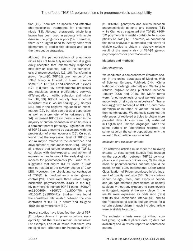
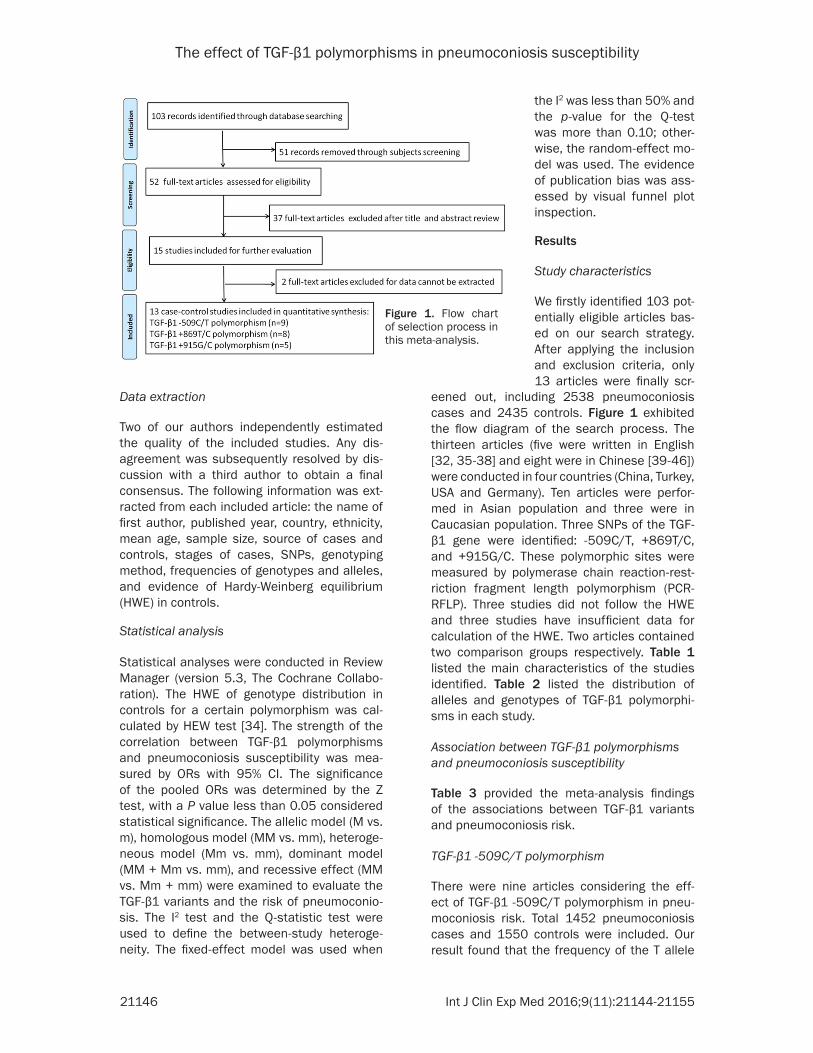
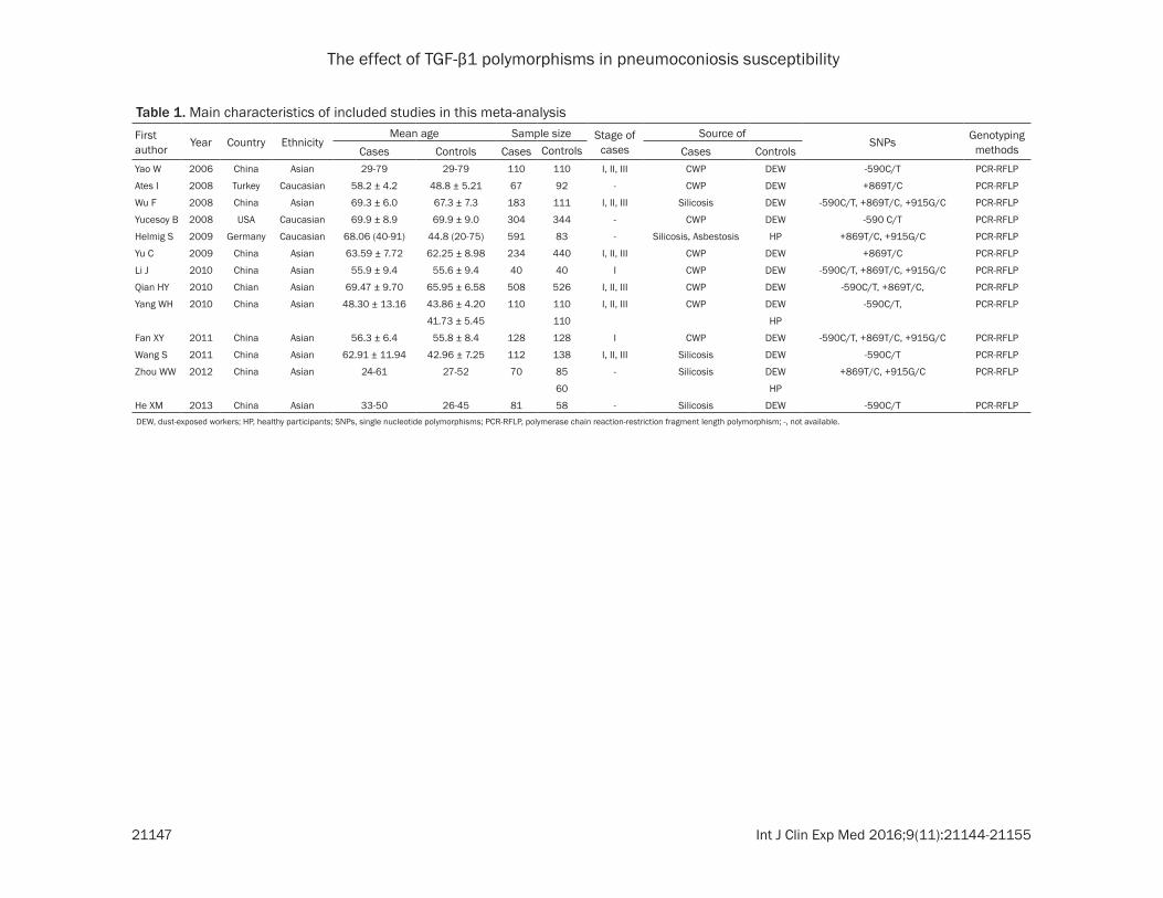
eened out, including 2538 pneumoconiosis cases and 2435 controls. Figure 1 exhibited the flow diagram of the search process. The thirteen articles (five were written in English [32, 35-38] and eight were in Chinese [39-46]) were conducted in four countries (China, Turkey, USA and Germany). Ten articles were perfor- med in Asian population and three were in Caucasian population. Three SNPs of the TGF-β1 gene were identified: -509C/T, +869T/C, and +915G/C. These polymorphic sites were measured by polymerase chain reaction-rest- riction fragment length polymorphism (PCR-RFLP). Three studies did not follow the HWE and three studies have insufficient data for calculation of the HWE. Two articles contained two comparison groups respectively. Table 1 listed the main characteristics of the studies identified. Table 2 listed the distribution of alleles and genotypes of TGF-β1 polymorphi- sms in each study.
Association between TGF-β1 polymorphisms and pneumoconiosis susceptibility
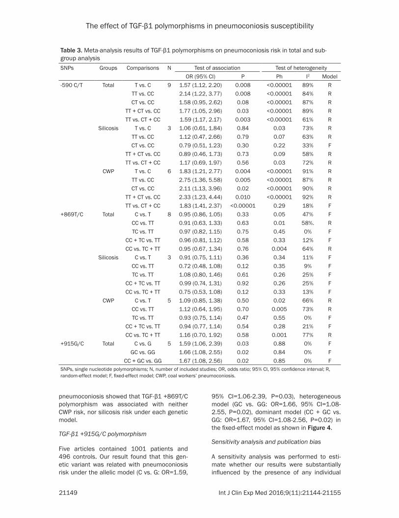
Table 3 provided the meta-analysis findings of the associations between TGF-β1 variants and pneumoconiosis risk.
TGF-β1 -509C/T polymorphism
There were nine articles considering the eff- ect of TGF-β1 -509C/T polymorphism in pneu-moconiosis risk. Total 1452 pneumoconiosis cases and 1550 controls were included. Our result found that the frequency of the T allele
Data extraction
Two of our authors independently estimated the quality of the included studies. Any dis-agreement was subsequently resolved by dis-cussion with a third author to obtain a final consensus. The following information was ext- racted from each included article: the name of first author, published year, country, ethnicity, mean age, sample size, source of cases and controls, stages of cases, SNPs, genotyping method, frequencies of genotypes and alleles, and evidence of Hardy-Weinberg equilibrium (HWE) in controls.
Statistical analysis
Statistical analyses were conducted in Review Manager (version 5.3, The Cochrane Collabo- ration). The HWE of genotype distribution in controls for a certain polymorphism was cal- culated by HEW test [34]. The strength of the correlation between TGF-β1 polymorphisms and pneumoconiosis susceptibility was mea-sured by ORs with 95% CI. The significance of the pooled ORs was determined by the Z test, with a P value less than 0.05 considered statistical significance. The allelic model (M vs. m), homologous model (MM vs. mm), heteroge-neous model (Mm vs. mm), dominant model (MM + Mm vs. mm), and recessive effect (MM vs. Mm + mm) were examined to evaluate the TGF-β1 variants and the risk of pneumoconio-sis. The I2 test and the Q-statistic test were used to define the between-study heteroge- neity. The fixed-effect model was used when
Figure 1. Flow chart of selection process in this meta-analysis.
the I2 was less than 50% and the p-value for the Q-test was more than 0.10; other-wise, the random-effect mo- del was used. The evidence of publication bias was ass- essed by visual funnel plot inspection.
Results
Study characteristics
We firstly identified 103 pot- entially eligible articles bas- ed on our search strategy. After applying the inclusion and exclusion criteria, only 13 articles were finally scr-
The effect of TGF-β1 polymorphisms in pneumoconiosis susceptibility
21147 Int J Clin Exp Med 2016;9(11):21144-21155
Table 1. Main characteristics of included studies in this meta-analysisFirst author Year Country Ethnicity
Mean age Sample size Stage of cases
Source ofSNPs Genotyping
methodsCases Controls Cases Controls Cases ControlsYao W 2006 China Asian 29-79 29-79 110 110 I, II, III CWP DEW -590C/T PCR-RFLP
Ates I 2008 Turkey Caucasian 58.2 ± 4.2 48.8 ± 5.21 67 92 - CWP DEW +869T/C PCR-RFLP
Wu F 2008 China Asian 69.3 ± 6.0 67.3 ± 7.3 183 111 I, II, III Silicosis DEW -590C/T, +869T/C, +915G/C PCR-RFLP
Yucesoy B 2008 USA Caucasian 69.9 ± 8.9 69.9 ± 9.0 304 344 - CWP DEW -590 C/T PCR-RFLP
Helmig S 2009 Germany Caucasian 68.06 (40-91) 44.8 (20-75) 591 83 - Silicosis, Asbestosis HP +869T/C, +915G/C PCR-RFLP
Yu C 2009 China Asian 63.59 ± 7.72 62.25 ± 8.98 234 440 I, II, III CWP DEW +869T/C PCR-RFLP
Li J 2010 China Asian 55.9 ± 9.4 55.6 ± 9.4 40 40 I CWP DEW -590C/T, +869T/C, +915G/C PCR-RFLP
Qian HY 2010 Chian Asian 69.47 ± 9.70 65.95 ± 6.58 508 526 I, II, III CWP DEW -590C/T, +869T/C, PCR-RFLP
Yang WH 2010 China Asian 48.30 ± 13.16 43.86 ± 4.20 110 110 I, II, III CWP DEW -590C/T, PCR-RFLP
41.73 ± 5.45 110 HP
Fan XY 2011 China Asian 56.3 ± 6.4 55.8 ± 8.4 128 128 I CWP DEW -590C/T, +869T/C, +915G/C PCR-RFLP
Wang S 2011 China Asian 62.91 ± 11.94 42.96 ± 7.25 112 138 I, II, III Silicosis DEW -590C/T PCR-RFLP
Zhou WW 2012 China Asian 24-61 27-52 70 85 - Silicosis DEW +869T/C, +915G/C PCR-RFLP
60 HP
He XM 2013 China Asian 33-50 26-45 81 58 - Silicosis DEW -590C/T PCR-RFLPDEW, dust-exposed workers; HP, healthy participants; SNPs, single nucleotide polymorphisms; PCR-RFLP, polymerase chain reaction-restriction fragment length polymorphism; -, not available.
The effect of TGF-β1 polymorphisms in pneumoconiosis susceptibility
21148 Int J Clin Exp Med 2016;9(11):21144-21155
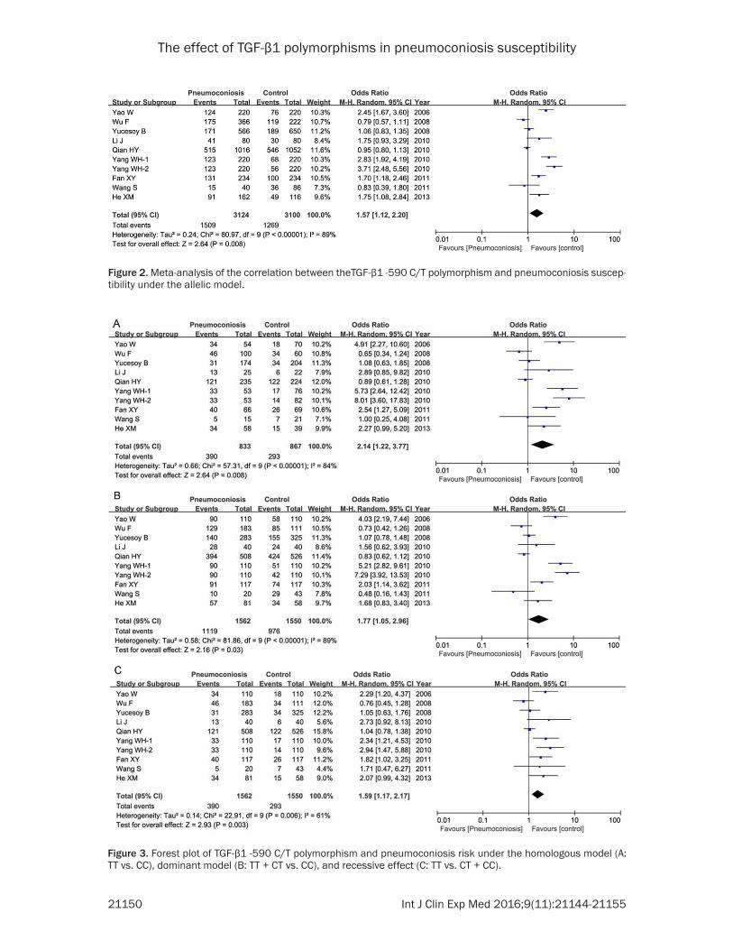
was higher in patients than that in controls (47.7% versus 40.9%), and the statistical analy-sis showed a significant difference in the rate of allele mutation between pneumoconiosis cases and controls (T versus C: OR=1.57, 95% CI=1.12-2.20, P=0.008) in the random-effect model as shown in Figure 2. This significant relationship was observed in homologous mo- del (TT vs. CC: OR=2.14, 95% CI=1.22-3.77, P=0.008), dominant model (TT + CT vs. CC: OR=1.77, 95% CI=1.05-2.96, P=0.03), and recessive effect (TT vs. CT + CC: OR=1.59, 95% CI=1.17-2.17, P=0.003) as well as shown in Figure 3. While no association was detected between TGF-β1 -509C/T polymorphism and pneumoconiosis risk under the heterogeneous model (CT vs. CC: OR=1.58, 95% CI=0.95-2.62, P=0.08). Subgroup analysis by types of this
disease showed that TGF-β1 -509C/T poly- morphism was significantly associated with CWP risk, not with silicosis risk (Table 3) under each genetic model.
TGF-β1 +869T/C polymorphism
Eight articles included 1810 cases and 1554 controls. Our result did not detect a signifi- cant correlation between TGF-β1 +869T/C poly-morphism and pneumoconiosis susceptibi- lity under any comparison models (C vs. T: OR=0.95, 95% CI=0.86-1.05, P=0.33; CC vs. TT: OR=0.91, 95% CI=0.63-1.33, P=0.63; TC vs. TT: OR=0.97, 95% CI=0.82-1.15, P=0.75; CC + TC vs. TT: OR=0.96, 95% CI=0.81-1.12, P=0.58; CC vs. TC + TT: OR=0.95, 95% CI=0.67-1.34, P=0.76). Subgroup analysis by types of
Table 2. Distribution information of alleles and genotypes of TGF-β1 polymorphisms in each included study in this meta-analysisFirst author Cases Controls-590 C/T CC CT TT C T CC CT TT C T HWE Yao W 20 56 34 96 124 52 40 18 144 76 0.121 Wu F 54 83 46 191 175 26 51 34 103 119 0.724 Yucesoy B 143 109 31 395 171 170 121 34 461 189 0.215 Li J 12 15 13 39 41 16 18 6 50 30 0.968 Qian HY 114 273 121 501 515 102 302 122 506 546 0.003 Yang WH-1 20 57 33 97 123 59 34 17 152 68 0.015 Yang WH-2 20 57 33 97 123 68 28 14 164 56 0.003 Fan XY 26 51 40 103 131 43 48 26 134 100 0.216 Wang S 10 5 5 25 15 14 22 7 50 36 0.945 He XM 24 23 34 71 91 24 19 15 67 49 0.051+869T/C TT TC CC T C TT TC CC T C Ates I 17 26 24 60 74 22 50 20 94 90 0.703 Wu F 52 83 48 187 179 24 56 31 104 118 0.991 Helmig S 197 302 92 696 486 32 36 15 100 66 0.690 Yu C 74 101 59 249 219 129 206 105 464 416 0.443 Li J 9 18 13 36 44 14 17 9 45 35 0.689 Qian HY 123 338 47 584 432 109 332 85 550 502 0.000 Fan XY 28 50 39 106 128 37 51 29 125 109 0.406 Zhou WW-1 34 32 4 100 40 36 34 15 106 64 0.394 Zhou WW-2 34 32 4 100 40 34 21 5 89 31 0.798+915G/C GG GC CC G C GG GC CC G C Wu F 181 2 0 364 2 111 0 0 222 0 - Helmig S 497 93 1 1087 95 73 10 0 156 10 0.843 Li J 32 8 0 72 8 34 6 0 74 6 0.876 Fan XY 83 34 0 200 34 97 20 0 214 20 0.599 Zhou WW-1 70 0 0 140 0 85 0 0 170 0 - Zhou WW-2 70 0 0 140 0 60 0 0 120 0 -HWE, Hardy-Weinberg equilibrium in controls; -, not applicable.
The effect of TGF-β1 polymorphisms in pneumoconiosis susceptibility
21149 Int J Clin Exp Med 2016;9(11):21144-21155
Table 3. Meta-analysis results of TGF-β1 polymorphisms on pneumoconiosis risk in total and sub-group analysisSNPs Groups Comparisons N Test of association Test of heterogeneity
OR (95% CI) P Ph I2 Model-590 C/T Total T vs. C 9 1.57 (1.12, 2.20) 0.008 <0.00001 89% R
TT vs. CC 2.14 (1.22, 3.77) 0.008 <0.00001 84% RCT vs. CC 1.58 (0.95, 2.62) 0.08 <0.00001 87% R
TT + CT vs. CC 1.77 (1.05, 2.96) 0.03 <0.00001 89% RTT vs. CT + CC 1.59 (1.17, 2.17) 0.003 <0.00001 61% R
Silicosis T vs. C 3 1.06 (0.61, 1.84) 0.84 0.03 73% RTT vs. CC 1.12 (0.47, 2.66) 0.79 0.07 63% RCT vs. CC 0.79 (0.51, 1.23) 0.30 0.22 33% F
TT + CT vs. CC 0.89 (0.46, 1.73) 0.73 0.09 58% RTT vs. CT + CC 1.17 (0.69, 1.97) 0.56 0.03 72% R
CWP T vs. C 6 1.83 (1.21, 2.77) 0.004 <0.00001 91% RTT vs. CC 2.75 (1.36, 5.58) 0.005 <0.00001 87% RCT vs. CC 2.11 (1.13, 3.96) 0.02 <0.00001 90% R
TT + CT vs. CC 2.33 (1.23, 4.44) 0.010 <0.00001 92% RTT vs. CT + CC 1.83 (1.41, 2.37) <0.00001 0.29 18% F
+869T/C Total C vs. T 8 0.95 (0.86, 1.05) 0.33 0.05 47% FCC vs. TT 0.91 (0.63, 1.33) 0.63 0.01 58%. RTC vs. TT 0.97 (0.82, 1.15) 0.75 0.45 0% F
CC + TC vs. TT 0.96 (0.81, 1.12) 0.58 0.33 12% FCC vs. TC + TT 0.95 (0.67, 1.34) 0.76 0.004 64% R
Silicosis C vs. T 3 0.91 (0.75, 1.11) 0.36 0.34 11% FCC vs. TT 0.72 (0.48, 1.08) 0.12 0.35 9% FTC vs. TT 1.08 (0.80, 1.46) 0.61 0.26 25% F
CC + TC vs. TT 0.99 (0.74, 1.31) 0.92 0.26 25% FCC vs. TC + TT 0.75 (0.53, 1.08) 0.12 0.33 13% F
CWP C vs. T 5 1.09 (0.85, 1.38) 0.50 0.02 66% RCC vs. TT 1.12 (0.64, 1.95) 0.70 0.005 73% RTC vs. TT 0.93 (0.75, 1.14) 0.47 0.55 0% F
CC + TC vs. TT 0.94 (0.77, 1.14) 0.54 0.28 21% FCC vs. TC + TT 1.16 (0.70, 1.92) 0.58 0.001 77% R
+915G/C Total C vs. G 5 1.59 (1.06, 2.39) 0.03 0.88 0% FGC vs. GG 1.66 (1.08, 2.55) 0.02 0.84 0% F
CC + GC vs. GG 1.67 (1.08, 2.56) 0.02 0.85 0% FSNPs, single nucleotide polymorphisms; N, number of included studies; OR, odds ratio; 95% CI, 95% confidence interval; R, random-effect model; F, fixed-effect model; CWP, coal workers’ pneumoconiosis.
pneumoconiosis showed that TGF-β1 +869T/C polymorphism was associated with neither CWP risk, nor silicosis risk under each genetic model.
TGF-β1 +915G/C polymorphism
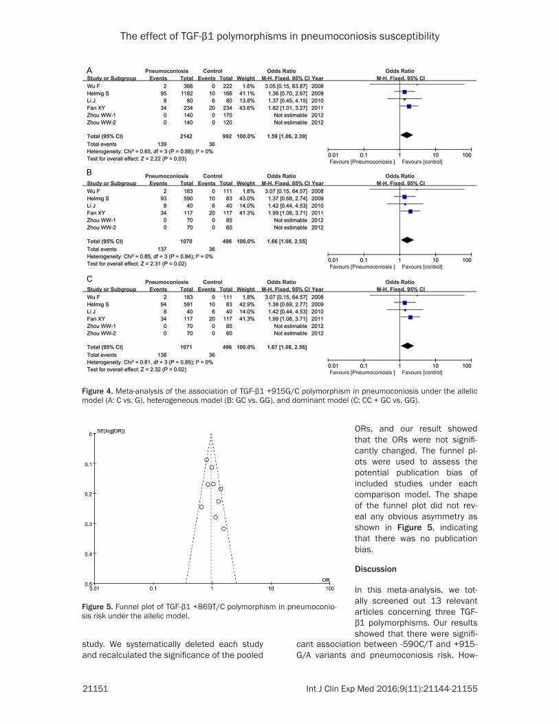
Five articles contained 1001 patients and 496 controls. Our result found that this gen- etic variant was related with pneumoconiosis risk under the allelic model (C vs. G: OR=1.59,
95% CI=1.06-2.39, P=0.03), heterogeneous model (GC vs. GG: OR=1.66, 95% CI=1.08-2.55, P=0.02), dominant model (CC + GC vs. GG: OR=1.67, 95% CI=1.08-2.56, P=0.02) in the fixed-effect model as shown in Figure 4.
Sensitivity analysis and publication bias
A sensitivity analysis was performed to esti-mate whether our results were substantially influenced by the presence of any individual
The effect of TGF-β1 polymorphisms in pneumoconiosis susceptibility
21150 Int J Clin Exp Med 2016;9(11):21144-21155
Figure 2. Meta-analysis of the correlation between theTGF-β1 -590 C/T polymorphism and pneumoconiosis suscep-tibility under the allelic model.
Figure 3. Forest plot of TGF-β1 -590 C/T polymorphism and pneumoconiosis risk under the homologous model (A: TT vs. CC), dominant model (B: TT + CT vs. CC), and recessive effect (C: TT vs. CT + CC).
The effect of TGF-β1 polymorphisms in pneumoconiosis susceptibility
21151 Int J Clin Exp Med 2016;9(11):21144-21155
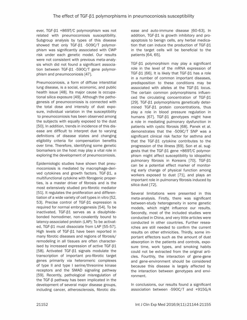
study. We systematically deleted each study and recalculated the significance of the pooled
cant association between -590C/T and +915- G/A variants and pneumoconiosis risk. How-
Figure 4. Meta-analysis of the association of TGF-β1 +915G/C polymorphism in pneumoconiosis under the allelic model (A: C vs. G), heterogeneous model (B: GC vs. GG), and dominant model (C: CC + GC vs. GG).
Figure 5. Funnel plot of TGF-β1 +869T/C polymorphism in pneumoconio-sis risk under the allelic model.
ORs, and our result showed that the ORs were not signifi-cantly changed. The funnel pl- ots were used to assess the potential publication bias of included studies under each comparison model. The shape of the funnel plot did not rev- eal any obvious asymmetry as shown in Figure 5, indicating that there was no publication bias.
Discussion
In this meta-analysis, we tot- ally screened out 13 relevant articles concerning three TGF-β1 polymorphisms. Our results showed that there were signifi-
The effect of TGF-β1 polymorphisms in pneumoconiosis susceptibility
21152 Int J Clin Exp Med 2016;9(11):21144-21155
ever, TGF-β1 +869T/C polymorphism was not related with pneumoconiosis susceptibility. Subgroup analysis by types of this disease showed that only TGF-β1 -509C/T polymor-phism was significantly associated with CWP risk under each genetic model. Our results were not consistent with previous meta-analy-sis which did not found a significant associa-tion between TGF-β1 -590C/T gene polymor-phism and pneumoconiosis [47].
Pneumoconiosis, a form of diffuse interstitial lung disease, is a social, economic, and public health issue [48]. Its major cause is occupa-tional silica exposure [49]. Although the patho-genesis of pneumoconiosis is connected with the total dose and intensity of dust expo- sure, individual variation in the susceptibility to pneumoconiosis has been observed among the subjects with equally exposed to the dust [50]. In addition, trends in incidence of this dis-ease are difficult to interpret due to varying definitions of disease states and changing eligibility criteria for compensation benefits over time. Therefore, identifying some genetic biomarkers on the host may play a vital role in exploring the development of pneumoconiosis.
Epidemiologic studies have shown that pneu-moconiosis is mediated by macrophage-deri- ved cytokines and growth factors. TGF-β1, a multifunctional cytokine with fibrogenic proper-ties, is a master driver of fibrosis and is the most extensively studied pro-fibrotic mediator [51]. It regulates the proliferation and differen-tiation of a wide variety of cell types in vitro [52, 53]. Precise control of TGF-β1 expression is required for normal embryogenesis [54]. To be inactivated, TGF-β1 serves as a disulphide-bonded homodimer, non-covalently bound to latency-associated protein (LAP); To be activat-ed, TGF-β1 must dissociate from LAP [55-57]. High levels of TGF-β1 have been reported in many fibrotic diseases and regions of fibrosis/remodeling in all tissues are often character-ised by increased expression of active TGF-β1 [58]. Activated TGF-β1 signals modulate the transcription of important pro-fibrotic target genes primarily via heteromeric complexes of type II and type I serine/threonine kinase receptors and the SMAD signaling pathway [59]. Recently, pathological misregulation of the TGF-β pathway has been implicated in the development of several major disease groups, including cancer, atherosclerosis, fibrotic dis-
ease and auto-immune disease [60-63]. In addition, TGF-β1 is growth inhibitory and pro-apoptosis to benign cells, any herbal medica-tion that can induce the production of TGF-β1 in the target cells will be beneficial to the patients [64, 65].
TGF-β1 polymorphism may play a significant role in the level of the mRNA expression of TGF-β1 [66]. It is likely that TGF-β1 has a role in a number of common important diseases, predisposition to these conditions may be associated with alleles at the TGF-β1 locus. The certain common polymorphisms influen- ced the circulating concentration of TGF-β1 [29]. TGF-β1 polymorphisms genetically deter-mined TGF-β1 protein concentrations, thus play a role in blood pressure regulation in humans [67]. TGF-β1 genotypes might have a role in mediating pulmonary dysfunction in patients with cystic fibrosis [68]. Panek et al. demonstrates that the -509C/T SNP was a significant clinical risk factor for asthma and that the TGF-β1 cytokine contributes to the progression of the illness [69]. Son et al. sug-gests that the TGF-β1 gene +869T/C polymor-phism might affect susceptibility to idiopathic pulmonary fibrosis in Koreans [70]. TGF-β1 can be a potential effect marker of monitor- ing early change of physical function among workers exposed to dust [71], and plays an important role in pulmonary fibrosis induced by silica dust [72].
Several limitations were presented in this meta-analysis. Firstly, there was significant between-study heterogeneity in some genetic models, which might influence our results. Secondly, most of the included studies were conducted in China, and very little articles were conducted in other countries. Further resea- rches are still needed to confirm the current results on other ethnicities. Thirdly, some im- portant effectors such as the amount of dust absorption in the patients and controls, expo-sure time, work types, and smoking habits could not be extracted from the original arti-cles. Fourthly, the interaction of gene-gene and gene-environment should be considered because this disease is largely affected by the interaction between genotypes and envi- ronment.
In conclusions, our results found a significant association between -590C/T and +915G/A
The effect of TGF-β1 polymorphisms in pneumoconiosis susceptibility
21153 Int J Clin Exp Med 2016;9(11):21144-21155
variants and pneumoconiosis risk, especially in CWP patients. Future well-designed, large-scale studies with more ethnicities are still needed to further evaluate the effects.
Disclosure of conflict of interest
None.
Address correspondence to: Dr. Qiong Ning, Dep- artment of Occupational Disease, Ji’nan Hospital, Room 63-1, Lishan Road, Lixia District, Ji’nan 250013, Shandong Province, China. Tel: +86-0531-58678136; Fax: +86-0531-58678102; E-mail: [email protected]
References
[1] Cullinan P and Reid P. Pneumoconiosis. Prim Care Respir J 2013; 22: 249-252.
[2] Lapp NL and Castranova V. How silicosis and coal workers’ pneumoconiosis develop-A cel-lular assessment. Occup Med 1993; 8: 35-56.
[3] Leslie KO and Wick MR. Practical pulmonary pathology: a diagnostic approach. Elsevier Health Sciences; 2011.
[4] Blanc PD and Seaton A. Pneumoconiosis Redux. Coal Workers’ Pneumoconiosis and Silicosis Are Still a Problem. Am J Respir Crit Care Med 2016; 193: 603-605.
[5] Joy G, Colinet J and Landen D. Coal workers’ pneumoconiosi prevalence disparity between Australia and the United States. Mining Engi-neering 2012; 64.
[6] Zhang M, Wang D, Zheng YD, Du XY and Chen SY. [Analyses on the characteristics and the trends of pneumoconiosis notified between 1997 and 2009, in China]. Zhonghua Lao Dong Wei Sheng Zhi Ye Bing Za Zhi 2013; 31: 321-334.
[7] Amar RK, Jick SS, Rosenberg D, Maher TM and Meier CR. Incidence of the pneumoconioses in the United Kingdom general population be-tween 1997 and 2008. Respiration 2012; 84: 200-206.
[8] Naghavi M, Wang H, Lozano R, Davis A, Liang X, Zhou M, Vollset SE, Ozgoren AA, Abdalla S and Abd-Allah F. Global, regional, and national age-sex specific all-cause and cause-specific mortality for 240 causes of death, 1990-2013: a systematic analysis for the Global Burden of Disease Study 2013. Lancet 2015; 385: 117-171.
[9] Shen CH, Lin TY, Huang WY, Chen HJ and Kao CH. Pneumoconiosis increases the risk of pe-ripheral arterial disease: a nationwide popula-tion-based study. Medicine 2015; 94: e911.
[10] Chuang CS, Ho SC, Lin CL, Lin MC and Kao CH. Risk of Cerebrovascular Events in Pneumo-
coniosis Patients: A Population-based Study, 1996-2011. Medicine 2016; 95: e2944.
[11] Ornek T, Atalay F, Erboy F, Altinsoy B, Tanriverdi H, Uygur F and Tor M. Is pneumoconiosis a fac-tor of severity in acute exacerbation of chronic obstructive pulmonary disease? Clin Ter 2013; 164: e473-477.
[12] Jun JS, Im Jung J, Kim HR, Im Ahn M, Han DH, Ko JM, Park SH, Lee HG, Arakawa H and Koo JW. Complications of pneumoconiosis: Radio-logic overview. Eur J Radiol 2013; 82: 1819-1830.
[13] Karkhanis VS and Joshi J. Pneumoconioses. Indian J Chest Dis Allied Sci 2012; 55: 25-34.
[14] Fishwick D. Pneumoconiosis. Medicine 2008; 36: 258-260.
[15] Yucesoy B, Vallyathan V, Landsittel DP, Sime-onova P and Luster MI. Cytokine polymor-phisms in silicosis and other pneumoconioses. Mol Cell Biochem 2002; 234: 219-224.
[16] Borm P and Schins R. Genotype and pheno-type in susceptibility to coal workers’ pneumo-coniosis. The use of cytokines in perspective. Eur Respir J Suppl 2001; 18: 127s-133s.
[17] Kim SJ, Glick A, Sporn M and Roberts A. Char-acterization of the promoter region of the hu-man transforming growth factor-beta 1 gene. J Biol Chem 1989; 264: 402-408.
[18] Horbelt D, Denkis A and Knaus P. A portrait of Transforming Growth Factor β superfamily sig-nalling: Background matters. Int J Biochem Cell Biol 2012; 44: 469-474.
[19] Massague J. The transforming growth factor-beta family. Annu Rev Cell Biol 1990; 6: 597-641.
[20] Penn JW, Grobbelaar AO and Rolfe KJ. The role of the TGF-β family in wound healing, burns and scarring: a review. Int J Burns Trauma 2012; 2: 18-28.
[21] Hinz B and McCulloch C. Integrin function in heart fibrosis: mechanical strain, TGF-β1 acti-vation and collagen glycation. Extracellular Matrix: Pathobiology and Signalling. DeGruyter Berlin, Germany; 2012. pp. 407-431.
[22] Graciarena M, Roca V, Mathieu P, Depino AM and Pitossi FJ. Differential vulnerability of adult neurogenesis by adult and prenatal inflamma-tion: Role of TGF-β1. Brain Behav Immun 2013; 34: 17-28.
[23] Zhang Y, Li L, Yu J, Zhu D, Zhang Y, Li X, Gu H, Zhang CY and Zen K. Microvesicle-mediated delivery of transforming growth factor β1 siR-NA for the suppression of tumor growth in mice. Biomaterials 2014; 35: 4390-4400.
[24] Su HT, Weng CC, Hsiao PJ, Chen LH, Kuo TL, Chen YW, Kuo KK and Cheng KH. Stem cell marker nestin is critical for TGF-β1-mediated tumor progression in pancreatic cancer. Mol Cancer Res 2013; 11: 768-779.
The effect of TGF-β1 polymorphisms in pneumoconiosis susceptibility
21154 Int J Clin Exp Med 2016;9(11):21144-21155
[25] Lee JS, Shin JH, Lee KM, Hwang JH, Baek JE, Kim JH and Choi BS. Serum Levels of TGF-β1 and MCP-1 as Biomarkers for Progressive Coal Workers’ Pneumoconiosis in Retired Coal Workers: A Three-year Follow-up Study. Ind Health 2014; 52: 129-136.
[26] Qu L, Wang HY and Tao L. Analysis on Contents of TGF-β and PDGF in Serum of Patients with Pneumoconiosis. Journal of Beihua University (Natural Science) 2013; 2: 12.
[27] Feng ZH and Li XZ. Detection of TGF-β_1 and PIIINP Levels in Dust-exposed workers. Occu-pation and Health 2009; 6: 013.
[28] Yuan BJ, Li C, Zou JM, Wang DM, Ma YH and Zhu XP. Changes of peripheral blood CD4~+CD25~(high) regulatory T cells and se-rum TGF-β1 levels in stage I coal worker’s pneumoconiosis. Chinese Journal of Industrial Medicine 2013; 1: 6.
[29] Grainger DJ, Heathcote K, Chiano M, Snieder H, Kemp PR, Metcalfe JC, Carter ND and Spec-ter TD. Genetic control of the circulating con-centration of transforming growth factor type β1. Hum Mol Genet 1999; 8: 93-97.
[30] Yao W, Wang Z, Wang M, Hao C and Wang N. [The relationship between the concentration of TGF-beta1 in serum and its gene polymor-phisms in CWP]. Sichuan Da Xue Xue Bao Yi Xue Ban 2005; 36: 827-829.
[31] Fan X, Li J, Wang X, Wang L, Bai Y, Yao S and Zhang S. [Relationship between gene polymor-phism of transforming growth factor-beta and pneumoconiosis]. Zhonghua Lao Dong Wei Sheng Zhi Ye Bing Za Zhi 2007; 25: 1-4.
[32] Qian H, Song Z, Wang M, Jia X, Li A, Yang Y, Shen L, Wang S, Ni C and Zhou J. Association of transforming growth factor-β1 gene variants with risk of coal workers’ pneumoconiosis. J Biomed Res 2010; 24: 270-276.
[33] ILO/UC. International Classification of radio-graphs of pneumoconiosis 1980, N° 22 re-vised. Occupational Safety and Health Series 1980.
[34] Elston R and Forthofer R. Testing for Hardy-Weinberg equilibrium in small samples. Bio-metrics 1977; 536-542.
[35] Yucesoy B, Johnson VJ, Kissling GE, Fluharty K, Kashon ML, Slaven J, Germolec D, Vallyathan V and Luster MI. Genetic susceptibility to pro-gressive massive fibrosis in coal miners. Eur Respir J 2008; 31: 1177-1182.
[36] Ates I, Suzen HS, Yucesoy B, Tekin IO and Kara-kaya A. Association of cytokine gene polymor-phisms in CWP and its severity in Turkish coal workers. Am J Ind Med 2008; 51: 741-747.
[37] Helmig S, Belwe A and Schneider J. Associa-tion of Transforming Growth Factor β1 Gene Polymorphisms and Asbestos-Induced Fibrosis and Tumors. J Investig Med 2009; 57: 655-661.
[38] Wu F, Qu Y, Tang Y, Cao D, Sun P and Xia Z. Lack of association between cytokine gene polymorphisms and silicosis and pulmonary tuberculosis in Chinese iron miners. J Occup Health 2008; 50: 445-454.
[39] WenHai Y, Jin Y, FaXuan W, YaNa W, Ling W, LiLi C and QiRu S. The distribution of TGF-β1 gene -509C/T polymorphism in coal worker’s pneumoconiosis patients in Ningxia area. Jour-nal of Environmental Occupational Medicine 2010; 27: 670-672.
[40] Fan XY, Ma QK, Yao SQ, Bai YP, Wang YH and Li J. Study on the relationship of genetic polymor-phism of pulmonary fibrosis and sensibility of pneumoconiosis. China Occupational Medi-cine 2011; 5: 12.
[41] Yao W, Hao CF and Wu YM. The relationship between concentration, gene polymorphisms of TGF-β_1 and the risk of CWP. China Occupa-tional Medicine 2006; 4: 009.
[42] Li J, Fan XY and Hao XH. Relationship between genetic polymorphism of transforming growth factor-β and susceptibility of coal worker’s pneumoconiosis. Chinese Journal of Industrial Medicine 2010; 5: 5.
[43] He XM, Chen J, Zhou WW and Huang XQ. Analy-sis on polymorphism of gene transforming growth factor-β1 C-509T for silicosis patients in Zhuang and Han nationalities people of Guangxi. China Occupational Medicine 2013; 4: 023.
[44] Zhou W, Huang X, Chen Z and Li R. Distribution of TGF-β1 T869C and +915C polymorphism in silicosis patients of Zhuang Nationality. Guangxi Medical Journal 2012; 34: 1441-1443.
[45] Wang S. Investigation on silicon levels in vivo and relationship between silicon level and ge-netic polymorphisms of TGF-β in workers ex-posed to silica [Master’s thesis]. Hebei United University 2011.
[46] Yu C, Li L and Qi F. Relationship of genetic poly-morphism of transforming growth factor-β and tumor necrosis factor in susceptibility of coal worker’s pneumoconiosis. Chinese Journal of Industrial Hygiene and Occupational Diseases 2009; 27: 240-242.
[47] Liu Q, Su W, Shan Y, Zhang Z, Xu G, Zhang W, Zhang H and Wang R. [Meta-analysis of asso-ciation of tumor necrosis factor alpha and transforming growth factor beta gene polymor-phisms with pneumoconiosis]. Zhonghua Lao Dong Wei Sheng Zhi Ye Bing Za Zhi 2012; 30: 587-592.
[48] Chan CC, Tsang EW, Siu A, Kwok H and Cheng A. Using WHO’s ICF Model on Service Needs of Patients with Pneumoconiosis. In: editors. Handbook of Vocational Rehabilitation and Disability Evaluation. Springer; 2015. pp. 355-369.
The effect of TGF-β1 polymorphisms in pneumoconiosis susceptibility
21155 Int J Clin Exp Med 2016;9(11):21144-21155
[49] Fishwick D and Barber C. Pneumoconiosis. Medicine 2012; 40: 310-313.
[50] Yucesoy B and Luster MI. Genetic susceptibility in pneumoconiosis. Toxicol Lett 2007; 168: 249-254.
[51] Borthwick LA and Wynn TA. IL-13 and TGF-β1: Core Mediators of Fibrosis. Current Pathobiol-ogy Reports 2015; 3: 273-282.
[52] Van Meeteren LA and Ten Dijke P. Regulation of endothelial cell plasticity by TGF-β. Cell Tis-sue Res 2012; 347: 177-186.
[53] Oh SA and Li MO. TGF-β: guardian of T cell function. J Immunol 2013; 191: 3973-3979.
[54] Bowen T, Jenkins RH and Fraser DJ. MicroR-NAs, transforming growth factor beta-1, and tissue fibrosis. J Pathol 2013; 229: 274-285.
[55] Henderson NC and Sheppard D. Integrin-medi-ated regulation of TGFβ in fibrosis. Biochim Biophys Acta 2013; 1832: 891-896.
[56] De Crescenzo G, Grothe S, Zwaagstra J, Tsang M and O’Connor-McCourt MD. Real-time moni-toring of the interactions of transforming growth factor-β (TGF-β) isoforms with latency-associated protein and the ectodomains of the TGF-β type II and III receptors reveals different kinetic models and stoichiometries of binding. J Biol Chem 2001; 276: 29632-29643.
[57] Van Laethem JL, Deviere J, Resibois A, Rick-aert F, Vertongen P, Ohtani H, Cremer M, Miya-zono K and Robberecht P. Localization of transforming growth factor β1 and its latent binding protein in human chronic pancreatitis. Gastroenterology 1995; 108: 1873-1881.
[58] Verrecchia F and Mauviel A. Transforming growth factor-beta and fibrosis. World J Gastro-enterol 2007; 13: 3056-3062.
[59] Valcourt U, Kowanetz M, Niimi H, Heldin CH and Moustakas A. TGF-β and the Smad signal-ing pathway support transcriptomic repro-gramming during epithelial-mesenchymal cell transition. Mol Biol Cell 2005; 16: 1987-2002.
[60] Principe DR, Doll JA, Bauer J, Jung B, Munshi HG, Bartholin L, Pasche B, Lee C and Grippo PJ. TGF-β: duality of function between tumor prevention and carcinogenesis. J Natl Cancer Inst 2014; 106: djt369.
[61] McCaffrey TA. TGF-betas and TGF-beta recep-tors in atherosclerosis. Cytokine Growth Factor Rev 2000; 11: 103-114.
[62] Epstein FH, Blobe GC, Schiemann WP and Lodish HF. Role of transforming growth factor β in human disease. N Engl J Med 2000; 342: 1350-1358.
[63] Bowen T, Jenkins RH and Fraser DJ. MicroR-NAs, transforming growth factor beta-1, and tissue fibrosis. J Pathol 2013; 229: 274-285.
[64] Lee CM, Park JW, Cho WK, Zhou Y, Han B, Yoon PO, Chae J, Elias JA and Lee CG. Modifiers of TGF-β1 effector function as novel therapeutic targets of pulmonary fibrosis. Korean J Intern Med 2014; 29: 281-290.
[65] Torres Lde S, Okumura JV, da Silva DG, Belini Júnior É, de Oliveira RG, Mimura KK, Lobo CL, Oliani SM, Bonini Domingos CR. Plasma levels of TGF-β1 in homeostasis of the inflammation in sickle cell disease. Cytokine 2016; 80: 18-25.
[66] Shah R, Hurley CK and Posch PE. A molecular mechanism for the differential regulation of TGF-β1 expression due to the common SNP− 509C-T (c. −1347 C>T). Hum Genet 2006; 120: 461-469.
[67] Li B, Khanna A, Sharma V, Singh T, Suthanthi-ran M and August P. TGF-β1 DNA polymor-phisms, protein levels, and blood pressure. Hypertension 1999; 33: 271-275.
[68] Arkwright PD, Laurie S, Super M, Pravica V, Schwarz MJ, Webb AK and Hutchinson IV. TGF-β1 genotype and accelerated decline in lung function of patients with cystic fibrosis. Thorax 2000; 55: 459-462.
[69] Panek M, Pietras T, Fabijan A, Zioło J, Wieteska Ł, Małachowska B, Fendler W, Szemraj J and Kuna P. Identification and association of the single nucleotide polymorphisms, C-509T, C+ 466T and T+ 869C, of the TGF-β1 gene in pa-tients with asthma and their influence on the mRNA expression level of TGF-β1. Int J Mol Med 2014; 34: 975-986.
[70] Son JY, Kim SY, Cho SH, Shim HS, Jung JY, Kim EY, Lim JE, Park BH, Kang YA and Kim YS. TGF-β1 T869C polymorphism may affect sus-ceptibility to idiopathic pulmonary fibrosis and disease severity. Lung 2013; 191: 199-205.
[71] He DD, Zhang ZD, Liu LF and Chen XM. Obser-vation of early change of expression level of serum transforming growth factor-β1 among workers exposed to dust. Occupation and Health 2014; 18: 15.
[72] Li J, Zheng QH, Hao XH, Zhu LH, Si DW, Li C and Liu YN. Expression of TGF-β1 and its receptor TβRI, TβRII in pulmonary fibrosis induced by silica dust in rats. China Occupational Medi-cine 2013; 4: 7.