Origin of high-rank groups of organisms
14
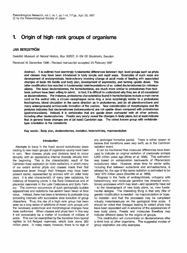
Paleontological Research, vol. 1, no. 1, pp.1-14, 7 Figs., April 30, 1997 © by the Palaeontological Society of Japan 1. Origin of high-rank groups of organisms JAN BERGSTROM Swedish Museum of Natural History, Box 50007, S-104 05 Stockholm, Sweden Received 16 December 1996; Revised manuscript accepted 23 February 1997 Abstract It is outlined how seemingly fundamental differences between high-level groups such as phyla and classes may have been introduced in fairly simple and rapid ways. Examples of such ways are development of endosymbiosis, heterochrony invoMng change of adult mode of feeding with associated changes of basic life habits and body plan, development of asymmetry, and turning upside-down. The latter modification has resulted in fundamental misinterpretations of so-called deuterostomes (or notoneur- alians). The basal deuterostomes, the hemichordates, are much more similar to protostomes than text- book authors have been willing to admit In fact, it is difficult to understand why they are at all considered as deuterostomes. For instance, protostome characteristics found in hemichordates include a main nerve cord on the ventral side, a circum-oesophageal nerve ring, a larva surprisingly similar to a protostome trochophora, blood circulation in the same direction as in protostomes, and (in all pterobranchians and many enteropneusts) schizocoelic formation of the coelom. New consideration of morphologies and life postures indicates that deuterostomes (notoneuralians) are not upside-down compared with protostomes (gastroneuralians). Instead, it is vertebrates that are upside-down compared with all other animals, including other deuterostomes. Fossils very poorly reveal the changes in body plans, but at least indicate that in general these changes are of (at least) Cambrian age. The oldest known group with vertebrate- type orientation is the conodonts. Key words: Body plan, deuterostomes, evolution, heterochrony, macroevolution Introduction Attempts to trace in the fossil record evolutionary steps leading to new major groups of organisms usually have been in vain. New classes, phyla and divisions tend to occur abruptly, with an appreciable internal diversity virtually from the beginning. This is the characteristic result of the Cambrian fossil explosion (or biotic radiation), in which many of our extant animal phyla and classes made their first appearance (even though their lineages may have been present earlier, represented by animals with an older body plan). It is also characteristic of many later radiations, for instance of flowering plants in the Early Cretaceous and of placental mammals in the Late Cretaceous and Early Terti- ary. The common occurrence of such geologically sudden appearances and radiations has seldom been taken at face value. Instead, there has been a tendency to postulate long preceding intervals with successive slow evolution of group characters. Thus, the rise of a high-rank group has been seen as a long series of additions of lower-rank groups until the necessary anatomical and morphological changes have been performed. When classes and phyla form in this way it will conceivably be a matter of hundreds of millions of years. This can be exemplified by the transition from typical reptiles to full-fledged mammals, which took some 200 million years. In many cases, however, there is no sign of any prolonged formative period. There is rather reason to believe that transitions were very swift, as in the Cambrian radiation event. It can be mentioned that molecular differences have been used to indicate an original radiation of coelomate animals 1,200 million years ago (Wray et a/. 1996). This estimation was based on extrapolation backwards of Phanerozoic evolutionary rates. However, when time for earlier splits, including that between eubacteria and archaebacteria, is allowed for, the age of the same radiation is estimated to be 'only' 670 million years (Doolittle et a/. 1966). Progress in the fields of endosymbiosis, ontogeny with heterochrony, and molecular genetics has revealed evolu- tionary processes which may lead-and apparently have led -to the development of new body plans, Le., new funda- mental designs. The interesting thing is that very little of genetic modification is needed-no macromutation or hope- ful monster-and that the processes may be completed virtually instantaneously on the geological time scale. It should be noted that lineages leading to extant phyla may have been separated well in advance of the appearances of new body plans. Fossils and molecules therefore may indicate different dates for the origins of groups. This contribution will concentrate on deuterostomes after a short look at other organisms. The suggested modes of group origination are only examples.
Transcript of Origin of high-rank groups of organisms
Paleontological Research, vol. 1, no. 1, pp.1-14, 7 Figs., April 30, 1997 © by the Palaeontological Society of Japan
1. Origin of high-rank groups of organisms
JAN BERGSTROM
Swedish Museum of Natural History, Box 50007, S-104 05 Stockholm, Sweden
Received 16 December 1996; Revised manuscript accepted 23 February 1997
Abstract It is outlined how seemingly fundamental differences between high-level groups such as phyla and classes may have been introduced in fairly simple and rapid ways. Examples of such ways are development of endosymbiosis, heterochrony invoMng change of adult mode of feeding with associated changes of basic life habits and body plan, development of asymmetry, and turning upside-down. The latter modification has resulted in fundamental misinterpretations of so-called deuterostomes (or notoneuralians). The basal deuterostomes, the hemichordates, are much more similar to protostomes than textbook authors have been willing to admit In fact, it is difficult to understand why they are at all considered as deuterostomes. For instance, protostome characteristics found in hemichordates include a main nerve cord on the ventral side, a circum-oesophageal nerve ring, a larva surprisingly similar to a protostome trochophora, blood circulation in the same direction as in protostomes, and (in all pterobranchians and many enteropneusts) schizocoelic formation of the coelom. New consideration of morphologies and life postures indicates that deuterostomes (notoneuralians) are not upside-down compared with protostomes (gastroneuralians). Instead, it is vertebrates that are upside-down compared with all other animals, including other deuterostomes. Fossils very poorly reveal the changes in body plans, but at least indicate that in general these changes are of (at least) Cambrian age. The oldest known group with vertebratetype orientation is the conodonts.
Key words: Body plan, deuterostomes, evolution, heterochrony, macroevolution
Introduction
Attempts to trace in the fossil record evolutionary steps leading to new major groups of organisms usually have been in vain. New classes, phyla and divisions tend to occur abruptly, with an appreciable internal diversity virtually from the beginning. This is the characteristic result of the Cambrian fossil explosion (or biotic radiation), in which many of our extant animal phyla and classes made their first appearance (even though their lineages may have been present earlier, represented by animals with an older body plan). It is also characteristic of many later radiations, for instance of flowering plants in the Early Cretaceous and of placental mammals in the Late Cretaceous and Early Tertiary. The common occurrence of such geologically sudden appearances and radiations has seldom been taken at face value. Instead, there has been a tendency to postulate long preceding intervals with successive slow evolution of group characters. Thus, the rise of a high-rank group has been seen as a long series of additions of lower-rank groups until the necessary anatomical and morphological changes have been performed. When classes and phyla form in this way it will conceivably be a matter of hundreds of millions of years. This can be exemplified by the transition from typical reptiles to full-fledged mammals, which took some 200 million years. In many cases, however, there is no sign of
any prolonged formative period. There is rather reason to believe that transitions were very swift, as in the Cambrian radiation event.
It can be mentioned that molecular differences have been used to indicate an original radiation of coelomate animals 1,200 million years ago (Wray et a/. 1996). This estimation was based on extrapolation backwards of Phanerozoic evolutionary rates. However, when time for earlier splits, including that between eubacteria and archaebacteria, is allowed for, the age of the same radiation is estimated to be 'only' 670 million years (Doolittle et a/. 1966).
Progress in the fields of endosymbiosis, ontogeny with heterochrony, and molecular genetics has revealed evolutionary processes which may lead-and apparently have led -to the development of new body plans, Le., new fundamental designs. The interesting thing is that very little of genetic modification is needed-no macromutation or hopeful monster-and that the processes may be completed virtually instantaneously on the geological time scale. It should be noted that lineages leading to extant phyla may have been separated well in advance of the appearances of new body plans. Fossils and molecules therefore may indicate different dates for the origins of groups.
This contribution will concentrate on deuterostomes after a short look at other organisms. The suggested modes of group origination are only examples.

2 Jan Bergstrom
Endosymbiosis
It now appears to be generally accepted textbook knowledge that most eukaryotic cells contain modified bacteria as endosymbiotic organelles (cf. for instance Margulis 1970; Taylor 1980,1994). One interesting category of endosymbionts is the coloured plastids, including the chloroplasts, which obviously stem from coloured bacteria. It is quite clear that such coloured bacteria have come to form endosymbionts not only once, but several times. The host organisms were protozoans. Thus there has been a repeated transition between two of the kingdoms that have been recognized of old: the Animalia and the Plantae, or in somewhat newer terms, the Protozoa and the Algae. It has been suggested that the chloroplasts of red algae stem from cyanophytes, those of green algae and land plants from prochlorophytes. There even seem to be cases where a protozoan has turned into an alga not by engulfing a coloured bacterium, but a eukaryotic alga. The result is something like a biological Russian doll, with the largest cell engulfing smaller cells which in turn had engulfed still smaller cells. One such group of complex organisms are the cryptomonads (Ludwig and Gibbs 1985; Douglas et al. 1991).
The endosymbiotic phenomenon could not have been detected on fossil material by palaeontologists. The stratigraphic control on its origination is vague at most, but most endosymbiosis events presumably took place in the Precambrian.
Heterochronic origins of animal phyla
In many marine coelomates the larva and adult lead two entirely different modes of life. Whereas the adult is benthic and may have one or the other mode of feeding and locomotion, the larva is pelagic and swims and feeds with the aid of cilia. Also many adult coelomates use cilia for feeding. Some of these collect their food directly from the water. Brachiopods, bryozoans, phoronids, endoprocts and pterobranchians (graptolites) are typical examples, with cilia situated on tentacles. Others collect at least part of their food from the sediment surface, although again with their cilia. In this category we find sipunculids, among others.
The question now is how these animals have developed their adult mode of feeding. We can first note that the tentaculated ciliary feeders belong to the most derived (most highly developed) of the three groups or levels of bilaterian animals, the three being acoelomates, pseudocoelomates and coelomates. Therefore there is every reason to believe that tentaculate ciliary feeding is a derived mode of feeding in adult bilaterian animals.
One way to develop ciliary feeding in adults with ciliaryfeeding larvae certainly would be by heterochronic retention of such feeding into the adult (Figure 1). Since heterochronic changes by themselves need to involve only minor genetic changes, they can reasonably occur in very short time. Logically, therefore, a shift to tentacle-feeding from grazing, mud-eating or hunting could lead to a tentaculate phylum from something fundamentally different within a million years, and presumably within a very much shorter
time, perhaps even 1000 years or less. Two groups are particularly instructive in this regard. The
first of them is the Endoprocta (Figure 1). An endoproct is virtually a trochophora larva on a stalk. The only characters in an endoproct that do not fit into such a larva are the stalk attachment and the reproduction ability (for instance Hyman 1951). Thus, we can easily believe that paedomorphic heterochrony made its adult characters unlike that of any other group, and particularly unlike that of the immediate ancestor, thus creating endoprocts as a discrete phylum. Since the endoproct characters are those of coelom ate larvae, endoprocts are coelomates by affinity (although not by construction), not pseudocoelomates. This is also borne out by the pattern of egg cleavage, which is most similar to that of annelids and similar animals.
The second instructive group is the Phoronida (for instance Hyman 1959). Phoronid larvae are typical coelomate planktic larvae with three transverse bands of cilia. One of the bands develops tentacles in the larva already before it settles on the sea bottom, and the adult tentacles develop from the same ciliary band. This demonstrates the continuity in equipment and feeding method from larva to adult.
Endoprocts and phoronids differ fundamentally in detail. For instance, the tentacles develop from a preoral ciliary band, the prototroch, in endoprocts, but from a postoral band, the metatroch, in phoronids. The message is the same, however: a fundamentally new animal, a new phylum, could be shaped by simple heterochrony.
Twisting
Another class of processes leading to new groups is twisting, that is, deformation of the body symmetry. The gastropods provide an interesting example of this simple change behind high-level taxonomic separation (Peel 1987, pp. 305-306). The diagnostic difference between gastropods and their ancestors, the monoplacophorans, is that the latter are bilaterally symmetrical, whereas in the former the shell with its enclosed soft-parts is rotated 180· on the foot. The evolutionary process is echoed in the embryology of the individual gastropod: the small larva is symmetrical, but at one stage the rotational torsion starts. It can start suddenly and be completed within minutes. Maybe it is a mechanical instability that causes the torsion, and did so when it was evolutionarily invented. The torsion gives gastropods a great advantage compared with monoplacophorans: the mantle cavity is shifted anteriorly and gives the animal the possibility to hide inside its shell.
Modern echinoderms, and even most fossil ones, have a nice pentameric symmetry. Their larvae are bilaterally symmetrical. Certainly their ancestors were symmetrical also as adults. There is general agreement that pentameric symmetry was reached via a stage with asymmetry distorting the ancestral bilateral asymmetry. This is so during ontogeny, and we know of many asymmetric forms from the Palaeozoic. Some of them reclined on their sides on the bottom. This is the case with the 'carpoids'. It is possible, even most probable, that the forms leading to pentameric echinoderms

Origin of high-rank groups of organisms
original adult
new adult
original life cycle
new life cycle
~ ...... -.. -:--• .. -:', ..... ;r ................ :. -~.-.
creeping larva
swimming larva
larva attaches
to substrate
Figure 1. Endoprocta as an example of possible origination of new phyla by heterochrony. The original life cycle is supposed to have had an adult with features of flatworms, molluscs and several types of marine larvae. By prolonging the larval type of feeding into the adult there would have been a need to extend the Ciliary band along tentacles to enhance the efficiency. A sessile mode of life would be optimal. It is easy to understand that the shift from the primary to the secondary life cycle would be fairly abrupt, and that the functional and morphological changes involved would remodel the adult so that relationships would be obscured - and so a new phylum would be born.
3

4 Jan Bergstrom
were attached. 'Carpoids', including 'calcichordates', being asymmetric recliners, may therefore represent blind ends in evolution. In any case, echinoderms provide still another example of new body plans created by small means, presumably with little genetic change and in short time.
Deuterostomes (notoneuralians)
The constructional gap between vertebrates and their relatives on one side, and 'protostomes' (or gastroneuralians) on the other, has long disturbed attempts to compare the two groups. As a result, the phylogenetic and evolutionary relationships have been poorly understood and hotly debated.
As the names indicate, students have thought that in protostomes/gastroneuralians the mouth has a primary position and the central nervous system is ventral. In deuterostomes/notoneuralians the mouth would have a secondary position and the central nervous system would be dorsal (for instance Nielsen 1995). In the following I occasionally use the terms neural side and cardial side when the terms dorsal and ventral may be confusing or irrelevant.
One attempt to understand the origination of the vertebrates has been to start from a protostome that was turned upside down, so that the originally ventral nerve cord became dorsal in vertebrates. An early proponent of this idea was Etienne Geoffroy St. Hilaire (1822), who suggested that the vertebrate origin was among the arthropods. Other mostly 19th Century authors (F. Leydig, C. Semper, A. Dohrn and A. Naef, see NObler-Jung and Arendt 1994) have tried to find an origin among annelid-type worms also by means of dorsoventral inversion. In the 20th Century, W. Patten (1912) has been intrigued by the idea of inversion. He believed in a derivation from chelicerate arthropods. However, on the whole the idea of inversion has had only a limited number of proponents. In reality, most zoologists have considered the idea to be of entirely historical interest.
Recently, however, there has been brought impressive molecular evidence that indicates, or rather proves, that vertebrates in fact are upside-down in comparison with 'protostomes'. It started with a contribution by Arendt and NObler-Jung (1994; see also 1996), where they demonstrated that genes controlling dorsoventral patterning of embryos show close correspondences between vertebrates and insects. The striking difference is that a gene complex (the achaete-scute complex) influencing neuronal precursor cells ventrally in insects (Drosophila) influences the same type of cells dorsally in vertebrates (Xenopus). In the other direction, decapentaplegic-related genes affect mesoderm and ectoderm dorsally in insects and ventrally in vertebrates. Holley et at. (1995) found that the sag (short gastrulation) gene, expressed in the region of the ventral nerve cord in insects, corresponds to the chordin gene, that is expressed in the mid-dorsal area in vertebrates, around the dorsal nerve cord. De Robertis and Sasai (1996) added further substance to the idea of inversion. Arendt and NObler-Jung (1996) also demonstrated a close longitudinal correspondence between the brains of insects and vertebrates, both in gene control and in functional respects. These hard facts demonstrate
that zoologists have been completely misled in producing our current view of the differences between protostomes and deuterostomes.
The new understanding of how a vertebrate must be oriented in order to be correctly compared with a protostome has prompted an attempt to see what this implies for other deuterostomes (Bergstrom, Viehweg and Naumann in prep.) Are all so-called deuterostomes 'upside-down' in comparison with protostomes, or only vertebrates, or some intermediate-sized assemblage? Between which groups did the inversion occur? How could this influence our ideas of protostome-deuterostome relationships?
We can first state that enteropneusts, cephalochordates and vertebrates are the only deuterostome groups with a dorsoventral life orientation that can be compared with that of protostome worms, molluscs and arthropods. It simply has no sense to compare pentaradiate echinoderms or sessile groups with a U-shaped gut with ordinary worms, since dorsal and ventral cannot be defined in a comparable way. In search for the solution of the orientation problem, I was struck by the fact that cephalochordates (amphioxus) tend to rest on their neural side, that is, upside down as the text-books see it. This raised two immediate questions: are they upside down in their life posture, or are they in textbook drawings? And if the textbook drawings are upsidedown, what are the implications for our anatomical comparisons with other animals, such as the hemichordates?
Anatomy, function and relationships
In this approach I am not suggesting any bold new ideas on the relationship between phyla. The deuterostomes, as delimited here, are what they are in most textbooks: hemichordates, echinoderms and chordates. There are strong reasons to believe that these groups are closely related. These reasons are well known and need not be repeated here.
I am not suggesting that anyone group evolved from any other group such as they are delimited today. For example, I do not believe that vertebrates evolved from modern cephalochordates, but from a common ancestor which had already achieved important derived characters, such as segmentation of musculature and nerve system. This means that the ancestor was able to swim, as are also modern cephalochordates, if only in short darts.
It is worth noting that all preceding steps on the evolutionary ladder within the deuterostomes-pterobranchs, enteropneusts, urochordates, cephalochordates-include ciliary feeding but not active search for food, and no eyes are involved in feeding. Vertebrates, on the other hand, have well-developed sensory organs including eyes for orientation, and they search their food actively, not by ciliary feeding. Therefore it is a logical conclusion that vertebrates have come into being by a radical shift of habits. As in cases referred to above, we see this shift also in the ontogeny, since larval lampreys live and feed just as the amphioxus.
This is not unique to vertebrates. Function is intimately tied to body plans. We must therefore expect evolution to

Origin of high-rank groups of organisms 5
have produced similar solutions over and over again. Repeated heterochronic retention of ciliary feeding resulting in a number of tentaculate phyla was mentioned above as one example. Unfortunately many scientists are so impressed by one or the other individual similarity that they overlook the overwhelming amount of parallel and convergent evolution that is clearly evident from the mosaic distribution of characters among animals.
Nielsen (1995) has produced one of the latest phylogenetic trees. His tree is founded on a series of hypothetical ancestral stages, partly larval, for which there is no evidence. His guiding idea is that two types of larval ciliary feeding, downstream and upstream food collecting, defines two phylogenetic groups. It is obvious that upstream ciliary collecting is closely tied to mesotroch ciliary feeding. This combination is found in brachiopods, phoronids, bryozoans, echinoderms, pterobranchs and enteropneusts, all ciliary feeders as adults. Endoprocts, which have prototroch ciliary feeding, have a downstream collecting system, like echiurids, annelids, nemerteans and molluscs, which are not ciliary feeders as adults. A downstream collecting system appears to be primitive among coelomates. Whenever animals have shifted to mesotroch ciliary feeding there appears also to have been a shift to upstream collecting. I do not understand why this is so, nor does Nielsen indicate any understanding of the phenomenon. There is no reason whatsoever that upstream collecting should not have evolved several times, and there is no justification for using downstream and upstream collecting systems to distinguish phylogenetic groups. Nielsen's results are not in line with either modern molecular phylogeny or molecular genetics. For instance, the very striking resemblance in genetic steering of morphogenesis between insects and vertebrates (Arendt and NObler-Jung 1994,1996; Holley et al. 1995) demonstrates that it is unreasonable to distinguish the Protostomia and Deuterostomia as two main groups, and unreasonable to regard ctenophores as a twig on the deuterostome branch.
There is also in Nielsen's approach the common tendency to generalize, and to give protostomes and supposed deuterostomes characters which they do not have, or that only some of them have. For instance, Nielsen (1994, Table 19) claims that phoronids, pterobranchs, enteropneusts, urochordates and cephalochordates have a dorsal central nervous system, and that pterobranchs, which lack a pelagic larva, have dipleurula-like ciliary bands. As stated elsewere, in many ciliary feeders there is no possibility to distinguish between dorsal and ventral sides, and regarding pterobranchs, enteropneusts, and apparently also cephalochordates, Nielsen is clearly incorrect (see below).
Enteropneusts, protostomes and the central nervous system
Vagile protostomes tend to have at least one ventral nerve cord, or a pair. An anterior part of it embraces the oesophagus. Although deuterostomes are said to have their central nervous system dorsally rather than ventrally, enteropneusts in fact have a well-developed ventral nerve cord, from the
anterior end of which commissures rise to embrace the oesophagus and join the (occasionally hollow) 'brain' tube in the mesosoma (Figure 2). There is also a dorsal nerve cord, but it is the ventral cord that is the thickest and that is in close association with the longitudinal musculature. It is notable that there is one or more dorsal (and lateral) cords also in certain protostome groups, such as the flatworms, nematodes and nemertines.
The central nervous system of enteropneusts is therefore typically protostomian in its character. It is commonly said that the hollowness of the 'brain' is found elsewhere only in chordates but no chordate has a hollow nerve tube on the cardial side of the body. Arendt and NObler-Jung (1996, p. 258) have rightly pointed out the true character of the enteropneust nervous system. It is obvious that enteropneusts are not upside-down in comparison with protostomes. Since they have a ventral mouth and a circum-oesophageal nerve commissure, it would appear to be a mistake to call them deuterostomes. Their mouth is most probably where it has always been in their ancestors, technically they are protostomes, whether or not they are related to vertebrates. And they are gastroneuralians, since the central nervous system is dominantly ventral.
Enteropneusts and chordates
The circum-oesophageal commissure is generally lost in chordates, but there is still a nerve ring in appendicularians (Urochordata), even if there is no longer any brain on the cardial side (Olsson et at. 1990). The existence of a nerve ring is most important for the comparison between hem ichordates and chordates. If the central nervous system is used to compare appendicularians with enteropneusts, it appears that the neural (dorsal) side of the former must correspond to the neural side of the latter, that is, the side that is considered as ventral in text-books. This is at odds with conventional wisdom, according to which the neural (dorsal) side of vertebrates, and therefore the neural side of appendicularians and other urochordates, corresponds to the cardial (dorsal) side of enteropneusts.
On the whole, however, urochordates are too derived to be conveniently compared with enteropneusts. Cephalochordates (amphioxus) can be more rewarding. When amphioxus is placed with its neural (biologically ventral, but conventionally dorsal) side down, a series of similarities with enteropneusts are apparent (Figures 2, 3, 4). First, the main part of the central nervous system is down in both. The direction of blood circulation is the same, and conforms also with the general protostomian pattern. On the upper side, both cephalochordates and enteropneusts have a pair of folds, called metapleural folds in the former, genital folds in the latter. In amphioxus (but not generally in vertebrates), the metapleural folds are interconnected by a transverse fold closing an atrial space around the pharynx. Between the folds are the pharyngeal tremata (gill pores), although in adult cephalochordates the individual tremata have disappeared and there is a single atrial opening posteriorly. The anus is on the upper side both in cephalochordates and in the larval tailed enteropneust (and in pterobranchs). In cross section,

6 Jan Bergstrom
A
circum-oesophageal nerve flng
m
c
¢:: ¢::
=>
¢::. ¢::
a
ens
a
ens pc
a
ens nc
Figure 2. Central nervous system and blood circulation in protostomes (A) and deuterostomes (B, C). A, Onychophora; B, Enteropneusta (with anus in semi-larval position); C, Cephalochordata (amphioxus). With amphioxus oriented as in nature, there is a basic similarity throughout. The main blood circulation is forwards on the dorsal side, backwards on the ventral side. The main stem of the central nervous system is ventral; in the anterior end there is a circum-pharyngeal ring except in amphioxus. The absence in amphioxus appears to be a secondary condition, since such a ring is known from urochordates (Oikopleura). The dorsal position of the anus in amphioxus has its counterpart in hemichordates (pterobranchians and larval enteropneusts).
there are paired longitudinal muscles in the lower part of the animal, whereas the reproductive organs are situated in the upper part. Between the gut and the nerve cord is the notochord. Correspondingly there is in some enteropneust species a pygochord (for instance in species of Balanog/ossus; cf. Figures 2, 3). This pygochord likely functions as a skeleton. The endostyle (and its derivatives), so characteristic of chordates, may correspond to the preoral ciliated organ of enteropneusts.
This impressive, and previously unexpected, list of similarities indicates that chordates may have their origin among animals very similar to extant enteropneusts. With swimming habits, there would be no need for a proboscis, and the
animal would immediately be much more ampioxus-like (although lacking, for instance, muscular segmentation). However, many readers may wonder how it would have been possible for sluggish bottom-living enteropneust-like animals to develop swimming powers and pelagic habits. Actually, whereas many enteropneusts are deposit-feeders, others are able to collect food directly from the water, and the species Glandiceps hacksi has been observed swarming at the surface in shallow water, feeding on phytoplankton (Brusca and Brusca 1990, p. 853).

Origin of high-rank groups of organisms 7
reproductive organ
alimentary canal
gill slit
muscle
pygochord
notochord
nerve cord
Figure 3. Idealized cross sections through A, enteropneust (Hemichordata) and B, amphioxus (Cephalochordata). Both are oriented as in nature, which means that the amphioxus is upside-down in comparison with textbook illustrations. There is a close correspondence in several structures and organs: pharynx with gill slits, paired dorsal 'wings' containing gonads (called genital wings for simplicity; in amphioxus, their technical term is metapleural folds), ventrally situated longitudinal muscles, main stem of central nervous system ventrally, and a stiffening rod (pygochord and notochord) between gut and nerve stem. The enteropneust musculature is usually poorly developed, but is massive in species of Saccoglossus. The enteropneust pygochord is not widely distributed, but occurs in speCies of Balanoglossus.
Filter-feeding cephalochordates, macrophagous vertebrates
We have to accept that we must orient amphioxus with its neural side down when we compare its anatomy with that of protostomes and enteropneusts. Since this is also the way it lives, it would be no problem, but for one thing: doing this, we place them upside-down in comparison with vertebrates, and we can hardly accept instead to place a fish or a mammal upside-down in order to facilitate comparison. However, we have localized the place in the evolutionary tree where reorientation must have occurred: it is inside the chordate tree, after cephalochordates (amphioxus) branched off. Vertebrates obviously are the only upside-down deuterostomes, the only real Notoneuralia.
Cephalochordates have several peculiarities of their own, but they are also similar to vertebrates in many respects, for instance in having a segmented musculature. One difference, however, is that they lack eyes, whereas vertebrates have a pair of anterior eyes. They have light-sensitive spots in the central nervous system which helps them determine the correct burrowing depth, but they cannot spot their food, and they do not need to because they are still filter-feeders like most hemichordates and urochordates. On the contrary, vertebrates find their food actively by the
use of sensory organs including eyes. How could a change from a passive filter-feeding organ
ism to a macrophagous swimmer come about? How does the reversal of body posture fit in, if at all ?
We do not know why a change came about. For the moment we just have to accept that it did. For an active localization of its food the original vertebrate apparently needed eyesight, since eyes were developed. Eyes most easily may develop from the light-sensitive nerve cord. For a primitive chordate without a circum-oesophageal ring and a cardial-side component of the brain, this means that the eyes take their origin below rather than above the mouth. For food search along the bottom this situation is inconvenient. The simplest way to solve the problem would be a reversal of body posture. We can now see that nature found this solution. Reversal would shift the mouth to the new ventral side. It is of course possible that the order of events was just opposite, with reversal preceeding eye development.
How likely is it that a swimming proto-vertebrate would be able to shift to an upside-down life? We can get some idea from a study of living amphioxus. My own observations of Branchiostoma lanceolatum indicate that it easily turns around when it meets a hindrance, such as aquarium glass. It turns immediately 180', and does not swim on its side.

8 Jan Bergstrom
mouth
gill pore
genital ~~+----- ----+~~:
WIng
genital ~I~----- ------+~~
WIng
gill pore
c
Figure 4. Ventral views of anterior body parts of enteropneusts (Hemichordata) and amphioxus (Cephalochordata). A, The enteropneust Balanog/ossus, cardial (dorsal) side. B-C, l...aJval and adult amphioxus (Branchiostoma), cardial (so-called ventral) side. Note the general similarity, partly disguised by asymmetry in the larval amphioxus and loss of individual gill pores in adult amphioxus. The genital wings of the enteropneust are similar to the metapleural folds in amphioxus and similarly enclose the gill pores. A, modified from Ruppert & Barnes (1994); B-C, modified from Herdman (in Harmer and Shipley 1932).
After a second or two, it tums over again-and then over again. When hiding in the bottom shell sand, it usually rests with the neural side down, but in exceptional cases it appears to be the other way around. To amphioxus, there-
fore, one or the other side up does not mean any impossible difference.
We have some idea on when it happened. The great radiation within coelomate phyla started around 540-530

Origin of high- rank groups of organisms 9
million years ago, at the beginning of the Cambrian. The oldest known cyclostomes lived during the Early Ordovician and are slightly less than 500 million years ago ; they had eyes. The conodont animals must have belonged to the vertebrate side of the evolutionary tree (see below). The oldest definite conodont animals we know of lived during the Late Cambrian, some 505 million years ago. Inversion and the vertebrate eye thus are some 505- 530 million years old.
Embryology
Reversing the adult means that also the egg and embryo have to be turned around 180°. Surprisingly, the edges closing the neural tube then must be considered as the lips of the urmund. In urochordates the definite mouth forms at the neuropore, that is, just at the anterior end of the urmund. This makes urochordates indistinguishable from protostomes in this allegedly profound character (Figure 5).
In cephalochordates and vertebrates the situation is more derived because of a secondary displacement of the mouth. However, it is notable that the nasal sack still forms from the neuropore, and that this sack is continuous with the gut in hagfish. Could it be the original mouth? Arendt and NCJbler- Jung (1996, p.258) found neural evidence that it corresponds to the original mouth, and thought that the
functional mouth in vertebrates is a new penetration. This is of course possible, but from a functional point of
view it appears likely that the new mouth in one way or another has evolved from the old. We know that in many enteropneusts the pharynx is divided into upper and lower portions by lateral infoldings of the pharynx wall. The upper channel leads water to the pharyngeal tremata ('g ill' pores), while the lower channel conveys food backwards to the midgut. A similar division could have separated the anteriormost part of the vertebrate forebears into an upper nasopharyngeal tube and a lower mouth cavity, both opening posteriorly into the pharynx. This appears to be the situation that is still present in myxinids (hagfishes), while in petromyzontids the upper tube is closed at the rear to form a nasal sack.
The situation in cephalochordates is less easy to interpret. Ontogenetically the definitive mouth forms as a new opening on the left side of the head, only to move later to the upper (so- called ventral) midline. This shift serves two purposes. First, it 'has to' move from its orig inal position on the lower (so-called dorsal) midline to let the notochord grow to the anterior end of the animal, where it serves as a support during burrowing. Second, for an animal concealed in the bottom sediment its position should be as high as possible, thus on the upper side. As for vertebrates it appears most
future mouth
neuropore; mouth in
urochordates
neural fold -----,.;,~~~4
--- blastopore lip
------ future anus ____ _
Figure 5. Idealized views of embryos from the neural side : left, protostome; right, urochordate deuterostome. In a protostome the future mouth is said to be formed by or at the anterior end of the closing blastopore, although there are numerous exceptions. The ventral nerve cord forms along the fused blastopore lips. In a deuterostome the central nerve cord similarly forms along a pair of fused folds, which in urochordates extend between the future mouth and anus. The similarity between the two embryo types is striking, but has not been appreciated because students 'knew' they were looking at opposite sides of the embryos.

10 Jan Bergstrom
likely that the mouth is not a completely new opening from an evolutionary point of view, but has shifted position. It may have moved over the anterior tip of the animal. If so, the lateral formation of the mouth may be a later adaptation, perhaps to lying down on one side, or to rotational swimming in the larvae for more effective ciliary feeding.
Fossil deuterostomes
Apart from echinoderms and vertebrates, no deuterostome group has yielded fossils useful for softpart reconstruction. The only other group that is abundant as fossils is the graptolites, to which the two genera of living pterobranchians should be counted. Unfortunately the Palaeozoic graptolites are known only from their colonial skeletons.
The Lower Cambrian Chengjiang fauna of China has yielded the worm-like animal Yunnanozoon lividum. In their original description, Hou et al. (1991) regarded it as slugshaped. Bolder interpretations and reconstructions have been issued since. Dzik (1995) and Chen et al. (1995) suggested that the animal is a deuterostome, the segmented bands being myomeres of the type found in cephalochordates and vertebrates. I hesitate to accept this interpretation. There are several reasons for this. For instance, the supposed myomeres lack the V-shape characteristic of segmented chordates. They are also notably dark, which is difficult to explain if they were myomeres. It is even more remarkable that there is occasionally a small overlap between adjoining 'segments'. Such an overlap is hardly compatible with the idea that the 'segments' are massive muscle blocks. At the edge the 'segment' in front occasionally stands out a little over the successive one. These features indicate that the 'segments' are not muscle blocks at all, but sclerites in the skin of the animal. Such sclerites are unknown in marine deuterostomes. It can be added
B
that the supposed gills have a ventral position, whereas the gill pores in deuterostomes (other than vertebrates) have a decidedly dorsal position. Moreover, Shu et at. (1996) pointed out that the supposed notochord is filled with gut contents.
Shu et al. (1996) recently described Cathaymyrus diadexus from the Chengjiang fauna as a cephalochordate. The single fossil specimen preserves a possible alimentary canal, a possible notochord, a possible pharynx perhaps with gill slits, and transverse bands that are interpreted as myomeres. No fins are visible. According to the authors the animal is judged to be a relative of amphioxus. This is not obvious from the illustrations. Additional material is needed before any well founded judgement on the significance of this fossil can be made.
The Middle Cambrian Pikaia graci/ens is more easily accepted as a chordate. It has the general appearance of an eel larva (Figure 6A). It appears to be segmented and to have typically V-shaped myotomes, a key character of cephalochordates and vertebrates. However, as long as it is unknown if it has eyes or jaws (teeth) it cannot be decided if it is a cephalochordate or a vertebrate. This diminishes its value in the discussion. It does not seem to be a conodont animal, since it is likely that the conodont teeth would be easily preservable and visible. On the other hand, the absence of shell sand in the Burgess Shale indicates that its mode of life was different from that of modern cephalochordates.
These fossils apparently can not give us much information about the origin and early evolution of deuterostome groups.
The conodont animals, or conodontophorids, are now known to have had a pair of eyes and a body musculature divided into V-shaped myomeres (Figure 6B; Purnell 1995). It is quite obvious that they are segmented chordates. The possession of eyes and teeth demonstrates that they were
~«(~~-~ Figure 6. Fossil segmented chordates, both oriented with the neural side up. A, Pikaia graci/ens, a possible
amphioxus relative from the Middle Cambrian Burgess Shale. Length about 4 cm. Drawn from Conway Morris (1982, PI. S). B, Clydagnathus windsorensis, a conodont animal from the Carboniferous of Scotland. Its eyes reveal that it belongs to the vertebrate lineage. Length about 4 cm. Simplified from Purnell (1995, Figure 1).

Origin of high-rank groups of organisms 11
not ciliary feeders, but hunters. This gives us the possibility to say that they were not cephalochordates, but belonged to the vertebrate side of the evolutionary tree. The oldest true conodonts are from the Late Cambrian. However, they were preceeded already in the Early and Middle Cambrian by so-called protoconodonts and paraconodonts, which are considered by many specialists to be ancestral to the conodonts (see for instance Bengtson 1976, 1983; Andres 1981; Dzik 1991).
Echinoderms are differentiated into some 20-25 classes. The classes are so distinct that it is generally almost impossible to be sure· of interrelationships. Four subphyla have been distinguished. One of them, the Homalozoa (the carpoids), include asymmetric to superficially bilaterally symmetric forms which rested with one side on the bottom. The other three phyla include more or less pentameric forms. The subphylum Asterozoa (with seastars and brittlestars) includes forms with true arms, that is, extensions of the body that contain organs such as gonads. The remaining subphyla, the Crinozoa and Echinozoa, consist of a variety of classes and are difficult to define. The basic similarities between pterobranchs (graptolites) and echinoderms makes it most likely that the latter have evolved from a pterobranchlike ancestor. However, nothing of that transition is preserved. As indicated above, a key to understanding the origination of echinoderms is the formation of asymmetry in association with a new mode of attachment (which was presumably lost in the Homalozoa). Another key invention is that of a calcitic endoskeleton. This made it possible for the animals to grow to much larger size than before. The presence of a calcareous skeleton apparently makes a preCambrian origin of the echinoderm body plan impossible. Virtually without exception, calcareous skeletons occur first in the Cambrian. One reason may be that the dissolved oxygen content was too low to form them. Rhoads and Morse (1971) have shown that at present, with an atmospheric oxygen ratio below 10% acids produced by anaerobic glycolysis will dissolve calcareous skeletons. Still, echinoderms with a calcitic skeleton were around already in the Early Cambrian.
Fossils which have been used frequently in attempts to explain vertebrate origins are the so-called calcichordates (for instance Jefferies et aI. 1987; Jefferies 1988; Woods and Jefferies 1992), more or less irregular to bilaterally symmetric forms with echinoderm-type calcitic skeleton. Most authors regard them as echinoderms forming the class Stylophora within the sub-phylum Homalozoa. According to the calcichordate hypothesis, concavities on the inner side of the skeleton housed a well-developed nervous system with a surprisingly large brain, and similarities with vertebrates include a tail (by other students considered to be a tentacle). The theory is considered controversial. It appears to be immensely more difficult to derive a cephalochordate-vertebrate origin from a 'calcichordate' than from an enteropneust. How, for instance, to evolve a muscular tail swinging sideways around a central endoskeleton from a vertically swinging pulling tool with, functionally speaking, a peripheral exoskeleton?
Deuterostomes a separate branch?
With the understanding of vertebrate inversion-and only vertebrate !-the discussion on 'protostomes' and 'deuterostomes' (or 'gastroneuralians' and 'notoneuralians') can be seen in a new light. There do not seem to exist any deuterostomes, in the sense that they should be animals with a newly formed mouth unrelated to the anterior end of the urmund. On the contrary, urochordate embryology demonstrates that the definitive mouth is formed exactly at the anterior end of the urmund. In this respect, 'deuterostomes' are 'protostomes'.
Of all the groups which now and then have been referred to as 'deuterostomes' or 'notoneuralians', only the vertebrates qualify as 'notoneuralians' in the sense that they have their neural side up (except in the upside-down swimming fish Synodontis batensoda). When other chordates are illustrated in the same posture, it is only by convention.
Hemichordates have a typical protostomian arrangement of their central nervous system, including an anterior dorsal part connected by a (fairly diffuse) circum-oesophageal ring with a posterior ventral nerve cord. Among chordates, some urochordates have a circum-oesophageal ring (as noted above), but the post-oesophageal nerve cord dominates strongly. Since no part of the central nervous system appears to be missing (Arendt and NObler-Jung 1996), the oesophagus appears to have moved forwards through the central nervous system with a resulting displacement of the brain from the cardial to the neural side of the body.
The textbooks repeat the characters said to be typical of deuterostomes. One such character is the mode of coelom formation, which should be enterocoelous. There is in fact much variation. For instance, pterobranchs have schizocoelous coelom formation, some enteropneusts are schizocoelous, others enterocoelous, echinoderms are usually enterocoelous, but some ophiuroids are schizocoelous, urochordates virtually lack coelom, etc. The enterocoelous condition of deuterostomes apparently is a myth, and the variation indicates that the mode of coelom formation has little if any bearing on animal relationships.
The idea that there is a typical 'deuterostomian' larva is also a myth. It is true that enteropneust larvae and certain echinoderm larvae are similar to each other (except for the mode of swimming !), but these are exceptions among the 'deuterostomes'. It is also true that these larvae are similar to trochophoras and trochophora-like larvae, said to be typical of 'protostomes' (Figure 7). In particular, the larvae of enteropneusts, molluscs and annelids share a rotating motion around their longitudinal axis during swimming. Echinoderm larvae do not.
Comparisons of genetic steering mechanisms (Arendt and NObler-Jung 1994, 1996; Holley et aI. 1995; De Robertis and Sasai 1996) demonstrate great affinities between 'deuterostomes' and advanced 'protostomes', such as arthropods. Other protostomes (pseudocoelomates, flatworms) are distinctly further apart. There is fairly good agreement in molecular phyletic trees that this is so (for instance, Turbeville et aI. 1992; Smothers et aI. 1994; Bergstrom 1994; Doolittle et al. 1996). Winnepenninckx and Backeljau (1996)

12 Jan Bergstrom
a a
act
~ ::;;:::"" ~ ~ -=
-;::::---
Figure 7. Comparison between typical idealized 'protostome' and 'deuterostome' larvae. A, the 'protostome' trochophora. B, the 'deuterostome' tornaria, present in enteropneusts. Except for the course of the two anterior ciliary bands (prototroch and metatroch), the two larval types are very similar. The similarities even include a rotating mode of swimming, unique to these larvae. Text-book authors, however, conventionally claim that these larvae are utterly dissimilar. a, anus; act, apical ciliary tuft; e, apical eye; m, mouth; mt, metatroch; pt, prototroch; tt, telotroch; vt, ventral Ciliary band or neurotroch.
tried different tree-making approaches and molecular models on 18S rRNA and found that these greatly influence the result. However, in 16 presented trees out of 16, deuterostomes were derived from protostomes, although from almost all possible positions. In other words, although there is no agreement on the branching details of the evolutionary tree, there is general agreement that deuterostomes are derived from protostomes.
There remains no evidence whatsoever to regard 'deuterostomes' ('notoneuralians') as being anything else than derived 'protostomes'. There are in fact very strong arguments against the old idea that 'protostomes' and 'deuterostomes' are two main branches of the animal kingdom. 'Deuterostomian' characters such as enterocoelous coelom, radial egg cleavage, indeterminate egg cleavage, ciliary feeding and functionally associated oligomery, and upstream collecting ciliary feeding appear to be end products in protostome evolution. These characters are irregularly
distributed among phyla. Having said this, I want to emphasize that hemichordates,
echinoderms and chordates belong together in a natural group. Even if the name Deuterostomia is a misnomer (like so many other names), this is of no significance for the practical use of the name. It does not cover the same groups as Nielsen's Notoneuralia, and his Neorenalia is just a younger and unnecessary synonym.
Conclusion
A major conclusion is that high-level systematiC units, including classes and phyla, may have formed through simple processes involving only small genetic changes. This indicates that classes, and particularly phyla, may have originated as fast as species. Processes include the formation of endosymbiosis in protists, and both heterochrony, torsion and body reorientation in metazoans.

Origin of high-rank groups of organisms
Among metazoans, most of the dramatic changes appear to have happened in the Cambrian.
Although many zoologists still see protostomes and deuterostomes more or less as sister-groups, there is general agreement among scientists dealing with molecular comparisons that deuterostomes are derived from protostomes.
The Deuterostomia are greatly misunderstood, since textbooks tend to overemphasize certain derived features. In fact, however, primitive deuterostomes, notably hem ichordates, have series of protostomian characteristics, such as schizocoel, ventrally positioned central nervous system with an anterior circum-oesophageal ring, vascular system with identical direction of circulation, and a pelagic larva that has great similarities with a molluscan and annelid trochophora. The striking differences between 'typical' deuterostomian and protostomian features are a result of the conventional upside-down orientation of chordates in virtually all comparative studies.
Parallellism and convergence in evolution are much more common phenomena than usually recognized. For instance at the phylum level this has repeatedly resulted in ciliary feeding, segmentation etc, yielding superficial similarities which often have prompted systematists to recognize superphyla such as Articulata and Tentaculata.
Acknowledgements
This study was supported by the Hierta-Retzius Fund and by the fund 'Till Broderna Jacob och Marcus Wallenbergs Minne". I am grateful to Wilfried W. Naumann and Jens Viehweg for discussions during cooperation on the upside down problem, and to Ragnar Olsson for inspiration and practical support.
Appendix: Outline of deuterostome classification
Deuterostomian superphylum HEMICHORDATA Bateson, 1885, emend. Fowler, 1892
Graptolithina Bronn, 1846 = Pterobranchia Lankester, 1877 Enteropneusta Gegenbaur, 1870 Xenoturbellida Westblad, 1949
ECHINODERMATA de Brugiere 1789 HOMALOZOA Whitehouse, 1941 (= Carpoidea Jaekel) Homostelea Gill and Caster, 1960 Homoiostelea Gill and Caster, 1960 Stylophora Gill and Caster, 1960 (= Calcichordata Jefferies) Ctenocystoidea Robison and Sprinkle, 1969
CRINOZOA Matsumoto, 1929 (8 classes) ASTEROZOA Haeckel in Zittel, 1895 (1 or 2 classes) ECHINOZOA Haeckel in Zittel, 1895 (perhaps 6 classes)
CHORDATA UROCHORDATA (TUNICATA) Appendicularia (Larvacea) Doliolariacea (or as Doliolida under Thaliacea 7) Tunicata (with tunic) Ascidiacea, sea squirts Thaliacea, salpa
CEPHALOCHORDATA or ACRANIA Leptocardia
VERTEBRATA (CRANIATA) Several classes including Conodontophorida
References
Andres, D., 1981: Beziehungen zwischen kambrischen Conodonten und Euconodonten. Berliner geowissenschaftliche Abhandlungen, A, vol. 32, p.19-31.
Arendt, D. and NObler-Jung, K., 1994: Inversion of dorsoventral axis 7 Nature, vol. 371, p.26.
Arendt, D. and NObler-Jung, K., 1996: Common ground plans in early brain development in mice and flies. BioEssays, vol. 18, part 3, p.255-259.
Bengtson, S., 1976: The structure of some Middle Cambrian conodonts, and the early evolution of conodont structure and function. Lethaia, vol. 9, p. 185-206.
Bengtson, S., 1983: The early history of the Conodonta. Fossils and Strata, vol. 15, p. 5-19.
Bergstrom, J., 1994: Ideas on early animal evolution. In, S. Bengtson, ed., Early life on Earth, Nobel Symposium, No. 84, p.460-466. Columbia University Press, New York.
Brusca, R.C. and Brusca, G.J., 1990: Invertebrates, xviii+ 922 p. Sinauer Associates, inc., Sunderland, Massachusetts.
Chen, J.-y., Dzik, J., Edgecombe, G.D., Ramskold, L. and Zhou, G.-q., 1995: A possible Early Cambrian chordate. Nature, vol. 377, p.720-722.
Conway Morris, S., ed., 1982: Atlas of the Burgess Shale. Palaeontological Association.
Doolittle, R.F., Feng, D.-F., Tsang, S., Cho, G. and Little, E, 1966: Determining divergence times of the major kingdoms of living organisms with a protein clock. Science, vol. 271, p.470-477.
Douglas, S.E, Murphy, C.A., Spencer, D.F. and Gray, M,W., 1991: Cryptomonad algae are evolutionary chimeras of two phylogenetically distinct unicellular eukaryotes. Nature, vol. 350, p.148-151.
Dzik, J., 1991: Evolution of oral apparatuses in the conodont chordates. Acta Palaeontologica Polonica, vol. 36, part 3, p. 265-323.
Dzik, J., 1995: Yunnanozoon and the ancestry of chordates. Acta Palaeontologica Polonica, vol. 40, part 4, p.341-360.
Geoffroy St. Hilaire, E, 1822: Considerations generales sur les vertebres. Memoires du Museum d'Histoire Naturelle, vol. 9, p. 89-119.
Harmer, S.F. and Shipley, A.E, eds., 1932: The Cambridge Natural History vol. 7, xvii+760 p. Macmillan and Co., Ltd., London.
Holley, SA, Jackson, P.D., Sasal, Y., Lu, B., De Robertis, EM., Hoffmann, F.M. and Ferguson, E.L., 1995: A conserved system for dorsal-ventral patterning in insects and vertebrates involving sog and chordin. Nature, vol. 376, p.249-253.
Hou, X. -g., Ramskold, L. and Bergstrom, J., 1991 : Composition and preservation of the Chengjiang fauna-a Lower Cambrian soft-bodied biota. Zoologica Scripta, vol. 20, part 4, p. 395-411,
Hyman, L.H., 1951: The invertebrates, vol. III. Acanthocephala, Aschelminthes, and Entoprocta. The
13
14 Jan Bergstrom
pseudocoelomate Bilateria, vi +572 p. McGraw-Hili Book Company, New York.
Hyman, L.H., 1959: The Invertebrates, vol. V. Smaller coelomate groups. Chaetognatha, Hemichordata, Pogonophora, Phoronida, Ectoprocta, Brachiopods, Sipunculida. The coelomate Bilateria, viii + 783 p. McGraw-Hili Book Company, New York.
Jefferies, R.P.S., Lewis, M. and Donovan, S.K., 1987: Protocystites menevensis-stem group chordate (Cornuta) from the Middle Cambrian of South Wales. Palaeontology, vol. 30, part 3, p. 429-484.
Jefferies, R.P.S., 1988: How to characterize the Echinodermata-some implications of the sister-group relationship between echinoderms and chordates. In, Paul, C.R.C. and Smith, A.B., Echinoderm phylogeny and evolutionary biology, p. 3-12. Clarendon Press, Oxford.
Ludwig, M. and Gibbs, S.P., 1985: DNA is present in the nucleomorph of cryptomonads: Further evidence that the chloroplast evolved from a eukaryotic endosymbiont. Protoplasma, vol. 127, p. 9-20.
Margulis, L., 1970: Origin of Eukaryotic Cells. 532 p. Yale University Press. New Haven, Conn.
Nielsen, C., 1994: Larval and adult characters in animal phylogeny. American Zoologist, vol. 34, p.492-501.
Nielsen, C., 1995: Animal evolution. Oxford University Press. 467 p.
NClbler-Jung, K. and Arendt, D., 1994: Is ventral in insects dorsal in vertebrates? Roux's Archives Developmental Biology, vol. 203, p. 357-366.
Olsson, R., Holmberg, K. and Lilliemark, Y. 1990: Fine structure of the brain and brain nerves of Oikopleura dioica (Urochordata, Appendicularia). Zoomorphology, vol. 110, p.1-7.
Patten, W., 1912: The evolution of the vertebrates and their kin. J.A. 373 p. Churchill, London.
Peel, J.S., 1987: Class Gastropoda. In, R.S. Boardman, A.H. Cheetham & A.J. Rowell, Fossil Invertebrates, p. 304-329. Blackwell, Palo Alto.
Purnell, M.A., 1995: Large eyes and vision in conodonts. Lethaia, vol. 28, p. 187-188.
Reisinger, E., 1960: Was ist Xenoturbella? Zeitschrift fiir wissenschaftliche Zoologie, vol. 164, p. 188-198.
De Robertis, E.M. and Sasai, Y., 1996: A common plan for dorsoventral patterning in Bilateria. Nature, vol. 380, p. 37-40.
Ruppert, E.E. and Barnes, R.D., 1994: Invertebrate Zoology (sixth edition), xii + 1056 p. Saunders College Publishing.
Rhoads, D.C. and Morse, J.W., 1971: Evolutionary and ecologic significance of oxygen deficient marine basins. Lethaia, vol. 4, p. 413-428.
Shu, D.-g., Conway Morris, S. and Zhang, X.-I., 1996: A Pikaia-like chordate from the Lower Cambrian of China. Nature, vol. 384, p.157-158.
Smothers, J.F., von Dohlen, C.D., Smith Jr., L.H. and Spall, R.D., 1994: Molecular evidence that myxozoan protists are metazoans. Science, vol. 265, p. 1719-1721.
Taylor, F.J.R., 1980: The stimulation of cell research by endosymbiotic hypotheses for the origin of eukaryotes. In, Schwemmler, W. and Schenk, H.E.A., Endocytobiology, p. 917-942. De Gruyter, Berlin.
Taylor, F.J.R., 1994: The role of phenotypiC comparisons of living protists in the determination of probable eukaryotic origin. In, Bengtson, S., Early Life on Earth. Nobel Symposium, no. 84, p. 312-326. Columbia University Press, New York.
Turbeville, J.M., Field, K.G. and Raff, R.A., 1992: Phylogenetic position of phylum Nemertini, inferred from 188 rRNA sequences: molecular data as a test of morphological character homology. Molecular Biology Evolution, vol. 9, part 2, p. 235-249.
Winnepenninckx, B. and Backeljau, T., 1996: 188 rRNA alignments derived from different secondary structure models can produce alternative phylogenies. Journal of Zoological Systematics and Evolutionary Research, vol. 34, p. 135-143.
Woods, I.S. and Jefferies, R.P.S., 1992: A new stem-group chordate from the Lower Ordovician of South Wales, and the problem of locomotion in boot-shaped cornutes. Palaeontology, vol. 35, part 1, p.1-25.
Wray, GA, Levinton, J.S. and Shapiro, L.H., 1996: Molecular evidence for deep Precambrian divergences among metazoan phyla. Science, vol. 274, p. 568-573.