Organizers - digital.library.unt.edu
88
International Workshop on Infrared Microscopy and Spectroscopy with Accelerator Based Sources July 8-11, 2003 Granlibakken Conference Center Lake Tahoe, CA, USA Organizers Michael C. Martin (ALS, LBNL) Todd I. Smith (HEPL, Stanford) Wayne R. McKinney (ALS, LBNL) Daniel Palanker (HEPL, Stanford) International Advisory Board Dimitri Basov (UC San Diego, USA) G. Lawrence Carr (NSLS, USA) Antonio Cricenti (ISM-CNR, Italy) Paul Dumas (LURE, France) Gian Piero Gallerano (Frascati, Italy) Hoi-Ying Holman (LBNL, USA) Fritz Keilmann (MPI für Biochemie, Germany) Yi-Chung Lo (SRRC, Taiwan) Tim May (CLS, Canada) Lisa Miller (NSLS, USA) George Neil (Jefferson Lab, USA) Jean-Michel Ortega (LURE, France) Paul Richards (UC Berkeley, USA) Ulrich Schade (BESSY, Germany) Gwyn Williams (Jefferson Lab, USA) Sponsors Major funding for this workshop was provided by Lawrence Berkeley National Laboratory, and Stanford University. WIRMS 2003 would also like to thank the following sponsors for their support: Oxford Instruments, MMR Technologies, Manning Applied Technologies, and Fiveash Data Management. July 8-11, 2003, Lake Tahoe, CA
Transcript of Organizers - digital.library.unt.edu

International Workshop on Infrared Microscopy and Spectroscopy with
Accelerator Based Sources
July 8-11, 2003
Granlibakken Conference Center
Lake Tahoe, CA, USA
Organizers Michael C. Martin (ALS, LBNL) Todd I. Smith (HEPL, Stanford)
Wayne R. McKinney (ALS, LBNL) Daniel Palanker (HEPL, Stanford)
International Advisory Board Dimitri Basov (UC San Diego, USA)
G. Lawrence Carr (NSLS, USA) Antonio Cricenti (ISM-CNR, Italy)
Paul Dumas (LURE, France) Gian Piero Gallerano (Frascati, Italy)
Hoi-Ying Holman (LBNL, USA) Fritz Keilmann (MPI für Biochemie, Germany)
Yi-Chung Lo (SRRC, Taiwan) Tim May (CLS, Canada) Lisa Miller (NSLS, USA)
George Neil (Jefferson Lab, USA) Jean-Michel Ortega (LURE, France) Paul Richards (UC Berkeley, USA) Ulrich Schade (BESSY, Germany)
Gwyn Williams (Jefferson Lab, USA)
Sponsors Major funding for this workshop was provided by Lawrence Berkeley National Laboratory, and Stanford University. WIRMS 2003 would also like to thank the following sponsors for their support: Oxford Instruments, MMR Technologies,
Manning Applied Technologies, and Fiveash Data Management.
July 8-11, 2003, Lake Tahoe, CA

Main Conference Center
GARDEN DECK
MOUNTAINDECK
ALUMNIROOM
PRE-FUNCTION AREA
GRANHALL
PAVILION
CEDARHOUSE
KITCHEN
MO
UN
TA
IN R
OO
M
MO
UN
TA
IN B
AL
LR
OO
M
LA
KE
RO
OM
BA
Y R
OO
M
SO
LA
RIU
MP
ay Ph
on
es
FRONTDESK
LOBBY
CEDARDECK
PINEVIEW
Courtview #1
Courtview #2
DiamondPeak
ScottPeak
SquawPeak
AgateBay
CrystalBay
EmeraldBay
HandicappedIncline
RestRoom
Rest
Ro
om
RestRoom
Exit
Exit Storage Area
RestRoom

3
International Workshop on Infrared Microscopy and Spectroscopy with
Accelerator Based Sources
Granlibakken Conference Center
Program
TUESDAY, 8 JULY 2003
6:00 PM - 8:00 PM Location: Cedar House
1. Dinner
8:00 PM - 10:00 PM Location: Cedar House
2. Welcome Reception
WEDNESDAY, 9 JULY 2003
7:30 AM - 8:45 AM Location: Cedar House
3. Breakfast
9:00 AM - 11:30 AM Location: Bay Room
4. Biomedical Spectroscopy H.-Y. Holman, Presiding
9:00 AM Welcoming Remarks
9:10 AM 4.1 Biomedical Applications at the Vanderbilt FEL Center D.W. Piston
9:40 AM 4.2 Infrared Spectra and Spectral Maps of Individual Cells, Cellular Com-ponents and Tissue Sections M. Diem
July 8-11, 2003, Lake Tahoe, CA
4
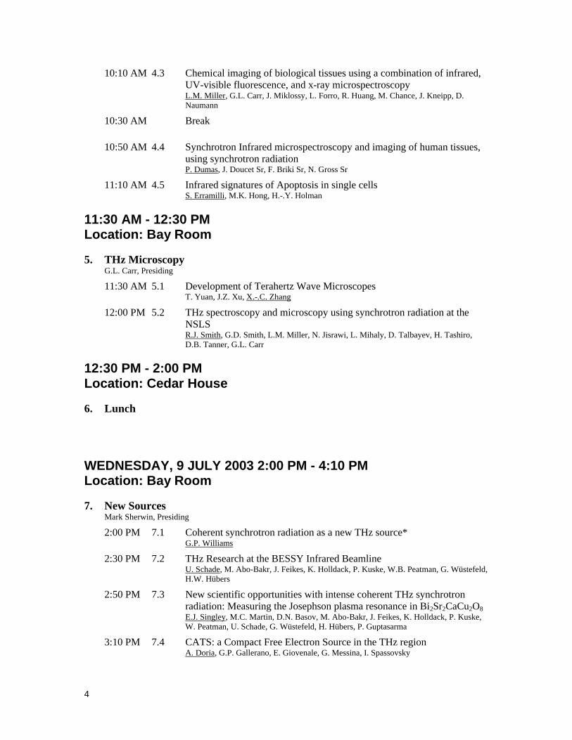
10:10 AM 4.3 Chemical imaging of biological tissues using a combination of infrared, UV-visible fluorescence, and x-ray microspectroscopy L.M. Miller, G.L. Carr, J. Miklossy, L. Forro, R. Huang, M. Chance, J. Kneipp, D. Naumann
10:30 AM Break
10:50 AM 4.4 Synchrotron Infrared microspectroscopy and imaging of human tissues, using synchrotron radiation P. Dumas, J. Doucet Sr, F. Briki Sr, N. Gross Sr
11:10 AM 4.5 Infrared signatures of Apoptosis in single cells S. Erramilli, M.K. Hong, H.-.Y. Holman
11:30 AM - 12:30 PM Location: Bay Room
5. THz Microscopy G.L. Carr, Presiding
11:30 AM 5.1 Development of Terahertz Wave Microscopes T. Yuan, J.Z. Xu, X.-.C. Zhang
12:00 PM 5.2 THz spectroscopy and microscopy using synchrotron radiation at the NSLS R.J. Smith, G.D. Smith, L.M. Miller, N. Jisrawi, L. Mihaly, D. Talbayev, H. Tashiro, D.B. Tanner, G.L. Carr
12:30 PM - 2:00 PM Location: Cedar House
6. Lunch
WEDNESDAY, 9 JULY 2003 2:00 PM - 4:10 PM Location: Bay Room
7. New Sources Mark Sherwin, Presiding
2:00 PM 7.1 Coherent synchrotron radiation as a new THz source* G.P. Williams
2:30 PM 7.2 THz Research at the BESSY Infrared Beamline U. Schade, M. Abo-Bakr, J. Feikes, K. Holldack, P. Kuske, W.B. Peatman, G. Wüstefeld, H.W. Hübers
2:50 PM 7.3 New scientific opportunities with intense coherent THz synchrotron radiation: Measuring the Josephson plasma resonance in Bi2Sr2CaCu2O8 E.J. Singley, M.C. Martin, D.N. Basov, M. Abo-Bakr, J. Feikes, K. Holldack, P. Kuske, W. Peatman, U. Schade, G. Wüstefeld, H. Hübers, P. Guptasarma
3:10 PM 7.4 CATS: a Compact Free Electron Source in the THz region A. Doria, G.P. Gallerano, E. Giovenale, G. Messina, I. Spassovsky

5
3:30 PM 7.5 CIRCE: a dedicated storage ring for Far-IR THz coherent synchrotron radiation F. Sannibale, J.M. Byrd, W.E. Byrne, M.C. Martin, W.R. McKinney, D.V. Munson, H. Nishimura, D.S. Robin, T. Scarvie, R.D. Schlueter, C.A. Steier, W.G. Thur, J.Y. Jung, W. Wan
3:50 PM 7.6 Stimulated–Superradiance FEL Oscillator A. Gover
WEDNESDAY, 9 JULY 2003 4:10 PM - 6:00 PM Location: Bay Room
8. Advanced Techniques George Neil, Presiding
4:10 PM Break
4:40 PM 8.1 High Field Electron Spin Resonance on Correlated Electron Systems L. Mihaly
5:10 PM 8.2 Non-linear far-IR spectroscopy with an FEL T. Dekorsy
5:40 PM 8.3 Enhancing the spatial resolution for synchrotron infrared microspectroscopy G.D. Smith, R.J. Smith, L.M. Miller, G.L. Carr
6:00 PM - 8:00 PM Location: Cedar House
9. Dinner
8:00 PM - 10:00 PM Location: Pavilion
10. Poster Session
10.1 Fourier-transform infrared spectroscopy of the freshwater blue-green algae Anabaena flos-aquae and Aphanizomenon flos-aquae- D. Sigee, A. Dean, K. White, M. Tobin
10.2 High-Pressure Synchrotron Infrared Studies of Mineral Systems Z. Liu, H.K. Mao, R. Hemley
10.3 Infrared micro- spectroscopy at the ANKA infrared edge radiation beamline: application to cement mineralogy B. Gasharova, Y.L. Mathis, K. Garbev, P. Stemmermann, D.A. Moss
10.4 Infrared characterization of environmental samples by pulsed photothermal spectroscopy W. Seidel, H. Foerstendorf, K.H. Heise, R. Nicolai, T. Dekorsy, J.M. Ortega, F. Glotin, R. Prazeres
10.5 Noise reduction efforts for the infrared beamlines at the Advanced Light Source T. Scarvie, N. Andresen, K. Baptiste, J. Byrd, M. Chin, M.C. Martin, W.R. McKinney, C. Steier

6
10.6 High Intensity Coherent THz Pulses at the NSLS DUV-FEL G.L. Carr, H. Loos, W.S. Graves, B. Sheehy
10.7 Infrared Microspectroscopy Station at BL43IR of SPring-8 Y. Ikemoto, T. Moriwaki, T. Hirono, H. Kimura, K. Kobayashi, S.I. Kimura, K. Shinoda, M. Matsunami, T. Nanba, N. Nagai
10.8 A microtron-injector for laboratoty-scale wide-band FIR FEL G.M. Kazakevitch, Y.U. Jeong, B.C. Lee, V.M. Pavlov, M.N. Kondaurov
10.9 Unusual In-Plane Anisotropy in a series of Detwinned Single Crystals of La2-xSrxCuO4 as viewed by Infrared Spectroscopy W.J. Padilla, M. Dumm, D.N. Basov, S. Komiya, Y. Ando
10.10 Synchrotron radiation-based FTIR-SM of latent human fingerprints - a novel forensic analysis T.J. Wilkinson, M.C. Martin, W.R. McKinney, D.L. Perry
Post-deadline Posters:
10.11 THz radiation studies on biological systems at the ENEA FEL Facility A.Doria, G. P. Gallerano, E. Giovenale, G. Messina, A. Lai, A. Ramundo-Orlando, V. Sposato, M. D’Arienzo, A. Perrotta, M. Romanò, M. Sarti, M. R. Scarfì, O. Zeni
THURSDAY, 10 JULY 2003
7:30 AM - 8:45 AM Location: Cedar House
11. Breakfast
9:00 AM - 11:30 AM Location: Bay Room
12. Environmental & Planetary Sciences David W. Piston, Presiding
9:00 AM 12.1 Evidence links the survival strategy of Arthrobacter to the dynamic fine-grain formation of chromium-ligands in aerobic environments H.-.Y. Holman, Z. Lin, N.V. Asatiani, T.L. Kalabegishvili, N.A. Sapojnikova, M.C. Martin, W.R. McKinney, D.L. Perry, N.Y. Tsibakhashvili
9:30 AM 12.2 New Developments in High-Pressure Synchrotron Infrared Spectroscopy R.J. Hemley, Z. Liu, H.K. Mao
10:00 AM 12.3 Synchrotron infrared spectroscopy of cosmic dust - direct comparison with astronomical data J.P. Bradley
10:30 AM Break
10:50 AM 12.4 Infrared Spectroscopy of Cosmic Dust in the Laboratory: An Effort to Identify the Minerals in Interstellar Grains G.J. Flynn, L.P. Keller

7
11:10 AM 12.5 Agricultural and Ecological Applications of Synchrotron IR Microscopy T.K. Raab, J.P. Vogel
11:30 AM - 12:30 PM Location: Bay Room
13. Applied IR Microscopy Paul Dumas, Presiding
11:30 AM 13.1 Highly resolved infrared microscopy in polymer science G. Ellis
12:00 PM 13.2 Darkfield Illumination Method for Infrared Microscopy using Synchrotron Radiation K. Nishikida
12:30 PM - 2:00 PM Location: Cedar House
14. Lunch
2:00 PM - 3:20 PM Location: Bay Room
15. Beyond the diffraction limit Todd I. Smith, Presiding
2:00 PM 15.1 Near-field optical microscopy: overview and perspectives G. Margaritondo, P. Perfetti, M. Luce, R. Generosi, N.H. Tolk, J.S. Sanghera, I.D. Aggarwal, G. Talley, A. Cricenti
2:30 PM 15.2 Laser Scanning Microscopy in the Vibrational Spectral Domain with One-Micrometer Resolution D. Palanker, D. Simanovskii, K. Cohn, T. Smith
3:00 PM 15.3 Infrared near-field Spectromicroscopy: Theoretical approach of the resolution limit for absorbing sample A. Dazzi, L. Salomon
3:20 PM - 6:00 PM Location: Bay Room
16. Facility News & Updates Gwyn P. Williams, Presiding
3:20 PM 16.1 Synchrotron Infrared Microspectroscopy beamlines projects at SOLEIL and at ESRF F. Polack, O. Chubar Sr, K. Scheidt, P. Elleaume Sr, J. Susini Sr, P. Dumas
3:35 PM 16.2 Performance and noise studies of infrared beamline at NSRRC of Taiwan light source Y.C. Lo, C. Wang, C.I. Chen, C.H. Chang, K.T. Hsu, D.S. Hung, K.L. Tsang, C.T. Chen
3:50 PM Break

8
4:15 PM 16.3 First experiments at SINBAD, the Synchrotron Infrared Beamline at DAFNE A. Marcelli, A. Nucara, E. Burattini, M. Piccinini, A. Grilli, A. Raco, G. Cinque, P. Calvani Sr, M. Cestelli Guidi
4:30 PM 16.4 The edge radiation infrared beamline ANKA-IR Y.L. Mathis, B. Gasharova, D.A. Moss
4:45 PM 16.5 The JLab THz/IR/UV Coherent Light Source Facility G. Neil, H.F. Dylla, G. Williams
5:00 PM 16.6 Wide-band FIR FEL Experimental Bench for Users Applications Y.U. Jeong, G.M. Kazakevitch, B.C. Lee, S.H. Park
5:15 PM 16.7 A new Synchrotron Infrared Beamline at ELETTRA S. Lupi, A. Nucara, P. Calvani, L. Quaroni
5:30 PM 16.8 Infrared Facility at the Canadian Light Source T. May
5:45 PM 16.9 The synchrotron IR program at the Swiss Light Source facility L. Degiorgi
6:00 PM 16.10 Results from the Infrared Beamline at the SRC C. Hirschmugl, R. Julian, R. Hansen
7:00 PM - 9:00 PM Location: Cedar House
17. Banquet Dinner
FRIDAY, 11 JULY 2003
7:30 AM - 8:45 AM Location: Cedar House
18. Breakfast
9:00 AM - 11:00 AM Location: Bay Room
19. Applied IR Spectroscopy Daniel Palanker, Presiding
9:00 PM 19.1 Microscopic Electro-Optical Studies on Blue Bronze, a Charge-Density-Wave Conductor J.W. Brill, R.C. Rai, V.A. Bondarenko
9:20 AM 19.2 Vibrational Lifetime of Hydrogen Defects in Silicon G. Luepke
9:50 AM 19.3 Semiconductors in strong, periodic Terahertz fields M. Sherwin
10:20 AM 19.4 Artificial Magnetic Response from Nonmagnetic Conductors at

9
Terahertz Frequencies W.J. Padilla, D.N. Basov, D.R. Smith, D. Yen, X. Zhang
10:40 AM Break
11:00 AM 19.5 Synchrotron far infrared reflectance spectroscopy in interfacial electrochemistry F. Hahn, C.A. Melendres
11:00 AM - 1:00 PM Location: Bay Room
20. Strongly Correlated Materials L. Mihaly, Presiding
11:20 AM 20.1 An infrared probe of inhomogeneous superconducting state in high-Tc cuprates D.N. Basov
11:50 AM 20.2 Synchrotron-based far-infrared ellipsometry on high-Tc superconductors A.V. Boris, C. Bernhard, N.N. Kovaleva, A.V. Pimenov, D. Munzar, Y.L. Mathis, B. Keimer
12:20 PM 20.3 Ultrashort THz pulses: a dynamical probe of insulating, conducting and superconducting phases R.A. Kaindl, M. Carnahan, D. Haegele, J. Orenstein, D. Chemla, J. Eckstein, S. Oh
12:40 PM 20.4 Time-resolved THz spectroscopy of MgB2 and ultra-thin á-MoGe films H. Tashiro, R.P.S.M. Lobo, D.B. Tanner, D.H. Reitze, J.M. Graybeal, S.-.I. Lee, G.L. Carr
1:00 PM - 2:00 PM Location: Cedar House
21. Lunch
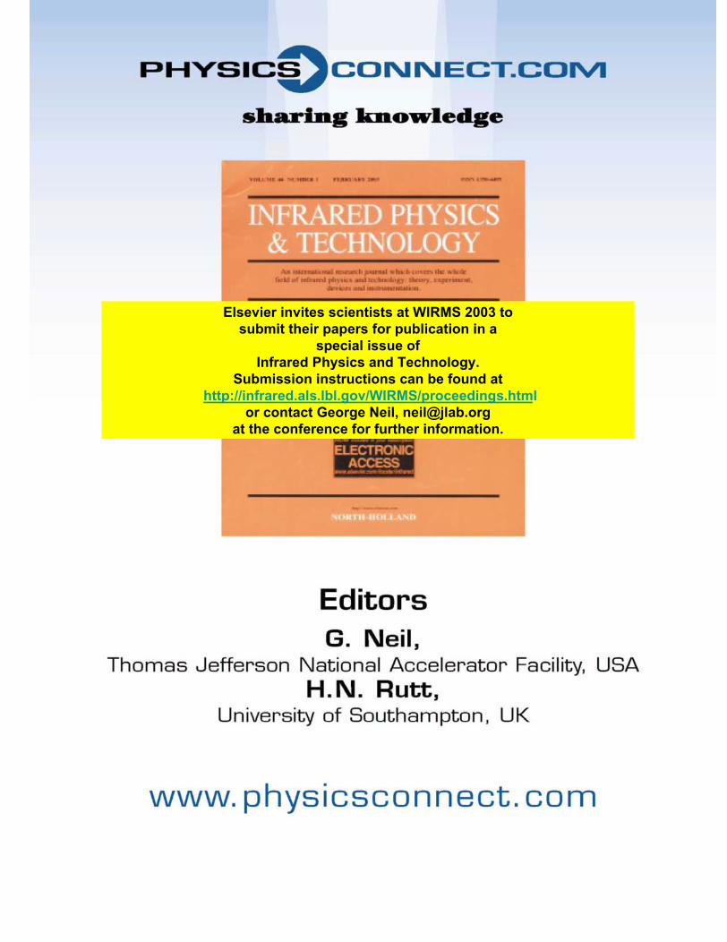
Elsevier invites scientists at WIRMS 2003 to submit their papers for publication in a
special issue of Infrared Physics and Technology.
Submission instructions can be found at http://infrared.als.lbl.gov/WIRMS/proceedings.html
or contact George Neil, [email protected] the conference for further information.

11
SESSION NO. 1, 6:00 PM Tuesday, 8 July 2003
Dinner Granlibakken Conference Center Cedar House
SESSION NO. 2, 8:00 PM Tuesday, 8 July 2003 Welcome Reception
Granlibakken Conference Center Cedar House
SESSION NO. 3, 7:30 AM Wednesday, 9 July 2003
Breakfast Granlibakken Conference Center Cedar House
SESSION NO. 4, 9:00 AM Wednesday, 9 July 2003
Biomedical Spectroscopy Granlibakken Conference Center Bay Room
H.-Y. Holman, Presiding

12
4.1. Biomedical Applications at the Vanderbilt FEL Center D.W. Piston, Vanderbilt University, USA
E-mail: [email protected] The Vanderbilt FEL Center has a wide array of on-going biomedical projects. Perhaps the most prominent has been
the application of 6.45 micron light to human surgery. Recent advances in fiber delivery are now opening up further applications of this surgical approach.
Another application of the IR FEL is the ultrashort-pulse infrared matrix-assisted laser desorption-ionization (IR-MALDI) mass spectrometry, holds promise to revolutionize the systematic analysis of proteins by increasing the rapidity of results and reducing the amount of sample required. Unlike conventional MALDI mass spectrometry, which cannot be performed with water-based solvents, IR-MALDI uses an aqueous matrix that more closely mimics the native protein environment. Further, analysis of water-based samples opens the possibility of studying protein expression directly from living cells, perhaps even from a single living cell. Development of IR-MALDI is a key component of our current biophotonics program.
We have also constructed a stand alone pulsed, tunable, monochromatic X-ray system designed for animal and human imaging. we use Compton Backscattering to generate monochromatic X-rays using a laser beam that is collided head-on into an electron beam. The laser scatters off the electron beam and becomes X-rays. This technology was originally developed using the FEL beam and the electron beam that pumps the FEL. Currently, we are developing applications for this X-ray source including mammography and protein crystallography.
13
4.2. Infrared Spectra and Spectral Maps of Individual Cells, Cellular Com-ponents and Tissue Sections
M. Diem, Hunter College, City University of New York, USA
E-mail: [email protected] During the past decade, several research groups have demonstrated that infrared mi-cro spectroscopy (IR-MSP) can
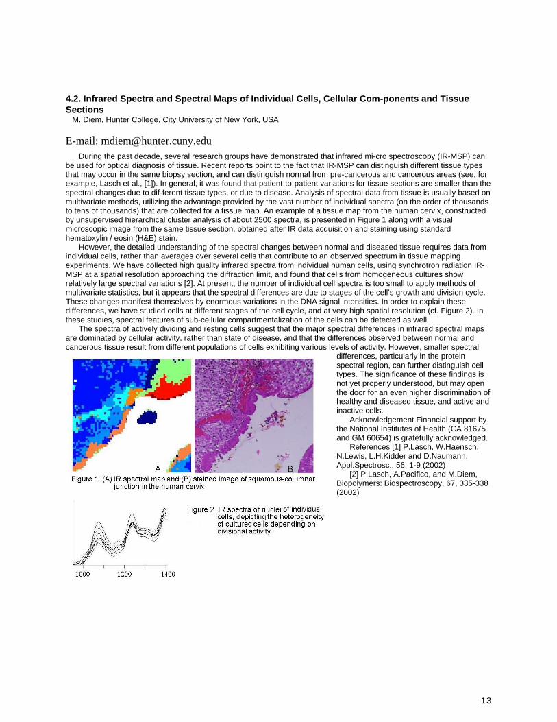
be used for optical diagnosis of tissue. Recent reports point to the fact that IR-MSP can distinguish different tissue types that may occur in the same biopsy section, and can distinguish normal from pre-cancerous and cancerous areas (see, for example, Lasch et al., [1]). In general, it was found that patient-to-patient variations for tissue sections are smaller than the spectral changes due to dif-ferent tissue types, or due to disease. Analysis of spectral data from tissue is usually based on multivariate methods, utilizing the advantage provided by the vast number of individual spectra (on the order of thousands to tens of thousands) that are collected for a tissue map. An example of a tissue map from the human cervix, constructed by unsupervised hierarchical cluster analysis of about 2500 spectra, is presented in Figure 1 along with a visual microscopic image from the same tissue section, obtained after IR data acquisition and staining using standard hematoxylin / eosin (H&E) stain.
However, the detailed understanding of the spectral changes between normal and diseased tissue requires data from individual cells, rather than averages over several cells that contribute to an observed spectrum in tissue mapping experiments. We have collected high quality infrared spectra from individual human cells, using synchrotron radiation IR-MSP at a spatial resolution approaching the diffraction limit, and found that cells from homogeneous cultures show relatively large spectral variations [2]. At present, the number of individual cell spectra is too small to apply methods of multivariate statistics, but it appears that the spectral differences are due to stages of the cell’s growth and division cycle. These changes manifest themselves by enormous variations in the DNA signal intensities. In order to explain these differences, we have studied cells at different stages of the cell cycle, and at very high spatial resolution (cf. Figure 2). In these studies, spectral features of sub-cellular compartmentalization of the cells can be detected as well.
The spectra of actively dividing and resting cells suggest that the major spectral differences in infrared spectral maps are dominated by cellular activity, rather than state of disease, and that the differences observed between normal and cancerous tissue result from different populations of cells exhibiting various levels of activity. However, smaller spectral
differences, particularly in the protein spectral region, can further distinguish cell types. The significance of these findings is not yet properly understood, but may open the door for an even higher discrimination of healthy and diseased tissue, and active and inactive cells.
Acknowledgement Financial support by the National Institutes of Health (CA 81675 and GM 60654) is gratefully acknowledged.
References [1] P.Lasch, W.Haensch, N.Lewis, L.H.Kidder and D.Naumann, Appl.Spectrosc., 56, 1-9 (2002)
[2] P.Lasch, A.Pacifico, and M.Diem, Biopolymers: Biospectroscopy, 67, 335-338 (2002)

14
4.3. Chemical imaging of biological tissues using a combination of infrared, UV-visible fluorescence, and x-ray microspectroscopy
L.M. Miller1, G.L. Carr1, J. Miklossy2, L. Forro3, R. Huang4, M. Chance4, J. Kneipp5, D. Naumann5, 1 Brookhaven National Laboratory, USA, 2 Temple University, 3 Swiss Federal Institute of Technology, 4 Albert Einstein College of Medicine, 5 Robert Koch Institute
E-mail: [email protected] Synchrotron infrared (IR) microspectroscopy is a valuable technique for examining the inherent chemical makeup of
biological cells and tissues at a spatial resolution unsurpassed by conventional IR microscopes. As a complementary technique, x-ray fluorescence microprobe can be used to image metal ions within biological tissue. For both techniques, the complex composition of biological tissues often benefits from sample visualization with fluorescence illumination. For example, immunofluorescence, where fluorochrome labels are attached to specific antibodies, has become a widespread and powerful technique in cell biology and immunology for visualizing targeted proteins.
In this work, technological improvements for combining infrared, UV-visible fluorescence, and x-ray microspectroscopy will be presented. In addition, biomedical applications to Alzheimer’s disease, scrapie, and bone disease will be discussed. This work was performed at Beamlines U10B and X26A at the National Synchrotron Light Source, Brookhaven National Laboratory. The NSLS is supported by the United States Department of Energy under contract DE-AC02-98CH10886. More information on the infrared programs at the NSLS can be found at http://infrared.nsls.bnl.gov.

15
4.4. Synchrotron Infrared microspectroscopy and imaging of human tissues, using synchrotron radiation
P. Dumas, J. Doucet Sr, F. Briki Sr, N. Gross Sr, LURE, France
E-mail: [email protected] Synchrotron radiation is a high-brightness infrared source, which is well suited to spectroscopy. The spectral range,
which extends from about 2.5 to 20 micron, so-called mid-infrared region, spans most of the vibrational mode frequencies necessary to a detailed identification of functional chemical groups. Brightness is crucial for infrared microscopy when one is trying to illuminate a small area as possible with as much light as possible. Accordingly, IR microscopic analysis have became diffraction-limited, typically half of the probed wavelength in confocal configuration. With enhanced spatial resolution, the imaging capability in Biological or biomedical studies has been also improved. The image quality obtained depends markedly on the signal-to-noise of the spectra acquired in diffraction-limited area of the sample (~6 10-3 absorbance unit or 0.4% transmittance for a 3x3 mm2 aperture, 32 scans, 8 cm-1 resolution). This is achieved with synchrotron-powered infrared microscope. High contrast imaging is achieved either using univariate analysis (chemical imaging), or multivariate analysis (hierarchical clustering, Principal Component Analysis, Fuzzy c-means cluster analysis …). Using the synchrotron infrared beamline at LURE (Mirage beamline), we have made a thorough study of human skin and hair sections. Highly localized lipids compounds have been identified and images in various regions of these tissues: Stratum Corneum for the skin sections, and cuticle and medulla for hair section. Using multivariate analysis and imaging, we have been able to discriminate between various lipids composition in different regions, as well as slightly different secondary peptide structure composition. Infrared microscopes can now be equipped with Focal Plane Array detector (FPA). We have also made a thorough comparison of the image quality obtained with a synchrotron powered IR microscope, and FPA-IR microscope, obtained on the same samples. This demonstrates the nice complementarities between the two approaches.

16
4.5. Infrared signatures of Apoptosis in single cells S. Erramilli1, M.K. Hong1, H.-.Y. Holman2, 1 Boston University, USA, 2 LBNL, USA
E-mail: [email protected] Apoptosis is a morphologically distinct form of cell death, associated with a number of characteristic molecular level
changes. Label-free techniques for detecting the onset of apoptosis are of considerable interest not only for cell biophysical studies, but also for assessing certain treatment modalities in which apoptosis can be deliberately induced in tumors. We show that infrared microspectroscopy provides us with several vibrational transitions that appear to characterize apoptosis. These changes are observed at the single cell level using synchrotron radiation based infrared microscopy, and are assigned to characteristic molecular transformations in the lipid membrane, as well as nucleic acid and protein secondary structure. Relating the observed infrared changes with other well established fluorescence and morphological assays suggests that infrared microspectroscopy can provide a tool for assaying the onset of apoptosis under physilogical conditions. Support from the Natonal Science Foundation is gratefully acknowledged.

17
SESSION NO. 5, 11:30 AM Wednesday, 9 July 2003 THz Microscopy
Granlibakken Conference Center Bay Room G.L. Carr, Presiding

18
5.1. Development of Terahertz Wave Microscopes T. Yuan, J.Z. Xu, X.-.C. Zhang, Rensselear Polytechnic Institute, USA
E-mail: [email protected]
Abstract Terahertz (THz) radiation, electromagnetic radiation in a frequency interval from 0.1 to 10 THz, has tremendous
potential as various materials and substances will allow their unique rotational and vibrational responses to be identified within this frequency. However, due to its long wavelength (0.3 mm at 1THz), the spatial resolution of a THz image is limited.
A microscope at THz frequency with sub-µm resolution will allow us to explore the rich spectroscopic signatures of molecular vibrations, rotations, and other low-energy transitions for microscopic sensing and imaging applications. Biological and organic compounds have distinct signatures within the THz region of the electromagnetic spectrum, such as molecular vibrational and rotational levels, and their chemical compositions can be examined by these THz wave microscopic systems. A THz wave microscope is capable of sensing and imaging structures at the cellular level with a µm or sub-µm spatial resolution, and may provide a new way for biomolecular spectroscopy and THz wave imaging.
We demonstrated an imaging setup by focusing a pumped fs-laser beam onto a small spot within an electro-optic (EO) crystal to generate a THz wave (THz wave). This near-field method has the potential to achieve resolution close to that of an optical microscope [1-5].
We will report the construction and preliminary results of current progress at Rensselaer’s Center for Terahertz Research on the development of THz wave microscopes. These microscopes generate and detect picosecond electromagnetic pulses (THz signals) by using nonlinear optical crystals or semiconductors, ultrafast laser pulses and computer analysis. To overcome the wavelength diffraction-limit, several methods are applied. One method uses a near-field imaging modality by focusing the optical beams into an EO crystal to generate (by optical rectification) and detect (by the EO effect) THz to a sub-micron resolution. The imaging area of the biomedical tissue, which is attached to the top of the EO crystal, is comparable to the optical focal spot, and is independent of the THz beam wavelength. The optimal conditions and damage threshold for optical focusing in a ZnTe crystal have been obtained, and 1/10 λ spatial resolution with the use of a 12 µm-thick ZnTe crystal has been demonstrated.
Figure 1 schematically illustrates one of the operation principles of the microscope. Because the fs-laser pump beam is tightly focused, optical damage in the emitter crystal is a major concern. In the experiment, we found the damage threshold of the ZnTe crystal to be about 100 GW/cm2, this number is lower than the data previously reported. The damage threshold limits the higher pump power. For example, when using a lens with numerical aperture 0.4, the focus spot diameter is about 2.5 µm and the maximum pump power of a 100 fs laser should be less than 10 mW. The measured result is in good agreement with the theoretical calculation.
Fig. 1. Schematic illustration of a THz wave microscope. A laser beam (pump beam) is modulated by an acoustic optical modulator and focused onto an EO crystal with an optical lens (NA=0.4). The focus diameter is about 2.6 µm, the total optical power is less than 10 mW. The transmitted THz wave (generated by optical rectification within the crystal) is detected by a collinear optical probe beam in the second EO crystal. A pair of balanced push-and-pull photodetectors is used to measure the optical probe beam.
During the process of optical rectification, a THz wave is generated along the whole pump beam path within the crystal. In order to obtain higher spatial resolution the emitter crystal has to be very thin. However, the trade-off is that the thinner the emitter crystal, the weaker THz wave generated. In addition, a very thin crystal is fragile. To avoid this is to use

19
a crystal that has poor phase-match between the laser beam and THz wave, so that the effective THz generation region is a very thin top layer.
We have used a thick and a thin ZnTe crystal as an emitter with a tightly focused optical beam. We have found that a thin crystal is more efficient for THz wave generation, as shown in Figure 2. This may be due to the higher order nonlinearity, but details need to be carefully studied. This enhancement will significantly benefit development of THz wave microscopy.
(a) (b)
Fig. 2. THz generation from ZnTe crystal from (a) a thick ZnTe crystal and (b) thin ZnTe crystal. The focal spot diameter is 2.5 µm.
We have tested the spatial resolution by using the experimental setup as shown in Figure 1 with a mask (a silver paint) on the surface of a 12 µm-thick ZnTe crystal. This arrangement is similar to setting up a sample close to the emitter crystal. A spatial resolution of 30 µm is demonstrated, which is 1/10 of the center wavelength of THz radiation, as shown in Figure 3.

20
(a) (b)
Fig. 3. (a) THz peak signal vs. scan position over a silver paint mark on a 12-µm thick ZnTe crystal (b) extended scale around 4.5 mm shows 30 µm spatial resolution for 10% to 90% signal variation.
Reference: 1. T. Yuan, S. Mickan, J.Z. Xu, D. Abbott and X.-C. Zhang, "Toward an apertureless electro-optic THz wave
microscope," CFD3, the Conference of Laser and Electro-Optics (CLEO), Long Beach, CA, May 24, 2002. 2. T. Yuan, J.Z. Xu and X.-C. Zhang, "THz wave microscope," paper #71, BECON 2002 – Sensors for Biological
Research and Medicine, Bethesda, MD, June 24, 2002. 3. J.Z. Xu, T. Yuan and X.-C. Zhang, "Pursuing THz microscope," WED13, International Symposium on Ultrafast
Phenomena and THz Wave, Beijing China July 24, 2002. 4. J. Z. Xu and X.-C. Zhang, "Optical rectification in an area with a diameter comparable to or smaller than the
center wavelength of THz radiation," Optics Letters, 27, 1067 (2002). 5. J.Z. Xu, T. Yuan and X –C. Zhang, "Optimal condition of THz optical rectification with a focused beam,"
Nonlinear Optics 2002, Hawaii Aug. 2002.
21
5.2. THz spectroscopy and microscopy using synchrotron radiation at the NSLS R.J. Smith1, G.D. Smith1, L.M. Miller1, N. Jisrawi1, L. Mihaly2, D. Talbayev2, H. Tashiro3, D.B. Tanner3, G.L. Carr1, 1 Brookhaven National Laboratory, USA, 2 Stony Brook University, USA, 3 University of Florida, USA
E-mail: [email protected] The synchrotron, as an infrared source, retains considerable advantages over thermal sources well into the far-
infrared, yet the its use in this spectral range has remained limited. This is partly due to the rather large angular collection required for efficient extraction from the synchrotron vacuum chamber, but also from a lack of instrumentation optimized for spectroscopy in this spectral range. Two IR beamlines at the NSLS, U4IR and U12IR, were designed to reach long wavelengths. At U4IR we have installed an infrared microspectrometer with a bolometer to explore the long wavelength performance, and have found that high quality spectra can be acquired to frequencies of 20 cm-1 (0.5 mm wavelength) with diffraction-limited performance. At U12IR, a Martin-Pupplett spectrometer reaches frequencies below 4 cm-1, although the quality of the spectra are limited due to presence of sharp spectroscopic features in the synchrotron's output. Some or all of these features are the result of an infrared source whose apparent dimensions are comparable to the electron beam chamber, in combination with multiple source points and highly reflecting materials, leading to strong interference effects.

22
SESSION NO. 6, 12:30 PM Wednesday, 9 July 2003
Lunch Granlibakken Conference Center Cedar House
SESSION NO. 7, 2:00 PM Wednesday, 9 July 2003
New Sources Granlibakken Conference Center Bay Room
Mark Sherwin, Presiding

23
7.1. Coherent synchrotron radiation as a new THz source* G.P. Williams, Jefferson Lab, USA
E-mail: [email protected] Studies of materials over increasingly wider spectral ranges and on shorter time scales lead to deeper understandings
of the fundamental mechanisms responsible for their behavior. In the infrared, many of these studies demand light sources of ever-increasing peak and average brightness across a broad band of wavelengths covering 4 orders of magnitude, and in regions where thermal sources are weak. Accelerators have played a major role in advancing the technology of such sources, particularly for applications to IR microscopy and spectroscopy. We will describe recent advances [1,2] which use coherent synchrotron radiation [3] to improve the performance of these sources considerably. Specifically in Nature we reported the production of high power (20 watts average, ~1 Megawatt peak) broadband THz light from the coherent emission off sub-picosecond bunches of relativistic electrons. The work was done at the Thomas Jefferson National Accelerator Facility. Electron bunches passing through a dipole magnet exhibit super-radiant synchrotron radiation emission at THz wavelengths when the bunch length is shorter than the wavelength of the emitted light. The radiation is essentially similar to the THz radiation produced by ultrafast laser techniques - spatially coherent, short duration pulses with transform-limited spectral content. The high intensity is easily understood from Larmor's formula as being due to the relativistic enhancement. In our experiment with 40 MeV electrons, the ratio of the mass of the electrons to their rest mass was 78, and the enhancement was the 4th power of this, namely 784.
In the talk the physics of these sources will be described in detail, together with theoretical calculations and their experimental verification. We will also describe a new THz facility at Jefferson Lab which has higher peak and average power than we reported before [2]. This striking improvement in THz power has potential applications in driving a new class of non-linear phenomena via the strong electric fields, and in studies of linear dynamics in the time or frequency domain. Further applications are likely to be in imaging by allowing full-field, real-time frame capture.
*In collaboration with George Neil (JLab), Kevin Jordan (JLab), Larry Carr (BNL), Mike Martin (LBNL) and Wayne McKinney (LBNL)
1. G.L. Carr, M.C. Martin, W.R. McKinney, K. Jordan, G.R. Neil and G.P. Williams "High Power Terahertz Radiation from Relativistic Electrons", Nature 420 153-156 (2002).
2. M. Abo-Bakr, J. Feikes, K. Holldack, P. Kuske, W. B. Peatman, U. Schade, G. Wüstefeld, and H.-W. Hübers "Brilliant, Coherent Far-Infrared (THz) Synchrotron Radiation", Phys. Rev. Lett. 90, 094801 (2003)
3. Carol J. Hirschmugl, Michael Sagurton and Gwyn P. Williams, "Multiparticle Coherence Calculations for Synchrotron Radiation Emission", Physical Review A44, 1316, (1991).
This work was supported primarily by the U.S. Dept. of Energy under contracts DE-AC02-98CH10886 (Brookhaven National Laboratory), DE-AC03-76SF00098 (Lawrence Berkeley National Laboratory) and DE-AC05-84-ER40150 (Thomas Jefferson National Accelerator Facility). The JLab FEL is supported by the Office of Naval Research, the Air Force Research Laboratory, the Commonwealth of Virginia and the Laser Processing Consortium. We are indebted to our colleagues at each institution for critical support without which these experiments would not have been possible.

24
7.2. THz Research at the BESSY Infrared Beamline U. Schade1, M. Abo-Bakr1, J. Feikes1, K. Holldack1, P. Kuske1, W.B. Peatman1, G. Wüstefeld1, H.W. Hübers2, 1 Berliner Elektronenspeicherring-Gesellschaft für Synchrotronstrahlung m.b.H, Germany, 2 Deutsches Zentrum für Luft- und Raumfahrt, Germany
E-mail: [email protected]
Synchrotron radiation sources have proven themselves many times over as brilliant emitters of radiation in the VUV, the soft and the hard x-ray regions of the spectrum. In recent years, their strength as a unique source of IR radiation has also become apparent and is increasingly being exploited at synchrotron radiation facilities around the world. In particular, IR radiation in the mid infrared wavelength region from incoherent synchrotron radiation sources has found increasing use in research by means of Fourier transform spectroscopy on biological tissues down to single cells, high-pressure and micro-sample measurements and in investigations on surfaces and thin films applying infrared ellipsometry with a high lateral resolution. The infrared beamline at BESSY, IRIS, is a multipurpose beamline which is equipped with two Fourier transform spectrometer, a microscope as well as an ellipsometer and provides useful IR intensities over a broad spectral range [1].
Also in the far infrared region with energies between 10 and 400 cm-1 incoherent synchrotron radiation is an excellent broadband source of high brilliance and power in comparison with standard thermal sources. However, for wavenumbers beyond 10 cm-1 where thermal sources are not anymore feasible for spectroscopic applications the low net transmittance of the extracting optics of an infrared beamline caused by diffraction due to the larger natural opening angle of the radiation and the finite sizes of the optical elements involved may limit the practical application of the incoherent synchrotron radiation.
This counts for the part of the electromagnetic spectrum between microwaves and thermal black body radiation. Between these limits no powerful radiation has been available until recently. Therefore, this region of the electromagnetic spectrum was referred to as the 'THz-gap'. The gap could be closed recently by fs-table-top-lasers which are not particularly powerful. The only powerful sources so far have been free electron lasers or diodes. These sources, however, show a small bandwidth and hence are not useful for spectroscopic applications. Coherent synchrotron radiation (CSR) from LINACs and storage rings is a tool which overcomes these limitations. It offers powerful and broadband radiation in the THz-range. For the first time CSR was observed 1989 in Japan at Tohoku-300-MeV LINAC. Recently an average power of 20 W was reported from the LINAC at Jefferson Laboratory [2]. CSR was also detected at some electron storage rings in the last years, but only as bursting radiation, indicating that bunch instabilities are involved in the emission process. During the past few years, at BESSY a new technique to generate stable, coherent sub-THz and THz-radiation from the electron storage ring has been developed [3]. THz-radiation is emitted by relativistic electrons radially accelerated by magnetic fields, as a part of the synchrotron radiation spectrum ranging from X-rays to THz-radiation. Normally, the phases of the electro-magnetic waves are not correlated and the power of the radiation is linearly increasing with the number of radiating electrons. In this incoherent radiation process the emitted THz power is low. For the transition from incoherent to coherent emission process, three length parameters have to be considered: bunch length, radiation wavelength, and cutoff wavelength. For radiation with wavelength longer than the bunch, phases of the waves become independent of the emission point within the bunch and all phases become equal. The bunch can be considered as a single macroparticle and the emitted waves add up coherently. Their field intensity grows linearly and their power quadratically with the number of electrons leading to a dramatic enhancement in the emitted power since there are 108 to 109 electrons involved in this process. The cutoff of the vacuum chamber sets a limit to this process.
Initial tests of the feasibility of using the coherent synchrotron radiation in scientific applications at the IRIS beamline have been made [4]. As an example, the Josephson plasma resonance in the sub-THz region of optimally doped Bi2Sr2CaCu2O8 could be measured for the first time [5]. The production of stable, high power, coherent synchrotron radiation at THz and sub-THz frequencies at BESSY opens a new region in the electromagnetic spectrum which can be applied for imaging, spectroscopic and microscopic methods in solid state physics, biology, and medicine.
Acknowledgments We are indebted to our colleagues of BESSY for discussions and support. The work is supported by the Bundesministerium für Bildung und Forschung and by the Land Berlin.
References [1] U. Schade et al., Rev. Sci. Instr., 73, 1568-1570 (2002). [2] L. Carr et al., Nature 420, 153 (2002). [3] M. Abo-Bakr, et al., Phys. Rev.Lett. 88, 254801 (2002). [4] M. Abo-Bakr, et al., Phys. Rev.Lett. 90, 094801 (2003). [5] J. Singley et al., (2003), to be published.

25
7.3. New scientific opportunities with intense coherent THz synchrotron radiation: Measuring the Josephson plasma resonance in Bi2Sr2CaCu2O8
E.J. Singley1, M.C. Martin1, D.N. Basov2, M. Abo-Bakr3, J. Feikes3, K. Holldack3, P. Kuske3, W. Peatman3, U. Schade3, G. Wüstefeld3, H. Hübers4, P. Guptasarma5, 1 Advanced Light Source, Lawrence Berkeley Laboratory, USA, 2 University of Califonia, San Diego, USA, 3 Berliner Elektronenspeicherring-Gesellschaft für Synchrotronstrahlung m.b.H, Germany, 4 Institut für Weltraumsensorik und Planetenerkundung, Germany, 5 University of Wisconsin-Milwaukee, USA
E-mail: [email protected] Infrared spectroscopy has been employed to investigate the c-axis reflectivity of Bi2Sr2CaCu2O8 in the sub-THz
frequency region. In order to reach this challenging frequency range a novel synchrotron source has been employed. Working in a special low momentum compaction mode of operation where the electron bunch shape is significantly shortened and distorted, stable broadband coherent (super-radiant) very far-IR radiation is produced with orders of magnitude more intensity than conventional thermal and synchrotron sources. Using this source for reflectivity measurements we have been able to observe the Josephson Plasma Resonance (JPR) in optimally doped Bi2Sr2CaCu2O8 for the first time. This source allows us to investigate charge dynamics in this extremely anisotropic superconductor, and opens up the possibility to study other highly correlated systems in this critical low energy region.

26
7.4. CATS: a Compact Free Electron Source in the THz region A. Doria, G.P. Gallerano, E. Giovenale, G. Messina, I. Spassovsky, ENEA, Italy
E-mail: [email protected] A Coherent Advanced THz Source (CATS) has been realised at the ENEA FEL laboratories in Frascati. The CATS
project exploit the coherent spontaneous emission from short bunches of relativistic electrons. This FEL source utilizes a 3 MeV RF linac to generate the electron beam, which is injected into a magnetic undulator composed of 16 periods, each 2.5 cm long. A second RF structure, called Phase Matching Device (PMD), is inserted between the linac and the undulator and is controlled in phase and amplitude to correlate the electron distribution in energy as a function of time in the bunch. In this way the contributions to the total radiated field by individual electrons in the bunch are added in phase, leading to a manyfold enhancement of the coherent emission. The radiation emitted during the first set of running tests can be tuned from 480 µm up to 800 µm just acting on the relative RF phese between the Linac and the PMD. The characteristics of the source and of the radiation generated, together with the potentialities will be presented.

27
7.5. CIRCE: a dedicated storage ring for Far-IR THz coherent synchrotron radiation F. Sannibale, J.M. Byrd, W.E. Byrne, M.C. Martin, W.R. McKinney, D.V. Munson, H. Nishimura, D.S. Robin, T. Scarvie, R.D. Schlueter, C.A. Steier, W.G. Thur, J.Y. Jung, W. Wan, Advanced Light Source, Lawrence Berkeley National Laboratory, USA
E-mail: [email protected] We present the concepts for a storage ring dedicated to and optimized for the production of stable coherent
synchrotron radiation (CSR) over the far-infrared terahertz wavelength range from 200 µm to about a cm. CIRCE (Coherent InfraRed CEnter) will be a 66 m circumference ring using the ALS injector and will be located on top of the existing ALS booster synchrotron shielding. This area provides enough floor space for both the ring and the beamlines. We present a model for CSR emission in which the stable bunch distortion induced by the synchrotron radiation field is used to significantly extend the CSR emission towards high frequencies. In this configuration a photon flux gain of 6 - 8 orders of magnitude was calculated for CIRCE when compared with the best existing sources. Additionally, the particular design of the dipole vacuum chamber allows a greater transmission of the far-infrared. We believe that such a source can be constructed for a modest cost.

28
7.6. Stimulated–Superradiance FEL Oscillator A. Gover, Tel-Aviv University, Israel
E-mail: [email protected] The synchrotron undulator radiation of an electron beam is substantially enhanced when the electron beam is
prebunched – either periodically, at a frequency (f) within the spectral range of the synchrotron undulator radiation [1], or in single bunches of duration shorter than the optical period of the radiation frequency (T=1/f) [2]. In the first case the emitted radiation is entirely monochromatic and coherent, and in the second case its spectral range is equal to the undulator radiation bandwidth f/Nu (Nu – number of undulator periods). In both cases, the emission power is proportional to the number of electrons per bunch (Nb) squared [3,4], instead of just being proportional to the number of electrons or the beam current, as is the case for conventional synchrotron undulator radiation [5].
This kind of enhanced radiation emission is a phenomenon of superradiance [6], because it stems from coherent
constructive interference (summation) of the electric field amplitudes of the radiation wavepackets emitted by all electrons. Thus, the E field amplitude of the total radiation field is proportional to the number of electrons in the bunch Nb (or the beam bunching current Ib). Consequently the total emitted radiation energy or power is proportional to Nb
2 (or Ib2).
A landmark experiment carried out recently in TJL [7] demonstrated that this scheme can provide very high intensity in
the THz regime with a magnetic dipole (“half a wiggle period”), emitting Coherent Synchrotron Radiation (CSR). Of course, such radiation will be substantially more intense and monochromatic, if a full wiggler is used [8]. In this case, single bunches have a disadvantage relative to periodic bunching, because of the slippage effect. However, this can be overcome by using a “zero-slippage” dispesive waveguide scheme [2] or a short train of periodic bunches [9].
Yet, a many orders of magnitude enhancement of the radiation from a bunched beam is still possible, if the super-
radiant emitting bunches will be stimulated by a radiation field to emit. This stimulated superradiance process was demonstrated by us experimentally in the microwave regime [10] and would be extremely interesting if it could be explored within a resonator in an oscillator configuration. Preliminary analysis of this yet-non existing radiation scheme, was presented by us in [11]. In order to realize it with single bunches, three technical problems must be solved:
a) Good synchronization between the bunch repetition rate and the cavity round trip time.
b) Use of schemes to reduce the slippage effect as discussed before.
c) Providing a scheme for bringing the oscillator to enter the stable “high energy- extraction saturation-state” in the system that is bistable in nature.
I will present the unique characteristics of this new kind of high efficiency high power oscillator and technical ways to realize it.
References
1. I. Schnitzer and A. Gover, “The Prebunched Free Electron Laser in Various Operating Gain Regimes”, Nucl. Inst. & Meth. In Phys. Res, A237 1240140 (1985).
2. A. Gover, F. V.Hartemann, G.P. lesage, N.C. Luhmann, R.S. Zhang, C. Pelllegrini, “Time and Frequency Domain analysis of superradiant coherent synchrotron radiation in a wave-guide free electron laser”, Phys. Rev. Lett., 72, 1192-1195 (1994).
3. Y. Pinhasi, A. Gover, “A unified analysis of spontaneous and super-radiant emissions in free electron lasers”, Nucl. Inst. & Meth. In Phys. Res. A. 393, 343-347 (1997).
4. M. Arbel, A.L. Eichenbaum, Y. Pinhasi, Y. Lurie, M. Tecimer, A. Abramovich, H. Kleinman, A. Gover, “Super-radiance in a prebunched beam free electron maser”, Nucl. Inst. & Meth in Phys. Res. A445, 247-252 (2000).
5. H. Motz , R.N. Whitehurst, J. Appl. Phys. 22, 826 (1953). 6. R. H. Dicke, Phys. Rev. 93, 99 (1954). 7. G.L. Carr et al, “High power terahertz radiation from relativistic electrons” Nature, 420, 153 (Nov. 2002). 8. http://www.frascati.enea.it/thz-bridge/fel-cats.htm 9. J. G. Neumann, P.G. O’Shea, D. Demske, W.S. Graves, B. Sheehy, H. Loos, G.L. Carr, “Electron beam modulation
using a laser driven photocathode”, 24th Int’l FEL Conf., Chicago, Sept. (9-13) 2002. 10. M. Arbel, A. Abramovich, A.L. Eichnebaum, A. Gover, H. Kleinman, Y. Pinhasi, I.M. Yakover, “Super-radiant and
stimulated super-radiant emission in a pre-bunched beam free electron maser”, Phys. Rev. Lett., 86, 2561-2564 (2001).
11. M.V. Krongauz, Y. Pinhasi, M. Tecimer, A. Gover, “Bi-stable and post-saturation optimization in a pre-bunched free electron laser”, Nucl. Inst. & Meth. In Phys. Res. A 445, 28-33 (2000).

29
SESSION NO. 8, 4:00 PM Wednesday, 9 July 2003
Advanced Techniques Granlibakken Conference Center Bay Room
George Neil, Presiding

30
8.1. High Field Electron Spin Resonance on Correlated Electron Systems L. Mihaly, Brookhaven National Laboratory, USA
E-mail: [email protected] LaMnO3 exhibits an extensively-studied antiferromagnetic (AF) transition at TN=141K. Electron spin resonance (ESR)
has been studied in a stoichiometric single crystal of LaMnO3 in the temperature range of 4.2K-250K. The frequency range of our instrument covers a broad far-infrared to infrared band, with a lower cut-off frequency of 4cm-1 (120GHz), diffraction-limited by the sample size. Magnetic fields up to 14T have been applied in several different directions: along the crystallographic b direction (the easy axis direction in the AF state) and perpendicular to it. The field dependence of the resonance is fully mapped. The low temperature results are described by the AF resonance theory of Kittel and Keffer, but corrections have to be made for the canted AF structure of the compund. Strong deviations from the theory are evident at temperatures close to TN.

31
8.2. Non-linear far-IR spectroscopy with an FEL T. Dekorsy, Forschungszentrum Rossendorf, Germany
E-mail: [email protected]
The non-linear optical properties of solid state materials in the THz frequency region are to a large extent unexplored due to missing high-intensity coherent light sources in this frequency range. Although recent advances in table-top laser based THz systems have been enormous, free-electron lasers (FEL) are at present still the only tunable lasers which provide high peak intensities and a sufficient narrow spectral width to perform nonlinear spectroscopy at THz frequencies. For solid-state materials a large number of elementary excitations lie in the THz frequency, like plasmons, phonon-polaritons, magnons, intersubband transitions in nanostructures, the superconducting gap in high-temperature superconductors, etc. The non-linear interaction of electromagnetic radiation in resonance with these excitations allows to gain deeper insight into basic physical properties.
Recently we investigated the dispersion of the second order nonlinear susceptibility in thin GaAs crystals below the optical phonon resonance via second harmonic generation (SHG) experiments with an FEL [1]. These experiments provide insight into the relative contributions of higher-order cohesive lattice forces to χ(2). The nonlinear optical susceptibility in polar semiconductors in the THz range is strongly influenced by the presence of optical phonons and should exhibit several peculiarities, i.e. a strong resonant enhancement of the SHG at half the frequency of the TO phonon (8.0 THz in GaAs) and at the TO phonon itself, and a zero-crossing for frequencies between 4.0 THz and the TO phonon resonance due to the cancellation of higher order ionic and electronic contributions. The first frequency doubling experiments below the phonon frequency of a semiconductor were performed on GaAs with a FIR gas laser operating in the frequency range from 0.6 THz to 1.7 THz [2]. However, the frequency of this laser system was too far away from the predicted resonance at half the phonon frequency to see any resonance enhancement. The zero-crossing of the second-order susceptibility theoretically expected around 5.1 THz could also not be observed.
The experiments are performed with the FEL FELIX (Nieuwegein, Netherlands), which delivers picosecond pulses at a macro-bunch repetition rate of 10 Hz and a micro-bunch repetition rate of 25 MHz with 100 micro-pulses per macro-pulse. The radiation frequency is tuned between 4 THz to 6 THz with a spectral width (FWHM) of 0.2 THz to 0.25 THz and a micro-pulse pulse energy between 4 and 8 µJ. The radiation was focused on thin GaAs films of several µm thickness only. Such thin samples are necessary because of the large phase mismatch around the lattice resonance. The SHG intensity was measured with a high sensitivity liquid He cooled Ge:Ga detector. In order to detect the SHG generated in the sample without background the FEL radiation has to be purified from higher harmonics produced in the undulator and the fundamental has to be blocked before the detector. These requirements are achieved with a crystalline quartz plate before the sample and a thick CsBr crystal before the detector, respectively, which define a window from 4.4 to 5.6 THz where the signal can clearly be attributed to SHG from the sample.
We could observe both the resonance and the zero-crossing of χ(2) below the Reststrahlen-band. From the value obtained for the zero-crossing of the nonlinear susceptibility we conclude that the contribution of the phonon interaction through the second-order lattice dipole moment has to be significantly smaller and the contribution from the third-order lattice potential anharmonicity has to be larger than determined previously [3]. Besides the relevance for the THz nonlinear susceptibility these terms are also important for two-phonon sidebands in the infrared absorption, phonon decay [3,4] and for a quantitative description of Raman spectra [5]. We propose that SHG below the optical phonon resonance is an elegant method to quantitatively determine the higher-order potential contributions to the nonlinear susceptibility - without the need for the determination of absolute conversion efficiencies.
References [1] T. Dekorsy et al., "Infrared-phonon-polariton resonance of the nonlinear susceptibility in GaAs", Phys. Rev. Lett. 90,
055508 (2003). [2] A. Mayer and F. Keilmann, "Far-infrared nonlinear optics. I. c(2) near ionic resonance ," Phys. Rev B 33, 6954
(1986). [3] C. Flytzanis, " Infrared dispersion of second order electric susceptibilities in semiconducting compounds," Phys.
Rev. B 6, 1264 (1972). [4] C. Flytzanis, "Dominant second-order dipole-moment contribution in the infrared absorption of III-V compounds,"
Phys. Rev. Lett. 29, 772 (1972). [5] S. Go, H. Bilz, and M. Cardona, " Bond charge, bond polarizability, and phonon spectra in semiconductors," Phys.
Rev. Lett. 34, 580 (1975).

32
8.3. Enhancing the spatial resolution for synchrotron infrared microspectroscopy G.D. Smith, R.J. Smith, L.M. Miller, G.L. Carr, Brookhaven National Laboratory, USA
E-mail: [email protected] By comparing the results of diffraction analysis with measurements on known specimens, we demonstrate that the
spatial resolution for synchrotron infrared microspectroscopy is controlled by diffraction. Images of small circular geometries show interesting artifacts that are in agreement with diffraction predictions. We also note that the diffraction patterns for grazing incidence and ATR methods show strong deviations from a simple Airy disk. Lastly, we discuss opportunities for enhancing the resolution through PSF deconvolution, solid immersion lenses, and non-traditional approaches.

33
SESSION NO. 9, 6:00 PM Wednesday, 9 July 2003
Dinner Granlibakken Conference Center Cedar House
SESSION NO. 10, 8:00 PM Wednesday, 9 July 2003
Poster Session Granlibakken Conference Center Pavilion

34
10.1. Fourier-transform infrared spectroscopy of the freshwater blue-green algae Anabaena flos-aquae and Aphanizomenon flos-aquae-
D. Sigee1, A. Dean1, K. White1, M. Tobin2, 1 Manchester University, United Kingdom, 2 Daresbury Laboratory, United Kingdom
E-mail: [email protected] This study was carried out to obtain information on the molecular characteristics of two freshwater blue-green algae.
FTIR spectroscopy has considerable potential for the study of environmental samples, allowing high resolution analysis of single species within mixed populations of biota.
Mixed phytoplankton samples from a freshwater eutrophic lake were obtained from a range of depths within the water column, deposited on infrared reflectance slides and air-dried for FTIR analysis. FTIR spectra from colonies of Anabaena and Aphanizomenon showed clear absorbance bands, and were analysed in relation to qualitative (molecular assignments) and quantitative (areas values, correlations) parameters. Results showed that:
1. FTIR spectra from both algae showed a similar range of band assignments, indicating close molecular similarity. In both cases bands were: Band 1 (3029-3639)- mainly water, 2(2809-3012) lipid CH2, 3 (1583-1709) amide I, 4 (1381-1585) amide II, 5 (1425-1477) lipid C-CH3, 6 (1357-1423) lipid n-(CH3)3, 7 (1191-1356) nucleic acid >P=O, 8 (1134-1174) glycogen, 9 (1072-1099)glycogen, nucleic acid, 10 (980-1072) glycogen.
2. For each species, comparison (Kruskal Wallis test) of separate depth samples (n=20) showed no consistent differences in mean band areas, and indicated that all depth samples could be regarded as part of a single (aggregate) population within the water column.
3. Analysis of the aggregate population (n=80) for each species showed that the distribution of most band areas approximated to a normal distribution. Some did not, and non-parametric analysis was therefore used in subsequent tests.
4. Comparison of the two species (Mann Whitney test) showed that all molecular species of Anabaena were present at consistently higher levels compared to Aphanizomenon. This apparent difference between species was attributed to differences in specimen thickness, since normalisation to band 1 (mainly water of hydration) gave much closer values. Anova of normalised values gave significant interspecies differences only in respect of nucleic acid (band 7) and glycogen (bands 8,10).
5. For each species, Spearman correlation analysis of normalised data indicated distinctive patterns of band correlation within spectra. Factor analysis confirmed the Spearman results, and indicated that bands could be separated into two major groups – Group I (bands 2-7: 40-60% of sample variance), and Group II (bands 8-10: 20% of sample variance).
In this study, FTIR analysis provides novel information on the molecular composition of two ecologically important blue-green algae. The overall analysis suggests close qualitative and quantitative similarities between the two species, with little significant difference between samples taken at different depths in the water column. FTIR spectra from the blue green algae show key differences from the data previously obtained with environmental samples of the green alga Pediastrum, taken from the same lake (Sigee et al., (2002). Eur. J. Phycol. 37, 19-26).

35
10.2. High-Pressure Synchrotron Infrared Studies of Mineral Systems Z. Liu, H.K. Mao, R. Hemley, Geophysical Laboratory, Carnegie Institution of Washington, USA
E-mail: [email protected] High-pressure spectroscopy provides crucial and often unique information on the properties of Earth and planetary
materials from near-surface conditions to those of the deepest interiors. Vibrational infrared/Raman spectroscopy, for example, provides detailed information on bonding properties of crystals, glass, and melts, thereby yielding a microscopic description of thermochemical properties. Synchrotron infrared sources are well suited to high pressure investigations in which both small sample area and a narrow beam are required in order to generate extremely high pressure with a diamond anvil cell. The dedicated high-pressure beam line U2A on the VUV ring of the National Synchrotron Light Source, Brookhaven National Laboratory is an integrated facility for a wide range of microspectroscopic studies from ambient to ultrahigh pressures and at variable temperatures. Recently, the beamline has been upgraded to further improve the performance in far-IR range. The facility thus permits systematic high-pressure studies addressing a range of problems in Earth and planetary science. These studies include high pressure (and variable temperature) studies of planetary gases and ices; minerals of the Earth's crust, mantle, and core; geochemical reactions; glasses and melts; organic geochemistry; surfaces and interfaces and whole-rock samples; and extraterrestrial samples. Examples of recent experiments include studies of gypsum, coesite, OH-clinohumite, and OH-chondrodite.

36
10.3. Infrared micro- spectroscopy at the ANKA infrared edge radiation beamline: application to cement mineralogy
B. Gasharova, Y.L. Mathis, K. Garbev, P. Stemmermann, D.A. Moss, Forschungszentrum Karlsruhe, Germany
E-mail: [email protected] At the ANKA facility in Karlsruhe, we have been developing mineralogical applications based on FT-IR micro-
spectroscopy. Our aim is to address the need for more sophisticated investigations by using the advantages of the synchrotron edge radiation (SER) in the infrared spectral range compared to conventional laboratory sources: higher flux in the far IR and higher spatial resolution because of the higher brilliance in the complete IR domain.
One of our research directions is the study of new and not fully understood mineral crystal structures, transitions from semi-amorphous into crystalline state, mechanisms of incorporation of toxic ions into the mineral crystal structures. This will complement our results obtained by XRD, SEM methods, etc.
Another important need in many mineralogical applications is the identification of individual minerals as a function of spatial distribution. Images of the highest spatial resolution have been obtained using an atomic force microscopy. Unfortunately, there is no ability to differentiate crystallochemical differences with this technique. On the other hand, techniques such as (E)SEM provide some chemical information albeit at modest spatial fidelity. This trade-off between crystallochemical specifity and spatial fidelity means that we must often combine techniques in order to address analytical needs.
Infrared microscopy combines the rich crystallochemical specifity for samples even in amorphous state associated with vibrational spectroscopy. By using synchrotron-based infrared microspectroscopy we can investigate samples down to the diffraction limit. This extends a mainstream characterization tool into a new region of use.
The use of this technique can provide much new insight into the nature of mineral systems and mineral surface reactions. Examples with application to cement mineralogy that demonstrate the power of this technique will be discussed.
In this presentation we will focus on C-S-H phases, the main products of hydration of cement materials. These are amorphous calcium silicate hydrates (C-S-H), which are mostly characterized by their Ca/Si (C/S) ratio. The composition of the phases within the CaO-SiO2-H2O system varies over a large C/S range (0.6 to 2). Their properties determine to a large extend the physical properties of the whole system. Some critical points related to C-S-H that we would like to address using SER-FTIR will be presented: - carbonation of fresh and hardened cement pastes, - structure and incorporation of heavy metals in C-S-H phases, etc.

37
10.4. Infrared characterization of environmental samples by pulsed photothermal spectroscopy W. Seidel1, H. Foerstendorf1, K.H. Heise1, R. Nicolai1, T. Dekorsy1, J.M. Ortega2, F. Glotin2, R. Prazeres2, 1 Research Centre Rossendorf, Germany, 2 LURE, France
E-mail: [email protected] The low concentration of toxic radioactive metals in environmental samples often limits the interpretation of results of
infrared studies investigating the interaction processes between the metal ions and environmental compartments. For the first time, we could show that photothermal infrared spectroscopy performed with a pulsed free-electron laser can provide reliable infrared spectra throughout a distinct spectral range of interest. In this model investigation, we provide vibrational absorption spectra of a rare earth metal salt dissolved in a KBr matrix and a natural calcite sample obtained by thermal beam deflection technique and FT-IR spectroscopy, respectively. General agreement was found between all spectra of the different recording techniques. Spectral deviations were observed with samples containing low concentration of the rare earth metal salt indicating a lower detection limit of the photothermal method as compared to conventional FT-IR spectroscopy. Furthermore, the photothermal method provide spatial information of a sample surface. This may result in a microspectrometric technique for determining the distribution of metal species on mineral surfaces. First experiments exploring the spatial resolution of photothermal spectroscopy were carried out by scanning the surface of a germanium substrate showing a localized region where O-ions were implanted. The border range of this region was investigated by recording time curves of the deflection signal at distinct positions of the substrate surface with a constant free-electron laser wavelength of 11.6 micrometer.
38
10.5. Noise reduction efforts for the infrared beamlines at the Advanced Light Source T. Scarvie, N. Andresen, K. Baptiste, J. Byrd, M. Chin, M.C. Martin, W.R. McKinney, C. Steier, Lawrence Berkeley National Lab, USA
E-mail: [email protected] The quality of infrared microscopy and spectroscopy data collected at synchrotron based sources is strongly
dependent on noise. We have successfully identified and suppressed several noise sources affecting Beamline 1.4.2, 1.4.3, and 1.4.4 at the Advanced Light Source (ALS), resulting in significant reductions to the noise in the users' FTIR spectra. In this paper, we present our methods of noise source analysis and the techniques used to reduce the noise and its negative effect on the infrared beam quality. These include analyzing and changing physical mounts to better isolate portions of the beamline optics from low-frequency environmental noise, and modifying the input signals to the main RF system. We also discuss the relationship between electron beam energy oscillations at a point of dispersion and infrared beamline noise.

39
10.6. High Intensity Coherent THz Pulses at the NSLS DUV-FEL G.L. Carr, H. Loos, W.S. Graves, B. Sheehy, Brookhaven National Laboratory, USA
E-mail: [email protected] Electron accelerators for short wavelength free-electron lasers (FELs) are designed to produce extremely short
electron bunches (often less than 1 ps). The coherent superposition of the the radiated fields from such short bunches leads to a large enhancement in the very far-infrared (THz) spectral range. The NSLS Deep-UV FEL linac provides ~300 fs electron bunches with nearly 1/2 nC of charge (more than 109 electrons), and produces large THz pulses as dipole or transition radiation (about 1 µJ each). The qualities of such pulses and their use for a number of scientific measurements (e.g., "all THz pump-probe") will be presented.
40
10.7. Infrared Microspectroscopy Station at BL43IR of SPring-8 Y. Ikemoto1, T. Moriwaki1, T. Hirono1, H. Kimura1, K. Kobayashi1, S.I. Kimura2, K. Shinoda3, M. Matsunami4, T. Nanba4, N. Nagai5, 1 SPring-8/JASRI, Japan, 2 Institute for Molecular Science, Japan, 3 Osaka City Univ, Japan, 4 Kobe Univ, Japan, 5 Toray research center, Japan
E-mail: [email protected]
Infrared synchrotron radiation (IRSR) has high brilliance compared to the black body source. The brilliance is extremely important for microscopy, because the light is illuminate as small area as possible. The infrared microspectroscopy station at BL43IR of SPring-8 has been in operation since May 2000, and has users from the variety of fields such as physics, chemistry, earth science and medical science. In this paper, we examined the capability of the microscope in the far-IR region. The spatial resolution in far-IR region is found to be almost the diffraction limit depending on the wavenumber.
BL43IR at SPring-8 is designed exclusively for the infrared materials research[1]. The spectrometer is FTIR (Bruker 120HR/X). The spectral range is between 100 and 20000 cm-1. It has four different kinds of experimental stations, infrared surface science station[2], absorption and reflection spectroscopy station[3], magneto-optical spectroscopy station[4] and infrared microspectroscopy station[4]. The vertical divergence of IRSR becomes large with decreasing wavenumber and small with increasing the radius of curvature of the electron orbital. IRSR in BL43IR has the low divergence because of the large radius of curvature, 39.3 m. This is an advantage for the microspectroscopy, especially for far infrared region.
Figure 1 shows the schematic illustration of the optical path in the infrared microscopy station. We designed an original microscope for this station. Between the microscope and FTIR, there is an optical window to separate the vacuum of FTIR and air pressure. The window is chosen from quartz, BaF2, KRS-5 and polyethylene film depending on the wavenumber. All optical paths in the microscope are purged with dry air. By changing the angle of SM1, both of the transmission and the reflection spectra can be measured. The mirror SM2 is a parabolic one and the light is focused on the lower aperture. The mirror SM4 is also a parabolic one and the light is reflected down to the sample stage by a half-area mirror SM5. The light is focused on the sample stage by the Schwartzschild mirrors (x8, NA=0.5), SM6 and SM7. The upper aperture is located at the focal point of SM7. The improved aperture consists of four blades that can be slid independently. The detectors are a Si-bolometer in the far-infrared region, an MCT in the mid-infrared region, an InSb in mid- to near-infrared region, and a Si-photodiode in the near-infrared to visible region. The important advantage of our microscope is a long working distance, 100 mm. With this advantage, many kinds of instruments can be installed, such as a x-y mapping stage, flow-type cryostat (Oxford microstat-He, 4.2-400 K), high temperature DAC ( ~ 1000 K, ~ 30 GPa), low temperature DAC (10-400 K, ~20GPa), and so on.
In order to measure the spectra in the far-IR region, we used polyethylene film for the window between the microscope and FTIR, Si-bolometer for the detector, and Mylar-3.5 micron for the beam splitter in FTIR. The spectral region is expanded down to 100 cm-1. The spatial resolution is estimated by using the edge of the Au mirror. We didnÕt use any apertures. The reflection profile at the edge of the Au mirror is differentiated and the full width at half maximum is regarded as the spot size at the sample position of the microscope. The spot size is 50 micron at 600 cm-1 and 70 micron at 300 cm-
1. Figure 2 shows the absorption spectrum of alumina poly-crystal, measured at room temperature. The resolution was 2 cm-1. The diameter of the poly-crystal was about 40 micron. There are several types of alumina crystals, such as alpha- delta- and theta-alumina, which are prepared by heating aluminum hydroxides crystals. Each type of alumina crystals have its own phonon modes in the far-IR spectra region. The modes are assigned to the stretching motions of the octahedral AlO6 and tetrahedral AlO4 units. Figure 2 has the structures at 330, 360 and 560 cm-1, and the shape of the spectrum is a typical one of the theta-alumina. If we choose the samples that have appropriate absorbance, the diameter of 40 micron is found to be enough to measure the far-IR spectrum in our station even without any apertures.
The infrared microspectroscopy station at BL43IR covers very wide wavenumber region from 20000 down to 100 cm-1. The spatial resolution is close to the diffraction limit in the whole spectral region. The users can measure the absorption and reflection spectra under various environments such as different temperatures, pressures, and so on. This station is expected to provide new experimental fields of materials science.
[1]H. Kimura et al., Nucl. Instr. And Meth. A 467-468 (2001) pp. 441; S. Kimura et al., Nucl. Instr. and Meth. A 467-468 (2001) pp. 437.
[2]M. Sakurai et al., Nucl. Instr. And Meth. A 467-468 (2001) pp. 1477. [3]H. Okamura et al., Nucl. Instr. And Meth. A 467-468 (2001) pp. 1465. [4]S. Kimura et al., Nucl. Instr. and Meth. A 467-468 (2001) pp. 893.

41
42
10.8. A microtron-injector for laboratoty-scale wide-band FIR FEL G.M. Kazakevitch1, Y.U. Jeong, B.C. Lee, V.M. Pavlov4, M.N. Kondaurov, 1 Korea Atomic Energy Research Institute (KAERI), South Korea, 2 Budker Institute of Nuclear Physics (BINP), Novosibirsk, Russia
E-mail: [email protected]
Abstract An inexpensive and compact magnetron-driven m icrotron-injector has been developed for a laboratory-scale Far Infrared (FIR) Free Electron Laser (FEL)
tunable in the wavelength range of 100-200 µm. The microtron provides the extracted current of 40-50 mA in 5.5 µs-duration macro-pulse with low values of the emmitance and the energy spread by the total energy variable in the range of 4.9-7 MeV. The bunch repetition rate during the macro-pulse is stabilized on the level of 3-5⋅10-5 by stabilization of the magnetron frequency through the wave reflected from accelerating cavity. The microtron-injector provides stable operation of the compact FIR FEL tunable in the full-scale range with the extracted FIR power of 40-50 W by the FIR macro-pulse duration of 2.5-4 µs. Main results of the investigations and the microtron parameters important for the FIR FEL operation are presented and discussed.

43
10.9. Unusual In-Plane Anisotropy in a series of Detwinned Single Crystals of La2-xSrxCuO4 as viewed by Infrared Spectroscopy
W.J. Padilla1, M. Dumm1, D.N. Basov1, S. Komiya2, Y. Ando2, 1 University of California San Diego, USA, 2 Central Research Institute of Electric Power Industry, Japan
E-mail: [email protected] The in plane electrodynamics are investigated for the high Tc superconducting family La2-xSrxCuO4 by infrared
spectroscopy. Lightly doped untwined single crystals (x=0-0.06) are characterized and the electrodynamic response is found to be anisotropic in all samples including the parent compound La214. We argue the anisotropy is a result of the formation of spin stripes and these new results shed light on the emergence of conducting state in a prototypal doped Mott-Hubb insulator.

44
10.10. Synchrotron radiation-based FTIR-SM of latent human fingerprints - a novel forensic analysis
T.J. Wilkinson, M.C. Martin, W.R. McKinney, D.L. Perry, Lawrence Berkeley National Laboratory, USA
E-mail: [email protected] Synchrotron-based FTIR spectromicroscopy has been used to characterize and analyze latent human fingerprints.
Fingerprints are an important part of the forensic arsenal, but are coming under increased legal scrutiny due to their apparent lack of statistical proof of their uniqueness. Also, various reports indicate that the latent fingerprints of pre-pubescent children disappear from detectable surfaces much faster than those of adults. Samples are being obtained from 160 individuals -- adults, adolescents and children. Using synchrotron-based FTIR-SM the fingerprints are being examined, characterized and studied for a wide variety of compounds, and with the aim of being able to distinguish between children and adults, and ultimately, between individuals.
45
SESSION NO. 11, 7:30 AM Thursday, 10 July 2003
Breakfast Granlibakken Conference Center Cedar House
SESSION NO. 12, 9:00 AM Thursday, 10 July 2003
Environmental & Planetary Sciences Granlibakken Conference Center Bay Room
David W. Piston, Presiding

46
12.1. Evidence links the survival strategy of Arthrobacter to the dynamic fine-grain formation of chromium-ligands in aerobic environments
H.-.Y. Holman1, Z. Lin1, N.V. Asatiani2, T.L. Kalabegishvili2, N.A. Sapojnikova2, M.C. Martin1, W.R. McKinney1, D.L. Perry1, N.Y. Tsibakhashvili2, 1 Lawrence Berkeley National Laboratory, USA, 2 Georgian Academy of Sciences, Georgia
E-mail: [email protected] The Gram-positive aerobes Arthrobacter are ubiquitous in geologic materials and are known for their ability to tolerate
the widespread environmental contaminants, including the powerful oxidizing and toxic agent hexavalent chromium(VI) anions. However, little is known about their survival mechanisms and potential roles in the bioremediation and geochemical cycles of chromium. Here we present evidence that a survival mechanism for a chromium(VI)-tolerant Arthrobacter (Arthrobacter oxydans) is provided by the production of a positively-charged surface protein and a capsule that act as sites for the immobilization and chemical transformation of chromium(VI) anions-. A detailed microbiological and spectroscopic study of the temporal chromium(VI) anion interactions with Arthrobacter oxydans links this well-orchestrated survival strategy to the fine-grain formation of chromium(V)- and chromium(III)-ligand complexes externally via a transformation pathway that is consistent with the single- and two-electron reduction of Cr(VI) to Cr(III) in biological systems. These results suggest a more advantageous survival strategy than the one previously known, and that the chromium(VI)-tolerant members of the widespread Arthrobacter may be an important but currently unrecognized agent in the biogeochemical cycle and remediation of chromium.
Acknowledgement: Financial support for this research was provided by the U.S. Department of Energy (DOE) under
Contract No. DE-AC03-76SF00098, the International Science and Technology Center (ISTC) under Grant No. G-348, and the Berkeley Laboratory Directed Research and Development (LDRD) Program.

47
12.2. New Developments in High-Pressure Synchrotron Infrared Spectroscopy R.J. Hemley1, Z. Liu2, H.K. Mao1, 1 Geophysical Laboratory, Carnegie Institution of Washington, USA, 2 Geophysical Laboratory, Carnegie Institution of Washington, Beamline U2A, National Synchrotron Light Source, USA
E-mail: [email protected]
Understanding the effects of pressure on materials is fundamental to an expanding range of problems in the physical and even biological sciences. Microspectroscopic probes, including those that utilize synchrotron radiation, are among the most important techniques for characterizing materials to very high pressures. The P-T range of these studies extends well into the megabar (>100 GPa) range and over a broad range of temperatures. These experiments are carried out using a variety of devices based on the diamond-anvil cell. The high brightness of synchrotron infrared radiation is ideally suited to many of these studies. These investigations complement optical laser spectroscopy, x-ray diffraction and spectroscopy, and transport measurements carried out on the same materials.
The dedicated high-pressure beam line U2A on the VUV/IR ring of the National Synchrotron Light Source, Brookhaven National Laboratory, is an integrated facility for infrared and optical microspectroscopy from ambient to multimegabar pressures and at variable temperatures. The beam line has high IR brightness, particularly at long wavelengths, with its 40 x 40 mrad aperture. The facility includes a FT-IR vacuum spectrometer (Bruker IFS 66v/S) along with a nitrogen purged high-pressure, long working distance microscopes for high pressure (and variable temperature) applications, a vacuum microscope system for far IR high-pressure measurements and a commercial, high-magnification infrared microscope for diffraction-limited micro-infrared measurements of samples at ambient and high pressures. Recently, the beamline has been upgraded to further improve the performance in far-IR range. Companion laser and grating spectrograph systems also permit a variety of complementary optical experiments (e.g., Raman, visible absorption/emission/reflectivity, and ruby fluorescence) to be performed concurrently on the same samples.
A broad range of materials have been studied to date over a wide range of P-T conditions. These materials include mineral and molecular systems, where pressure effects on vibrational, electronic and magnetic excitations have been documented. An overview of recent examples will be discussed. In simple molecular systems, a new class of molecular phases of nitrogen has been documented that is characterized by strong intermolecular interactions and infrared vibron absorption. Studies of more complex chemical systems reveal additional phenomena. The properties of the unusual high-pressure compound nitrosonium nitrate were investigated. At temperatures below 180 K, the NO+NO3
- species was found to persist on quenching to atmospheric pressure. The results contrast with the behavior of CO2, which forms a polymeric phase at comparable P-T conditions. High-pressure studies of various ices continue, with new phases being uncovered by these methods. Studies of clathrates under pressure reveal polymorphism and multiple occupancy of the of the guest molecules in the structure. These techniques have also been applied to biological molecules under pressure. High-pressure synchrotron far-infrared spectroscopy measurements down to 20 cm-1 have been used to characterize the low-frequency dynamics of model heme Fe-CO compounds, including the long-sought doming mode in which the iron atom moves out of the porphyrin plane.

48
12.3. Synchrotron infrared spectroscopy of cosmic dust - direct comparison with astronomical data
J.P. Bradley, Lawrence Livermore National Laboratory, USA
E-mail: [email protected] No Abstract

49
12.4. Infrared Spectroscopy of Cosmic Dust in the Laboratory: An Effort to Identify the Minerals in Interstellar Grains
G.J. Flynn, SUNY-Plattsburgh, USA, L.P. Keller, NASA Johnson Space Center, USA
E-mail: [email protected]
Astronomical spectroscopy provides information on the types and abundances of matter present in a variety of astrophysical environments. The infrared region of the spectrum is particularly useful in characterizing the dust because the stretching, bending, rocking, wagging, and other modes of molecular bonds as well as lattice modes of solids are located in the infrared. The Infrared Space Observatory (ISO) obtained spectroscopic data over the 2.5 to 200 micron wavelength range. To interpret the astronomical spectra it is necessary to have laboratory spectra of well-characterized materials that match the astronomical spectra. Interplanetary dust particles (IDPs), fragments from asteroids and comets, collected by NASA from the Earth's stratosphere, range from about 5 to 50 micrometers in size. Some IDPs exhibit isotopic compositions distinctly different from Solar System materials. Messenger (2000) reported that one group of IDPs, called "cluster IDPs" because they break into numerous fragments on impact with the collection surface, frequently have non-solar D/H and 15N contents. More recently, Messenger et al. (2003) reported that about 1% of the silicate grains in cluster IDPs show O isotopic anomalies consistent with them being interstellar. We have begun a systematic infrared survey of the minerals in IDPs and a comparison of the spectra with the features detected by infrared astronomical observations.
The infrared spectra of the IDPs were measured using two microscope-based Fourier Transform InfraRed instruments, a Spectra-Tech IRus and a Nicollet Continuum, installed on the U4-IR and the U10B infrared beamlines of the VUV storage ring of the National Synchrotron Light Source at Brookhaven National Laboratory. The Continuum has about 1,000 times the infrared flux over the wavelength range from 2.5 to about 25 microns, with the upper limit on wavelength determined by the transmission of KBr beamsplitter. The IRus has been modified, replacing the KBr beamsplitter with a silicon beamsplitter, extending its range into the far infrared.
The IDPs are generally dominated by silicates: anhydrous (olivine or pyroxene), hydrated, or glassy (non-crystalline). Each of these silicates has a distinct infrared spectrum. Bradley (1994) suggested that the glassy silicates found in IDPs might be the common silicate in the interstellar medium. These silicates, called GEMS (for Glass with Embedded Metal and Sulfide), are typically about 0.5 microns in diameter. We have examined GEMS in one fragment of a cluster IDP, L2011*B6, that is dominated by Fe-sulfide, but contains two small lobes of GEMS and some associated carbonaceous material. The infrared spectrum of these GEMS is an excellent match to the broad 9.5 microns feature of the interstellar silicate (Bradley et al., 1999). This is the first time any single, naturally-occurring material has matched the width and shape of the amorphous interstellar silicate feature.
Some cold, dense molecular clouds show an emission feature, in their ISO spectra, from 20 to 26 microns. This feature was attributed to FeO based on calculated FeO spectra (Bouwman et al., 2000). However, FeO is thermodynamically unstable and is rare in meteorites, so it is an unlikely candidate. Sulfur is depleted in the gas phase in some cold molecular clouds, but the sulfur host was not known. We measured the infrared absorption spectra of the common Fe-sulfide, pyrrhotite, in the IDPs as well as terrestrial samples (Keller et al., 2002). The terrestrial pyrrhotite is a good match to the ISO feature, while those in the two IDPs exhibit a somewhat narrower absorption feature. The difference might be due to composition, since pyrrhotite can range from FeS to Fe 0.8 S. The identification of pyrrotite may have significant implications for astrobiology, since Cody et al. (2000) suggest that sulfide grains may have served as catalysts in the synthesis of organic molecules.
The C-H stretching absorption features, near 3.4 microns, have been detected in interstellar grains. Li and Greenberg (1997) proposed that the most abundant type of interstellar grain is a glassy silicate mantled by refractory organic matter. Transmission Electron Microscope examination of L2011*B6 shows carbonaceous material associated with the GEMS. However, because of the small amount of carbonaceous material in the ultramicrotome section (only about 70 nm thick), we have not been able to obtain an infrared spectrum. Other IDPs contain carbonaceous units large enough to be examined by infrared spectroscopy. These spectra show the C-H stretching absorptions characteristic of aliphatic hydrocarbons (Flynn et al., 2000). When we compare the C-H3 to C-H2 absorption ratio in the IDPs to that measured in interstellar grains by Pendleton et al. (2002) we find that the interstellar grains have a significantly higher C-H3 to C-H2 ratio, suggesting that the interstellar aliphatic chains are, on average, shorter then those in the dominant organic matter in the IDPs.
Using the IRus we measured the spectra of two IDPs and mineral standards to 100 microns. Common meteoritic minerals have diagnostic absorption features in the far-IR: plagioclase has absorptions at 26 and 45 microns, fassaite at 62 microns, gehlenite at 39 and 66 microns, and hibonite at 82 microns. L2005*A4, a fragment of a D-rich cluster IDP, shows spectral features consistent with forsterite, clinoenstatite, and amorphous olivine, and its far-IR spectrum is

50
remarkably similar to that of Comet Hale-Bopp (Keller and Flynn, 2003). A similar result was obtained by Molster et al. (2003).
References: J. Bouwman et al., Astron. Astrophys., 360, 213 (2000). J. P. Bradley, Science, 265, 925 (1994). J. P. Bradley et al., Science, 285, 1716 (1999). G. Cody et al. Science, 289, 1337 (2000). G. J. Flynn et al. in "A New Era in Bioastronomy," ASP Conf. Series 213, 191 (2000). L. P. Keller et al., Nature, 417, 148-150(2002). L. P. Keller and G. J. Flynn, Lunar & Planet. Sci. XXXIV, Abs.# 1875 (2003). A. Li and J. M. Greenberg, Astron. Astrophys., 323, 566 (1997) S. Messenger, Nature, 404, 968 (2000). S. Messenger et al., Science, in press (2003). F. Molster et al., Lunar & Planet. Sci. XXXIV, Abs.#1148. Y. J. Pendleton and L. J. Allamandola, Astrophys, J. Suppl., 138, 75 (2002).

51
12.5. Agricultural and Ecological Applications of Synchrotron IR Microscopy T.K. Raab, Carnegie Institution of Washington, USA, J.P. Vogel, United States Department of Agriculture, USA
E-mail: [email protected]
The chemical composition of both the leaf surface as well as the small, diffusible molecules exuded to the soil from roots determine the nature of plant communication. By the year 2010, plant scientists hope to establish the function of all of the 25,000+ genes encoded in the genome of the model organism, Arabidopsis thaliana, a small mustard sequenced a few years ago. Parallel with this international effort, groups are assembling genomic information for rice, maize, and several legumes. Due to the diffraction-limited spectroscopic information that may be collected using synchrotron-based IR microscopes, metabolic and structural information may be gathered on transgenic Arabidopsis plants created by T-DNA insertional inactivation, overexpression, or chemical mutagenesis. At ALS Beamline 1.4.3, we have investigated two major classes of mutants. The first class involved the chemical elucidation of changes in the cell wall composition of leaves and stems from Arabidopsis T-DNA mutants that had altered expression of genes encoding Cellulose Synthases, or a closely related family of so-called "Cellulose synthase-like" genes. Several of these enzymes have been demonstrated to synthesize the major classes of biological polymers (cellulose, pectins, hemicelluloses) which form the basis for both the paper/pulp industry and future renewable "biofuels". In a related set of experiments, utilizing our understanding of IR signatures of cell wall polysaccharides, we have characterized the function of two families of genes involved in providing fungal disease resistance to the powdery mildew pathogen, Erysiphe cichoracearum. The initial cloning of PMR6 (powdery mildew resistance), demonstrated that the gene product was a pectate lyase enzyme, involved in the cleavage of certain polysaccharide linkages. Using cell-specific imaging in the 6-12 micron wavelength region of the IR and multivariate methods, we demonstrated a very clear alteration in the 3-dimensional network of the cell wall of Arabidopsis. The significance of this finding lies in the complete novelty of this disease-resistance pathway, which is entirely independent of previously characterized signaling pathways in plants. A second genetic locus, PMR5, encodes a novel, plant-specific protein that is excreted to the outside of the plant cell, and which, based on very similar visual and IR spectroscopic phenotypes, appears to interact with the PMR6 enzyme. In these experiments, synchrotron IR imaging was essential in suggesting new experiments, and uncovered interactions between mutants that could not have been identified by existing molecular biology methods.
Synchrotron radiation and infrared-transmitting windows (e.g. ZnSe) can be used to spatially localize and chemically identify small organic molecules actively produced and released by live plants into soil-containing microcosms. These compounds are the agents of many different processes. Some small phenolic compounds assist plants in solubilizing nutrients from infertile soils. Such a strategy is essential for productive cropping in the upland tropics of South America, and savannah regions in Western Africa. Screening of deep-rooted local grasses and legumes (plants that fix atmospheric N2) are now being conducted with agronomists in Colombia.
From an ecological perspective, some of the protein and complex carbohydrates that are actively deposited in the soil by plants, can serve as either "feeding attractants" to beneficial bacteria, building material for healthy organic soils, or both. Improved soil structure ultimately feeds back to increase "greenhouse gas" sequestration capacity in ecosystems. We view synchrotron IR facilities as high spectral/spatial resolution "test beds" for identifying promising mid-IR wavelengths for real-time soil-based sensors.

52
SESSION NO. 13, 11:30 AM Thursday, 10 July 2003
Applied IR Microscopy Granlibakken Conference Center Bay Room
Paul Dumas, Presiding

53
13.1. Highly resolved infrared microscopy in polymer science G. Ellis, CSIC, Institute of Polymer Science and Technology, Spain
E-mail: [email protected] The potential of accelerator-based IR sources for high-resolution microspectroscopy studies of polymeric materials will
be discussed. A series of examples of heterogeneous polymer systems will be presented including polymer blends and composites, and reinforced polymeric matrices from a series of studies undertaken at the MIRAGE beamline, LURE, Paris, France. Special emphasis will be placed on the information available from the interphase region of liquid crystal polymer fibre-reinforced polypropylene model composites.

54
13.2. Darkfield Illumination Method for Infrared Microscopy using Synchrotron Radiation K. Nishikida, Thermo Electron Corporation, USA
E-mail: [email protected]
So far all microspectroscopic measurements in the infrared region have been performed under the brightfield illumination. Although it is common in white light optical microscopy, darkfield illumination has never been attempted in the infrared region. Sampling aperture(s), which is necessary to restrict the area of observation in the case of brightfield Infrared microscopes, is not needed any more in the darkfield infrared illumination method, because the sample itself functions as the mask. The fact that the aperturte is unnecessary is extremely attractive to observe small particles or particles with awkward shape.
Thermo Nicolet’s Continuum microscope was modified to block the passing infrared beam when there is no sample in the sample position, and to allow detection of the deviated infrared beam when a sample scatters, reflects, and refracts the highly focused infrared beam from Synchrotron source.
It is found that the darkfield infrared microscope gives rise to more meaningful data below 1200 cm-1 for sphere(s) smaller than diffraction limit. As predicted in terms of Mie scattering, it was found that the wavenumber at the highest scattered intensity depended on the radius of the sphere. Mie scattering calculations using polystyrene optical constants confirmed this dependency, as shown below. Also it was found that darkfield illumination infrared microscope with synchrotron radiation source enables us to measure a single sphere as small as 3 micron diameter.

55
SESSION NO. 14, 12:30 PM Thursday, 10 July 2003
Lunch Granlibakken Conference Center Cedar House
SESSION NO. 15, 2:00 PM Thursday, 10 July 2003
Beyond the diffraction limit Granlibakken Conference Center Bay Room
Todd I. Smith, Presiding
15.1. Near-field optical microscopy: overview and perspectives G. Margaritondo1, P. Perfetti2, M. Luce2, R. Generosi2, N.H. Tolk3, J.S. Sanghera4, I.D. Aggarwal4, G. Talley4, A. Cricenti2, 1 EPFL, Switzerland, 2 Istituto di Struttura della Materia, CNR, Italy, 3 Vanderbilt University, USA, 4 Naval Research Laboratory, USA
E-mail: [email protected] Near-field microscopy has rapidly evolved from a novel technique to a powerful instrument for the study of materials
science and biological systems. The use of advanced infrared sources of the free electron laser type is an important ingredient in this evolution. We will review a number of recent results obtained with this approach in diverse fields of materials science. The experiments specifically concern studies of defects at the platinum silicide - silicon interface and of boron clusters implanted in silicon. High resolution well beyoin the diffraction limit of far-field microscopy was tested, in particular, by studying microcircuits. Specifically important were the experiments performed in the "spectroscopic" mode of near-field microscopy. This is the approach that reaches chemical selectivity by detecting specific vibrational modes with high lateral resolution. The approach was successfully tested in the case of diamond fils and of AlGaAs-GaAs quantum wires. The presentation will finally analyze the likely future develolpments of this technique and in particular the opportunities opened by new advanced photon sources


57
immersion lens. In this technique a transient Fresnel SIL was created by projecting a demagnified image of a lithographic mask with blocked and open zones onto the surface of a semiconductor with the second harmonic of the Ti:Sa laser. Illuminated regions of the semiconductor became opaque to the IR light and thus worked as "dark" zones, while shadowed areas remained transparent and worked as "open" zones of the diffractive element. For simultaneous imaging in the visible and the mid-IR, the cells are deposited or grown on a semiconductor substrate, such as GaP, that is transparent in IR and a part of the visible spectrum. 3) Coherent Anti-Stokes Raman Scattering (CARS) microscopy. While scanning near-field infrared microscopes can, and already do, provide infrared vibrational imaging with sub-wavelength resolution, they are intrinsically microscopes with two-dimensional resolution. Recently Coherent Anti-Stokes Raman Scattering (CARS) microscopy has attracted a lot of interest as a technique permitting 3D vibrational imaging in liquid medium with high spatial and spectral resolution. Since this technique uses Raman scattering stimulated by visible light, its spatial resolution is limited by the wavelength of the pump laser, which is typically 1/10th of the vibrational transition itself. In addition, since visible light is not absorbed by water, the cells can be imaged in a liquid medium and it is not limited by its depth. CARS is a third order nonlinear optical process that involves interaction between a pump beam of frequency ωp, a Stokes beam of frequency ωs, and a signal at the anti-Stokes frequency of 2ωp-ωs
generated in the phase matching direction. The vibrational contrast in CARS microscopy is created when the frequency difference ωp-ωs between the pump and the Stokes beams is tuned to be resonant with a Raman-active molecular vibration. CARS signal is free of autofluorescence background due to the anti Stokes shifting, and is more efficient than conventional Raman scattering due to stimulated generation of signal. CARS microscopy complements the near-field infrared imaging capabilities by allowing for: - 3D imaging of biological samples in aqueous solutions, - higher spatial resolution, up to 400 nm, - extended range of vibrational frequencies from 4000 cm-1
to 1000 cm-1.
SAMPLE
FILTER
OUTPUT
ωp
ωas
ωs
DICHROIC MIRROR
DICHROIC MIRROR
Figure 3. Schematic of a CARS setup with epi-detection. The pump (ωp) and the Stokes (ωs) laser beams are focused on the sample, the back-scattered anti-Stokes frequency is filtered out and focused on onto an avalanche photodiode (APD) detector.

58
15.3. Infrared near-field Spectromicroscopy: Theoretical approach of the resolution limit for absorbing sample
A. Dazzi, LURE, France, L. Salomon, LPUB, France
E-mail: [email protected]
The aim of our work is to develop near-field spectromicroscopy in the infrared spectral range. The combination of our FEL source, CLIO (3 - 120 µm), with a PSTM (Photon Scanning Tunnelling Microscope) set-up, allow studies in a wide spectral range, spanning over the molecular fingerprint region of functional molecular group motions. Sub-wavelength resolution of the near field has been demonstrated in the visible range. This resolution improvement is very attractive if it can be associated with spectroscopy.
To understand the behaviour of the diffracted infrared light, we have model by an homogeneous layer in which a buried localised absorbing region is enclosed, the later is characterised by two narrow absorption bands, similar to those of the polymer that was previously studied experimentally 1 . Calculations have been made using the R-matrix propagation algorithm based on the differential theory of grating. They show that the shape of the diffraction electric field is similar to the shape of the absorbing region only when the size of this region is larger than the wavelength (fig.1). When it is smaller one has to discuss the concept of lateral resolution in such a microscopy and the mains parameters limiting this resolution. The resolving power is a usual characteristic of an optical instrument but in this case we show that this parameter is closely related to the absorbing region size and shape. Our spectral analysis reveals that the absorption bands are not only localised above the absorbing region but can be detected as well away from the source. The bands may also shifted by several cm-1 in some cases. This shift seems to be created only by the dispersion of the real part of the refractive index induced by the bands of absorption, rather than by the imaginary part. Interestingly, it appears that the profile result in the linear sum of the real and imaginary part of the refraction index. Then, the diffracted electric field of absorbing region smaller than the wavelength value exhibits a large spreading (fig.2). Therefore, constant wavelength mappings are practically impossible, at least in this configuration, although spectroscopic studies may bring valuable information about local properties of the samples. The advantage over the far field is that the spectroscopic contrast is not diluted over an area much larger than the absorbing region. One must be careful, however, of possible overlaps of spectra of neighbour regions. Also, in order to get a correct spectrum of a sample it is necessary to collect the electric field much closer to the surface (< λ/10) than the near-field extension (~ λ).
Our theoretical results demonstrate that the use of near-field microscope is not straightforward and has to be done with a rigorous and cautious way to get relevant data. Different near-field configuration may lead to somewhat different conclusions. A solution to improve the resolution could be to illuminate the sample with a wide angular aperture in order to destruct the spatial coherence giving rise to the above mentioned diffraction patterns or to generate the evanescent field inside the sample, such as in the SNOM configuration. We plan to extend our numerical simulations to this case.
1 Infrared near-field study of a localized absorption in a thin film, N. Gross, A. Dazzi, J.M. Ortega, R. Andouart, R. Prazeres, C. Chicanne, J.-P. Goudonnet, Y.Lacroute, C. Boussard, G. Fonteneau and S. Hocdé, EPJ-AP, 16, 91 (2001)

59

60
SESSION NO. 16, 3:20 PM Thursday, 10 July 2003
Facility News & Updates Granlibakken Conference Center Bay Room
Gwyn P. Williams, Presiding

61
16.1. Synchrotron Infrared Microspectroscopy beamlines projects at SOLEIL and at ESRF F. Polack1, O. Chubar Sr1, K. Scheidt2, P. Elleaume Sr2, J. Susini Sr2, P. Dumas3, 1 Synchrotron SOLEIL, France, 2 ESRF, France, 3 LURE, France
E-mail: [email protected] The development and realization of infrared (IR) beamlines for microscopic experiments are planned at the National
synchrotron facility SOLEIL and the European Synchrotron Radiation Facility (ESRF). The under-construction facility SOLEIL (expected operational date: January 2006), and the running facility ESRF operate at two different electron energy: 2.75 GeV and 6 GeV respectively. At the ESRF, the extraction geometry being severely limited by design constraints, edge radiation will be collected using a 16 mrad horizontal (+4 to –12), 8.54 mrad ( 4.27) vertical collection angle. At SOLEIL, edge radiation will also be collected, as well as a non-negligible portion of the conventional constant field radiation: 78 mrad horizontal (+14 to –64 mrad) and 12 mrad vertical ( 6 mrad). In both facilities, the calculated photons flux, in the infrared region, is about the same. Both beamlines will use slotted mirrors to extract the IR beam ( located at 1.35m and 3.20 m from the source at SOLEIL and ESRF respectively) and deviate it toward the focusing optics. At SOLEIL, a pair of toroid mirrors will be used to reshape the elongated beam before focalisation on the diamond window. Beam profile calculation and performances of the microscope beamline will be presented. A different extraction scheme is being installed at the ESRF. A large diamond window will be positioned right after the first extracting mirror, in order to isolate the optical elements from the storage ring ultra high vacuum. A pair of toroid and cylindrical mirrors appears to be the best compromise to reshape the beam and accommodate it with the acceptable dimension for accurate focus using Schwartzchild type objective of the IR microscope. At the ESRF, a particular emphasis is made on the sample holder that allows same-sample analysis using different techniques of microscopy and microspectroscopy. The holder is designed to be compatible with both the IR microscope and the X-ray microscope currently in use at ESRF. It will allow the precise reconstruction of alignment and positioning within the two machines, so that parallel microspectroscopy or imaging measurements can be executed on the same sample position.

62
16.2. Performance and noise studies of infrared beamline at NSRRC of Taiwan light source Y.C. Lo, C. Wang, C.I. Chen, C.H. Chang, K.T. Hsu, D.S. Hung, K.L. Tsang, C.T. Chen, National Synchrotron Radiation Research Center, Taiwan
E-mail: [email protected] The instrumentation and performance of infrared beamline, BL14A, at the National Synchrotron Radiation Research
Center (NSRRC) in Taiwan is described. This beamline collects 70 x 35 mrad of synchrotron radiation in the horizontal and vertical directions, respectively, and covers a wavelength range from 2 to 30 um which provides optimal performance capability down to ~400 cm-1 (25 microns). The optical design consists of one water-cooled plane mirror, two high-order corrected polynomial bendable mirrors and a set of steering and collimating mirrors. Two high-order bendable polynomial mirrors are designed and constructed to effectively collect and focus the 70 mrad extented arc radiation source onto a focal point.In order to focus effectively the extended arc source of bending magnet, a special Kirkpatrick-Baez mirror system which uses two high-order polynomial mirrors has been designed and fabricated. To achieve high mechanical stability,, the photo beam is reflected downwards 90 degrees by the first mirror and the mirror holder is attached to the massive bending magnet girder. The stability of mirror manipulators were in-situ measured and analyzed. The synchrotron infrared emission at BL14A is compared to standard thermal source under different sizes of pinhole diameter and becomes advantageous for use of synchrotron radiation at pinhole sizes of approximately below 30 um. In addition, the signal to noise ratio (SNR) of the synchrotron radiation in the mid infrared can be 300 to 500 times greater than that from a blackbody when small pinhole sizes below 10 um are measured. This report also gives the results of our studies on beamline noise and investigations into the source of noise exciting the electron beam. We have solved most of the noise problems observed at the infrared beamline at NSRRC. The primary source of high frequency peaks of the noise spectrum appeared at frequencies 3.5 kHz and 4.1 kHz have been identified as the power supply system for the magnet corrector in one of the undulator insertion devices in the storage ring. A series of observations and solutions to the noise signals observed at infrared beamline will be also discussed in this report.

63
16.3. First experiments at SINBAD, the Synchrotron Infrared Beamline at DAFNE A. Marcelli1, A. Nucara2, E. Burattini1, M. Piccinini1, A. Grilli1, A. Raco1, G. Cinque1, P. Calvani Sr2, M. Cestelli Guidi2, 1 Laboratori Nazionali di Frascati - INFN, Italy, 2 INFM and Universita' La Sapienza, Italy
E-mail: [email protected] DAFNE is the electron-positron collider of Laboratori Nazionali of Frascati INFN of Frascati (Italy) which operates as a
Phi-meson factory for experiments of high energy physics. Both its high beam current (up to 1.8 A) and its low energy (0.51 GeV) are ideal for extracting infrared synchrotron radiation (IRSR). SINBAD, the Synchrotron INfrared Beamline At DAFNE, is now fully operating. The IRSR brilliance, as measured in the far infrared at the sample position, has been compared with that of the mercury lamp of the interferometer. For lambda=100 microns and through an aperture of 1 mm, the gain in intensity is larger than 20, a figure that can be further improved in the near future. The agreement with beamline design calculations is excellent. The linear polarization degree reaches 80% in the mid infrared with the slit in front of the bending magnet full open, while the beam stability allows for long interferogram accumulation. Specific software is being developed to this purpose. Preliminary measurements on diamond anvil cells are also presented.

64
16.4. The edge radiation infrared beamline ANKA-IR Y.L. Mathis, B. Gasharova, D.A. Moss, Forschungszentrum Karlsruhe, Germany
E-mail: [email protected]
ANKA is a new synchrotron radiation facility at the Forschungszentrum Karlsruhe, a large government research center in the southwest of Germany. The acronym stands for Angstrom Source Karlsruhe. The electron storage ring is 110.4 m in circumference and was designed to store a 2.5 GeV electron beam at a nominal current of 400 mA. The machine has been in operation since September 2000 and is now running routinely at full energy and 200 mA current.
On modern synchrotron light source optimized for the production of x-rays, extraction of infrared radiation from poses a particular challenge: the natural vertical divergence of synchrotron radiation emitted from particles circulating in the constant magnetic field part of a bending magnet increases with the wavelength of the radiation, while the maximum acceptance angle for extraction of the radiation is limited by design constraints.
The solution to this conflict implemented at the ANKA-IR beamline is to use edge radiation rather than conventional bending magnet radiation as the source. Edge radiation is emitted as the electrons enter and leave a magnetic field, and is characterized by a much tighter spatial distribution than bending magnet radiation. This makes it possible to extract all of the radiation across the entire infrared range down to 100 cm-1 through a vertical acceptance angle of only 15 mrad: with a conventional bending magnet source, the natural divergence would exceed this acceptance angle at frequencies below 2500 cm-1.
In this presentation we will detail the beamline design and experimental set-ups, compare the theoretical predictions and measurements of the photon beam characteristics, and report on the most recent experiments performed.

65
16.5. The JLab THz/IR/UV Coherent Light Source Facility G. Neil, H.F. Dylla, G. Williams, Jefferson Lab, USA
E-mail: [email protected]
Jefferson Lab is in the process of building an upgrade to our Free-Electron Laser Facility with broad wavelength range and timing flexibility. The facility will have two cw free-electron lasers, one in the infrared operating from 1 to 14 microns [1] and one in the UV/VIS operating from 0.25 to 1 micron. In addition, there will be beamlines for Thompson-backscattered femtosecond X-rays, and broadband THz radiation produced by coherent synchrotron emission (see G. Williams, this conference). The average power levels for each of these devices will exceed any other available sources by at least 2 orders of magnitude: > 1 kW in the UV, > 10 kW in the IR, and > 100 W in the terahertz region. Timing of the sub-picosecond pulses can be continuously mode-locked at least 4 different repetition rates or in macropulse mode with pulses of a few microseconds in duration with a repetition rate of many kHz. Light in each of these bands will be transported to User Labs for applied and scientific applications. The status of the commissioning of this facility and a review of its capabilities will be presented.
[1] “A 10 kW IRFEL Design For Jefferson Lab”, D. Douglas et al., Proceedings of the 2001 Particle Accelerator Conference, Chicago, IL.
*This work supported by the Office of Naval Research, the Joint Technology Office, the Commonwealth of Virginia, the Air Force Research Laboratory, and by DOE Contract DE-AC05-84ER40150.
66
16.6. Wide-band FIR FEL Experimental Bench for Users Applications Y.U. Jeong, G.M. Kazakevitch, B.C. Lee, S.H. Park, Korea Atomic Energy Research Institute, South Korea
E-mail: [email protected] A compact FIR FEL driven by a magnetron-based microtron has been upgraded to extend the wavelength range from
0.1-0.16 mm to reach 0.3 mm. The wide-band FIR radiation is transported to an experimental bench through a 8-m-length vacuum-channel with a collimating lens and gold-coated mirrors. The radiation is well collimated and slightly focused having converging angle less than 1 mrad through the experimental bench. The minimum spot size (FWHM) of the radiation in the focal point is less than 7 mm with the wavelength of 0.11 mm. The spot size for the wavelength of 0.2 mm is less than 10 mm. The pulse width and macropulse power of the lasing signals measured at the experimental bench are approximately 3 microseconds and 10 W, respectively. The FIR radiation is divided by a FIR beam splitter(crystal quartz) and detected by calibrated pyroelectric detectors or liquid-Helium-cooled Ga:Ge detectors. The scheme allows us high accuracy measurement of the pulse energy with fluctuation less than 1%. If we use the liquid-Helium cooled Ga:Ge detectors, the signal to noise ratio is more than 10^7. The spectral width of the laser is 0.7% of the central wavelength and we can improve the resolution more than 10 times with monochromators. The FIR experimental bench with stable, wide-band, high power FIR laser source and high performance diagnostic tools for the wavelength range will be used for the applications to the FIR imaging, molecular spectroscopy, solid-state physics, and so on.

67
16.7. A new Synchrotron Infrared Beamline at ELETTRA S. Lupi1, A. Nucara1, P. Calvani1, L. Quaroni2, 1 Coherentia-INFM and Universita' La Sapienza, Italy, 2 Sincrotrone Trieste, Italy
E-mail: [email protected] The construction of a beamline for infrared microscopy at ELETTRA, the third-generation storage ring in Trieste (Italy),
has been approved in 2000 and funded in 2002 for commissioning in 2004. The beamline will extract radiation from a bending magnet under 70x25 mrads and will also exploit the edge effect. We present here the calculations of the optics and preliminary measurements of the radiation emitted by ELETTRA in the midinfrared. To this purpose, an existing beamline for the diagnostics of the electron beam with a single focusing quartz lens has been used. In spite of this rough setup, the experiment pointed out a linear dependence of the intensity on the electron beam current, an excellent signal-to-noise ratio, and a good short- and long-term photon beam stability.

68
16.8. Infrared Facility at the Canadian Light Source T. May, Canadian Light Source Inc, Canada
E-mail: [email protected]
Canadian Light Source Inc. is constructing two beamlines for infrared spectroscopic science of biological, polymeric, structural and electronic materials. One will be dedicated to mid-infrared spectromicroscopy, and the other to far-infrared gas-phase high-resolution spectroscopy. Instrument improvements will be pursued as part of the continuing facility development. The beamlines will span from visible to 2000 micron wavelengths (15,000 cm-1 to 5 cm-1). A special bend magnet port design will provide a 55-milliradian square acceptance at the front end of both infrared beamlines. A method for extracting the light from the vacuum, using a two-part mirror and photon mask to circumvent the high heat load is based on the BESSY II design. The layout and features of the beamlines will be presented.
First light and commissioning is expected in 2004 for both mid and far infrared beamlines. These facility-owned beamlines will operate 24/7. Members of the Canadian infrared spectroscopy community are planning to develop a high-level infrared spectroscopy facility at the Canadian Light Source, to be known as the Canadian Consortium for Synchrotron Infrared Spectroscopy (CCSIRS). The purpose of CCSIRS is to provide a comprehensive facility to perform spectroscopic experiments using synchrotron IR radiation that will be accessible by the entire Canadian spectroscopy community. In light of the national character of the facility, the beamline teams have representation from the three major spectroscopic communities: academic, industrial and government. CLS is owned by the University of Saskatchewan in Saskatoon.

69
16.9. The synchrotron IR program at the Swiss Light Source facility L. Degiorgi, Paul Scherrer Institut (PSI) and ETH Zurich, Switzerland
E-mail: [email protected] The construction of the Swiss Light Source (SLS) attracted our interest for the development and realization of an
infrared (IR)-beamline for optical experiments. The SLS facility certainly belongs to the new synchrotron generation and should make possible state-of-the-art optical investigations for several (international) research groups. Our IR-beamline at SLS will be used in connection with a wealth of optical techniques (far-infrared (FIR) Fourier spectrometry, IR-microscopy, IR-ellipsometry, tunable IR-ultraviolet (UV) source for Raman spectroscopy etc.). The facility should be exploited for a variety of projects in solid state physics as well as in biophysics. I will present our project for the IR-beamline and its present status and developments. Moreover, I will address its scientific motivation and expose possible research topics, which we intend to tackle with this new powerful light source. Further information on the IR-beamline project at SLS can be obtained by consulting the technical report at the web.-link: http://www.solidphys.ethz.ch/spectro/new.htm.
16.10. Results from the Infrared Beamline at the SRC C. Hirschmugl1, R. Julian2, R. Hansen2, 1 University of Wisconsin-Milwaukee, USA, 2 Synchnrotron Radiation Center, USA
E-mail: [email protected] A summary of recent results from the microspectroscopy facility will be presented. In addition, a comparison of edge and bending magnet radiation will also be discussed showing both theoretical predictions and measured fluxes.

70
SESSION NO. 17, 7:00 PM Thursday, 10 July 2003
Banquet Dinner Granlibakken Conference Center Cedar House

71
SESSION NO. 18, 7:30 AM Friday, 11 July 2003
Breakfast Granlibakken Conference Center Cedar House
SESSION NO. 19, 9:00 AM Friday, 11 July 2003
Applied IR Spectroscopy Granlibakken Conference Center Bay Room
Daniel Palanker, Presiding

72
19.1. Microscopic Electro-Optical Studies on Blue Bronze, a Charge-Density-Wave Conductor J.W. Brill, R.C. Rai, V.A. Bondarenko, University of Kentucky, USA
E-mail: [email protected] A charge-density-wave (CDW) is a periodic modulation of the electron density in a crystal. In several materials, the
CDW can be made to “slide” by application of a small electric field; the resulting non-Ohmic response is accompanied by a variety of other anomalous electronic and elastic properties. Recently, it was discovered that the infrared properties of the CDW conductor blue bronze (K0.3MoO3) are also affected by CDW sliding, giving rise to an electro-optic response at much smaller electric fields and over a much wider spectral range than for conventional electro-optic materials. This electro-optic response is believed to be caused by strain of the CDW in the applied electric field and is extremely position dependent, varying on a length-scale of ~ 0.1 mm. Because the effects are small, however, (typical changes in transmittance, at voltages near the threshold for CDW depinning, are ~ 0.1%, with relative changes in reflectance an order of magnitude smaller), modulation spectroscopic techniques with an infrared microscope are being used to illuminate the spatial and temporal dependence of CDW deformations under different experimental conditions. Measurements of the spatially dependent modulated reflectance and tranmittance spectra, using tunable IR diode lasers with powers < 1 mW as sources, have shown that phonon frequencies and bandwidths are also affected by the CDW deformations, with changes on the order of 0.01 cm^-1. This effect has also been used to search for new intragap states associated with CDW current injection at the contacts, and has set new upper limits on the density*cross-sections for such states. Microscopic measurements with a high intensity infrared source would allow a more complete characterization of the position, electric field, and wavelength dependence of this unique electro-optic effect, as well as provide more precise limits on such midgap states.

73
19.2. Vibrational Lifetime of Hydrogen Defects in Silicon G. Luepke, College of William & Mary, USA
E-mail: [email protected] Vibrational lifetimes of hydrogen- and deuterium-related stretch modes in crystalline silicon are measured by high-
resolution infrared absorption spectroscopy and pump-probe transient bleaching technique using the Jefferson Lab. Free-Electron Laser (FEL). The lifetimes are found to be extremely dependent on the defect structure, ranging from 2 to 295 ps. Against conventional wisdom, we find that lifetimes of Si-D modes typically are longer than for the corresponding Si-H modes. Vibrational lifetimes have been obtained in the temperature range from 5 K to room temperature for the Si-H stretch modes of the H2* and HV-VH(110) defects in crystalline Si. The 2062-cm-1 mode of the interstitial-type defect H2* has a short lifetime, T1=4.2 ps at 5 K, whereas the lifetime of the 2072.5-cm-1 mode of the vacancy-type complex HV•VH(110) is two orders of magnitude longer, T1=295 ps. The temperature dependencies of the lifetimes of the two defects are found also to be extremely different, showing that the two modes have different decay channels. The 2062-cm-1 mode of H2* decays into vibrational modes of 165 cm-1, which are likely to be TA phonons. In contrast, the 2072.5-cm-1 mode appears to decay into LA phonon modes of 343 cm-1. Our results show that the multi-phonon coupling strengths depend strongly on the structure of the defect, i.e. highly distorted interstitial-type defects have larger coupling constants and couple to lower frequency modes than vacancy-type defects. The potential implications of the results on the physics of electronic device degradation are discussed.

74
19.3. Semiconductors in strong, periodic Terahertz fields M. Sherwin, UCSB, USA
E-mail: [email protected] I will give an overview of experiments on semiconductor physics at UCSB's FEL.

75
19.4. Artificial Magnetic Response from Nonmagnetic Conductors at Terahertz Frequencies W.J. Padilla1, D.N. Basov1, D.R. Smith1, D. Yen2, X. Zhang2, 1 University of California San Diego, USA, 2 University of California Los Angeles, USA
E-mail: [email protected] Artificial materials that exhibit electric and magnetic response have been the subject of a vast amount of interest
recently, due to the demonstration of left-handed metamaterials at microwave frequencies. Left-handed metamaterials are structured materials with simultaneously negative values of the electric permittivity and magnetic permeability. Although this has been demonstrated in the microwave regime the phenomena has yet to be pursued at higher frequencies. In particular, it is known that there is a theoretical high frequency limit that such magnetic materials can exist. Motivated by the recent attention to artificially structured media and the upper frequency limit, we show here that a magnetic response can be achieved in a medium composed of non-magnetic conducting scattering elements at terahertz frequencies. This nontrivial result sets a new upper bound realizable for left-handed phenomenon and indicates that metamaterials may help bridge the terahertz gap.

76
19.5. Synchrotron far infrared reflectance spectroscopy in interfacial electrochemistry F. Hahn, C.A. Melendres, University of Poitiers – CNRS, France
E-mail: [email protected] Synchrotron Far Infrared Reflectance Spectroscopy (SFIRS) has been found to be a very useful technique for the « in
situ » investigations of the structure of the electrochemical interface. Two areas of application have been explored initially: corrosion and electrocatalysis. We will present results of studies on the nature of anodically formed surface films on metals and adsorbed ions in aqueous solution environments as a function of applied potential. The surface films on copper have been found to consist of Cu2O, CuO and Cu(OH)2 depending on the potential. We have also obtained the far IR spectra of halides (Cl- and Br-) and oxyanions (sulphate, phosphate and hydroxide) adsorbed at a gold electrode “in situ”. Further applications to the study of the anodic oxidation of small organic molecules, as well as the use of infrared microspectroscopy and ellipsometry to electrochemical systems and problems, are being explored.

77
SESSION NO. 20, 11:00 AM Friday, 11 July 2003
Strongly Correlated Materials Granlibakken Conference Center Bay Room
L. Mihaly, Presiding

78
20.1. An infrared probe of inhomogeneous superconducting state in high-Tc cuprates D.N. Basov, University of Califonia, San Diego, USA
E-mail: [email protected] We report on the interlayer far infrared response for a series of La2-xSrxCuO4 crystals with 0.08 < x < 0.20 focusing on
the survey of the Josephson plasmon resonance (JPR). The analysis of the JPR mode provides information on the local variation of the superfluid density within the CuO2 planes. Our results uncover presence of macroscopic regions with characteristic length of 250 Angstroms within which superconductivity is strongly depressed or completely depleted. An examination of the doping trends suggests that development of superconducting inhomogeneities is triggered by the formation of the unidirectional spin density wave state in La2-xSrxCuO4 at x=1/8. Similar behavior is detected in YBa2Cu3O6.6 crystals doped with Zn.

79
20.2. Synchrotron-based far-infrared ellipsometry on high-Tc superconductors A.V. Boris1, C. Bernhard1, N.N. Kovaleva1, A.V. Pimenov1, D. Munzar2, Y.L. Mathis3, B. Keimer1, 1 MPI-FKF, Germany, 2 Masaryk University, Czech Republic, 3 Forschungszentrum Karlsruhe, Germany
E-mail: [email protected]

80
20.3. Ultrashort THz pulses: a dynamical probe of insulating, conducting and superconducting phases
R.A. Kaindl1, M. Carnahan1, D. Haegele1, J. Orenstein1, D. Chemla1, J. Eckstein2, S. Oh2, 1 Lawrence Berkeley National Lab, USA, 2 University of Illinois at Urbana-Champaign, USA
E-mail: [email protected] New insights into intrinsic quasiparticle interactions in many-particle systems can be gained by studying the dynamics
of their low-energy excitations. In the realm of new proposed sources of far-infrared light, we will discuss recent time-resolved optical-pump terahertz-probe studies that determine the quasiparticle dynamics in two different systems. The first study follows the picosecond dynamics of Cooper pairs in the high-Tc superconductor Bi-2212. After initial depletion of the superconducting condensate, its time-evolution is monitored with terahertz pulses. The condensate is shown to follow a fast bimolecular re-formation kinetics, connected to the microscopic recombination of quasiparticles. In the second part, we discuss how fundamental processes of a different many-particle system, the electron-hole gas in GaAs quantum wells, become detectable in the transient terahertz conductivity. Resonant optical creation of bound electron-hole pairs (excitons) induces a low-energy oscillator linked to transitions between internal 1s and 2p exciton states. The terahertz conductivity dynamics reveals transient insulating and conducting phases determined by exciton formation, relaxation and ionization.

81
20.4. Time-resolved THz spectroscopy of MgB2 and ultra-thin α-MoGe films H. Tashiro1, R.P.S.M. Lobo2, D.B. Tanner1, D.H. Reitze1, J.M. Graybeal3, S.-.I. Lee4, G.L. Carr2, 1 University of Florida, USA, 2 Brookhaven National Laboratory, USA, 3 Lucent Technologies, USA, 4 POSTECH, South Korea
E-mail: [email protected] The combination of pulsed THz synchrotron radiation and a synchronized laser is used to perform pump-probe
spectroscopy of two interesting metallic superconductors: amorphous MoGe and MgB2. A short laser pulse is used to break Cooper pairs, creating a transient population of excess quasiparticles that can be sensed as changes in the superconductor's transmission or reflection. The quasiparticle relaxation takes place on a > 200 ps time scale (due to phonon bottle-neck effects) and is well-matched to the duration of typical synchrotron radiation pulses. Our study of MoGe focuses on the competition between disorder and superconductivity, which can be controlled through film thickness. For MgB2, we find that a two-gap analysis is necessary to explain the experimental results.

82
SESSION NO. 21, 1:00 PM Friday, 11 July 2003
Lunch Granlibakken Conference Center Cedar House
Post-Deadline Posters Granlibakken Conference Center Pavilion
SESSION NO. 10, 8:00 PM Wednesday, 9 July 2003
83
10.11. THz radiation studies on biological systems a t the ENEA FEL Facility A.Doria 1, G. P. Gallerano1, E. G iovenale 1, G. Messina1, A. Lai1, A. Ramundo-Orlando2, V. Sposato 2, M. D’Arienzo3, A. Perrotta3, M. Romanò3, M. Sarti3, M. R. Scarfì3, O. Zeni3, 1ENEA – FIS-ACC, Via E. Fermi 45, 00044 Frascati, Italy, 2ICEmB at INEMM-CNR, Via Del Fosso del Cavaliere, 100, 00133 Roma, Italy, 3ICEmB at CNR-IREA, Via Diocleziano 328, 80124 Napoli, Italy.
E-mail: [email protected] The study of potential health hazard induced by electromagnetic radiation in the THz region (100 GHz -
20 THz) is one of the objectives of the EU funded project THz-BRIDGE. Despite the recent technological applications of THz radiation in biology and biomedicine, which are based on the specific spectroscopic fingerprints of biological matter in this spectral region, very little is known about its interaction with biological systems. A variety of techniques and biological assays are employed within THz-BRIDGE to clarify such issues.
Several in vitro studies indicate that m illimeter waves radiation may alter structural and functional
properties of the cell membrane [1]. A promising approach for evaluating alteration induced by electromagnetic fields (EMFs) exposure is to use a simple membrane model system, such as liposomes. Previous studies indicated that cationic liposomes loaded with carbonic anhydrase offers a good system for evaluating permeability alteration of lipid bilayer induced by EMFs both at 2.45 GHz and at extremely low frequency (7-13 Hz) [2,3]. Studies at higher frequencies are desirable both in the CW and pulsed regime. For the present study a Compact Free Electron Laser [4] (FEL) has been used to generate coherent radiation at 130 GHz. The FEL radiation is composed of 4 µs pulses at a repetition rate that can be typically varied between 1 and 10 Hz. The radiation is transported to a dedicated user room by means of a special mm-wave transmission line composed of an evacuated copper light pipe and appropriate delivery optics. The useful average power incident on the samples is several mW .
The investigations performed so far indicate a significant alteration of liposome permeability following
exposure to 4 µs pulses of 130 GHz radiation at a repetition rate of 7 Hz. We also report on possible chromosome and DNA damage in human peripheral blood lymphocytes,
which has been investigated by applying the cytokinesis block micronucleus (MN) technique and the alkaline single-cell gel (SCG) / comet assay respectively, after THz irradiation. In this case the results obtained indicate that the experimental conditions adopted do not alter the parameters investigated, suggesting absence of chromosomal and DNA damage. In fact, by comparing sham-exposed cultures with exposed ones, both MN frequency and tail factor resulted unaffected, as assessed by two tailed paired Student’s t test (p>0,05 in all cases).
References: 1. Pakhomov A. G., et al (review) Bioelectromagnetics19 : 393-413 (1998). 2. Ramundo-Orlando, A., Mossa, G., and d’Inzeo, G. Bioelectromagnetics 15 : 303-313 (1994). 3. Ramundo-Orlando A., Morbiducci U., Mossa G., d’Inzeo, G. Bioelectromagnetics 21 : 491-498
(2000). 4 . Gallerano G.P., Doria A., Giovenale E., Renieri A. Infrared Phys. and Tech. 40 , 161-174 (1999) 5 M .R. Scarfì et al. THz Exposure of whole blood for the study of biological effects on human
lymphocytes, to appear in J. Biol. Physics, 29 (2) 171-177 (2003) This work has been carried out with financial support from the Commission of the European
Communities, specific RTD programme "Quality of Life and Management of Living Resources", QLK4-CT2000-00129 "Tera-Hertz Radiation In Biological Research, Investigation on Diagnostics and study on potential Genotoxic Effects".

84
85
Author Index
Abo-Bakr, M. 24, 25
Aggarwal, I.D. 55
Ando, Y. 43
Andresen, N. 38
Asatiani, N.V. 46
Baptiste, K. 38
Basov, D.N. 25, 43, 75, 78
Bernhard, C. 79
Bondarenko, V.A. 72
Boris, A.V. 79
Bradley, J.P. 48
Briki, F. 15
Brill, J.W. 72
Burattini, E. 63
Byrd, J. 38
Byrd, J.M. 27
Byrne, W.E. 27
Calvani, P. 63, 67
Carnahan, M. 80
Carr, G.L. 14, 21, 32, 39, 81
Cestelli Guidi, M. 63
Chance, M. 14
Chang, C.H. 62
Chemla, D. 80
Chen, C.I. 62
Chen, C.T. 62
Chin, M. 38
Chubar, O. 61
Cinque, G. 63
Cohn, K. 56
Cricenti, A. 55
D’Arienzo, M. 83
Dazzi, A. 58
Dean, A. 34
Degiorgi, L. 69
Dekorsy, T. 31, 37
Diem, M. 13
Doria, A. 26, 83
Doucet, J. 15
Dumas, P. 15, 61
Dumm, M. 43
Dylla, H.F. 65
Eckstein, J. 80
Elleaume, P. 61
Ellis, G. 53
Erramilli, S. 16
Feikes, J. 24, 25
Flynn, G.J. 49
Foerstendorf, H. 37
Forro, L. 14
Gallerano, G.P. 26, 83
Garbev, K. 36
Gasharova, B. 64, 36
Generosi, R. 55
Giovenale, E. 26, 83
Glotin, F. 37
Gover, A. 28
Graves, W.S. 39
Graybeal, J.M. 81
Grilli, A. 63
Gross, N. 15
Guptasarma, P. 25
Haegele, D. 80
Hahn, F. 76
Hansen, R. 72
Heise, K.H. 37
Hemley, R.J. 35, 47

86
Hirono, T. 40
Hirschmugl, C. 69
Holldack, K. 24, 25
Holman, H.-.Y. 16, 46
Hong, M.K. 16
Hsu, K.T. 62
Huang, R. 14
Hübers, H. 25
Hübers, H.W. 24
Hung, D.S. 62
Ikemoto, Y. 40
Jeong, Y.U. 42, 66
Jisrawi, N. 21
Julian, R. 72
Jung, J.Y. 27
Kaindl, R.A. 80
Kalabegishvili, T.L. 46
Kazakevitch, G.M. 42, 66
Keimer, B. 79
Keller, L.P. 49
Kimura, H. 40
Kimura, S.I. 40
Kneipp, J. 14
Kobayashi, K. 40
Komiya, S. 43
Kondaurov, M.N. 42
Kovaleva, N.N. 79
Kuske, P. 24, 25
Lai, A 83
Lee, B.C. 42, 66
Lee, S.-.I. 81
Lin, Z. 46
Liu, Z. 35, 47
Lo, Y.C. 62
Lobo, R.P.S.M. 81
Loos, H. 39
Luce, M. 55
Luepke, G. 73
Lupi, S. 67
Mao, H.K. 35, 47
Marcelli, A. 63
Margaritondo, G. 55
Martin, M.C. 25, 27, 38, 44, 46
Mathis, Y.L. 36, 64, 79
Matsunami, M. 40
May, T. 68
McKinney, W.R. 27, 38, 44, 46
Melendres, C.A. 76
Messina, G. 26, 83
Mihaly, L. 21, 30
Miklossy, J. 14
Miller, L.M. 14, 21, 32
Moriwaki, T. 40
Moss, D.A. 36, 64
Munson, D.V. 27
Munzar, D. 79
Nagai, N. 40
Nanba, T. 40
Naumann, D. 14
Neil, G. 65
Nicolai, R. 37
Nishikida, K. 54
Nishimura, H. 27
Nucara, A. 63, 67
Oh, S. 80
Orenstein, J. 80
Ortega, J.M. 37
Padilla, W.J. 43, 75
Palanker, D. 56
Park, S.H. 66
Pavlov, V.M. 42
Peatman, W. 25
Peatman, W.B. 24

87
Perfetti, P. 55
Perrotta, A. 83
Perry, D.L. 44, 46
Piccinini, M. 63
Pimenov, A.V. 79
Piston, D.W. 12
Polack, F. 61
Prazeres, R. 37
Quaroni, L. 67
Raab, T.K. 51
Raco, A. 63
Rai, R.C. 72
Ramundo-Orlando, A.
83
Reitze, D.H. 81
Robin, D.S. 27
Romanò, M. 83
Salomon, L. 58
Sanghera, J.S. 55
Sannibale, F. 27
Sapojnikova, N.A. 46
Sarti, M. 83
Scarfì, M.R. 83
Scarvie, T. 27, 38
Schade, U. 24, 25
Scheidt, K. 61
Schlueter, R.D. 27
Seidel, W. 37
Sheehy, B. 39
Sherwin, M. 74
Shinoda, K. 40
Sigee, D. 34
Simanovskii, D. 56
Singley, E.J. 25
Smith, D.R. 75
Smith, G.D. 21, 32
Smith, R.J. 21, 32
Smith, T. 56
Spassovsky, I. 26
Sposato, V. 83
Steier, C. 38
Steier, C.A. 27
Stemmermann, P. 36
Susini, J. 61
Talbayev, D. 21
Talley, G. 55
Tanner, D.B. 21, 81
Tashiro, H. 21, 81
Thur, W.G. 27
Tobin, M. 34
Tolk, N.H. 55
Tsang, K.L. 62
Tsibakhashvili, N.Y. 46
Vogel, J.P. 51
Wan, W. 27
Wang, C. 62
White, K. 34
Wilkinson, T.J. 44
Williams, G.P. 23, 65
Wüstefeld, G. 24, 25
Xu, J.Z. 18
Yen, D. 75
Yuan, T. 18
Zeni, O. 83
Zhang, X. 75
Zhang, X.-.C. 18

88
Major funding for this workshop was provided by Lawrence Berkeley National Laboratory and Stanford University. WIRMS 2003 would also like to thank the following sponsors for their support.
http://www.oxford-instruments.com/
http://www.mmr.com/
http://www.appl-tech.com/
http://www.fdmspectra.com/