Organization of the GABAergic system in the rat hippocampal formation · PDF...
18
THE JOURNAL OF COMPARATIVE NEUROLOGY 280254-271 (1989) Organization of the GABAergic System in the Rat Hippocampal Formation: A Quantitative Immunocytochemical Study WALTER WOODSON, LILIANA NITECKA, AND YEHEZKEL BEN-ARI INSERM Unite 029, HGpital de Port-Royal, 123 Bd de Port-Royal, 75014 Paris, France ABSTRACT Specific antibodies against gamma aminobutyric acid (GABA) and glu- tamic acid decarboxylase (GAD) were used to study the organization of the GABAergic system in the rat hippocampal formation. Both the number of GABA-like-immunoreactive (Li) somata and neuropil density were assessed in semithin sections. Cell counts revealed that approximately 11% of the hippocampal neuronal population showed GABA-Li within the various planes of section. Each layer in the hippocampal formation had a characteristic organiza- tion of GABA-Li elements. In Ammon’s horn, 80-95% of the neuronal so- mata within the apical and basal dendritic regions were GABA-Li positive. Within the pyramidal cell layer 54% of the cells were GABA-Li in the CA1 to CA3 subfields of Ammon’s horn and only 3% were GABA-Li within that portion of the pyramidal cell layer that inserts into the hilus. Only slight differences in the density of the GABA-Li neuropil were observed within the CA1-CA3 dendritic regions. Restricted to the stratum lucidum was a dense band of GABA-Li label. Counts of immunoreactive grains localized on the perimeter of pyramidal (CA1-CA3) and granule somata revealed more ter- minal boutons on the CA3 cells than on CA1 and granule neuronal somata. A topographical distribution of GABA-Li somata and neuropil was found in the fascia dentata: There the label particularly concerned its suprapyr- amidal and rostrolateral portions. Approximately 40% of neurons in the molecular layer, 60% in the polymorph layer, and 18% within the hilar region were GABA-Li. Within the granule cell layer only 2% of the neurons were GABA-Li positive. Distinct differences in the density of the GABA-Li neuropil were present in the molecular, pericellular granular, and hilar regions of the fascia dentata. While the morphology of GABA-Li neuronal somata varied according to their hippocampal layer, the most heterogeneous cell types were found in the regio inferior of the hippocampus. There we have identified neurons that are reminiscent of the inferior region interneuron described in Golgi mate- rial by Amaral and Woodward (Brain Res. 124:225-236, ’77). Moreover, particularly in the sagittal plane, we have identified oval, triangular, and round cells and that have processes oriented in a parallel arrangement, appearing to be aligned along the granule cell mossy fibers. Key words: gamma aminobutyric acid, interneurons, quantitative organization Accepted May 27, 1988. Address reprints requests to Y. Ben-Ari, INSERM Unite 029, Hdpital de Port-Royal,123 Boulevard de Port-Royal,75014 Paris, France. Walter Woodson’s present address is University of California, San Diego, Department of Neurosciences, M008, La Jolla, CA 92093. Liliana Nitecka’s present address is Academy of Medicine, Gdansk, Poland. 0 1989 ALAN R. LISS, INC.
Transcript of Organization of the GABAergic system in the rat hippocampal formation · PDF...

THE JOURNAL OF COMPARATIVE NEUROLOGY 280254-271 (1989)
Organization of the GABAergic System in the Rat Hippocampal Formation:
A Quantitative Immunocytochemical Study
WALTER WOODSON, LILIANA NITECKA, AND YEHEZKEL BEN-ARI INSERM Unite 029, HGpital de Port-Royal, 123 Bd de Port-Royal, 75014 Paris, France
ABSTRACT Specific antibodies against gamma aminobutyric acid (GABA) and glu-
tamic acid decarboxylase (GAD) were used to study the organization of the GABAergic system in the rat hippocampal formation. Both the number of GABA-like-immunoreactive (Li) somata and neuropil density were assessed in semithin sections. Cell counts revealed that approximately 11% of the hippocampal neuronal population showed GABA-Li within the various planes of section.
Each layer in the hippocampal formation had a characteristic organiza- tion of GABA-Li elements. In Ammon’s horn, 80-95% of the neuronal so- mata within the apical and basal dendritic regions were GABA-Li positive. Within the pyramidal cell layer 5 4 % of the cells were GABA-Li in the CA1 to CA3 subfields of Ammon’s horn and only 3% were GABA-Li within that portion of the pyramidal cell layer that inserts into the hilus. Only slight differences in the density of the GABA-Li neuropil were observed within the CA1-CA3 dendritic regions. Restricted to the stratum lucidum was a dense band of GABA-Li label. Counts of immunoreactive grains localized on the perimeter of pyramidal (CA1-CA3) and granule somata revealed more ter- minal boutons on the CA3 cells than on CA1 and granule neuronal somata.
A topographical distribution of GABA-Li somata and neuropil was found in the fascia dentata: There the label particularly concerned its suprapyr- amidal and rostrolateral portions. Approximately 40% of neurons in the molecular layer, 60% in the polymorph layer, and 18% within the hilar region were GABA-Li. Within the granule cell layer only 2% of the neurons were GABA-Li positive. Distinct differences in the density of the GABA-Li neuropil were present in the molecular, pericellular granular, and hilar regions of the fascia dentata.
While the morphology of GABA-Li neuronal somata varied according to their hippocampal layer, the most heterogeneous cell types were found in the regio inferior of the hippocampus. There we have identified neurons that are reminiscent of the inferior region interneuron described in Golgi mate- rial by Amaral and Woodward (Brain Res. 124:225-236, ’77). Moreover, particularly in the sagittal plane, we have identified oval, triangular, and round cells and that have processes oriented in a parallel arrangement, appearing to be aligned along the granule cell mossy fibers.
Key words: gamma aminobutyric acid, interneurons, quantitative organization
Accepted May 27, 1988. Address reprints requests to Y. Ben-Ari, INSERM Unite 029,
Hdpital de Port-Royal, 123 Boulevard de Port-Royal, 75014 Paris, France.
Walter Woodson’s present address is University of California, San Diego, Department of Neurosciences, M008, La Jolla, CA 92093.
Liliana Nitecka’s present address is Academy of Medicine, Gdansk, Poland.
0 1989 ALAN R. LISS, INC.

HIPPOCAMPAL GABAERGIC SYSTEM
Whereas much of our present knowledge on the morphol- ogy of the neuronal types that make up the hippocampal interneuronal plexus is derived from the classical Golgi studies of Cajal ('11) and Lorente de NO ('34), additional neuronal types in the hilar region have been carefully de- scribed by Amaral('78). In the hippocampal formation, this plexus has been proposed to mediate tonic inhibition, which includes the cIassical recurrent inhibitory feedback cir- cuitry (Andersen et al., '64; Andersen, '75) and direct feed- forward inhibition via a portion of the hippocampal commissural projection (McNaughton et al., '78; Buzsaki and Czeh, '81; BuzSaki and Eidelberg, '81). More recently, Frotscher et al. ('84) have provided direct anatomical evi- dence in support of the latter physiological studies.
The inhibitory role of gamma (y)-aminobutyric acid (GABA) in the vertebrate central nervous system is now well established (see Roberts, '80, for a review). Biochemical methods have been used to identify GABA in the hippocam- pal formation by measuring the activity of its synthesizing enzyme, L-glutamate decarboxylase, GAD (Storm-Math- isen, '72, '76; Nadler et al., '74). Presently, immunocyto- chemical methodology is the most commonly used approach with which to identify stores of this neurotransmitter; its localization in the hippocampal formation has been demon- strated with antibodies against GAD (Barber and Saito, '76; Ribak et al., '78; Kohler and Chan-Palay, '83; Seress and Ribak, '83; Somogyi et al., '84; Kosaka et al., '84; Ottersen and Storm-Mathisen, '84; Kunkel et al., '861, against GABA-transaminase, which converts GABA and ketoglutarate to succinic semialdehyde and glutamate (Barber and Saito, '76; Nagai et al., '83), and against GABA (Ottersen and Storm-Mathisen, '84; Somogyi and Hodgson, '85; Somogyi et al., '85; Gamrani et al., '86; Sloviter and Nilaver, '87).
The above biochemical and immunocytochemical studies agree that 1) all hippocampal layers contain GABAergic markers and 2) high concentrations of GABA-Li and GAD- Li in the puncta are observed in the pyramidal cell sub- fields of Ammon's horn and the dentate granule cell layer and within the outer third of the dentate molecular layer. In addition, an electron microscopic analysis has shown that GABAergic axonal processes synapse upon both the initial segment of the axon of the dentate granule cells (Kosaka et al., '84) and the CA1 pyramidal cell axon @om- ogyi et al., '84; Kunkel et al., '86).
255
However, our present state of knowledge concerning the organization of the hippocampal GABAergic system is de- ficient notably in respect to the topographic distribution of GABA-Li along various hippocampal axes. Furthermore, the organization has not been mapped quantitatively and the possibility of GABA-Li gradients has not been exam- ined. This study provides data on these issues.
a ar ael aem CA1- CA3
fimb hf ir Pl sl slm sg sm
SP sr sub
cc
so
Abbreviations
alveus area retrosplenalis area entorhinalis lateralis area entorhinalis medialis subfields of Ammon's horn
corpus collusum fimbria hippocampal fissure interface region polymorph layer stratum lucidum stratum lacunosum moleculare stratum granulosum stratum moleculare stratum oriens stratum pyramidale stratum radiatum subiculum
MATERIALS AND METHODS Adult Wistar rats (n = 41) were used in the present study,
in which both thick and semithin sections were prepared. As each procedure differed, a detailed description of these protocols is provided below.
Thick sections Tissue preparation. The rats were anesthetized with
phenobarbital (35 mg/Kg) and perfusion fixation was initi- ated by intracardial flow of 150 ml of saline (9/1,000) fol- lowed by 200 ml of 5% glutaraldehyde in 0.1 M cacodylate buffer (pH 7.6). Following the removal of the brains, the tissue was postfixed for 1 hour in the same fixative, and vibratome sections of the entire hippocampal formation were cut in the frontal plane a t 50-pm thickness. The sec- tions were then collected in cold 0.1 M cacodylate buffer (pH 7.6) and stored overnight at 4°C.
Immunocytochemical staining. In the present exper- iments antiserum directed against GABA (Immunotech, France, and courtesy of Dr. Geffard, CNRS, France) has been used. In parallel experiments GAD antibodies (cour- tesy of Dr. Tappaz, CNRS, France) have been used to eval- uate the adequacy of GABA immunocytochemistry as a marker of GABAergic systems. The protocol used for the latter marker has been described by Oertel et al. ('81a,b).
For GABA immunocytochemistry the immunoperoxidase bridge technique (Sternberger, '79) has been performed as follows. The sections were washed in 0.1 M Tris buffer (pH 7.6) and exposed to the primary antibody (anti-GABA) at a dilution of 1:5,000 overnight a t 4°C on a rotating appara- tus. The next morning the sections were left at room tem- perature and then rinsed 3 x 10 minutes in 0.1 M Tris (pH 7.6). Afterward, the sections were exposed to the secondary antibody (goat antirabbit, IgG) at a dilution of 1:200 for 3 hours at room temperature. Following the same rinsing procedures as indicated above, the sections were incubated with the peroxidase-antiperoxidase (PAP) complex at 1:600 dilution for 1 hour at room temperature. The sections were then rinsed as above and exposed to diaminobenzidine (DAB, Sigma: 25 mg/100 ml) in 0.1 M Tris buffer containing 1% HzOZ for approximately 20 minutes. The sections were then mounted on gelatin-coated slides and coverslipped either without counterstaining or after exposure to Nissl stain.
Semithin sections Tissue preparation. The rats were anesthetized with
phenobarbital (35 mgkg) and perfusion fixation was initi- ated by intracardial flow of 100 ml of saline (9/1,000) followed by 200 ml of 5% glutaraldehyde-0.5% paraform- aldehyde in 0.1 M Tris buffer (pH 7.6). Following the re- moval of the brains, the tissue was postfixed for 1 hour (in the same fixative), Afterward, the hippocampi of both sides were cut into 1-mm-thick blocks, serially ordered from cau- dal to rostra1 (frontal plane), medial to lateral (sagittal plane), and dorsal to ventral (horizontal plane). The speci- mens were dehydrated in graded alcohol and embedded in

256 W. WOODSON ET AL.
Fig. 1. Montage of a 50-pm-thick section preparation showing the distri- 1 bution for GABA-Li cells at the midtemporal level. Notice the ventral hippocampal polymorph layer as compared to the dorsal hippocampal re- gion. Frontal section. X 38.

HIPPOCAMPAL GABAERGIC SYSTEM 257
Figs. 2-7. Line drawings of semithin sections in frontal (Figs. 2,3: caudal to rostral, respectively), sagittal (Figs. 4, 5 : medial to lateral, respectively) and horizontal (Figs. 6, 7: dorsal to ventral, respectively) planes. While the large dots of various shape represent GABA-Li neuronal somata, the pep- pering indicates label confined to the neuropil. The number of dots depicts the actual amount of GABA-Li cells within one section. Within the neuropil fiber orientation is illustrated either by lines or small broken lines. The bar in Figures 2 and 6 represent 100 pm. Whereas the arrows in Figure 4 within the stratum oriens, interface region, and hilar pyramidal region point to areas also shown in photomicrographs in Figures 9C, 10D, and 13A, respec- tively, the arrow in Figure 5 within the regio inferior of the hippocampus points to a similar cell arrangement shown in photomicrograph in Figure 13E.
araldite (Fullman, 501). Following the embedding proce- dures, the blocks were cut 1 pm thick on a Riegert-Jung ultramicrotome and stained with toluidine blue to obtain sections showing the full surface of the block. Afterward, the sections were mounted on gelatin-coated slides placed on a hot plate at 50°C and collected in four series. While the first slide was always stained with toluidine blue, the other three were used for control and immunocytochemical staining.
To remove the araldite, the slides were processed as fol- lows. They were dipped in a 6% solution of the sodium methylate (15-30 minutes), then in a 1:l methanol-benzene solution (2 minutes), in pure acetone (2 x 2 minutes), in distilled water (2 x 5 minutes), in 2 mM CaC12 + 0.02 M Tris buffer at room temperature (5 minutes), in 2 mM CaClz + 0.02 M Tris + pronase (0.001 mg/ml) at 37°C (5 minutes), and in 0.1 M Tris buffer (pH 7.6) at 37°C (5 minutes). The slides were subsequently exposed to 3% H202 for 10 min- utes to block nonspecific activity and then exposed to a solution of Tris buffer containing 0.1% Triton X-100 (4 x 5 minutes).
Immunocytochemical staining. The immunoperoxidase bridge technique (Sternberger, '79) has been used to detect GABA-like immunoreactivity (GABA-Li). The sections were exposed to anti-GABA antisera at a dilution of 1:2,000 for
Figure 2
at least 24 hours at room temperature. After incubation the slides were washed in 0.1 M Tris buffer (pH 7.6) and then in 0.1% Triton X-100 in Tris buffer (4 x 5 minutes) and then exposed to the secondary antibody (goat antirabbit, IgG) at a dilution of 1: lOO for 45 minutes at room tempera- ture. Afterward, the slides were again washed and exposed to Triton X-100, subsequently exposed to PAP at a dilution of 1:200 for 1 hour at room temperature, and then washed in 0.1 M Tris (pH 7.6) followed by four washes of 5 minutes each in 0.1% Triton X-100. The slides were then exposed to diaminobenzidine (30 mg/100 ml, Sigma) in 0.1 M Tris buffer containing 1% H20z for 20-30 minutes. After an- other wash, the slides were exposed to 0.04% osmium te- troxide for 20 seconds, dehydrated, and coverslipped with Permount.
In some experiments, instead of using the PAP complex the sections were treated with glucose oxidase-antiglucose oxidase (GAG, Jackson immunoresearch) in order to illu- minate background labelling (see Clark et al., '82, for de- tails on this technique).
Specificity of the immunoreaction The process of the production of the antiserum anti-GABA
and the biochemical controls of its specificity and cross- reactivity with other amino acids have been described (see

258
Figure 3
Figure 4

HIPPOCAMPAL GABAERGIC SYSTEM 259
Figure 5
cc
Figure 6

260 W. WOODSON ET AL.
Figure 7 v
Sequela et al., ’84). For the histochemical controls of the idine blue. The total number of cells with visible nucleoi in antibody specificity two procedures were used: 1) the omis- each layer of the hippocampal formation was counted under sion of the primary antibody followed by all other immu- magnifications of 160 and 250 X. This number was then nocytochemical steps and 2) incubation of the slides with compared to the adjacent section incubated in the GABA primary antisera absorbed with synthetic antigen (courtesy antibody.
Comments on the GABA-Li label of Dr. Geffard, CNRS, France) at a dilution of 1:2,000.
In our material, the staining intensities of GABA-Li neu- Data analysis Following examination of the slides, camera lucida draw- ronal somata varied between light and dark brown: a
ings of semithin sections were prepared, in each plane, and greater immunoprecipitate was generally found within the both labelled neuropil and neuronal somata were plotted nucleus as compared to the cytoplasm. However, these dif- onto the outlined sections. As the intensity and granular ferences are usually attributed to variations in the physio- size of the GABA-Li neuropil differed according to the layer logical state of the neuron (see Ottersen and Storm- within the hippocampal formation, the outlined drawings Mathisen,’84). As the pattern of GABA-Li confined to the in Figures 2 to 7 only illustrate the general pattern seen in neuropil had a granular appearance we shall use the term the section. Density measurements were then conducted in GABA-Li grains in the following description. Finally, de- order to quantify the organization. For the quantitative pending on the degree to which the araldite had been re- analysis three different cases in each plane of section (i.e. 2 moved, the immunoreactive product confined to GABA-Li frontal, 2 sagittal, and 2 horizontal) were sampled. The elements was either light or dark. However, this did not entire quantitative analysis was performed with a micro- appear to have an effect either on the number of GABA-Li scope attached to a Nachet NS 1000 module computer neuronal somata or on the amount of GABA-Li positive equipped with graphics tablet. This allowed the visual im- grains in the neuropil.
RESULTS age under the microscope to be projected onto the screen. For the calculation of GABA-Li grain density (see the fol- lowing section) in the neuropil, a box representing a surface The results were based on cases that underwent GAD and area of 100 pm2 was made on the screen under a magnifi- GABA immunocytochemistry. Whereas both antibodies re- cation of 1,OOOx. The semithin sections were then placed vealed a similar pattern of GABA-Li labelling within the onto the microscope stage and the box was micromanipu- hippocampal formation, a much more extensive label was lated over 5 different locations within each layer of the observed with the GABA antiserum, which avoids the use hippocampal formation under the same magnification. Each of colchicine to enhance labelling (see Ribak et a1.,’78). GABA-Li-positive grain within the neuropil (regardless of Therefore, the following description is primarily based on size) was then entered into the box. Only the sections which semithin sections incubated with the latter antibody. had a homogeneous dark immunoprecipitate were exam- ined (see following section). Nomenclature
The calculation-of the percent of GABA-Li neuronal so- In the present study the hippocampal formation includes mata was made from semi-thin sections stained with tolu- the pyramidal subfields of Ammon’s horn, and the fascia

HIPPOCAMPAL GABAERGIC SYSTEM 261
dentata. The subiculum was not included in our quantita- tive analysis. Whereas, the pyramidal cell layer of Am- man's horn is subdivided into CA1, CA2, and CA3 according to Lorente de NO (’34), CA2 is only considered as a narrow “transitional zone’’ between CA1 and CA3, having slightly larger cells and a looser arrangement of neuronal somata than CA1. Within the CA1-CA2 pyramidal cell dendritic field the term “interface region” refers to the boundary between the stratum radiatum and the stratum lacunosum moleculare. The polymorph layer is the subgranular zone boarding the hilus. This region contains many GABA-Li neuronal somata. The fascia dentata hilar region is subdi- vided on the basis of the pattern of GABA-Li label into (see Fig. 10F) 1) the subdivision of the hilar region adjacent to the polymorph layer, which is very immunoreactive in neu- ropil label, and 2) the portion of the hilar region just ventral to the above zone containing less immunoreactive product within the neuropil. However, both hilar subdivisions con- tain very few GABA-Li neuronal somata. The hilar pyra- midal cell layer creates a distinct aggregate of cells surrounded by GABA-Li pericellular networks. The term “suprapyramidal blade of the fascia dentata” refers to the portion of the granule cell layer facing the hippocampal fissure, and “infrapyramidal blade” indicates that portion immediately above the third ventricle.
In referring to the various planes of sectioning of the hippocampal formation it is necessary to view this struc- ture as a twisted “c” in form along its rostrocaudal axes. The extreme septa1 and temporal poles are oriented rostro- dorsal and caudoventral, respectively.
Distribution of GABA-Li neuronal somata General Organization. GABA-Li neuronal somata were
observed in all layers of the hippocampal formation (see Figs. 1 to 7), and bands of cells were found in the superficial stratum oriens, stratum pyramidale, interface region, and the polymorph layer. Rostrocaudally the greatest number of GABA-Li positive cells were observed at midseptotem- poral levels (Fig. 1). The most characteristic feature of the distribution of GABA-Li in the sagittal plane was the pres- ence of many GABAergic processes in the pyramidal cell layer of the hilar region (Fig. 4). Within this area and the stratum lucidum, a unique assemblage of GABA-Li neu- ronal somata were observed (see below).
Within the dorsal fascia dentata topographical differ- ences in the distribution of GABA-Li neuronal somata were observed and 3 gradients can be noted: a dorsoventral (fron- tal plane, Figs. 2, 3), a rostrocaudal (sagittal plane, Figs. 4, 5), and a mediolateral (horizontal plane, Fig. 7). The three planes of section showed that more GABA-Li neuronal so- mata were in the suprapyramidal blade (Figs. 2, 3, 5), particularly in its rostra1 (Figs. 4, 5) and lateral (Fig. 7) portions. While at midseptotemporal levels (frontal plane, Fig. 1) many GABA-Li neuronal somata were also observed particularly in the suprapyramidal blade, more cells were found in the ventral hippocampal formation. A slight dor- soventral topography was found in other hippocampal lay- ers in which more GABA-Li cells were observed in the regio superior of both the alveus-stratum oriens (compare Figs. 6 and 7) and the pyramidal cell layer subfields of Ammon’s horn (Figs. 2,3).
Quantitatiue analysis. Cell counts performed in the hip- pocampal formation in frontal, sagittal, and horizontal semithin sections revealed that the total number of tolu- idine blue stained neuronal somata ranged between 1,300
to 2,300. As the number of GABA-Li cells in adjacent sec- tions varied between 130 and 250, approximately 11% of the total neuronal population exhibit GABA-Li. In this connection, we have found only a small variation in the total number of granule cells to pyramidal neurons of Am- mon’s horn in the different planes (0.69 f 0.2, as compared to an estimated ratio of 0.66 suggested in Schlessinger et al., ’75).
We then proceeded to estimate the percentage of GABA- Li cells within each layer of the hippocampal formation. In as much as only slight differences in this percentage were observed in the different planes, the values shown in Figure 8 represent a grouping of frontal, sagittal, and horizontal cases. As noted in Figure 8, in the pyramidal cell dendritic subfields approximately 90% of the neurons were labelled with the antibody. Only slight differences were observed within the pyramidal cell layer of subfields CA1-CA3 (5- 8%), in spite of the differences between the planes of section in the total number of pyramidal neurons (900, 600, and 300 neurons in the sagittal, frontal and horizontal planes, respectively). A low percentage of nonpyramidal neurons had been suspected in a recent study (see Boss et al., ’87).
In contrast, the fascia dentata revealed differences in the distribution of GABA-Li-positive cells. The dendritic re- gions of the fascia dentata contained only a few cells stained in toluidine blue, most of which were found in the supra- pyramidal blade. Of these, approximately 42% were GABA- Li positive. In the polymorph layer of the suprapyramidal blade, an average of 50 toluidine-blue-stained cells were observed, of which approximately 60% were labelled for GABA-Li (in agreement with Seress and Ribak, ’83; see the Discussion). In contrast, the polymorph layer of the infra- pyramidal blade had a lower percentage of GABA-Li cells (approx. 20% out of 25-30 toluidine-blue-stained cells).
Within the two subdivisions of the hilar region we found that approximately 18% of the cells were GABA-Li positive, As for the portion of the pyramidal cell layer which inserts into the hilus, a slightly lower percentage (approx. 3%, not illustrated) was observed as compared to the CA1-CA3 subfields of Ammon’s horn. However, this value could con- ceivably be greater if the ventral hippocampus at midtem- poral levels had been counted (see Fig. 1).
The stratum granulosum had the lowest percentage of GABA-Li neuronal somata within the hippocampal forma- tion (approx. 2%; this value includes the GABA- Li positive cells in the polymorph layer which has many labelled cell bodies). Also, a difference was observed between the supra- pyramidal and infrapyramidal blades: a higher value was found in the former as compared to the latter (2.9%/586 counted cells and 0.71%/699 in the suprapyramidal and infrapyramidal blades, respectively). The above differences between the supra, and infrapyramidal blades can be ob- served in Figures 1-3, 5, and 7 (see discussion concerning the horizontal section).
Morphology of GABA-Li neurons. We first examined whether the GABA-Li neurons belonged to a homogeneous population in terms of size. With the exception of the super- ficial stratum oriens and the CA3 subfield of the pyramidal cell layer, the surface area of GABA positive neurons did not significantly differ (approximately 104 f 12 pm’). In the alveus-stratum oriens GABA-Li neurons had smaller somata (70 f. 28 pm’), whereas GABA-Li-positive neurons of the CA3 pyramidal cell region were larger (143 +_ 15 pm’).

W. WOODSON ET AL. radiatum and lucidum, respecting the border between the two regions. Pyramidal cell dendrites surround both sides of this neuronal type. In addition, GABA-Li neurons were present in the pyramidal portion of the hilar region; these neurons, which were particularly conspicuous in the sagit- tal plane (Fig. 13), had processes oriented along the many GABA-Li processes in this region. It appears that both the cells and processes may be aligned along the granule cell mossy fibers.
As mentioned above, the immunoreactive label within the stratum lucidum appeared as a uniform band of GABA- Li label (Figs. 1-5, 7). This label was present in sections incubated with either the peroxidase-antiperoxidase (which has a high background staining due to endogenous perox- idic activity) or glucose oxidase-anti-glucose-oxidase (which eliminates background staining as it is not a mammalian enzyme). Moreover, this pattern of labelling was observed in our GAD material (Fig. 11E). It seems likely that the various GABA-Li neuronal types observed in the regio in- ferior contribute to the label in the mossy fiber region (see discussion).
Organization of the GABA-Li neuropil General organization. The most distinct topography was
observed within the fascia dentata. GABA-Li grains were more numerous in the dorsal (frontal and sagittal planes, (Figs. 2, 3, 5) and lateral (horizontal plane, Fig. 7) portions of the molecular and granule (pericellular networks) layers and hilar region. Moreover, GABA-Li grains and fibers were more numerous in the alveus of the regio superior as compared to the inferior portion (Figs. 2-5, 7). The abun- dance of fibers within the regio superior is evident in the dorsal hippocampus (Fig. 6). It is also noticed that possible GABA-Li grains were present within a region that may correspond to the area retrosplenalis.
Quantitatiue analysis. The calculation of GABA-Li within the neuropil was performed only in our semithin sections, as the visibility of GABA-Li-positive grains was remarkably clear. In that the various cases were rather similar in density for the different hippocampal layers, the values presented in Figure 8 are a grouping of the results obtained from several cases in different planes of section. These results are in keeping with biochemical studies in which GAD activity was measured within each layer of the hippocampal formation (see Discussion).
Within the pyramidal cell dendritic subfields of Ammon's horn a more-or-less homogenous grain density was observed (Fig. 8). In this area, the interface region and the alveus had the highest grain densities (26 f 6 and 22 f 2 respec- tively, per 100 pm'). Furthermore, a slightly higher density was found in the stratum radiatum of CA3Ja as compared to that of CA1-CAB (25 f 6 and 19 & 3 respectively).
In contrast to the dendritic subfields of Ammon's horn, differences in grain density were observed within the den- dritic regions of the fascia dentata. Here, not only was there a difference between the outer third and the inner two- thirds of the dentate molecular layer of the suprapyramidal blade (40 f 4 and 26 f 1, respectively, Fig. 8), but we have also observed a slightly lower value in the infrapyramidal (approx. 221100 pm') and rostromedial (27 f 3) portions of the dentate molecular layer (not illustrated, see Figs. 2, 3, 5, 7). In addition, differences in the density of GABA-Li grains were found between the two subdivisions of the hilar region (33 & 5 and 21 + 3/100 pm2, respectively, Fig. 8). Moreover, the hilar region showed a slight difference be-
45 r 1 40
35 t h i 40 - 35 - 30 - 25 - 20 - 15 - 10 - 5 1
Fig. 8. Results obtained from semithin sections in frontal, sagittal, and horizontal planes illustrating both the density of GABA-Li grains and the percentage of GABA-Li neuronal somata within each layer of the hippocam- pal formation. Notice the distinct difference between the hippocampus and the fascia dentata with respect to the organization of GABA-Li. The grain density measurement above PL refers to the subdivision of the hilar region adjacent to the subgranular polymorph zone, and h refers to the ventral subdivision.
Morphological features of GABA-Li-positive cells differed according to the hippocampal layer. Thus, within the stra- tum oriens many GABA-Li neurons were horizontal cells with processes generally oriented parallel to the surface of the alveus (Fig. 9). Labelled neuronal somata within the CA1-CA2 pyramidal cell layer were often observed to have long processes descending into the stratum radiatum (Fig. 10A,B). At the CA1-CAB interface region two types of GABA-Li-positive cells have been found. The first was a round-oval-shaped cell having a thick process directed to- ward the above pyramidal cell layer (Fig. 1OC). The second type of neuron was a rather triangular shaped cell having a process oriented towards the interface region (Fig. 1OC).
Three neuronal types were observed in stratum radiatum of CA3 in the vicinity of the stratum lucidum; these neu- rons most likely contribute to the intense immunoreactive label present in the mossy fiber region (see below and Discussion). The first was a round-oval-shaped cell located just at the interface between the stratum lucidum and stratum radiatum. This neuron had a thick dendritic pro- cess extending from the top of the neuron directed toward the stratum radiatum, with an axon extending from the side of the neuron directed toward the stratum lucidum (Fig. 11D,E). This cell seems to resemble the inferior region interneuron described in Golgi material by Amaral and Woodward ('77). A second cell type was observed to have a thick process leaving from the side of the soma oriented toward and penetrating into the stratum lucidum (Fig. 12A). This process becomes totally embedded within the GABA- Li label in the stratum lucidum. A third GABA-Li-positive neuron was triangular shaped, embedded within the deep portion of the stratum lucidum, having its apex facing this region and its base bordering the stratum radiatum (Fig. 12B). The thick processes of this cell type leave from both sides of its base oriented along the border between strata

HIPPOCAMPAL GABAERGIC SYSTEM 263
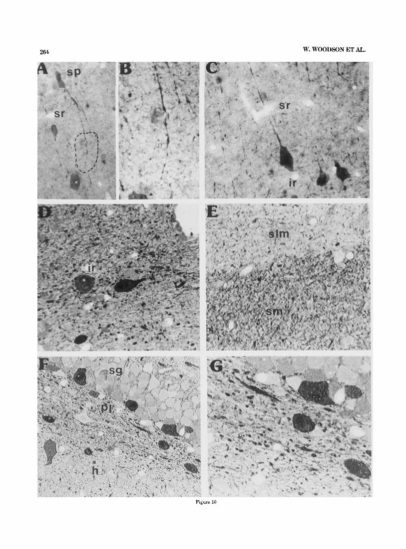
Fig. 9. Photomicrographs showing GABA-Li in the subicular and CA1 regions of Ammon’s horn in semithin sections. A,& LOW and high magnifi- cations of GABA-Li in the CAl-subicular region. Notice the distinct differ- ence in GABA-Li grain size in B. Photomicrographs taken from the section illustrated in Figure 4. Sagittal plane. x 230, and X 1,032, respectively. C,D: Label of GABA-Li in the CA1 region of the stratum oriens. GABA- GABA interactions are evident on the lighter stained GABA-Li cells. A
high magnification of the labelled horizontal cell is shown in D. Within the superficial portion of the pyramidal layer in C, pockets of GABA-Li grains are observed (arrows). Sagittal plane. x 131 and x 2,100, respectively. E: High magnification of a similar GABA-Li-positive cell within the stratum oriens; however, no GABA-Li terminals are seen on its perimeter. Sagittal plane. ~ 9 4 4 .
tween its lateral and medial portions in the horizontal plane (34 f 3 and 27 6/100 pm2, respectively, not illus- trated, see Fig. 7).
Finally, we have counted the number of GABA-Li-posi- tive grains attached to the perimeter of pyramidal and granule neuronal somata ( Fig. 14). These data indicated a significant difference ( P < .01) in the number of terminal grains from granule cell to CAI-CA2 pyramid to CA3 neu- ron (granule cell: approx. 21 grains1 soma; CA1-CAB: ap- prox. 34 and CA3 approx. 43).
In conclusion, these results (Figs. 8, 14) show that 1) whereas few variations in GABA-Li grain density are found within the dendritic regions of Ammon’s horn, distinct dif- ferences are observed in the molecular layer and the hilar region of the fascia dentata; 2) the number of GABA-Li- positive grains attached to the neuron is correlated to the perimeter of the somata.
Morphology of the GABAergic neuropil. In our semithin material both coarse and fine GABA-Li grains were evident (Figs. 9B, 10D-G). The arrangement of the coarse GABA- Li grains within the interface region was very distinct from
the label within either the stratum radiatum or the stra- tum lacunosum moleculare (compare Fig. 10C-E). This la- bel was often continuous and similar to the prominent GABA-Li grains within the stratum radiatum of CA3fa (see Figs. 2, 4, 5 , 7). It is noticed in Figure 8 that the density within the two regions was very similar.
In Figure 10E, there is a considerable difference in the amount of immunoreactive product between the molecular layers of Ammon’s horn and the fascia dentata which can be observed in our grain density measurements (see Fig. 8). Moreover, Figure 10F shows a striking distinction between the two subdivisions of the hilar region in respect to im- munoreactive product. Possibly related is that many cut GABA-Li processes are located just under and aligned par- allel to the granule cell layer (Fig. 1OG). As already men- tioned there is also a considerable difference in the grain density measurements between the two areas.
Several additional points concerning the GABAergic neu- ropil deserve emphasis. In agreement with Kunkel et al. (’86) we have observed GABA-Li somata surrounded by GABA-Li-positive terminals in the stratum oriens and the
264 W. WOODSON ET AL.
Figure 10

HIPPOCAMPAL GABAERGIc SYSTEM 265
interface region (Figs. 9C,D, 10D). In Figure 9E a similar GABA-Li-positive neuron is located within the stratum or- iens; however, no immunoreactive grains were seen on its cell body, suggesting that only certain GABA-Li cells in this layer receive GABAergic input. Furthermore, each layer had a characteristic grain pattern, Thus within the stratum oriens of CA1-CAB the grains, primarily of the coarse type, were basically oriented parallel to the surface of the alveus (Figs. 2-7, 9C). It was noticed that this label is continuous with the fiberlike arrangements within the fimbria (Figs. 2, 3, 7). The subiculum and to a lesser extent the stratum oriens of CA3 had a scattered distribution of GABA-Li grains (Figs. 2-7). Within the stratum pyrami- dale, in addition to the classical pericellular terminal net- works, “packets” of GABA-Li grain accumulations were in the pyramidal layer and at its border with the stratum oriens (Figs. 3, 9C). These packets were more frequent within the regio superior of the hippocampal formation.
Finally, there were two distinguishing features concern- ing the labelling in the stratum radiatum of CA1-CAB: 1) All planes of section showed GABA-Li grains to be oriented toward the above pyramidal cell layer (Figs. 2,4,5,7, lOC). It would seem that this immunoreactive labelling is largely due to the cut processes of GABAergic neurons located within both the CA1-CAB pyramidal cell layer and inter- face region (see Figs. 1OA-C), and 2) Varicose swellings on GABAergic processes were frequent within this region (Fig. 10B).
DISCUSSION General organization of GABA-Li in the
hippocampal formation The present description of GABA-Li in the hippocampal
formation is confirmatory of the many studies which have used various GABAergic markers to identify this inhibitory neurotransmitter (Storm-Mathisen, ‘72; Nadler et al., ’74; Barber and Saito, ’76; Ribak et al., ’78; Kohler and Chan- Palay, ’83; Seress and Ribak, ’83; Nagi, et al., ’83; Somogyi et al., ’84; Kosaka et al., ’84, ’85; Ottersen and Storm- Mathisen,’84; Somogyi and Hodgson, ’85; Gamrani et al., ’86; Kunkel et al., ’86; Storm-Mathisen and Ottersen, ’86; Sloviter and Nilaver, ’87). However, the comparison be- tween the various planes and the quantitative estimation of both GABA-Li-positive cells and neuropil density have provided additional information.
1) Approximately 11% of the “total population” of hippo- campal neuronal somata in our sections are GABA-Li posi- tive. If this estimate is applicable throughout the hippocampal formation, then of the approximately 1,300,000 neuronal somata in the adult Wistar rat (see Boss et al.,
~
Fig. 10. Photomicrographs demonstrating the distribution of GABA-Li in the CA1 pyramidal and fascia dentata regions. A,B: Typical GABA-Li “basket” neuron with process extending into the stratum radiatum. The circled region in A is enlarged in B. Frontal plane. x371 and ~ 9 1 2 , respec- tively. C: GABA-Li neuronal somata within the interface region. The oval and semitriangular neurons extend their processes into the stratum radia- turn. Frontal plane. x320. D: GABA-Li neuronal and neuropil label within the interface region. Sagittal plane. x 1,000. E: Illustration showing the distinct difference of immunoreactive product between the CA1-CAP and fascia dentata molecular layers. Sagittal plane. x900, Nomarski optics. F,G Low and high magnifications of GABA-Li in the hilar region and polymorph layer. In F a clear difference is found between the GABA-Li in the different subdivisions of the hilar region. Also notice the thick process just under the subgranular polymorph layer. Sagittal plane. ~ 4 5 0 and x900, respectively; Nomarski optics.
’871, over 100,000 cells could exhibit GABA-Li throughout its 5.6-mm rostrocaudal extent. (See Amaral, ’78, concern- ing the hippocampal length.) However, this point awaits clarification. The highest percentage of these neurons are found in the stratum oriens, stratum radiatum, interface region, and the polymorph layer.
Furthermore, 5 4 % of the cells within the pyramidal cell layer of subfields CA1-CA3 are GABA-Li positive, thus supporting the observation of Boss et al. (‘87) that the number of nonpyramidal neurons is quite small. In this connection a value of 3% is observed within the hilar pyra- midal cell layer and 1-2% within the fascia dentata granule cell layer. Whereas the total percentage of GABA-Li neu- ronal somata within the pyramidal cell layer of Ammon’s horn is quite similar to the 20% estimation of the total percentage of nonpyramidal neurons (Braitenberg and Schuz, ’83), our estimate of 1-2% in the granule cell layer is just under the value (5%) given by Seress and Pokorny (‘81). The percentage found in the suprapyramidal poly- morph layer (60%), not the “hilar region,” is in agreement with earlier studies (See Seress and Ribak, ’83, concerning their percentage within the “hilar region”.) These observa- tions reinforce the classical view that most of the (somatic) GABAergic innervation originates from the extra-pyrami- dal (or granular) layers where most of the GABAergic neu- rons are located.
2) Only slight differences in GABA-Li neuronal somata percentage (and grain density, see below) are found within the pyramidal cell dendritic subfields, which is in contrast to distinct differences in these GABA-Li elements within the dendritic regions of the fascia dentata. Thus, the 80- 95% estimate for the pyramidal cell dendritic subfields justifies the notion that many of the heterogeneous popula- tion of cells described with Golgi stains in Amaral’s (’78) zone 2 (see his Fig. 1A) are presumably GABA-Li positive. It was his conclusion, in fact, that these cells would mainly operate as interneurons. However, it is evident that these cells do give rise to long associational projections (see Koh- ler and Chan-Palay, ’83). Along these lines, there are clear fiberlike arrangements within the superficial stratum or- iens-alveus of the CA1-CA2 region. Coarse and fine GABA- Li grains (suggestive of termination) are also found in the stratum oriens of CA3 just next to and continuous with the label in the fimbria and in the subiculum. Both of these regions are targets for the termination of associational- commissural projections (Swanson et al., ’78). Our finding of GABA-Li fiber arrangements in the fimbria (in agree- ment with Ottersen and Storm-Mathisen, ’84) might indi- cate that at least some of the commissural projection is GABAergic, as has already been suggested by physiological study (Buzkaki and Czeh, ’81). However, 60% would be well above our estimation (see Seress and Ribak, ’83).
The grain density in the pyramidal cell dendritic sub- fields varies between 20 and 25 grains per 100 pm’. This distribution is to be compared with the measurements of GAD activity per dry weight (Storm-Mathisen, ’76) which indicate that there is a homogeneous distribution in most hippocampal layers with the exception of the distal den- dritic segment in the stratum lacunosum moleculare where higher values were found. Also very similar to these GAD activity measurements is the rather large difference be- tween the GABA-Li grain counts within the outer (40 * 4) and inner (26 * 1) portions of the dentate molecular layer. Interestingly, as the stratum radiatum extends into the suprapyramidal hilar region (see Amaral, ’78), our grain

266 W. WOODSON ET AL.
Fig. 11. Photomicrographs illustrating the general distribution of GABA- Li and GAD-Li in a thick section (50 pm) preparation. A Montage showing GABA-Li within the CAI strata radiatum - lacunosum moleculare den- dritic and hilar region. Notice the numerous GABA-Li-positive neuronal somata within the interface and polymorph regions. GABA-Li within the neuropil appears a s a dust-like homogeneous label in contrast to the clear granular appearance in semithin sections. ~ 8 0 . The box in the hilar region is enlarged in R: Bar equals 220 pm. C: Photomicrograph showing the
intense GABA-Li label in the stratum lucidum. Notice the cell (arrow) resembling the Amaral and Woodward ('78) interneuron within the stratum radiatum. x220. D: High magnification of a inferior region interneuron stained with anti-GABA antibody. Note the thick dendrites (arrowheads) directed toward the stratum radiatum. The thin process, presumably an axon, penetrates the stratum lucidum. Bar equals 100 gm. E: GAD immu- nocytochemical demonstration of a similar interneuron in the stratum luci- dum. Notice the intense GAD-Li label in the axonal hillock. ~ 2 4 0 .

HIPPOCAMPAL GABAERGIC SYSTEM 267
Fig. 12. Photomicrographs showing GABA-Li cell types at the stratum radiatum - stratum lucidum interface. A Oval-shaped cell with thick process (arrow) extending into the stratum lucidum. Sagittal plane. ~400. B: Triangular-shaped neuron with processes oriented parallel to the inter-
density measurements show a similarity between the two areas. Finally, when expressed in terms of the number of GABA-Li terminal grains per soma we find an increase from granule cell to CAl-CM pyramid to CA3 neuron, suggesting that the amount of GABAergic innervation on the somata perimeter depends largely on the available membrane surface.
Gradients of GABA-Li distribution in the fascia denata
A most distinct topographical pattern of GABA-Li is found in the fascia dentata. Here, the different planes of section suggest several gradients in which the number of cells decrease from dorsal to ventral and from lateral to medial: 1) frontal: dorsal (suprapyramidal) to ventral (infrapyrami- dal); 2) sagittal: rostrodorsal to rostroventral and 3) horizon- tal: rostrolateral to rostromedial. As the lateral and medial portions in horizontal sections are equivalent to the dorsal and ventral portions, respectively, in the frontal and sagit- tal planes (see Schlessinger et al., ’75), the GABA-Li gra- dients are completely in accord with each other.
The above GABA-Li topographical arrangements bear close similarity to both the neurogenic gradients of granule cell formation and the neurogenesis and distribution of mossy fibers. A consideration of each of these points is therefore valid.
It is well known that a vast majority of the dentate granule cells develop postnatally (Schlessinger et al., ’75; Bayer, ’80). Schlessinger et al. and Bayer have delineated several neurogenic gradients which are observed during granule cell ontogenesis. These are 1) a dorsal (suprapyr- amidal) to ventral (infrapyramidal) gradient (i.e., granule cell layer proliferation begins in the dorsal blade prenatally and then within the ventral blade postnatally), 2) an ectal to endal gradient (Bayer, ’80), which is equivalent to the lateral to medial definition of Schlessinger et al. (‘751, and
face between the strata radiatum and lucidum (arrows). Notice the sur- rounding pyramidal cell processes embedded within the GABA-Li-positive stratum lucidum. Sagittal plane. ~ 9 0 0 , Nomarski optics.
According to Bayer (‘80) the postnatal granule cell forma- tion in the medial or ventral blade lags behind that of the dorsal blade by approximately a few days. Moreover, these authors state that “the earlier’’-forming granule and hilar cells are influenced by these gradients.
It is therefore evident that the gradients defined in our GABA-Li material closely resemble the above neurogenic gradients. This seems to provide evidence which points to the idea that the earlier-forming regions within the fascia dentata affect the postnatal distribution of GABA-Li. How- ever, a possible differential GABA-Li cell death within the fascia dentata certainly cannot be discounted.
Amaral and Dent (‘81) have shown similar neurogenic gradients for the maturation of mossy fibers. Seemingly, the overall postnatal distribution of GABA-Li may addi- tionally be influenced by this gradient as the chronological establishment of recurrent inhibition is most likely of im- portance. Furthermore, the slight rostral inclination of mossy fibers (Lynch et al., ’73; Swanson et al., ’78,) may also influence the rostral accumulation of GABA-Li within the polymorph layer.
The fact that the regio superior pyramidal cell dendritic subfields of the stratum oriens contain more GABA-Li neu- ronal somata than the regio inferior is not compatible with any specific neurogenic gradient. However, this difference may be related to a differential migration of GABA-Li- positive cells, since a recent study suggests that the highest amount of GABA-Li neurons in the hippocampal formation is found in the regio superior within the superficial stratum oriens aligned within and just beneath the germinal zone during the first postnatal days (Rosenberg, et al., in preparation).
GABA-Li labelling in the stratum lucidum The present results indicate that stratum lucidum is en-
riched in GABA-Li label. Furthermore, in agreement with a hrief earlier immunocvtochemical descrhtion (Barber and 3) a temporal to septa1 gradient (Schlessinger et al., ’75). ._ _ _ _ _ _ ._

268 W. WOODSON ET AL.
Figure 13

HIPPOCAMPAL GABAERGIC SYSTEM
20 I I 1 I
1
Number of grains surrounding a neuron
Fig. 14. Results illustrating GABA-Li grains surrounding granule (slanted lines) and pyramidal (CAl, solid lines, and CA3, hatched lines) neurons. P < .01. Student’s t-test.
Saito, ’761, we found that GAD is also present in this zone, suggesting that the labelling is due to a conventional dense GABAergic innervation and not GABA, which would be formed by other routes not involving GAD as suggested by Storm-Mathisen and Ottersen (‘86).
To account for this labelling, the following possibilities must be considered. First, the labelling may reside in the mossy fibers themselves, as suggested by Ottersen and Storm-Mathisen (‘84). This possibility can be excluded, since following a complete destruction by colchicine of the gran- ule cells and the mossy fibers, both GABA-Li-positive neu- rons and the GABA-Li labelling in the stratum lucidum are unaffected (Robain, et al., in preparation). Second, there is evidence which suggests that the mossy fibers release an excitatory amino acid (Storm-Mathisen, ’81). Therefore, it is probable that the labelling is due to a dense network of GABAergic elements that originate from interneurons or projecting GABAergic neurons. As seen in our material, there is in the vicinity of the stratum lucidum a heteroge- neous population of GABA-Li-positive neurons, which have processes intimately related to the label found in the mossy fiber region. The possible alignment of GABA-Li processes along the mossy fibers (compare Fig. 13 A-C to Fig. 8 of Braitenberg and Schuz, ’83) is of interest as this finding, to our knowledge, has not been reported. However, electron microscopic studies are necessary to resolve the nature of the GABA-Li label within the stratum lucidum.
Fig. 13. Photomicrographs demonstrating GABA-Li neuronal types within the hilar region and CA3 pyramidal cell layer. A-C: Low and high magnifications of GABA-Li neuronal somata in the hilar pyramidal layer. Notice the parallel orientation of the GABA-Li processes. The arrowhead and stemmed arrow in A indicate the triangular and bipolar neurons shown at high magnification in B and C. The arrows in C and D indicate immuno- reactive processes within this region. Sagittal plane. A, x256; B and C: x900, Nomarski optics. D: A round GABA-Li neuron within the hilar pyramidal region having processes oriented within the GABA-Li-positive label. (arrowhead). Sagittal plane. ~ 9 0 0 , Nomarski optics. E: GABA-Li neuronal arrangement in the hilar pyramidal layer. Sagittal plane. X900, Nomarski optics. F: The frequently observed round GABA-Li neuron in the CA3 pyramidal cell layer. Horizontal plane. X500.
269
Morphology of GABA-Li neurons Morphological details of GABA-Li neuronal somata were
quite evident in our material. The most important points to be emphasized include the existence of GABA-GABA inter- actions and the presence of a heterogeneous population of GABA-Li neuronal somata within the hippocampal forma- tion, particularly as concerns the regio inferior. In respect to the former point, GABA-Li terminals cover both the soma and processes of GABA-Li-positive cells in the stra- tum oriens (in agreement with Kunkel et al., ’86). It was noted that these interactions are basically observed on the lighter-stained GABA-Li-positive neuronal somata, which most likely reflect the physiological state of the neuron (see Ottersen and Storm-Mathisen, ’84). A possible source of these GABA-Li terminals is from long associational-com- missural projections in CA1 and CA2 (see Kohler and Chan- Palay, ’83).
As GABA-GABA connections have additionally been re- ported in the pyramidal cell layer and visual cortex (see Roberts, ’80, for a review), this may be a general organiza- tional arrangement within the central nervous system. The functional significance of GABA-GABA interactions would most likely be related to disinhibition.
The morphological features of many hippocampal neu- rons have been described in Golgi material by Ram6n y Cajal (‘ll), Lorente de N6 (’34), Amaral and Woodword (‘77), and Amaral (‘78). Figures 6-8 of Lorente de NO (’34) show neurons that resemble the immunoreactive cells in our material. According to these figures, while the horizontal GABAergic interneurons of the stratum oriens basically have dendritic processes ramifying within the layer, the basket cell neurons within the pyramidal layer of CA1 and CA2 and interneurons at the stratum radiatudstratum lacunosum moleculare interface of the same region have dendritic processes oriented opposite to each other ramify- ing within the stratum radiatum.
Neither the above Golgi studies nor our material showed morphological details of GABA-Li-positive cells in the stra- tum radiatum of the CA3/a region. In contrast, our material did reveal excellent morphological details of labelled neu- ronal somata in both the CA3h,c stratum radiatum and the hilar pyramidal cell layer. In addition to our possible inferior region interneuron (see below), we could identify at the interface between the strata radiatum and lucidum a triangular neuron. This has not been described in Golgi material. As the processes of this GABA-Li neuron are uniquely aligned parallel to the interface between the two areas, it is possible that this cell type functions specifically at the interface. Indeed, as this cell belongs to Amaral’s (‘78) zone 2, it would fit his description of the neuronal constituents of this area, which are specialized to function at the interface between the fascia dentata and the hippocampus.
Within the hilar pyramidal cell layer, we have observed a heterogeneous population of very distinctive GABA-Li neurons, particularly in the sagittal plane. These cells are round, oval, and triangular and are characterized by hav- ing thick processes oriented parallel to the many GABA-Li processes in this area. It is our tentative belief that these cells could represent different variations of Amaral’s (‘78) long-spined multipolar cell (see his Fig. 25). According to him, this cell type is highly unique in that it has a very extensive longitudinal spread of dendrites.
Finally, we could additionally identify a neuron that re- sembles Amaral and Woodward’s (‘77) inferior region inter-

270 W. WOODSON ET AL. neuron. This cell type is in an interesting position since it may receive input from the entorhinal cortex (i.e., the per- forant path) and possibly commissural and cholinergic sep- tal afFerents (see Amaral and Woodward, '77). There is also some evidence that this neuron may receive mossy fiber termination (Amaral and Woodward, '77), and there is an- atomical evidence that mossy fibers directly innervate the GABAergic pyramidal basket cells of the CA3 region (Frotscher '85). Therefore, the inferior region interneurons constitute an obvious candidate for the important feed for- ward inhibition described in CA3 neurons (Buzkaki and Czeh, '81; Alger and Nicoll, '82; Frotscher et al., '84).
These neurons are probably destroyed following severe limbic seizures produced by kainate, along with the de- struction of the pyramidal cell layer of CA3 (see Ben-Ari, '85). This procedure spares the mossy fibers, and there is electrophysiological evidence of a decrease of hippocampal GABAergic neurons in kindled rats (Kamphuis et al., '87). This would tend to enhance excitability and is likely to contribute to the presence of epileptic discharge. Recently, however, Sloviter ('87) has proposed the loss of a basket- cell-activating system in the pathology of the epileptic state.
CONCLUSION The organization of GABAergic system in the hippocam-
pal formation is highly complex, in keeping with the mu- tiplicity of effects of GABA described in electrophysiological experiments (see Andersen et al., '82). Furthermore, the hippocampal GABAergic cell population is heterogeneous and probably consists of many different specialized types, such as the axoaxonal cell (see Somogyi et al., '841, and the unique assemblage of GABA-Li cell types which appear aligned along the mossy fibers. The heterogeneity of GABA- Li cells is further reflected by their coexistence with various neuropeptides: Cholecystokinin octapeptide (CCK), vasoac- tive intestinal polypeptide, neuropeptide y, and somatosta- tin (Somogyi et al., '84; Kosaka et al., '85; Kohler et al., '86). It also appears that GABA-Li somatostatin and CCK cells are distributed differently within the hippocampal layers (Somogyi et al., '84;, Sloviter and Nilaver, '87). Clearly this heterogeneity must be taken into consideration in order to comprehend better the physiological role of the GABAergic system in the hippocampal formation.
ACKNOWLEDGMENTS This work was supported by a grant to Walter Woodson
from the Ministgre des Relations Exterieure, French Gov- ernment (SGG/6564). The authors would like to thank Drs. 0. Robain, D. Amaral, and L. Swanson for making helpful comments during the preparation of this manuscript. We thank Drs. H. J. Karten and K. Keyser for use of their Nomarski optics and Dr. Toru Shimizu for computer analy- sis. Also, the secretarial assistance of S. Bauhurlet and E. Watelet was greatly appreciated.
LITERATURE CITED Alger, B.E., and R. Nicoll(1982) A feed forward inhibition in rat hippocam-
pus pyramidal cells studied in vitro. J. Physiol. (Lond.) 328r105-123. Amaral, D.G. (1978) A Golgi study of cell types in the hilar region of the
hippocampus in the rat. J. Comp. Neurol. 182t851-914. Amaral, D.G., and J.A. Dent (1981) Development of the mossy fibers of the
dentate gyrus: I. A light and electron microscopic study of the mossy fibers and their expansions. J. Comp. Neurol. 195:51-86.
Amaral, D.G., and D.J. Woodward (1977) A hippocampal interneuron ob- served in the inferior region. Brain Res. 124:225-236.
Andersen, P. (1975) Organization of hippocampal neurons and their inter- connections. In R.L. Isaacson and K.H. Pribram (eds): The Hippocampus. New York: Plenum Press, Vol. 1, pp. 155-175.
Andersen, P., B. Bie, and T. Ganes (1982) Distribution of GABA sensitive areas on hippocampal pyramidal cell. Exp. Brain Res. 45:357-363.
Andersen, P., J.S. Eccles, and Y. LByning (1964) Location of postsynaptic in- hibitory synapses of hippocampal pyramids. J. Neurophysiol. 27:592-607.
Barber, R., and K. Saito (1976) Light microscopic visualization of GAD and GABA-T immunocytochemical preparations of rodent CNS. In E. Rob- erts, T.N. Chase, and D.B. Tower (eds): GABA in Nervous System Func- tion. New York: Raven Press, pp. 113-132.
Bayer, S.A. (1980) Development of the hippocampal region in the rat. I. Neurogenesis examined with 3H-thymidine autoradiography. J. Comp. Neurol. 19Ot87-114.
Ben-Ari, Y. (1985) Limbic seizures and brain damage produced by kainic acid: Mechanism and relevance to human temporal lobe epilepsy. Neu- roscience 14t375-403.
Boss, B.D., K. Turlejski, B.B. Stanfield, and W.M. Cowan (1987) On the number of neurons in fields CA1 and CA3 of the hippocampus of Spra- gue-Dawley and Wistar rats. Brain Res. 406280-287.
Braitenberg, V., and A. Schuz (1983) Some anatomical comments on the hippocampus. In W. Seifert (ed.): Neurobiology of the hippocampus, London: Academic Press, pp. 21-37.
Buzsaki, G., and G. Czeh (1981) Commissural and perforant path interac- tions in the rat hippocampus: Field potentials and unitary activity. Exp. Brain Res. 43:429-438.
Buzsaki, G., and E. Eidelberg (1981) Commissural projection to the dentate gyrus of the rat: Evidence for feed-forward inhibition. Brain Res. 23Ot346-350.
Clark, C.A., E.C. Downs, and F.J. Primus (1982) An unlabeled antibody method using glucose oxidase-antiglucose oxidase complexes (GAG): A sensitive alternative to immunoperoxidase for the detection of tissue antigens. J. Histochem. Cytochem. 3Or27-34.
Frotscher, M., C.S. Leranth, K. Lubbersk, and W.H. Oertel (1984) Commis- sural afferents innervate glutamate decarboxylase immunoreactive nonpyramidal neurons in the guinea-pig hippocampus. Neurosci. Lett. 46:137-143.
Frotscher, M. (1985) Mossy fibers form synapses with identified pyramidal basket cells in the CA3 region of the guinea-pig hippocampus: A com- bined Golgi-electron microscope study. J. Neurocytol. 14t245-259.
Gamrani, H., B. Onteniente, P. Sequela, M. Geffard, and A. Callas (1986) Gamma-aminobutyric acid immunoreactivity in the rat hippocampus. A light and electron microscopic study with anti-GABA antibodies, Brain Res. 264:30-38.
Kamphuis, W., W.J. Wadman, R.M. Buijs, F.H. Lopes, and D.A. Silva (1987) The development of changes in hippocampal GABA immunoreactivity in the rat kindling model of epilepsy: A light microscopic study with GABA antibodies. Neuroscience 2433-446.
Kohler, C., and V. Chan-Palay (1983) Gamma-aminobutyric acid interneu- rons in the rat hippocampal region studied by retrograde transport of glutamic acid decarboxylase antibody after in vivo injections. Anat. Embryol. (Berl.) 163:53-66.
Kohler, C., L. Eriksson, S. Davies, and V. Chan-Palay (1986) Neuropeptide Y innervation of the hippocampal region in the rat and monkey brain. J. Comp. Neurol. 244t384-400.
Kosaka, T., K. Hama, and J.-Y. Wu (1984) GABAergic synaptic boutons in the granule cell layer of rat dentate gyrus. Brain Res. 293;353-359.
Kosaka, T., K. Kosaka, K. Tateishi, Y. Hamaoka, N. Yanaihara, J.-Y. Wu, and K. Hama (1985) GABAergic neurons containing CCK-8-like and/or VIP-like immunoreactivities in the rat hippocampus and dentate gyrus. J. Comp. Neurol. 239:420-430.
Kunkel, D.D., A.E. Hendrickson, J.-Y. Wu, and P.A. Schwartzkroin (1986) Glutamic acid decarboxylase (GAD) immunocytochemistry of develop- ing rabbit hippocampus. J. Neurosci. 6(2):541-552.
Lorente de NO, R. (1934) Studies on the structure of the cerebral cortex 11. Continuation of the study of the ammonic system. J. Psychol. Neurol. (Leipz.) 46: 113-177.
Lynch, G.S., S. Mosko, T. Parks, and C.W. Cotman (1973) Relocation and hyperdevelopment of the dentate gyrus commissural system after ento- rhinal lesions in immature rats. Brain Res. 5Ot174-178.
McNaughton, B.L., R.M. Douglas, and G.V. Goddard (1978) Synaptic en- hancement in fascia dentata. Cooperativity among coactive afferents. Brain Res. 157:277-293.
Nadler, J.V., C.W. Cotman, and G.S. Lynch (1974) Biochemical plasticity of short-axon interneurons: Increased glutamate decarboxylase activity in the denervated area of rat dentate gyrus following entorhinal lesions. Exp. Neurol. 45t403-413.
Nagai, T., P.L. McGeer, and E.G. McGeer (1983) Distribution of GABA-T intensive neurons in the rat forebrain and mid-brain. J. Comp. Neurol.

HIPPOCAMPAL GABAERGIC SYSTEM 271
218:220-238. Oertel, W.H., D.E. Schmechel, E. Mugnaini, and M.L. Tappaz (1981a) Im-
munocytochemical localization of glutamate decarboxylase in ra t cere- bellum with a new antiserum. Neuroscience 6:2715-2735.
Oertel, W.H., D.E. Schmechel, M.L. Tappaz, and J.J. Kopin (1981b) Produc- tion of a specific antiserum to rat brain glutamic acid decarboxylase by injection of an antigen antibody complex. Neurosience 6:2689-2700.
Ottersen, O.P., and J. Storm-Mathisen (1984a) Neurons containing or accu- mulating transmitter amino acids. In A. Bjorklund., T. HokfeIt, and M.J. Kuhar (eds): Handbook of Chemical Neuroanatomy. Vol. 3, Part II. Amsterdam: Elsevier, pp. 141-246.
Ottersen, O.P., and J. Storm-Mathisen (19841-1) Glutamate- and GABAcon- taining neurons in the mouse and rat brain as demonstrated with a new immunocytochemical technique. J. Comp. Neurol. 229t374-392.
Ramon y Cajal, S. (1911) Histologie du Systeme Nerveux de 1’Homme et des Vertebres. Vol. 2. Paris: Maloine.
Ribak, C.E., J.E. Vaughn, and K. Saito (1978) Immunocytochemical localiza- tion of glutamic acid decarboxylase in neuronal somata following colcbi- cine inhibition of axonal transport. Brain Res. 14Ot315-332.
Roberts, E. (1980) a-aminobutyric acid (GABA): A major inhibitory trans- mitter in the vertebrate nervous system. In R. Levi-Montalcini (ed): Nerve Cells, Transmitter, and Behavior. Rome: Pontificial Academy of Sciences.
Schlessinger, A.R., W.M. Cowan, and D.I. Gottlieb (1975) An autoradio- graphic study of the time of origin and the pattern of granule cell migration in the dentate gyms of the rat. J. Comp. Neuro1.159:149-176.
Sequela, P., M. Geffard, R.M. Bujis, and M. Le Moal (1984) Antibodies against a-aminobutyric acid Specificity studies and immunocytochemi- cal results. Proc. Natl. Acad. Sci. USA, 8It3888-3892.
Seress, L., and J. Pokorny (1981) Structure of the granular layer of the rat dentate gyrus. A light microscopic and Golgi study. J. Anat. 133(2):181- 195.
Seress, L., and C.E. Ribak (1983) GABAergic cells in the dentate gyrus appear to be local circuit and projection neurons. Exp. Brain Res. 50t173- 182.
Seress, L., and C.E. Ribak (1985) A combined Golgi-electron microscopic study of non-pyramidal neurons in the CA2 area of the hippocampus. J. Neurocytol. 14t717-730.
Sloviter, R.S. (1987) Decreased hippocampal inhibition and a selective loss of interneurons in experimental epilepsy. Science 235t73-77.
Sloviter, R.S., and G. Nilaver (1987) Immunocytochemical localization of GABA, cholecystokinin, vasoactive intestinal polypeptide and somato- statin-like immunoreactivity in the area dentata and hippocampus of the rat. J. Comp. Neurol. 256t42-60.
Somogyi, P., and A.J. Hcdgson (1985) Antisera to y-aminobutyric acid. 111. Demonstration of GABA in Golgi impregnated neurons and conven- tional electron microscopic sections of cat striate cortex. J. Histochem. Cytochem. 33249-257.
Somogyi, P., A.J. Hodgson, J.W. Chubb, B. Penke, and A. Erdei (1985) Antisera to a-aminobutyric acid 11. Immunocytochemical application to the central nervous system. J. Histochem. Cytochem. 33.240-248.
Somogyi, P., A.J. Hodgson, A.D. Smith, M.G. Nunzi, A. Gorio, and J.-V. Wu (1984) Different populations of GABAergic neurons in the visual cortex and hippocampus of cat contain somatostatin or cholecystokinin-immu- noreactive material. J. Neurosci. 4,2590-2603.
Somogyi, P., A.D. Smith, M.G. Nunzi, A. Gorio, H. Takagi, and J:Y. Wu (1984) Glutamate decarboxylase immunoreactivity in the hippocampus of the cat: Distribution of immunoreactive synaptic terminals with special reference to the axon initial segment of pyramidal neurons. J. Neurosci. 3:1450-1468.
Sternberger, L.A. (1979) Immunocytochemistry (2nd edition). New York Wiley and Sons.
Storm-Mathisen, J.C. (1972) Glutamate decarboxylase in the rat hippocam- pal region after lesions of the afferent fiber systems. Evidence that the enzyme is localized in intrinsic neurons. Brain Res. 40215-235.
Storm-Mathisen, J.C. (1976) Distribution of the components of the GABA system in neuronal tissue: Cerebellum and hippocampus. Effects of axotomy. In E. Roberts, T.N. Chase, and P.B. Tower (eds): GABA in Nervous System Functions. New York: Raven Press, pp. 149-165.
Storm-Mathisen, J. (1981) Autoradiographic and microchemical localization of high affinity glutamate uptake. In P.J. Roberts, J. Storm-Mathisen, and G.A.R. Johnston (eds): Glutamate: Transmitter in the Central Ner- vous System. Chichester: John Wiley and Sons, pp. 89-115.
Stom-Mathisen, J., and O.P. Ottersen (1986) Antibodies against amino acid neurotransmitters. In P. Panula., H. Paivarinta, and S. Soinila (eds): Neurochemistry: Modern Methods and Applications. New York: Alan R. Liss, Inc., pp. 107-136.
Swanson, L.W., J.M. Wyss, and W.M. Cowan (1978) The topographical or- ganization of intrahippocampal association pathways from Ammon’s horn and the dentate gyrus. 3. Comp. Neurol. 181t681-715.