Orbital disorders -...
19
Orbital disorders Otolaryngology online 9/25/2011 Dr T Balasubramanian This e book discusses orbital disorders from the perspective of otolaryngologist
Transcript of Orbital disorders -...
Orbital disorders
O t o l a r y n g o l o g y o n l i n e
9 / 2 5 / 2 0 1 1
Dr T Balasubramanian
This e book discusses orbital disorders from the
perspective of otolaryngologist
1
Drtbalu’s otolaryngology online
Patterns of orbital disorders
Introduction:
The effects caused by orbital diseases are governed by:
1. Pathophysiology of the disease process
2. Anatomic pattern of involvement (location of the lesion). This is more evident from
the fact that small tumors of orbital apex causes early symptoms due to involvement
of 2,3,4,5 and 6th cranial nerves. These patients will also manifest with progressive
vision loss during early course of the lesion. Laterally placed orbital Meningiomas
cause features of superior orbital syndrome which could manifest before or along
with loss of visual acuity.
Anatomy of orbit:
A careful study of anatomy of orbit is very important to an ENT surgeon because of its
proximity to the para nasal sinuses. A comprehensive knowledge of orbital and peri orbital
anatomy is necessary to understand the various disorders of this region and in its surgical
management.
The shape of the orbit resembles a four sided pyramid to begin with but as one goes
posterior it becomes three sided towards the apex. The volume of the orbital cavity in an
adult is roughly about 30cc. The rim of orbit in an adult measures about 40mm horizontally
and 35 mm vertically. The medial walls of orbit are roughly parallel and are about 25 mm
apart in an adult. The lateral walls of orbit angles about 90 degrees from each other.
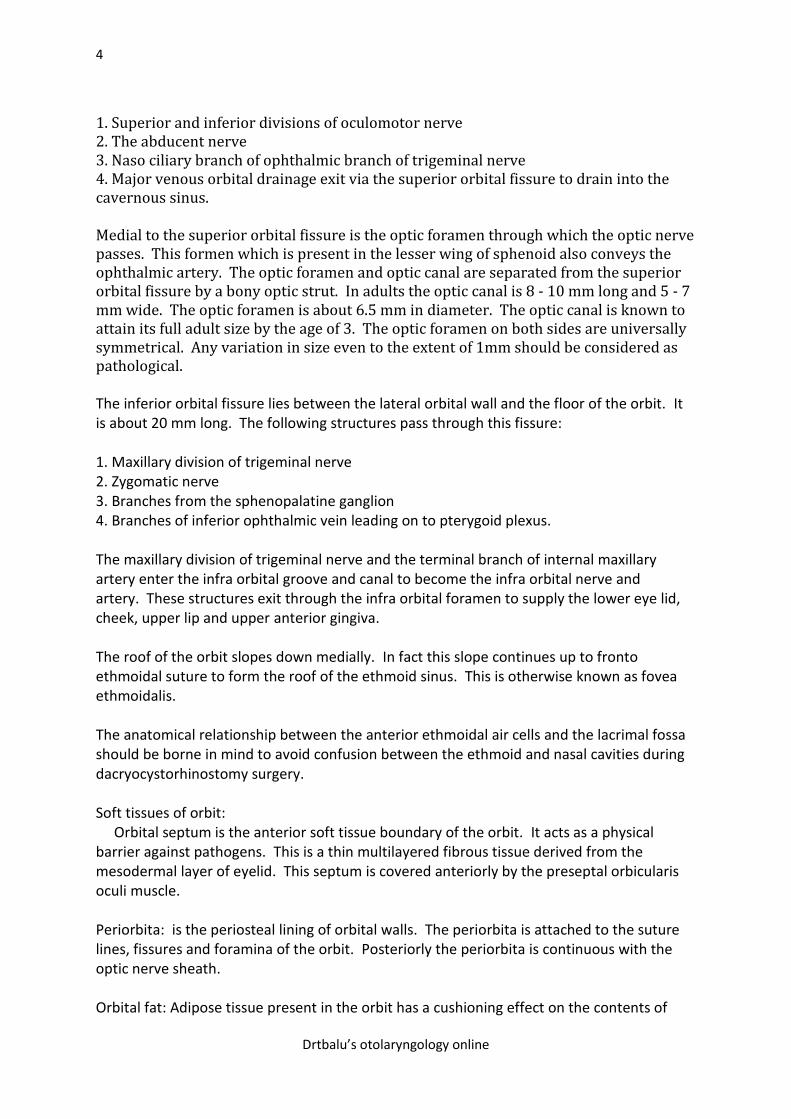
Osteology of orbit: The orbital rim is more or less spiral with its two ends
overlapping medially on either side of lacrimal fossa. The inferior orbital rim is formed by
the maxillary bone medially and zygomatic bone laterally.
The zygomatic bone forms the lateral orbital rim, while the frontal bone forms the superior
orbital rim. The superior rim is commonly indented by a small notch known as the supra
orbital notch. This notch is invariably present at the junction of medial and lateral 1/3. The
supra ortbital nerve and artery pass through this notch to reach the forehead.
The medial portion of the orbital rim is formed by the frontal process of maxilla and the
maxillary portion of the frontal bones. A depression known as the lacrimal fossa is formed
in the infero medial orbital rim. This fossa is formed by the maxillary and lacrimal
bones. This lacrimal fossa is bounded by two projections of bones i.e. the anterior lacrimal
crest of maxillary bone and the posterior lacrimal crest of lacrimal bone. This fossa houses
the nasolacrimal sac. This fossa opens in to the naso lacrimal canal through which the naso
lacrimal duct traverses.
2
Drtbalu’s otolaryngology online
The naso lacrimal duct is 3 - 4 mm in diameter, courses in an infero lateral and slightly
posterior direction towards the inferior turbinate under which it opens into the inferior
meatus. This duct is roughly 12mm long. All the walls of the lacrimal duct except its medial
wall is formed by the maxillary bone. The medial wall is formed by the lateral nasal wall
inferiorly and the descending process of lacrimal bone superiorly.
In the frontal process of maxilla just anterior to the lacrimal fossa a fine groove known as the
sutura longitudinalis imperfecta of Weber. This suture runs parallel to the anterior lacrimal
crest. Small branches of infraorbital artery pass through this groove to supply the nasal
mucosa. The presence of these vessels should be anticipated in any lacrimal sac surgery to
avoid unneccessary troublesome bleeding.
Embryology of orbit: The walls of the orbit formed by 7 bones, are embryologically derived
from neural crest cells. Ossification of the orbit is complete at birth except at its
apex. Except the lesser wing of sphenoid which is cartilagenous the other bones develop by
intramembranous ossification.
The roof of the orbit is mostly formed by the frontal bone, only the posterior 1.5 cms of the
roof is formed by the lesser wing of the sphenoid bone. The optic foramen through which the
optic nerve traverses is located in the lesser wing of the sphenoid bone. The optic nerve
enters the orbit at an angulation of 45 degrees.
The lacrimal gland fossa is located in the lateral portion of the orbital roof, while the
trochlear fossa is located in the anterio medial portion of the orbital roof.
The medial wall of the orbit is formed from anterior to posterior by:
1. frontal process of maxilla
2. lacrimal bone
3. ethmoid bone
4. lesser wing of sphenoid bone
The thinnest portion of the medial wall is the lamina papyracea which separates the ethmoidal
sinuses from the orbit. It is one of the components of ethmoid bone. Infections from
ethmoidal sinus can easily breach this paper thin bone and affect the orbital contents. The
medial wall of the orbit is thicker posterior where the sphenoid bone is present and anteriorly
where the posterior lacrimal crest is present.
The fronto ethmoidal suture line marks the approximate level of ethmoidal sinus roof, hence
any dissestion above this line may expose the cranial cavity. The anterior and posterior
ethmoidal foramina through which branches of ophthalmic artery (anterior and posterior
ethmoidal arteries) and branches of naso ciliary nerve passes are present in this suture. The
anterior ethmoidal foramen is located at a distance of 24 mm from the anterior lacrimal crest,
while the posterior ethmoidal foramen is located at a distance of 36mm from the anterior
lacrimal crest.
A vertical suture that runs between the anterior and posterior lacrimal crests is the anastomotic area between the maxillary and the lacrimal bone. If this suture is located more anteriorly it indicates a predominance of lacrimal bone, while a more posteriorly
3
Drtbalu’s otolaryngology online
placed suture line indicates a predominance of maxillary bone in the anastomotic relationship. The lacrimal bone at the level of lacrimal fossa is pretty thin (106 micrometer). This bone can be easily penetrated during dacryocystorhinostomy surgery. If the maxillary component is predominant it becomes difficult to perform the osteotomy in this area to access the sac because the maxillary bone is pretty thick. Hence lacrimal bone predominance makes it easy to expose the sac during dacryocystorhinostomy. The floor of the orbit is the shortest of all its walls and is bordered laterally by infra orbital fissure. Medially the floor is bounded by the maxillo ethmoidal strut. The floor of the orbit is almost entirely formed by the orbital plate of maxilla, palatine contributes to a small portion of the floor posteriorly. Zygoma also makes a small contribution to it anterolaterally. The infra orbital groove becomes a canal anteriorly, through this groove passes the infra orbital nerve and artery. The floor of the orbit medial to the infra orbital groove is thin because of the expansion of the maxillary sinus. With the growth of facial bones the infra orbital foramen migrates to about 6-10mm below the infra orbital rim. The lateral wall of the orbit is formed mainly by the greater wing of sphenoid bone with contributions from zygoma and zygomatic process of frontal bone anteriorly. The recurrent meningeal branch of middle meningeal artery may be seen coursing through a foramen in the suture line between the frontal and sphenoid bones. This artery forms a anastomosis between the external and internal carotid arterial systems. Roughly 4 - 5 mm behind the lateral orbital rim and 1 cm inferior to the frontozygomatic suture is the lateral tubercle of Whitnall. The following structures gets attached to this tubercle: 1. Lateral canthal tendone 2. Lateral rectus check ligament 3. Suspensory ligament of lower eyelid (Lockwoods ligament). 4. Orbital septum 5. Lacrimal gland fascia. The frontal process of zygomatic bone and the zygomatic process of frontal bone are thick and they protect the globe from lateral trauma. Just behind this facial buttress area the posterior zygomatic bone and the orbital plate of greater wing of sphenoid are thinner thus making the zygomatico sphenoid suture a convenient land mark for lateral orbitotomy. The zygomatico facial and zygomatico temporal nerves and vessels pass through the lateral wall of the orbit to reach the cheek and temporal regions. Posteriorly the lateral wall thickens and meets the temporal bone which forms the lateral wall of the cranial cavity. When lateral orbitotomy is being done only 12 - 13 mm separate the posterior aspect of lateral orbitotomy to that of the middle cranial fossa. This distance could still be shorter in females. Superior orbital fissure: is a linear notch between the greater and lesser wings of sphenoid. The superior portion of the fissure is narrower and here the lacrimal, frontal and trochlear nerves passes through outside the annulus of zinn. The annulus of zinn is a ring of fibrous tissue surrounding the optic nerve at its entrance into the apex of orbit. This ring gives origin to the extra ocular muscles. The following structures pass through the superior orbital fissure within the annulus of zinn:
4
Drtbalu’s otolaryngology online
1. Superior and inferior divisions of oculomotor nerve 2. The abducent nerve 3. Naso ciliary branch of ophthalmic branch of trigeminal nerve 4. Major venous orbital drainage exit via the superior orbital fissure to drain into the cavernous sinus. Medial to the superior orbital fissure is the optic foramen through which the optic nerve passes. This formen which is present in the lesser wing of sphenoid also conveys the ophthalmic artery. The optic foramen and optic canal are separated from the superior orbital fissure by a bony optic strut. In adults the optic canal is 8 - 10 mm long and 5 - 7 mm wide. The optic foramen is about 6.5 mm in diameter. The optic canal is known to attain its full adult size by the age of 3. The optic foramen on both sides are universally symmetrical. Any variation in size even to the extent of 1mm should be considered as pathological.
The inferior orbital fissure lies between the lateral orbital wall and the floor of the orbit. It is about 20 mm long. The following structures pass through this fissure: 1. Maxillary division of trigeminal nerve 2. Zygomatic nerve 3. Branches from the sphenopalatine ganglion 4. Branches of inferior ophthalmic vein leading on to pterygoid plexus. The maxillary division of trigeminal nerve and the terminal branch of internal maxillary artery enter the infra orbital groove and canal to become the infra orbital nerve and artery. These structures exit through the infra orbital foramen to supply the lower eye lid, cheek, upper lip and upper anterior gingiva. The roof of the orbit slopes down medially. In fact this slope continues up to fronto ethmoidal suture to form the roof of the ethmoid sinus. This is otherwise known as fovea ethmoidalis. The anatomical relationship between the anterior ethmoidal air cells and the lacrimal fossa should be borne in mind to avoid confusion between the ethmoid and nasal cavities during dacryocystorhinostomy surgery. Soft tissues of orbit: Orbital septum is the anterior soft tissue boundary of the orbit. It acts as a physical barrier against pathogens. This is a thin multilayered fibrous tissue derived from the mesodermal layer of eyelid. This septum is covered anteriorly by the preseptal orbicularis oculi muscle. Periorbita: is the periosteal lining of orbital walls. The periorbita is attached to the suture lines, fissures and foramina of the orbit. Posteriorly the periorbita is continuous with the optic nerve sheath. Orbital fat: Adipose tissue present in the orbit has a cushioning effect on the contents of
5
Drtbalu’s otolaryngology online
orbit. The extra ocular muscles of orbit arise from the annulus of zinn and are responsible for the movement of the globe. These muscles are: lateral and medial rectus Superior and inferior rectus Superior and inferior oblique The lacrimal system: The main lacrimal gland is located in the supero temporal portion of orbit. It lies in the shallow lacrimal fossa of the frontal bone. The gland is composed of numerous secretory units known as acini which progressively drain in to small and larger ducts. The gland measures 20 mm by 12 mm. A fibrous band incompletely divides the lacrimal gland into two lobes i.e. posterior larger orbital lobe and a smaller anterior palpebral lobe. 2 - 6 ducts from the orbital lobe pass through the palpebral lobe joining with the ducts from the palpebral lobe to form 6 - 12 tubules to empty into the supero lateral conjunctiva. Hence damage to the palpebral lobe may block drainage from the entire gland. About 20 - 40 accessory lacrimal glands of Krause are located in the superior conjunctival fornix, about half this number is located over the lower fornix. The lacrimal gland is innervated by branches from 5th and 7th cranial nerves, sympathetic supply to lacrimal gland is via the nerves from the superior cervical ganglion. The parasympathetic fibers are supplied via the 6th nerve. Sensory supply is via the branches of trigeminal nerve.
The lacrimal excretory system begins at a 0.3 mm at the medial end of each eyelids known
as the punctum. These puncta are directed posteriorly. The punctal opening widens into
ampulla, which is perpendicular to the eye lid margin. The ampulla makes a sharp turn to
drain into the canaliculi. The canaliculi measures 0.5 - 1mm in diameter and courses parallel
to the lid margins. The superior canaliculus is 8 mm long and the inferior canaliculus is 10
mm long. In majority of individuals the superior and inferior canaliculi merge into a
common canaliculi before draining into naso lacrimal sac. The opening of common
canaliculi into the naso lacrimal sac is known as the common internal punctum. There is a
valve at the junction of common canaliculus and lacrimal sac at the common internal
punctum level. This is known as the Rosenmuller valve. Another valve known as the valve
of Hasner is found at the lower end of the naso lacrimal duct at the level of inferior meatus
of nose.
If this Hasner's valve is imperforate in new born infants it causes congenital naso lacrimal
obstruction.
The lacrimal sac resides in the lacrimal fossa. It measures about 12 - 15 mm vertically, and 4
- 8 mm antero posteriorly.
6
Drtbalu’s otolaryngology online
Features of orbital disorders depend on either the functional effects or mass effects caused
by the disorder. Functional effects interfere with motor, sensory or secretory functions of
orbital structures.
Mass effects create problems by shifting / displacement of orbital structures because they
occupy space. This feature is known as positive effect. These lesions can also increase
space by causing bone expansion. This feature is known as negative effect. Some of the
orbital lesions can cause scar formation. This can also compromise functions of various
orbital structures. This is also known as negative effect.
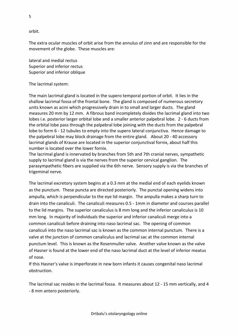
Ethmoidal mass lesions cause lateral displacement of orbit – Positive effect.
Destruction of orbital floor causes enophthalmos and downward displacement – Negative
effect.
Desmoplastic lesions like metastatic carcinoma may tether and trap structures causing
entrapment problems.
Photograph showing bony anatomy of orbit
7
Drtbalu’s otolaryngology online
Picture showing medial displacement of orbit
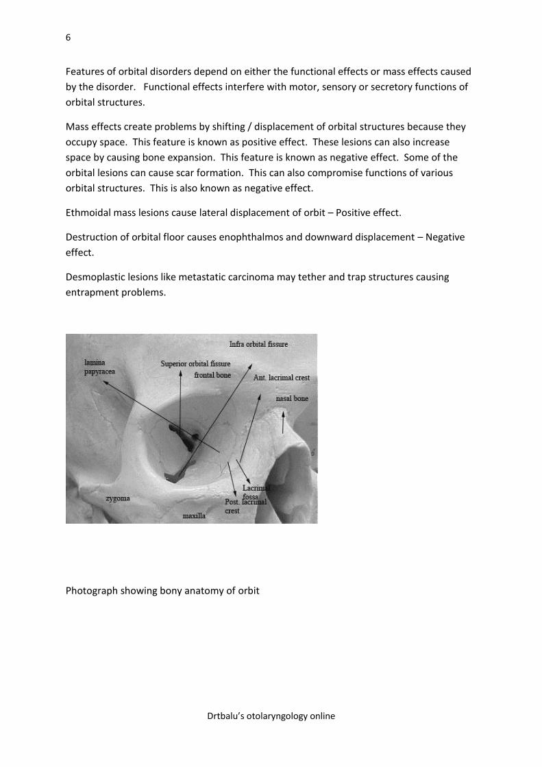
CT scan coronal showing proptosis
8
Drtbalu’s otolaryngology online
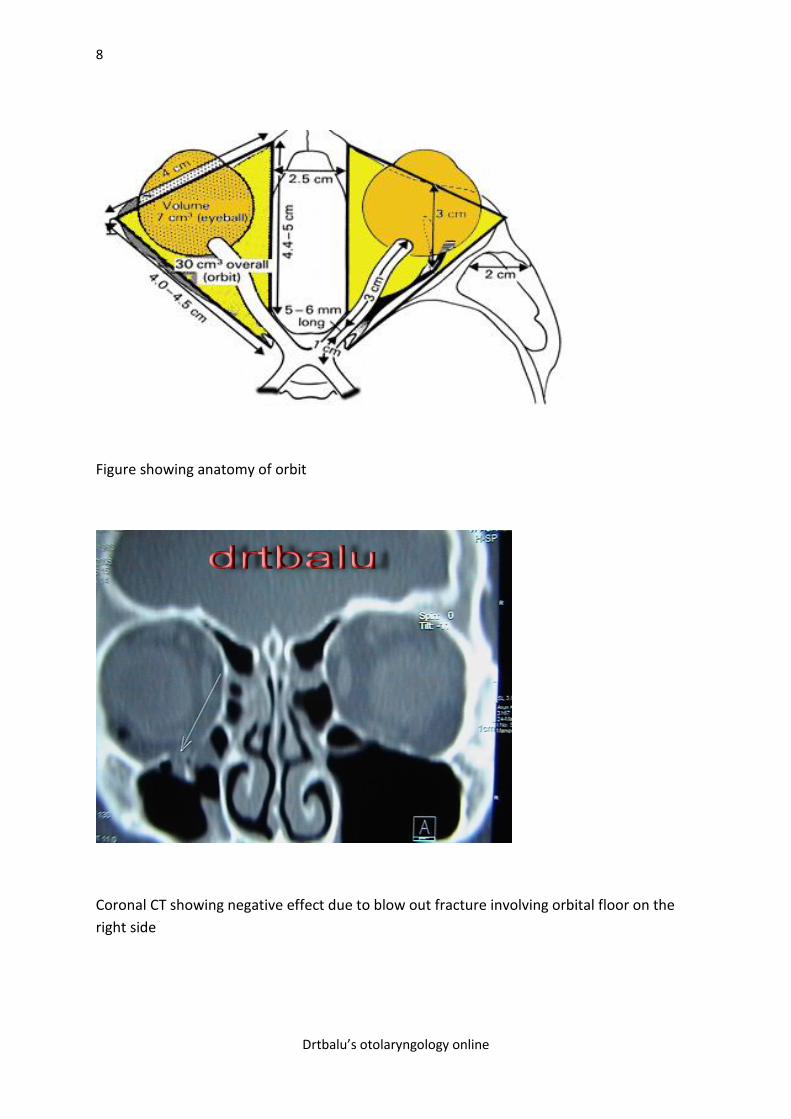
Figure showing anatomy of orbit
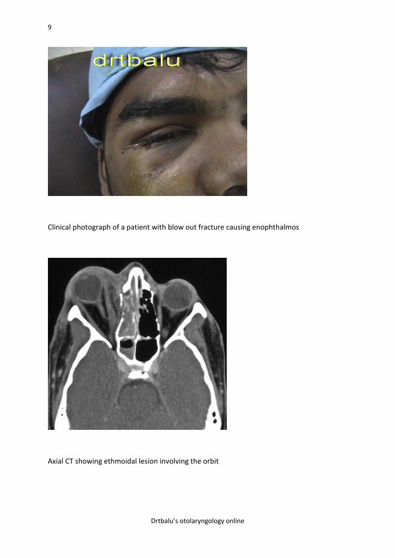
Coronal CT showing negative effect due to blow out fracture involving orbital floor on the
right side
9
Drtbalu’s otolaryngology online
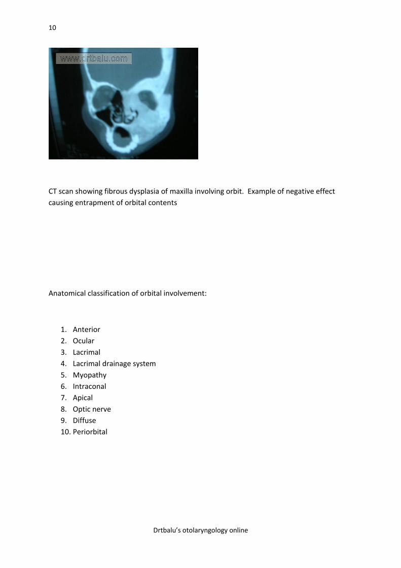
Clinical photograph of a patient with blow out fracture causing enophthalmos
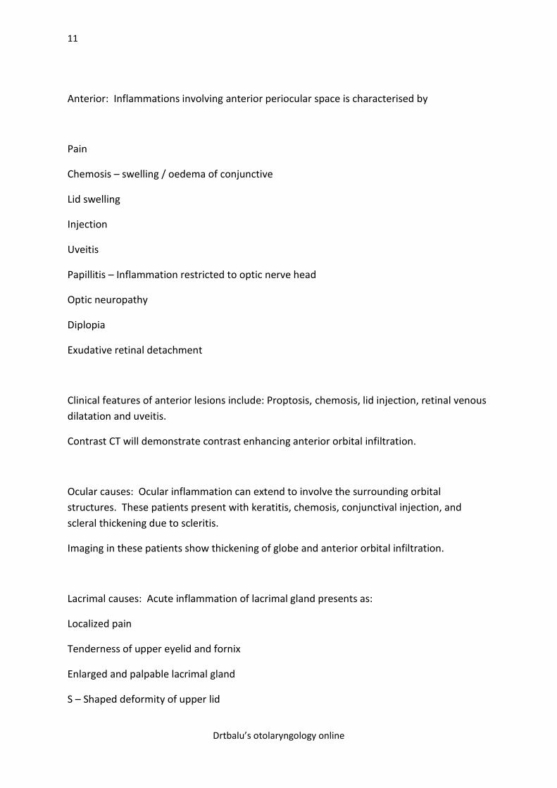
Axial CT showing ethmoidal lesion involving the orbit
10
Drtbalu’s otolaryngology online
CT scan showing fibrous dysplasia of maxilla involving orbit. Example of negative effect
causing entrapment of orbital contents
Anatomical classification of orbital involvement:
1. Anterior
2. Ocular
3. Lacrimal
4. Lacrimal drainage system
5. Myopathy
6. Intraconal
7. Apical
8. Optic nerve
9. Diffuse
10. Periorbital
11
Drtbalu’s otolaryngology online
Anterior: Inflammations involving anterior periocular space is characterised by
Pain
Chemosis – swelling / oedema of conjunctive
Lid swelling
Injection
Uveitis
Papillitis – Inflammation restricted to optic nerve head
Optic neuropathy
Diplopia
Exudative retinal detachment
Clinical features of anterior lesions include: Proptosis, chemosis, lid injection, retinal venous
dilatation and uveitis.
Contrast CT will demonstrate contrast enhancing anterior orbital infiltration.
Ocular causes: Ocular inflammation can extend to involve the surrounding orbital
structures. These patients present with keratitis, chemosis, conjunctival injection, and
scleral thickening due to scleritis.
Imaging in these patients show thickening of globe and anterior orbital infiltration.
Lacrimal causes: Acute inflammation of lacrimal gland presents as:
Localized pain
Tenderness of upper eyelid and fornix
Enlarged and palpable lacrimal gland
S – Shaped deformity of upper lid
12
Drtbalu’s otolaryngology online
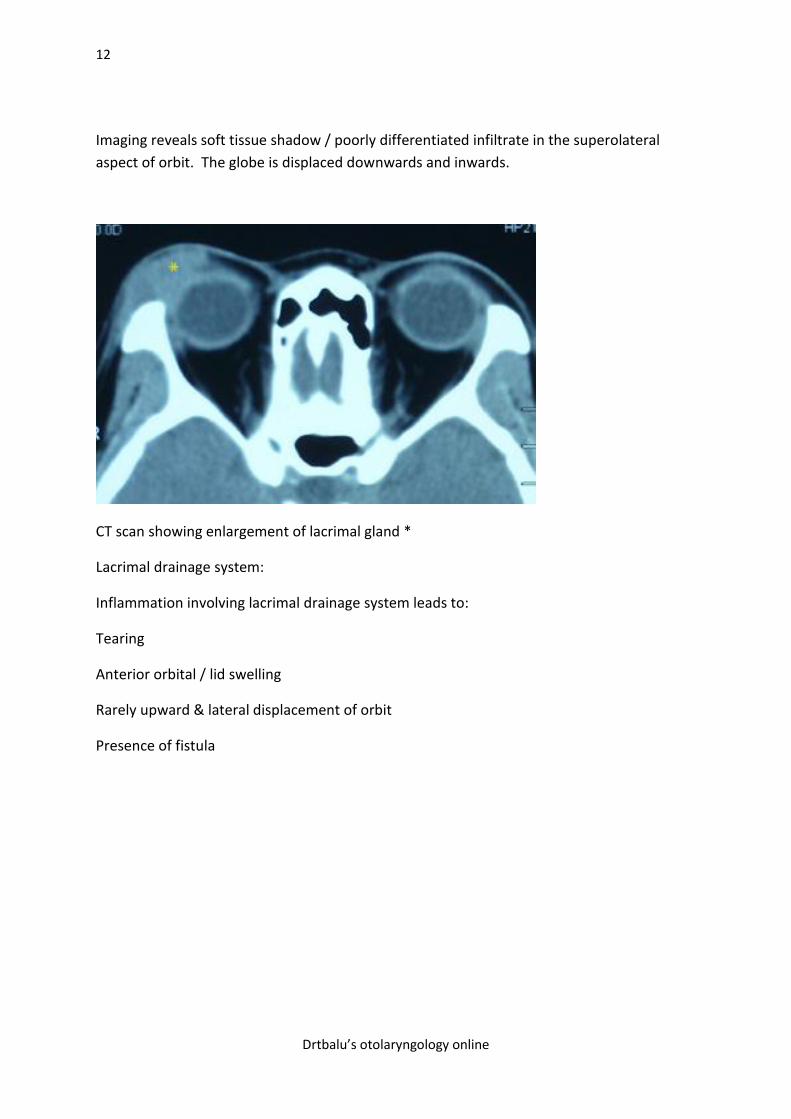
Imaging reveals soft tissue shadow / poorly differentiated infiltrate in the superolateral
aspect of orbit. The globe is displaced downwards and inwards.
CT scan showing enlargement of lacrimal gland *
Lacrimal drainage system:
Inflammation involving lacrimal drainage system leads to:
Tearing
Anterior orbital / lid swelling
Rarely upward & lateral displacement of orbit
Presence of fistula
13
Drtbalu’s otolaryngology online
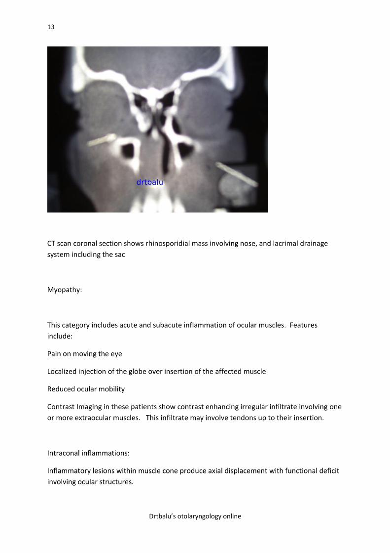
CT scan coronal section shows rhinosporidial mass involving nose, and lacrimal drainage
system including the sac
Myopathy:
This category includes acute and subacute inflammation of ocular muscles. Features
include:
Pain on moving the eye
Localized injection of the globe over insertion of the affected muscle
Reduced ocular mobility
Contrast Imaging in these patients show contrast enhancing irregular infiltrate involving one
or more extraocular muscles. This infiltrate may involve tendons up to their insertion.
Intraconal inflammations:
Inflammatory lesions within muscle cone produce axial displacement with functional deficit
involving ocular structures.
14
Drtbalu’s otolaryngology online
Apical lesions:
Perineural apical inflammation produces less proptosis. Pain is also negligible. These
patients have early optic atrophy and vision loss. Inflammation of orbital apex can also
involve superior orbital fissure. Superior and inferior ophthalmic vein obstruction can cause
superior orbital syndrome. There is classic involvement of 3rd, 4th, 6th and ophthalmic
branch of trigeminal nerve. This causes painful eye movement, vision loss and
ophthalmoplegia.
Diffuse lesions:
This is similar to anterior lesions but the clinical signs are more profound.
Periorbital lesions:
Diseases involving paranasal sinuses face and intracranial cavity can affect orbit due to their
contiguity. These may include diffuse cellulitis, fistulisation, recurrent inflammation and
acute proptosis secondary to subperiosteal abscess.
Definition: Proptosis is defined as abnormal protrusion of the eyeball. It is also used
interchangeably with exophthalmos. Purists are comfortable defining exophthalmos as
proptosis associated with lid lag.
Clinical evaluation:
The mnemonic VEIN is helpful in remembering the causes of proptosis.
V - Vascular causes
E - Endocrine causes
I - Inflammation and infective causes
N - Neoplastic causes
Vascular causes of proptosis:
Vascular causes of proptosis can be classified into arterial and venous causes. Venous causes
are due to the formation of dilated veins known as varices. Patients with these varices give a
classic history of positional proptosis (proptosis varying with positions) or proptosis being
induced by valsalve manuver. In patients with long standing varices there is also an
associated orbital fat atrophy leading on to a transient stage of enophthalmos. In these
15
Drtbalu’s otolaryngology online
patients a valsalva manuver may reveal proptosis. CT scan performed with jugular venous
compression or during a valsava manuver may prove diagnostic. Surgical intervention in
these patients may prove disastrous, hence observation and treatment of complications is
advisable.
In dural venous sinus fistula the shunt is low flow in type and proptosis is insiduous in onset,
high index of suspicion is necessary in diagnosing these patients.
Carotid cavernous fistula (high flow shunts) may arise as a result of trauma or spontaneously.
These patients have subjective bruits, proptosis, chemosis and vision loss. The conjunctival
vessels become arteriolised assuming a cock screw pattern. A fistula of spontaneous
occurence has a better chance of spontaneous resolution, but in intractable cases the shunt
must be closed with a baloon or carotid artery ligation.
Endocrine causes of proptosis: are the most common cause of exophthalmos. The diagnosis is
fairly simple because it is invariable associated with lid signs like lid lag. The major
endocrine cause for proptosis is thyrotoxicosis. This condition is also known as Graves
disease.
Characteristic features of endocrine causes of proptosis:
1. Presence of lid lag / lid retraction
2. Presence of temporal flare in the upper eyelid
3. Presence of orbital congestion
CT scan of the orbit show enlarged extra ocular muscles, there may also be a bulging of
orbital septum due to protrusion of fat. This is pathognomonic of Grave's disease. TSH
estimation show elevated levels in the serum.
Inflammatory causes of proptosis: In inflammatory proptosis the lesion could be either an
idiopathic inflammatory orbital pseudotumor, or due to specific orbital inflammation.
Proptosis in these patients appear suddenly and acutely. These patients are invariably toxic
and febrile. Myositis of extra ocular muscles may cause pain when eyes are being moved.
There may also be associated acute dacryo adenitis. There may also be peri optic neuritis
causing blindness. Orbital inflammation, perioptic neuritis and dacryo adenitis are highly
responsive to oral prednisolone.
Inflammations involving the paranasal sinuses may involve the orbit causing proptosis. The
interveining walls between the medial orbital wall and the ethmoidal sinuses is paper thin
(lamina papyracea) which can be easily breached by infections from the ethmoidal sinuses
causing spread to the orbit. In proptosis caused by ethmoidal sinus pathology the eye is
pushed laterally, whereas proptosis due to maxillary sinus pathology causes deviation of the
eye upwards and outwards. In frontal sinus pathology the eye is deviated downwards and
outwards. Commonest sinus inflammatory cause for proptosis is the formation of mucoceles
in the paranasal sinuses. This commonly occurs in the fronto ethmoidal regions.
Neoplastic causes of proptosis: Neoplasms involving orbit may cause proptosis. Here the eye
is pushed directly forwads. This type of proptosis is known as axial proptosis. Tumors
16
Drtbalu’s otolaryngology online
involving the optic nerve can cause axial proptosis. These patients have pain free disease. The
only exception to lack of pain is patients with adenocystic carcinoma of lacrimal gland. These
patients have excessive pain because the tumor infiltrates the nerves.
Neoplastic lesions involving the paranasal sinuses can also cause proptosis. The common
benign tumor involving the sinuses causing proptosis are:
1. Inverted papilloma
2. Fungal infections involving the paranasal sinuses
3. Mucoceles involving the paranasal sinuses
4. Fibrous dysplasia of the maxilla
5. Osteomas involving the frontal and ethmoidal sinuses
6. Juvenile nasopharyneal angiofibroma
Malingnant tumors involving the nasal cavity and paranasal sinuses can involve the orbit
causing proptosis. This is because of the common intervening walls between the orbit and the
paranasal sinuses.
Pathophysiological approach to diagnosis of orbital diseases:
History and clinical examination should be directed at answering the following questions.
The Probable location of the disease.
Dynamic alterations caused by the disease.
Location of the lesion can be ascertained with reasonable accuracy by analysing mechanical
displacement of orbital structures. When a disease process shifts the orbital contents the
direction of the shift provides vital clue to the probable underlying pathological process. The
effect can be considered to be positive if the lesion occupies space and manages to push the
orbital contents away. The effect can be considered negative if the disease process manages
to pull the contents towards itself due to scar tissue formation. Another effect is known as
excavating effect which is caused due to atrophy of contents / enlargement of orbit secondary
to hypoplasia of adjacent structures. Fractures involving the floor of the orbit may cause
prolapse of orbital contents into the maxillary antrum causing enophthalmos. This is a classic
example of negative effect.
Physical examination should focus on accurate measurement of orbital displacement.
17
Drtbalu’s otolaryngology online
The following measurements should be made:
Measurement of horizontal displacement: This can be performed by measuring the distance
of the medial canthi from the centre of the nose. The patient should be looking in the axial
direction while this measurement is being performed.
Measurement of vertical displacement: This is done by recording the position of the globe
above or below the level of canthi.
Measurement of proptosis by using exophthalmometer.
Dynamic alterations of orbital contents:
This is rather difficult to quantify with reasonable degree of accuracy. It requires analysis of
temporal change and the abnormal process.
Temporal change: This can be gleaned from patient’s history. It includes diurnal variation of
the disease, its intermittency. This feature accounts for the fact that patients with thyroid
ophthalmopathy have more proptosis and lid oedema on getting up from the bed because of
accumulation of fluid on lying prone during night.
Rapidly increasing proptosis may indicate either intraorbital haemorrhage or acute fulminant
orbital inflammation.
Insidious and progressive proptosis is a feature of low grade orbital inflammation or
neoplasia (either benign or malignant).
Intermittent /dynamic changes like pulsations in the orbit may indicate bony orbital defect in
continuity with a vascular neoplasm.
Alterations in orbit during valsalva manoeuvre indicate bony defect in the orbit.
Abnormal process:
This includes:
Inflammatory effect – This is clinically characterised by pain, warmth and orbital swelling.
There may be associated pain during ocular movement. It can be classified into into acute /
subacute / chronic.
Mass effect - This is characterised by displacement of orbital structures. The direction of
displacement points to the location of the disease. It can also cause involvement of sensory
and neurovascular structures in the orbit.

18
Drtbalu’s otolaryngology online
Infiltrative change – This is characterised by destruction and entrapment of orbital contents.
It can cause ocular motility disturbances, paraesthesia, diplopia and optic atrophy.
Vascular change – This is characterised by alterations in size and structural integrity of blood
vessels of orbit. It causes venous dilatation, pulsations in the orbit. Changes following
valsalva manoeuvre is a classic feature of this disease process. These patients also will
manifest with ocular bruit due to blood flowing in the abnormal vessels. These patients show
episcleral venous dilatation, mild increase in ocular pressure, retinal venous dilatation and
tissue engorgement due to artero venous shunts.