Oral radiology seminar.pptx
22
Other bone diseases. (Central giant cell granuloma, Aneurysmal bone cyst) Chapter 23/part 3. By :Minas Salah
-
Upload
minas-salah -
Category
Documents
-
view
224 -
download
0
Transcript of Oral radiology seminar.pptx
8/10/2019 Oral radiology seminar.pptx
http://slidepdf.com/reader/full/oral-radiology-seminarpptx 1/22
Other bone diseases.(Central giant cell granuloma, Aneurysmal bone cyst)
Chapter 23/part 3.
By :Minas Salah

8/10/2019 Oral radiology seminar.pptx
http://slidepdf.com/reader/full/oral-radiology-seminarpptx 2/22
Other lesions of the bone.
Central giant cell granuloma (CGCG):
Synonyms: giant cell reparative granuloma, giant celllesion, giant cell tumor.
Mechanism: reactive lesion to an unknown yet stimulusor a neoplastic lesion (debate).
Imaging characteristics: similar to that of benign tumors,maxillary lesions have some aggressive malignant typecharacteristics (ill-defined borders)

8/10/2019 Oral radiology seminar.pptx
http://slidepdf.com/reader/full/oral-radiology-seminarpptx 3/22
Clinical features:
• common lesion in the jaws
• mostly affecting adolescents & young adults
(60% of cases occur < 20 years)
• most common sign is painless swelling
• may elicit tenderness on palpation
•
overlying mucosa purple• sometimes no symptoms
• usually slow growth.

8/10/2019 Oral radiology seminar.pptx
http://slidepdf.com/reader/full/oral-radiology-seminarpptx 4/22
Imaging features:
1-location:• Mx : Mn 1:2
• in first 2 decades epicenter anterior to first molarin Mn and anterior to cuspids in Mx
• older individual greater frequency in posterioraspect.
2-Periphery:
• well-defined in Mn
• no cortication
• ill-defined in Mx (almost malignant appearingboarders).

8/10/2019 Oral radiology seminar.pptx
http://slidepdf.com/reader/full/oral-radiology-seminarpptx 5/22
3-Internal structure:
• some show no evidence of internal structure
(especially small lesions),

8/10/2019 Oral radiology seminar.pptx
http://slidepdf.com/reader/full/oral-radiology-seminarpptx 6/22
• Others show subtle granular pattern of
calcification organized into ill-defined wispysepta.

8/10/2019 Oral radiology seminar.pptx
http://slidepdf.com/reader/full/oral-radiology-seminarpptx 7/22
8/10/2019 Oral radiology seminar.pptx
http://slidepdf.com/reader/full/oral-radiology-seminarpptx 8/22
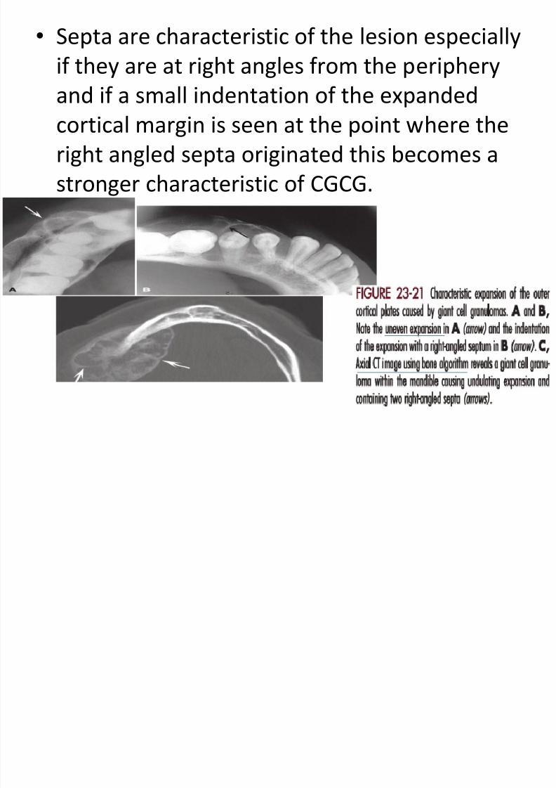
• Septa are characteristic of the lesion especially
if they are at right angles from the periphery
and if a small indentation of the expandedcortical margin is seen at the point where the
right angled septa originated this becomes a
stronger characteristic of CGCG.

8/10/2019 Oral radiology seminar.pptx
http://slidepdf.com/reader/full/oral-radiology-seminarpptx 9/22
4-effect on surrounding structure:
•
displaces and resorbs teeth• missing lamina dura
• ID canal displaced inferiorly
• strong propensity to expand the corticalboundaries of Mx & Mn.
8/10/2019 Oral radiology seminar.pptx
http://slidepdf.com/reader/full/oral-radiology-seminarpptx 10/22
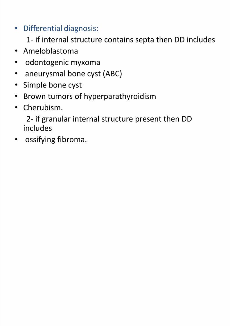
• Differential diagnosis:
1- if internal structure contains septa then DD includes• Ameloblastoma
• odontogenic myxoma
• aneurysmal bone cyst (ABC)
• Simple bone cyst
• Brown tumors of hyperparathyroidism
• Cherubism.
2- if granular internal structure present then DDincludes
• ossifying fibroma.

8/10/2019 Oral radiology seminar.pptx
http://slidepdf.com/reader/full/oral-radiology-seminarpptx 11/22
disease differentiating feature
ameloblastoma Older age group
Posterior Mn
Coarse curved, well defined trabeculae.
Odontogenic myxoma Older age group
Sharper straight septa
Don’t have the same propensity to expand.
Aneurysmal bone cyst Rare lesion
More often in the posterior aspect
Usually cause profound expansion.
Cherubisum Lesions are multiple
Epicenters located most posterior of Mx
and Mn.

8/10/2019 Oral radiology seminar.pptx
http://slidepdf.com/reader/full/oral-radiology-seminarpptx 12/22
• Management: CT scan (establish extent and
involvement), if in second decade of lifehyperparathyroidism should be considered
and test for elevated calcium level.
• Treatment: may include enucleation,curettage, resection of the jaw in some cases.
• recurrence is rare, but if occurs it is more
likely in Mx

8/10/2019 Oral radiology seminar.pptx
http://slidepdf.com/reader/full/oral-radiology-seminarpptx 13/22
Aneurysmal bone cyst (ABC)
Considered a reactive lesion of bone, however
there have been several chromosomal
translocations described that give some
credence to a neoplastic nature of the lesion.

8/10/2019 Oral radiology seminar.pptx
http://slidepdf.com/reader/full/oral-radiology-seminarpptx 14/22
Clinical features:
• 90%of jaw lesions in individuals younger than 30years old
• have a predilection for females
• fairly rapid bony swelling usually buccal or labial
• pain (occasional complaint)
• tender to palpation.
Imaging features:
1-location:• Mn:Mx 3:2
• molar and ramous region more involved thananterior region.

8/10/2019 Oral radiology seminar.pptx
http://slidepdf.com/reader/full/oral-radiology-seminarpptx 15/22
2-periphery and shape:
• well-defined periphery
• circular or hydraulic shape.
3-internal structure:
• small initial lesion shows no evidence ofinternal structure
• often the internal aspect has a multilocular
appearance• wispy ill-defined septa and at right angle to
the outer expanded border (as in CGCG).

8/10/2019 Oral radiology seminar.pptx
http://slidepdf.com/reader/full/oral-radiology-seminarpptx 16/22
4-effect on surrounding structure: can
displace and resorb teeth.

8/10/2019 Oral radiology seminar.pptx
http://slidepdf.com/reader/full/oral-radiology-seminarpptx 17/22

8/10/2019 Oral radiology seminar.pptx
http://slidepdf.com/reader/full/oral-radiology-seminarpptx 18/22
• Differential diagnosis:
• CGCG
• Amelobastoma
•
CherubismDiagnosis is based on biopsy, a hemorrhagic
aspirate favors diagnosis of ABC. CT scan will
determine the extent of the lesion.

8/10/2019 Oral radiology seminar.pptx
http://slidepdf.com/reader/full/oral-radiology-seminarpptx 19/22
disease Feature similar to ABC Differentiating feature
CGCG Internal granular septa Expand to a lesser degree
Ameloblastoma Expansion to the same
degree in posterior
mandible
Older age
cherubism Giant cell like feature Multifocal bilateral

8/10/2019 Oral radiology seminar.pptx
http://slidepdf.com/reader/full/oral-radiology-seminarpptx 20/22
• Management: surgical curettage and partial
resection.
• Recurrence rate is from 19% to about 50%
after curettage and approximately 11% after
resection.

8/10/2019 Oral radiology seminar.pptx
http://slidepdf.com/reader/full/oral-radiology-seminarpptx 21/22
• reference: oral radiology principles and
interpretations 7th edition by Stuart C. White
and Michael J. Pharoah.

8/10/2019 Oral radiology seminar.pptx
http://slidepdf.com/reader/full/oral-radiology-seminarpptx 22/22
THE END.