Oral pigmentations in Laugier-Hunziker syndrome: a case ... · 53 hard palate (Figure 2C), lower...
5
52 J Bras Patol Med Lab. 2018 Feb; 54(1): 52-56. CASE REPORT Oral pigmentations in Laugier-Hunziker syndrome: a case report and review of diagnostic criteria Pigmentações orais na síndrome de Laugier-Hunziker: relato de caso e revisão dos critérios de diagnóstico Luiz Arthur B. Silva; Rodrigo P. Mafra; Patrícia T. Oliveira; Ana Miryam C. Medeiros; Leão P. Pinto; Éricka Janine D. Silveira Universidade Federal do Rio Grande do Norte (UFRN), Rio Grande do Norte, Brazil. First submission on 10/23/17; last submission on 10/23/17; accepted for publication on 01/20/18; published on 02/20/18 ABSTRACT Laugier-Hunziker syndrome (LHS) is a rare mucocutaneous disorder, of unknown etiology, characterized by multiple hyperpigmented macules, dispersed mostly on the oral mucosa, occasionally associated with longitudinal ridging of the nails. The diagnosis requires exclusion of other conditions, such as Addison’s disease and Peutz-Jeghers syndrome. We report a case of a 34-year-old male patient, presenting with hyperpigmented macules on the lips, buccal mucosa and palate, as well as mild dark striations on toenails. After careful clinical and laboratorial investigations, the diagnosis of LHS was established. Given the lack of aesthetic complaints and symptoms, no treatment was necessary. Key words: hyperpigmentation; mouth mucosa; skin abnormalities. INTRODUCTION The Laugier-Hunziker syndrome (LHS) is a rare acquired mucocutaneous disorder of undetermined etiology (1) . It is a benign condition, with neither malignant potential (2, 3) nor systemic features (4) . LHS mainly affects middle-aged Caucasian women and presents with multiple greyish, brownish or black macules, with smooth surface and variable size that can be focal or diffuse. The main affected sites are lower lip, buccal mucosa and hard palate (5) . Less affected sites are gingiva, tongue, abdominal, genital and perianal region (2, 4) . Hyperpigmented longitudinal bands on finger and toenails are also quite frequent findings in patients with this syndrome (3, 5) . Histopathological characteristics of hyperpigmented areas found in LHS are unspecific and consist principally of increased melanin production by basal cells associated with pigmentary incontinence (1, 5) . For a conclusive diagnostic, the association of clinical and laboratory data is necessary for exclusion of systemic diseases, such as Addison’s disease, Peutz-Jeghers syndrome and McCune-Albright syndrome (3, 5) . Considering that oral lesions related to LHS are clinically similar to other pigmented lesions, correct diagnosis and management of this condition is essential to avoid erroneous conducts. The current work aims to report a case of LHS and discuss aspects of interest for diagnosis, highlighting clinical, laboratory and histopathological criteria, and comparing the findings with reports published in the latest 10 years in the scientific literature. CASE REPORT A 34-year-old male Black patient was referred to a service of oral diagnosis for evaluation of pigmented lesions in oral mucosa. During anamnesis, he stated the lesions were asymptomatic and had been noted more than 10 years before. He also reported to have undergone treatment with topical steroids on the lip for 30 days, with no remission of the lesions. Lymph nodes were not palpable, and the locoregional examination revealed brownish macules of ill-defined borders on the lower and upper lips (Figure 1). The clinical intraoral examination revealed diffuse bilateral black macules on buccal mucosa (Figure 2A and 2B), 10.5935/1676-2444.20180011
-
Upload
nguyentuyen -
Category
Documents
-
view
224 -
download
0
Transcript of Oral pigmentations in Laugier-Hunziker syndrome: a case ... · 53 hard palate (Figure 2C), lower...

52
J Bras Patol Med Lab. 2018 Feb; 54(1): 52-56.CaSE rEPort
Oral pigmentations in Laugier-Hunziker syndrome: a case report and review of diagnostic criteria
Pigmentações orais na síndrome de Laugier-Hunziker: relato de caso e revisão dos critérios de diagnóstico
Luiz Arthur B. Silva; Rodrigo P. Mafra; Patrícia T. Oliveira; Ana Miryam C. Medeiros; Leão P. Pinto; Éricka Janine D. Silveira
Universidade Federal do Rio Grande do Norte (UFRN), Rio Grande do Norte, Brazil.
First submission on 10/23/17; last submission on 10/23/17; accepted for publication on 01/20/18; published on 02/20/18
aBStraCt
Laugier-Hunziker syndrome (LHS) is a rare mucocutaneous disorder, of unknown etiology, characterized by multiple hyperpigmented macules, dispersed mostly on the oral mucosa, occasionally associated with longitudinal ridging of the nails. The diagnosis requires exclusion of other conditions, such as Addison’s disease and Peutz-Jeghers syndrome. We report a case of a 34-year-old male patient, presenting with hyperpigmented macules on the lips, buccal mucosa and palate, as well as mild dark striations on toenails. After careful clinical and laboratorial investigations, the diagnosis of LHS was established. Given the lack of aesthetic complaints and symptoms, no treatment was necessary.
Key words: hyperpigmentation; mouth mucosa; skin abnormalities.
introDuCtion
The Laugier-Hunziker syndrome (LHS) is a rare acquired mucocutaneous disorder of undetermined etiology(1). It is a benign condition, with neither malignant potential(2, 3) nor systemic features(4).
LHS mainly affects middle-aged Caucasian women and presents with multiple greyish, brownish or black macules, with smooth surface and variable size that can be focal or diffuse. The main affected sites are lower lip, buccal mucosa and hard palate(5). Less affected sites are gingiva, tongue, abdominal, genital and perianal region(2, 4). Hyperpigmented longitudinal bands on finger and toenails are also quite frequent findings in patients with this syndrome(3, 5).
Histopathological characteristics of hyperpigmented areas found in LHS are unspecific and consist principally of increased melanin production by basal cells associated with pigmentary incontinence(1, 5). For a conclusive diagnostic, the association of clinical and laboratory data is necessary for exclusion of systemic diseases, such as Addison’s disease, Peutz-Jeghers syndrome and McCune-Albright syndrome(3, 5).
Considering that oral lesions related to LHS are clinically similar to other pigmented lesions, correct diagnosis and management of this condition is essential to avoid erroneous conducts. The current work aims to report a case of LHS and discuss aspects of interest for diagnosis, highlighting clinical, laboratory and histopathological criteria, and comparing the findings with reports published in the latest 10 years in the scientific literature.
CaSE rEPort
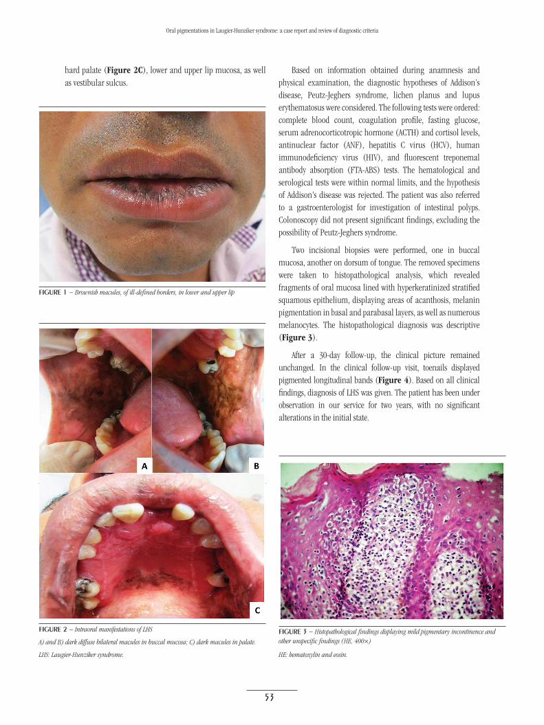
A 34-year-old male Black patient was referred to a service of oral diagnosis for evaluation of pigmented lesions in oral mucosa. During anamnesis, he stated the lesions were asymptomatic and had been noted more than 10 years before. He also reported to have undergone treatment with topical steroids on the lip for 30 days, with no remission of the lesions. Lymph nodes were not palpable, and the locoregional examination revealed brownish macules of ill-defined borders on the lower and upper lips (Figure 1). The clinical intraoral examination revealed diffuse bilateral black macules on buccal mucosa (Figure 2A and 2B),
10.5
935/
1676
-244
4.20
1800
11
53
hard palate (Figure 2C), lower and upper lip mucosa, as well as vestibular sulcus.
Based on information obtained during anamnesis and physical examination, the diagnostic hypotheses of Addison’s disease, Peutz-Jeghers syndrome, lichen planus and lupus erythematosus were considered. The following tests were ordered: complete blood count, coagulation profile, fasting glucose, serum adrenocorticotropic hormone (ACTH) and cortisol levels, antinuclear factor (ANF), hepatitis C virus (HCV), human immunodeficiency virus (HIV), and fluorescent treponemal antibody absorption (FTA-ABS) tests. The hematological and serological tests were within normal limits, and the hypothesis of Addison’s disease was rejected. The patient was also referred to a gastroenterologist for investigation of intestinal polyps. Colonoscopy did not present significant findings, excluding the possibility of Peutz-Jeghers syndrome.
Two incisional biopsies were performed, one in buccal mucosa, another on dorsum of tongue. The removed specimens were taken to histopathological analysis, which revealed fragments of oral mucosa lined with hyperkeratinized stratified squamous epithelium, displaying areas of acanthosis, melanin pigmentation in basal and parabasal layers, as well as numerous melanocytes. The histopathological diagnosis was descriptive (Figure 3).
After a 30-day follow-up, the clinical picture remained unchanged. In the clinical follow-up visit, toenails displayed pigmented longitudinal bands (Figure 4). Based on all clinical findings, diagnosis of LHS was given. The patient has been under observation in our service for two years, with no significant alterations in the initial state.
figurE 1 − Brownish macules, of ill-defined borders, in lower and upper lip
figurE 2 − Intraoral manifestations of LHS
A) and B) dark diffuse bilateral macules in buccal mucosa; C) dark macules in palate.
LHS: Laugier-Hunziker syndrome.
figurE 3 − Histopathological findings displaying mild pigmentary incontinence and other unspecific findings (HE, 400×)
HE: hematoxylin and eosin.
Oral pigmentations in Laugier-Hunziker syndrome: a case report and review of diagnostic criteria

54
figurE 4 − Longitudinal brownish bands in patient’s toenails
taBLE − LHS cases published in the literature in English in the latest 10 years (2007-2017)
Author SexAge
(years)Anatomical location Aspects of oral lesions Other manifestations
Present study M 34 Lip, buccal mucosa, palate Hyperpigmented macules Hyperpigmented longitudinal bands in toenailsVerma et al. (2017)(8) F 19 Lip, tongue, palate Hyperpigmented macules Longitudinal melanonychia in finger and toenails
Bhoyrul et al. (2016)(9) F 48 Lip Several brown to dark macules Hyperpigmentation of fingers
Kaçar et al. (2016)(10) M 23 Lip Multiple pigmentationLongitudinal hyperpigmented bands in nails,
punctiform pigmentation in the right eyeLalosevic et al. (2015)(7) F 63 Buccal mucosa, Regular brown pigmentation Longitudinal pigmented bands in finger and toenails
Mahmood et al. (2015)(3) F 46 Lip Hyperpigmented maculesLongitudinal hyperpigmentation in finger and
toenails; isolate macule in the conjunctivaErgun et al. (2013)(1) F 37 Lip, buccal mucosa, palate Brown to dark pigmentation -
Nikitakis et al. (2013)(5) F 55 Lip, buccal mucosa Multiple oral melanotic macules Ill-defined striae in fingernails
Wang et al. (2012)(2)
FM M
454036
Lip, buccal mucosa, gingiva, tongueGingiva
Lip
Diffuse hyperpigmentationWidely diffuse pigmentation
Multiple oral melanotic macules
Longitudinal hyperpigmentation in finger and toenailsBrown macule on conjunctiva
Brown macules in nails and interdigital spacesWondratsch et al. (2012)(6) F 61 Buccal mucosa, tongue Confluent pigmented macules Hyperpigmentation in vulva
Asati et al. (2011)(11) F 19 Lip, buccal mucosa, palate, tongueMild and multiple brown to dark macules, oval to irregular shape
Multiple freckles in skin of the periorbital region, nose, cheeks, fingertips and nails
Ma et al. (2011)(12) F 60 Lip, buccal mucosa, tongue Diffuse pigmentation Longitudinal pigmented bands in nails
Sachdeva et al. (2011)(13) F 50 Lip, tongue Brown maculesBrown macules in finger and toenails;
darkened bands in nailsJabbari et al. (2010)(14) F 45 Tongue Hyperpigmented macules Hyperpigmented macules in nails
Montebugn et al. (2010)(15) F 43 Lip, buccal mucosa, Diffuse brown macules Longitudinal pigmentation in nailsRangwala et al. (2010)(4) M 77 Lip Confluent brown macules Bilateral melanonychia striata in finger and toenails
Sendagorta et al. (2010)(16) F 76 Lip, buccal mucosa, palate, tongueMultiple brown to dark lenticular
maculesLinear melanonychia in finger and toenails
Tamiya et al. (2010)(17) F 61 Lip, buccal mucosa, Slightly brown to grayish blue macules,
oval to irregular shapeDiffuse longitudinal melanonychia
Aliagaoglu et al. (2008)(18) M 51 Lip, buccal mucosa, Multiple lenticular dark brown maculesIncreased pigmentation in right pre-tibial area;
pigmented macules in palmoplantar region
Kim et al. (2008)(19) F 54 Lip, buccal mucosa, tongueConfluent macules of brown
to dark aspectLongitudinal melanonychia in nails
Yago et al. (2008)(20)
M M
39
68
Lip, buccal mucosa, tongue, palate
Lip, buccal mucosa, tongue, palate
Multiple hyperpigmented macules
Multiple hyperpigmented macules
Well-defined brown macules in palmoplantar regions of toes and toenails
Multiple hyperpigmented macules in palmoplantar region; longitudinal pigmented bands in toenails
Gencoglan et al. (2007)(21) F 24 Lip, gingiva, tongue Bluish to brown maculesLongitudinal hyperpigmented macule in the vulva;
pigmented macules in palmoplantar area; melanonychia in toenails
LHS: Laugier-Hunziker syndrome; M: male; F: female.
DiSCuSSion
LHS is a rare condition of unknown pathogenesis, with a limited number of reported cases since its first description in 1970(6, 7). The Table summarizes the 25 cases published in the last decade (2007-2017) in the scientific literature in English available at PubMed(1-21). Although we report a case of LHS in a male patient, our review demonstrated that this condition is more common in women (72%), with a female/male incidence ratio of 2.75:1. The syndrome was observed to more commonly affect individuals between the fifth and seventh decades of life, with an average of 48.12 years and age ranging from 19 to 76 years.
Luiz Arthur B. Silva; Rodrigo P. Mafra; Patrícia T. Oliveira; Ana Miryam C. Medeiros; Leão P. Pinto; Éricka Janine D. Silveira
55
In all LHS cases listed in the Table, hyperpigmentation in the oral mucosa was identified, with this being the finding that frequently takes patients to search for specialized advice. The lip, especially the lower, is the anatomical location most commonly affected by hyperpigmentation (84%), followed by buccal mucosa (64%), tongue (48%), palate (28%) and gingiva (12%). Most patients identified in our survey (76%) presented more than one site of oral mucosa with hyperpigmentation. Out of the six patients that exhibited just one site of affected oral mucosa, five had lip lesions. In the present case, the patient exhibited hyperpigmentation on lip, palate, and buccal mucosa. As a general rule, oral mucosa lesions were described as multiple hyperpigmented macules of brownish-black or grayish color, well-delimited or diffuse with varied shapes.
Although most patients with LHS present hyperpigmentation of oral mucosa, pigmented lesions were also described in less common sites, as neck, thorax, abdomen, perianal and perivaginal mucosa, sclera and eyelids(7). Nails represent the most affected extraoral site, with some authors suggesting that this is a finding observed in up to 60% of individuals(3, 5, 14). In our review (Table), 20 out of the 25 patients (80%) presented linear hyperpigmentation of finger and/or toenails. Our patient also displayed mild nail affection. Other less frequent findings are summarized in the Table.
Although biopsies of oral mucosa pigmented lesions are a common clinical conduct, histopathological findings of hyperpigmented areas associated with LHS are unspecific. Foci of acanthosis and melanin pigmentation of varied degrees in basal cells of the epithelium(7) are commonly found, as in the case reported here.
The diagnosis of LHS is established based on exclusion of most frequent systemic conditions, such as Peutz-Jeghers syndrome Addison’s disease, and McCune-Albright syndrome(1). Peutz-Jeghers syndrome is characterized by the presence of gastrointestinal hemartomatous polyps with great potential for malignant transformation, besides frequent skin and mucosa
hyperpigmentation. The diagnosis of Peutz-Jeghers syndrome is given based on the identification of those polyps in association with familial history of the disorder(3). None of these was found in our patient. Addison’s disease, an endocrine disorder caused by insufficient cortisol and aldosterone production, is characterized by the increased pigmentation overlying joints, skin folds and mucosas. Other systemic findings include hypotension, dehydration and abdominal pain(3, 9). Upon proving normality of serum levels of cortisol in our patient and given the absence of other signs and symptoms, the hypothesis of Addison’s disease was rejected. McCune-Albright syndrome is characterized principally by hormone disorders associated with precocious puberty, polyostotic fibrous dysplasia, and cafe-au-lait macules(8). Such findings were also not observed in the conducted anamnesis.
Diffuse oral pigmentation can also be associated with systemic ingestion of medicines, such as tetracyclines, antimalarics, amiodarone, chemotherapeutical agents, oral contraceptives, phenothiazines, azidothymidine and ketoconazole(1); none of these drugs was used by the patient in question. Tobacco consumption can also induce increase in oral mucosa pigmentation, although generally, in these cases, the alteration is concentrated in the anterior gingiva and is not associated with pigmentation in other parts of the body(1, 5). This hypothesis was not considered in our case, since the patient denied tobacco consumption.
Given its benign behavior and absence of symptoms, hyperpigmentation associated with LHS does not require any treatment, although some patients complain because of esthetic reasons. In these cases, one can use cryotherapy and Q-switched Nd:YAG or Q-switched alexandrite laser(1, 6, 14). In our case, no measure was taken, as the patient did not voice esthetic complains.
We highlight the importance of rapid precise diagnosis of LHS, excluding other severe systemic conditions and avoiding unnecessary investigations and, principally, treatments that are inadequate for patients.
rESuMo
A síndrome de Laugier-Hunziker (SLH) é uma rara desordem mucocutânea, de etiologia indeterminada, caracterizada por múltiplas máculas hiperpigmentadas, dispersas principalmente na mucosa oral, por vezes associadas a estrias longitudinais nas unhas. O diagnóstico requer exclusão de condições como doença de Addison e síndrome de Peutz-Jeghers. Descrevemos o caso de um paciente do sexo masculino, 34 anos, com presença de máculas hiperpigmentadas em lábios, mucosa jugal e palato, além de discretas estrias enegrecidas nas unhas dos pés. Após minuciosa investigação clínica e laboratorial, foi estabelecido o diagnóstico de SLH. Dada a ausência de queixas estéticas e sintomatologia, nenhum tratamento foi necessário.
Unitermos: hiperpigmentação; mucosa bucal; anormalidades da pele.
Oral pigmentations in Laugier-Hunziker syndrome: a case report and review of diagnostic criteria

56
rEfErEnCES
1. Ergun S, Saruhanoğlu A, Migliari DA, Maden I, Tanyeri H. Refractory pigmentation associated with Laugier-Hunziker syndrome following Er:YAG laser treatment. Case Rep Dent. 2013; 2013: 1-4.
2. Wang WM, Wang X, Duan N, Jiang HL, Huang XF. Laugier-Hunziker syndrome: a report of three cases and literature review. Int J Oral Sci. 2012; 4(4): 226-30.
3. Mahmood T, Menter A. The Laugier-Hunziker syndrome. Proc (Bayl Univ Med Cent). 2015; 28(1): 41-2.
4. Rangwala S, Doherty CB, Katta R. Laugier-Hunziker syndrome: a case report and review of the literature. Dermatol Online J. 2010; 16(12): 9.
5. Nikitakis NG, Koumaki D. Laugier-Hunziker syndrome: case report and review of the literature. Oral Surg Oral Med Oral Pathol Oral Radiol. 2013; 116(1): 52-8.
6. Wondratsch H, Feldmann R, Steiner A, Breier F. Laugier-Hunziker syndrome in a patient with pancreatic cancer. Case Rep Dermatol. 2012; 4(2): 174-6.
7. Lalosevic J, Zivanovic D, Skiljevic D, Medenica L. Laugier-Hunziker syndrome – case report. An Bras Dermatol. 2015; 90(3): 223-5.
8. Verma B, Behra A, Ajmal AK, Sen S. Laugier-Hunziker syndrome in a young female. Indian Dermatol Online J. 2017; 8(2): 148-50.
9. Bhoyrul B, Paulus J. Macular pigmentation complicating irritant contact dermatitis and viral warts in Laugier-Hunziker syndrome. Clin Exp Dermatol. 2016; 41(3): 294-6.
10. Kaçar N, Yildiz CC, Demirkan N. Dermoscopic features of conjunctival, mucosal, and nail pigmentations in a case of Laugier-Hunziker syndrome. Dermatol Pract Concept. 2016; 6(1): 23-4.
11. Asati DP, Tiwari S. Laugier-Hunziker syndrome. Indian J Dermatol Venereol Leprol. 2011; 77(4): 536.
12. Ma DL, Vano-Galvan S. Hyperpigmentation in Laugier-Hunziker syndrome. CMAJ. 2011; 183(12): 1402.
13. Sachdeva S. Sachdeva S, Kapoor P. Laugier-Hunziker syndrome: a rare cause of oral and acral pigmentation. J Cutan Aesthet Surg. 2011; 4(1): 58-60.
14. Jabbari A, Gonzalez ME, Franks Jr AG, Sanchez M. Laugier Hunziker syndrome. Dermatol Online J. 2010; 16(11): 23.
15. Montebugnoli L, Grelli I, Cervellati F, Misciali C, Raone B. Laugier-Hunziker syndrome: an uncommon cause of oral pigmentation and a review of the literature. Int J Dent. 2010; 2010: 1-4.
16. Sendagorta E, Feito M, Ramírez P, Gonzalez-Beato M, Saida T, Pizarro A. Dermoscopic findings and histological correlation of the acral volar pigmented maculae in Laugier-Hunziker syndrome. J Dermatol. 2010; 37(11): 980-4.
17. Tamiya H, Kamo R, Sowa J, et al. Dermoscopic features of pigmentation in Laugier-Hunziker-Baran syndrome. Dermatol Surg. 2010; 36(1): 152-4.
18. Aliagaoglu C, Yanik ME, Albayrak H, Güvenç SC, Yildirim U. Laugier-Hunziker syndrome: diffuse large hyperpigmentation on atypical localization. J Dermatol. 2008; 35(12): 806-7.
19. Kim EJ, Cho SH, Lee JD. A case of Laugier-Hunziker syndrome. Ann Dermatol. 2008; 20(3): 126-9.
20. Yago K, Tanaka Y, Asanami S. Laugier-Hunziker-Baran syndrome. Oral Surg Oral Med Oral Pathol Oral Radiol Endod. 2008; 106(2): 20-5.
21. Gencoglan G, Gerceker-Turk B, Kilinc-Karaarslan I, Akalin T, Ozdemir F. Dermoscopic findings in Laugier-Hunziker syndrome. Arch Dermatol. 2007; 143(5): 631-5.
CorrESPonDing author
Éricka Janine Dantas da Silveira Departamento de Odontologia; Universidade Federal do Rio Grande do Norte; Avenida Senador Salgado Filho, 1787; Lagoa Nova; CEP: 59056-000; Natal-RN, Brasil; Phone/Fax: +55 (84) 3215-4138; e-mail: [email protected].
Luiz Arthur B. Silva; Rodrigo P. Mafra; Patrícia T. Oliveira; Ana Miryam C. Medeiros; Leão P. Pinto; Éricka Janine D. Silveira
This is an open-access article distributed under the terms of the Creative Commons Attribution License.