Oral Path Review Name this oral lesion: Varix (Varices, pl.) Sometimes called caviar tongue ...
75
Oral Path Review
-
Upload
jared-boone -
Category
Documents
-
view
217 -
download
3
Transcript of Oral Path Review Name this oral lesion: Varix (Varices, pl.) Sometimes called caviar tongue ...

Oral Path Review
Name this oral lesion:

Varix (Varices, pl.) Sometimes called
caviar tongue Distended vein (like a
hemorrhoid) No treatment usually
required Diff. diagnosis:
mucocele and hemangioma

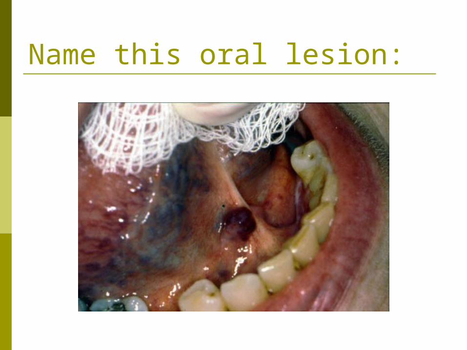
Name this oral lesion:

Mucocele Collection of saliva in the
oral mucosa Common symptom: gets
bigger, then smaller, bigger, etc.
Traumatic severance of salivary ducts
Treated by surgical excision of the entire gland that feeds the duct
Could be confused with salivary gland neoplasm, varix, and hemangioma

Name this oral lesion:
You should know this by now…

Torus palatinus and Torus mandibularis
Bony bumps in the mouth…midline if on the hard palate…single or bilateral for mandibular
Could be inherited…developmental overgrowths Won’t grow past their “programmed size” May require removal if they interfere with prosthetics
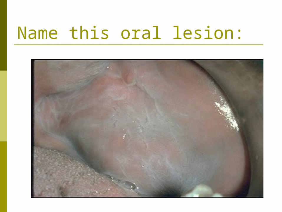
Name this oral lesion:
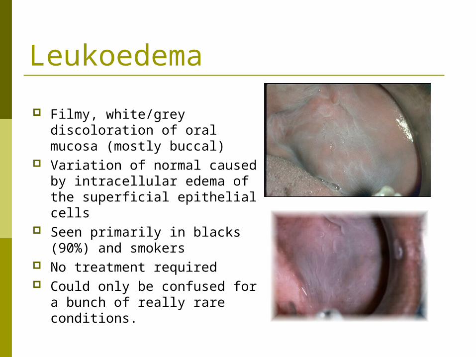
Leukoedema
Filmy, white/grey discoloration of oral mucosa (mostly buccal)
Variation of normal caused by intracellular edema of the superficial epithelial cells
Seen primarily in blacks (90%) and smokers
No treatment required Could only be confused for a
bunch of really rare conditions.

Name this oral lesion:

Papillary hyperplasia (PH) and/or Denture sore mouth (DSM)
PH and DSM may be the same thing, thought to be caused by Candida albicans
Can be small red spots. When it worsens, it turns bright red and produces the red, pebbly look of papillary hyperplasia.
Treatment with antifungals, but recurrence is common
Good oral/denture hygiene may help
Benign and unmistakable
See the book for other picturesand more detailed information.This can be highly variable inappearance.
Name this oral lesion:
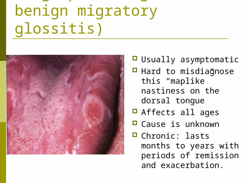
Geographic tongue (AKA: benign migratory glossitis)
Usually asymptomatic Hard to misdiagnose
this “maplike” nastiness on the dorsal tongue
Affects all ages Cause is unknown Chronic: lasts months to
years with periods of remission and exacerbation.
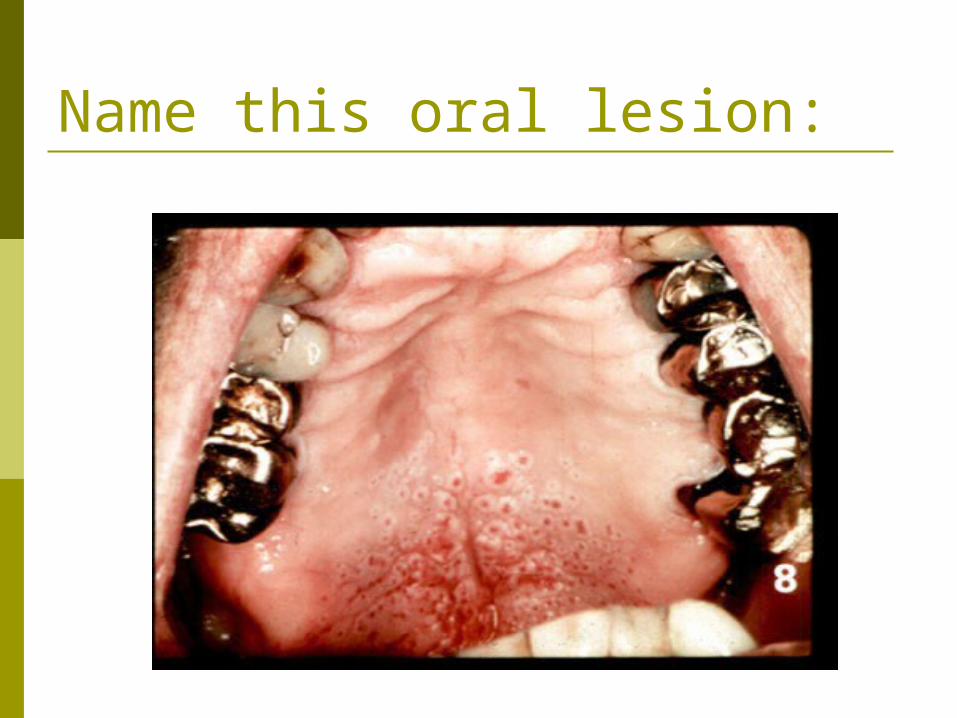
Name this oral lesion:
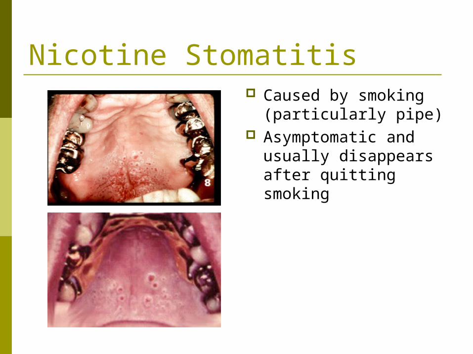
Nicotine Stomatitis Caused by smoking
(particularly pipe) Asymptomatic and
usually disappears after quitting smoking
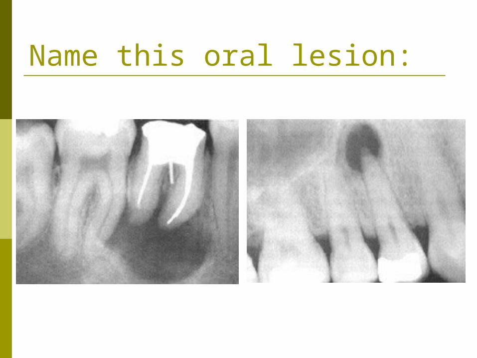
Name this oral lesion:
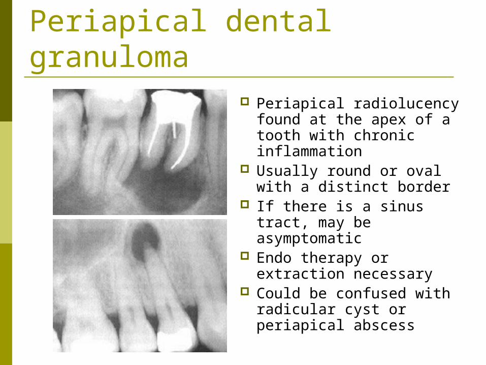
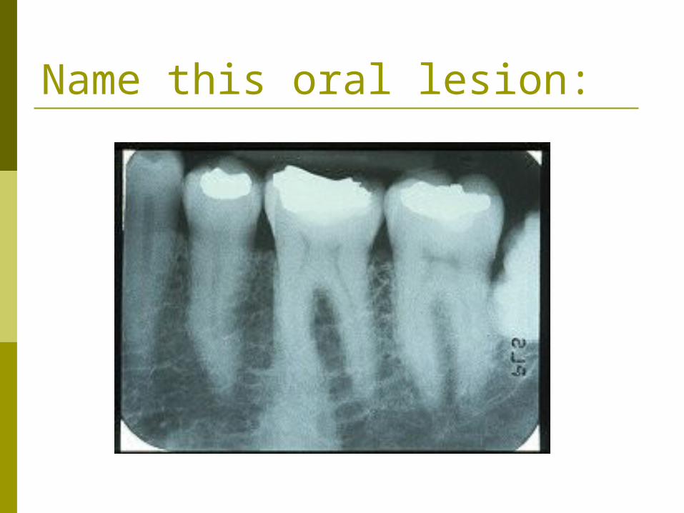
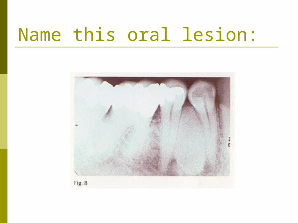
Periapical dental granuloma Periapical radiolucency
found at the apex of a tooth with chronic inflammation
Usually round or oval with a distinct border
If there is a sinus tract, may be asymptomatic
Endo therapy or extraction necessary
Could be confused with radicular cyst or periapical abscess

Name this oral lesion:
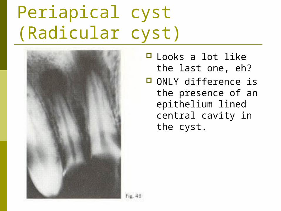
Periapical cyst (Radicular cyst)
Looks a lot like the last one, eh?
ONLY difference is the presence of an epithelium lined central cavity in the cyst.
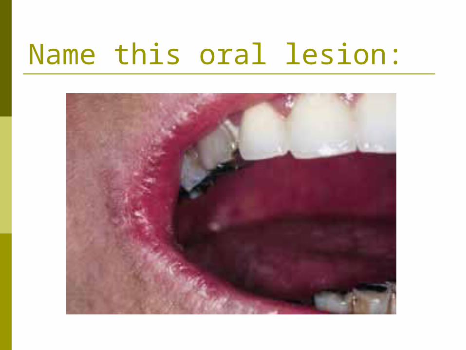
Name this oral lesion:

Angular cheilosis Dry corners of the
mouth May be caused by
slobber accumulating in the corners of the mouth in patients with a deep bite. Candida likes to hang out in the drool.
May be a riboflavin defficiency???
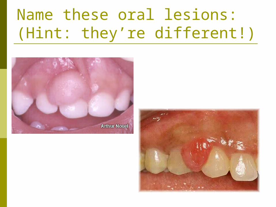
Name these oral lesions:(Hint: they’re different!)

Top: Periferal FibromaBottom: Pyogenic Granuloma
Usually in children & young adults
Histologically, this is mostly connective tissue
Can be excised, but may recur
Histologic examination may be the only way to distinguish this from Periferal Fibroma
Vascular and sometimes painful Common in pregnancy
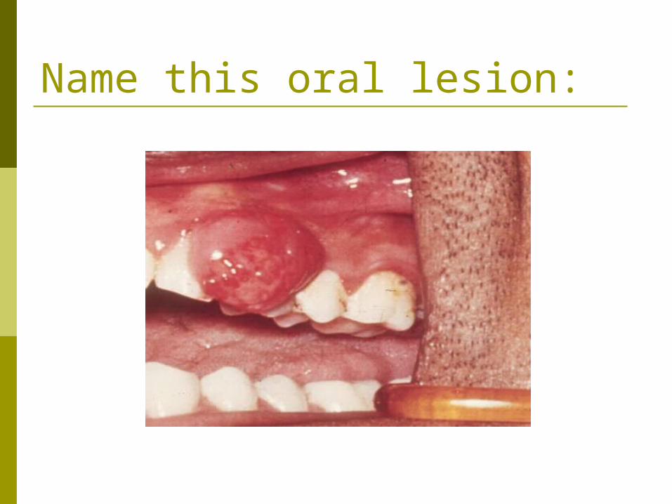
Name this oral lesion:
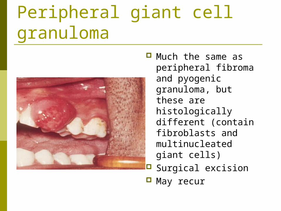
Peripheral giant cell granuloma
Much the same as peripheral fibroma and pyogenic granuloma, but these are histologically different (contain fibroblasts and multinucleated giant cells)
Surgical excision May recur

Name this oral lesion:
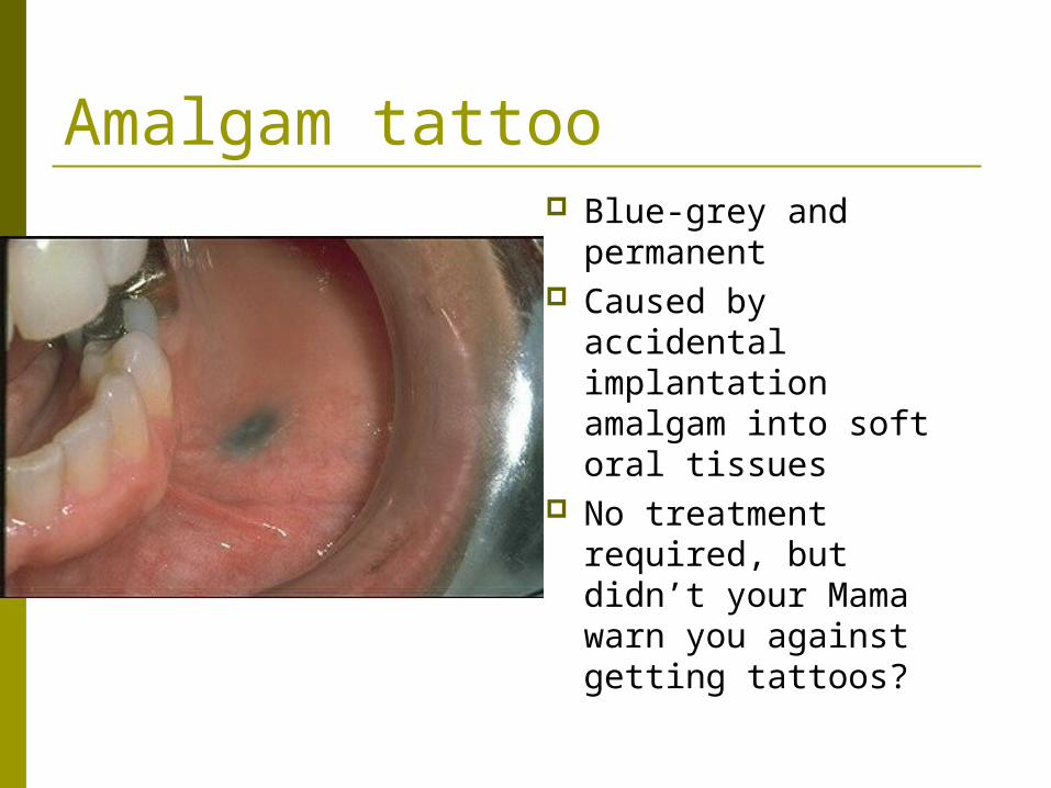
Amalgam tattoo Blue-grey and
permanent Caused by accidental
implantation amalgam into soft oral tissues
No treatment required, but didn’t your Mama warn you against getting tattoos?
Name this oral lesion:

Condensing Osteitis
Sclerotic reaction to infection commonly seen in young patients
Caused by infection of periapical tissues of low virulence
Treat only cases where the infection is symptomatic or carious in the associated tooth. Follow up with regular x-rays.
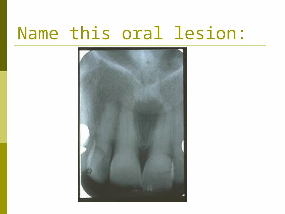
Name this oral lesion:
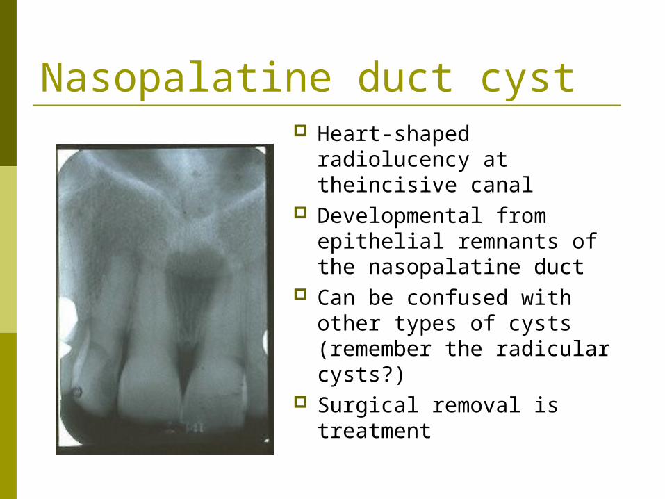
Nasopalatine duct cyst Heart-shaped radiolucency
at theincisive canal Developmental from
epithelial remnants of the nasopalatine duct
Can be confused with other types of cysts (remember the radicular cysts?)
Surgical removal is treatment

Name this oral lesion:

Dentigerous Cyst
Very common & found around an unerupted tooth
Most commonly around 3rd molars, but any tooth could be affected (rarely on deciduous teeth)
Large cysts can cause parasthesia and/or pain
Surgical enucleation should be followed up with histological examination
Name this oral lesion:

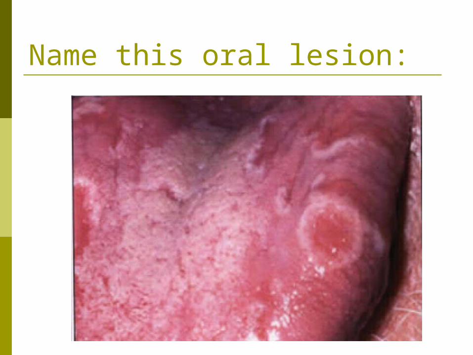
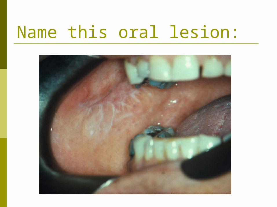
Lichen planus
Lacy, white lines are characteristic of reticular type. Erosive type and plaque type are variations.
Only erosive type requires treatment May predispose patient to oral cancer

Name this oral lesion:

Dilantin gingival hyperplasia
Can also be caused by cyclosporin and nephedapine (sp?)
Fibrous overgrowth of the gingiva, particularly the anterior (as opposed to the posterior and lingual areas)
Scrupulous dental hygiene recommended
Overgrowth requires surgical removal. Or ceasing to take the anticonvulsant may cause gradual recession within one year.
Name this oral lesion:
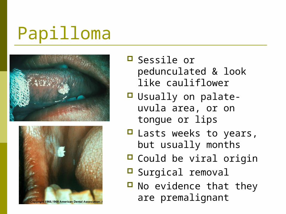
Papilloma Sessile or pedunculated
& look like cauliflower Usually on palate-uvula
area, or on tongue or lips Lasts weeks to years, but
usually months Could be viral origin Surgical removal No evidence that they
are premalignant
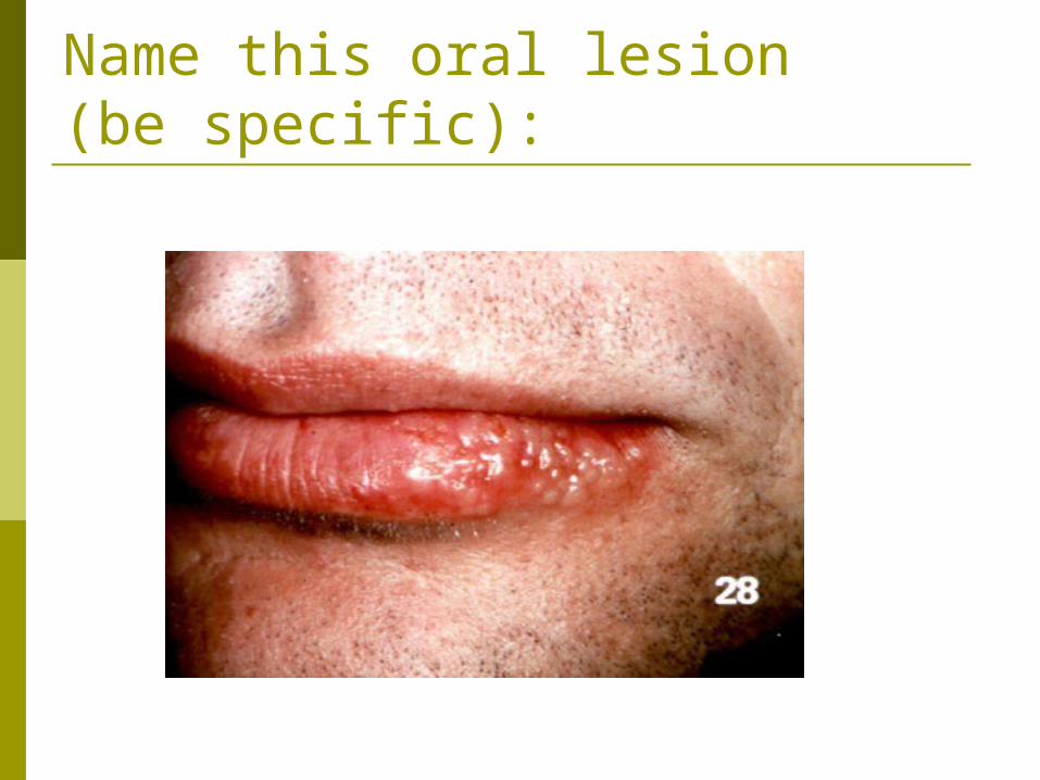
Name this oral lesion (be specific):

Herpes Labialis
Recurrent oral infection caused by herpesvirus
Recurrences vary from person to person and are thought to be triggered by exposure to sunlight, fever, trauma, and other irritants.
Virus “hides” in the nearest ganglion and lies dormant
Acyclovir ointment shortens healing time …systemic acyclovir doesn’t work for oral herpes
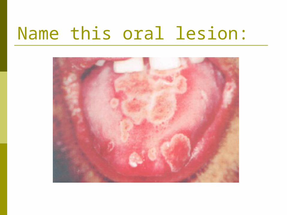
Name this oral lesion:

Primary herpetic stomatitis
Generalized involvement of the oral cavity infected with herpesvirus
Usually seen in children
Blisters break easily and are VERY painful (can’t eat or drink)
Accompanied by fever

Name this oral lesion:

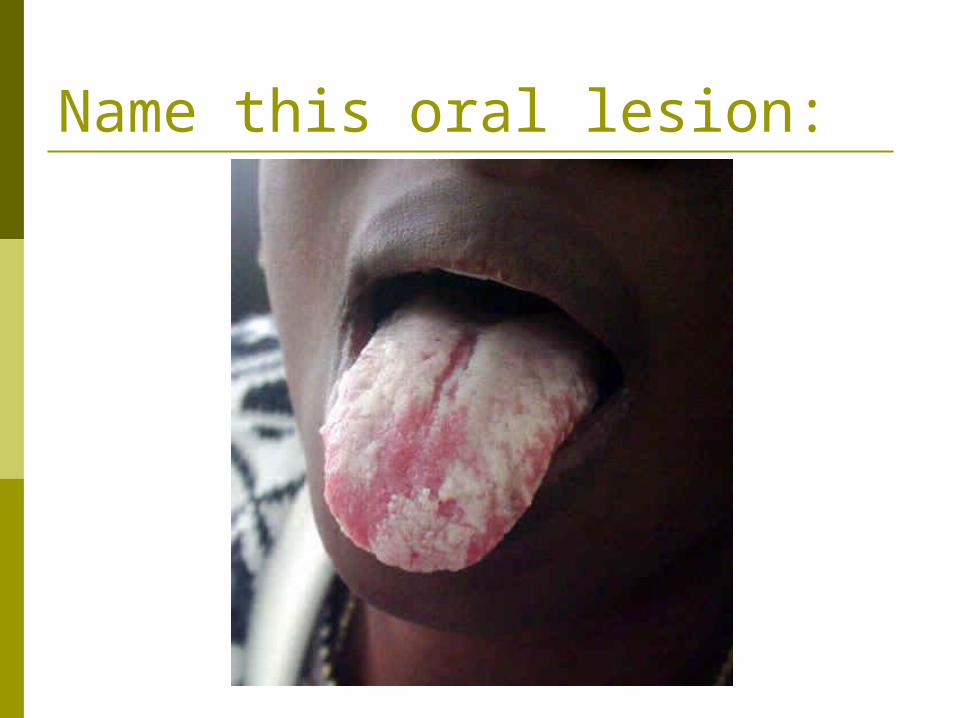
Fordyce granules
Ectopic sebaceous glands caused by a developmental anomaly
Seen in greatest numbers during puberty Most common on buccal mucosa No treatment required
Name this oral lesion:
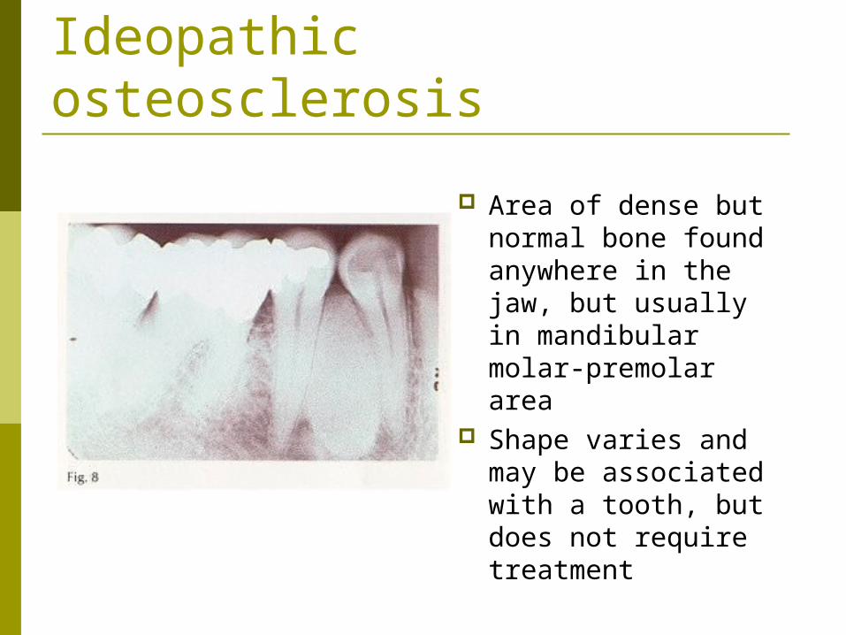
Ideopathic osteosclerosis
Area of dense but normal bone found anywhere in the jaw, but usually in mandibular molar-premolar area
Shape varies and may be associated with a tooth, but does not require treatment
Name this oral lesion:

Candidosis (Candidiasis, Moniliasis, Thrush)
Infection with Candida albicans
Involved mucus membrane develops a white, necrotic slough.
White lesions can be wiped off, leaving a bleeding, white surface
Occurs in the very young, very old, those with reduced resistance and those on long-term antibiotic therapy or immunosuppression (AIDS victim shown here)

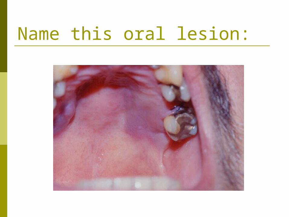
Name this oral lesion:
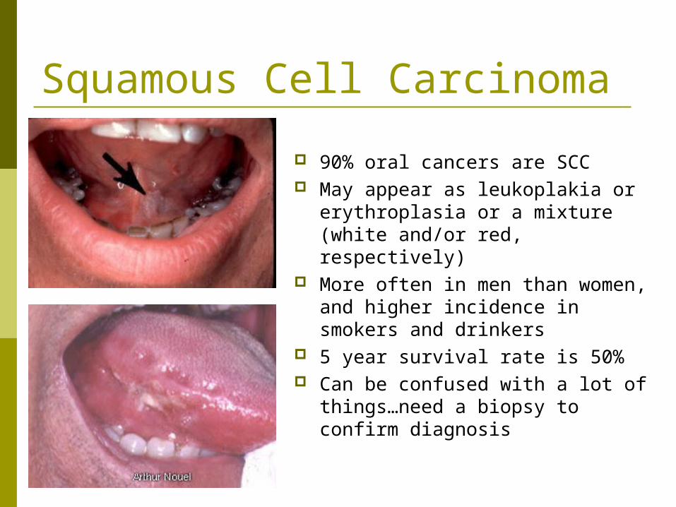
Squamous Cell Carcinoma
90% oral cancers are SCC May appear as leukoplakia or
erythroplasia or a mixture (white and/or red, respectively)
More often in men than women, and higher incidence in smokers and drinkers
5 year survival rate is 50% Can be confused with a lot of
things…need a biopsy to confirm diagnosis
Name this oral lesion:
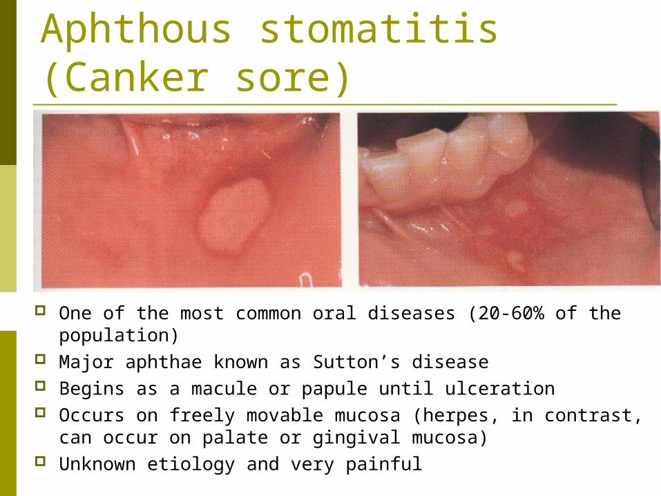
Aphthous stomatitis (Canker sore)
One of the most common oral diseases (20-60% of the population)
Major aphthae known as Sutton’s disease Begins as a macule or papule until ulceration Occurs on freely movable mucosa (herpes, in contrast, can
occur on palate or gingival mucosa) Unknown etiology and very painful
Name this oral lesion:

Kaposi’s Sarcoma Found in AIDS patients Appear as red or
purple bruises, but can progress to a hemorrhagic mass
Painful and invasive Radiation therapy
Name this oral lesion:
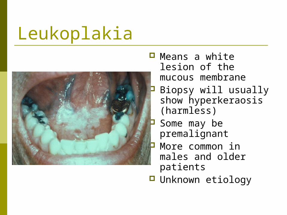
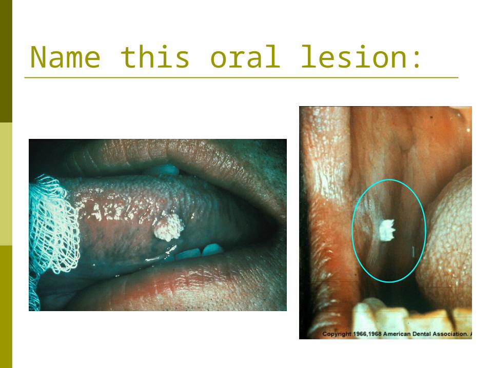
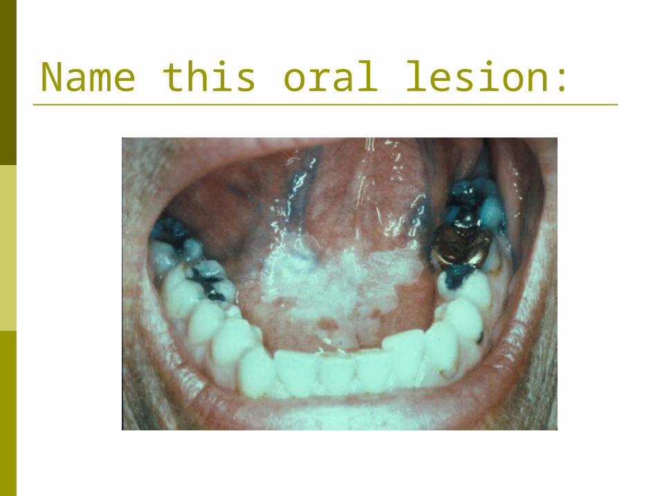
Leukoplakia Means a white lesion
of the mucous membrane
Biopsy will usually show hyperkeraosis (harmless)
Some may be premalignant
More common in males and older patients
Unknown etiology

Name this oral lesion:

Hairy leukoplakia Found in AIDS patients and
other immunocompromised patients
Usually on the lateral tongue
Caused by Epstein-Barr virus
Can easily be confused with candidiasis
Treat with anti-fungal first…if it doesn’t heal, then it is most likely hairy leukoplakia
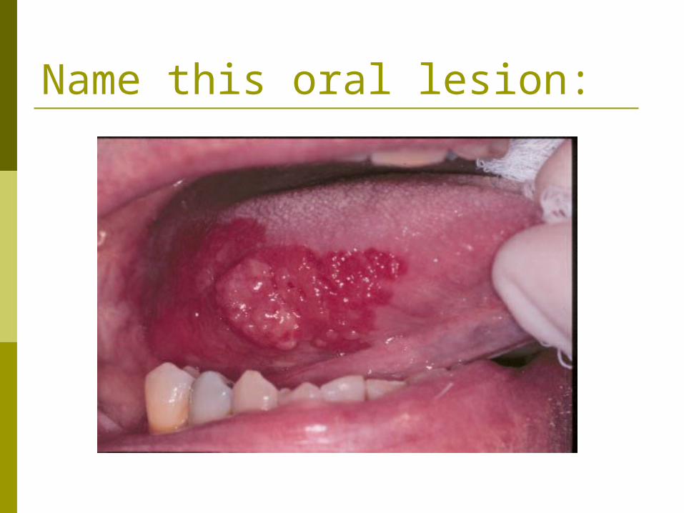
Name this oral lesion:
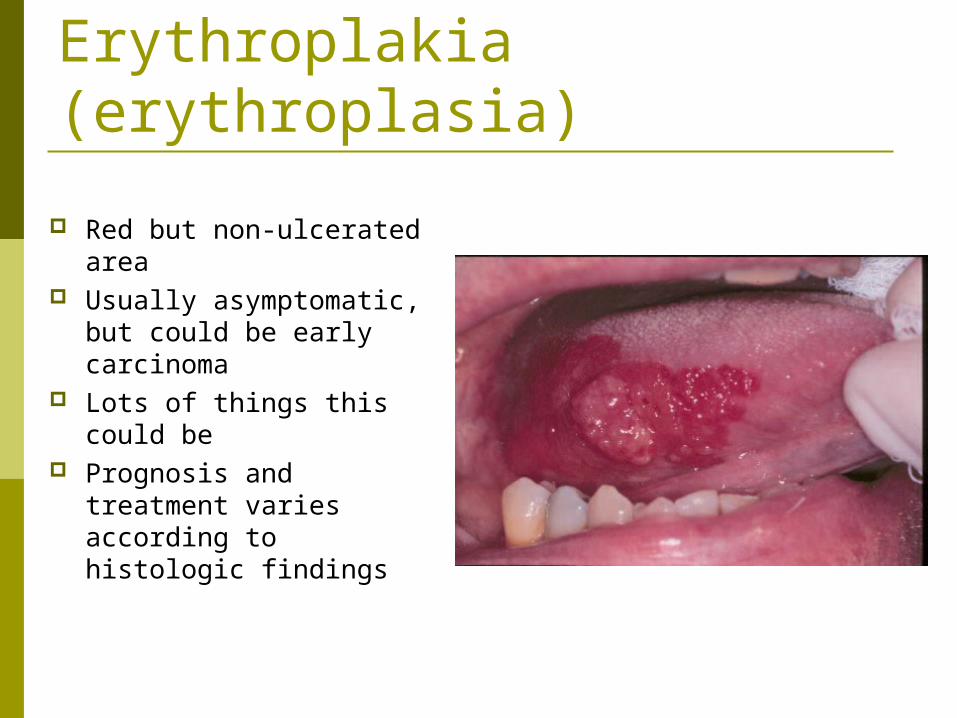
Erythroplakia (erythroplasia)
Red but non-ulcerated area
Usually asymptomatic, but could be early carcinoma
Lots of things this could be
Prognosis and treatment varies according to histologic findings

Name this oral lesion:

Epulis Fissuratum Two or more folds of
soft tissue separated by a central groove caused by an ill-fitting denture
Inflamatory hyperplasia that can surgically removed (denture border should also be reduced to prevent recurrence)
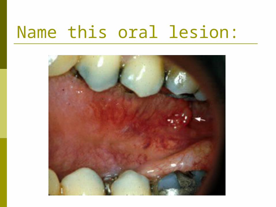
Name this oral lesion:
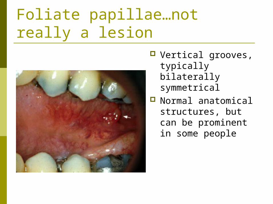
Foliate papillae…not really a lesion
Vertical grooves, typically bilaterally symmetrical
Normal anatomical structures, but can be prominent in some people
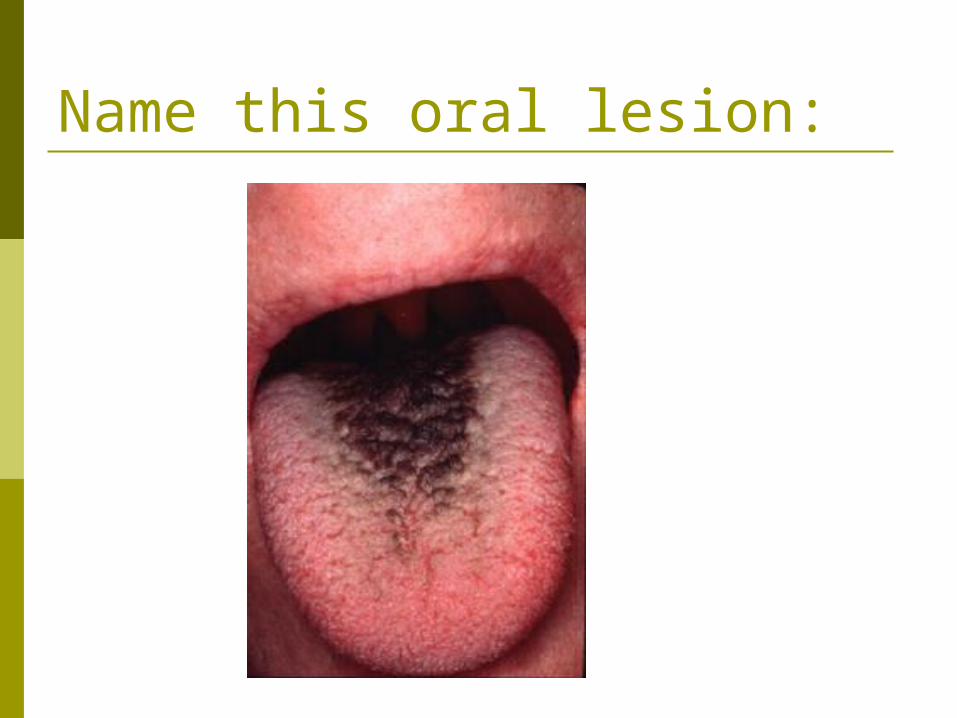
Name this oral lesion:
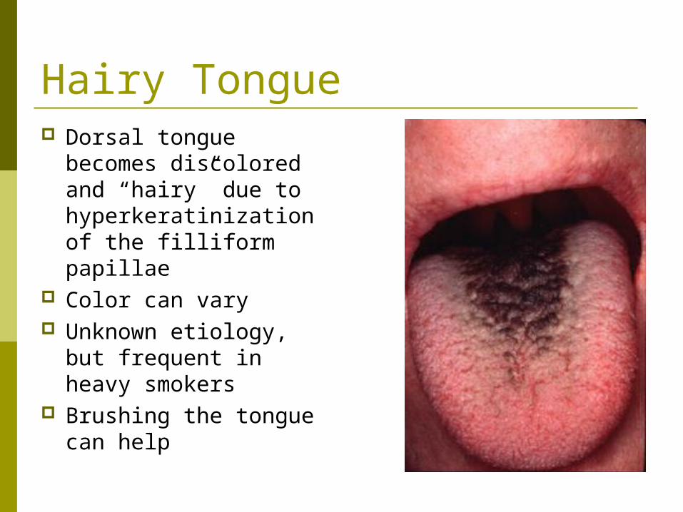
Hairy Tongue Dorsal tongue
becomes discolored and “hairy” due to hyperkeratinization of the filliform papillae
Color can vary Unknown etiology, but
frequent in heavy smokers
Brushing the tongue can help
Name this oral lesion:

Periapical cemental dysplasia (listed in the book as cementoma but this is a misnomer; AKA periapical cementodysplasia) Lesion around the apices
of vital teeth Principally in the lower
anteriors; mostly women; more common in blacks
Asymptomatic and does not require treatment
Begins as proliferation of benign fibrous connective tissue… cementum forms… the mass becomes mineralized

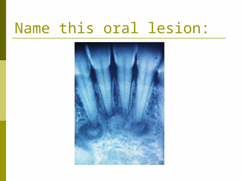
Name this oral lesion:
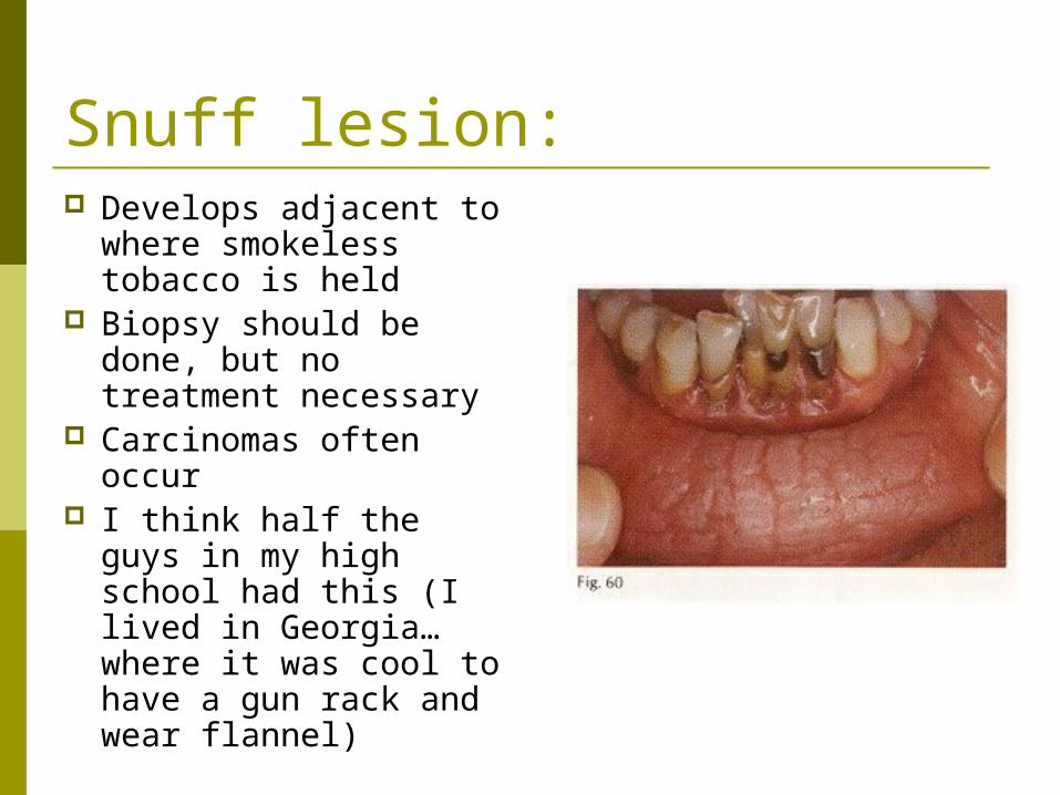
Snuff lesion: Develops adjacent to
where smokeless tobacco is held
Biopsy should be done, but no treatment necessary
Carcinomas often occur
I think half the guys in my high school had this (I lived in Georgia…where it was cool to have a gun rack and wear flannel)

Name this oral lesion:
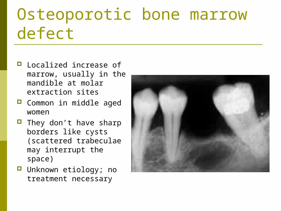
Osteoporotic bone marrow defect
Localized increase of marrow, usually in the mandible at molar extraction sites
Common in middle aged women
They don’t have sharp borders like cysts (scattered trabeculae may interrupt the space)
Unknown etiology; no treatment necessary
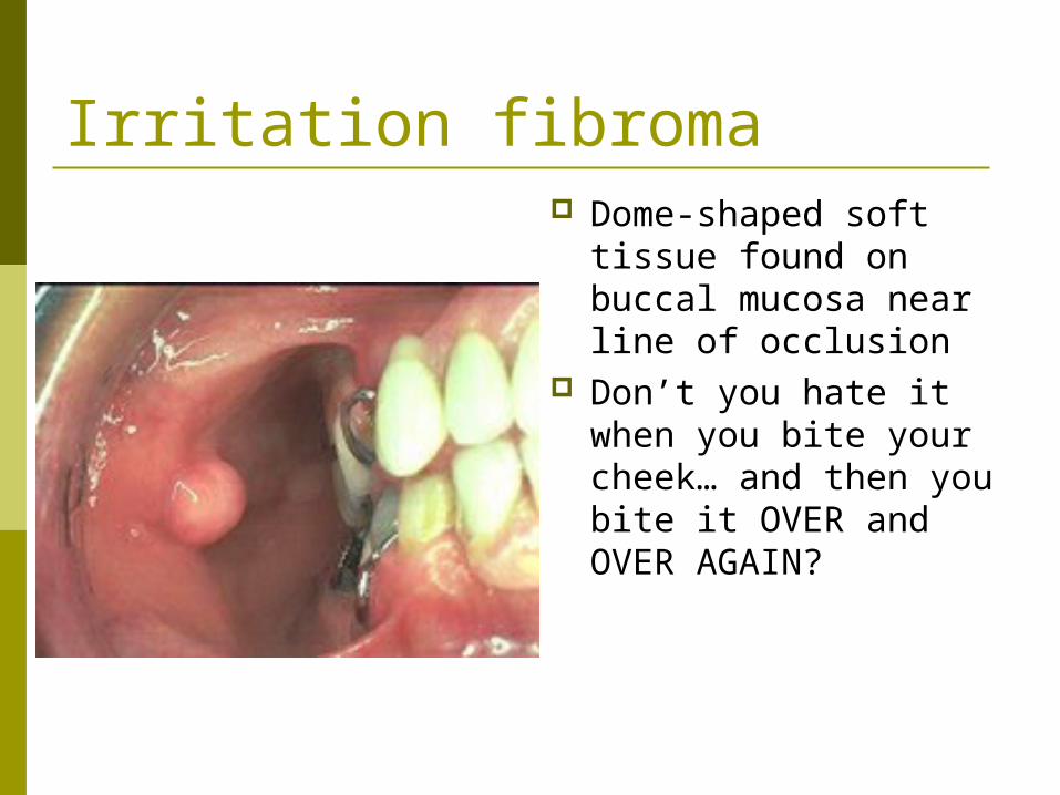
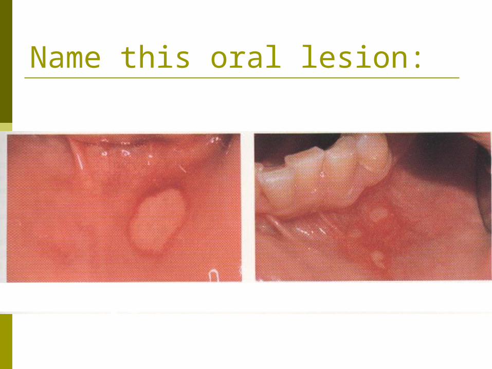
Irritation fibroma Dome-shaped soft
tissue found on buccal mucosa near line of occlusion
Don’t you hate it when you bite your cheek… and then you bite it OVER and OVER AGAIN?

Good luck on
finals!!!


![Clinics in Surgery Research Article · arteriovenous fistula and arteriovenous malformation (AVM) [1,2,4]. ... varix or varicose vein. The en bloc excision of scalp tissues affected](https://static.fdocuments.net/doc/165x107/5e2ad073f1470369d613c87e/clinics-in-surgery-research-arteriovenous-fistula-and-arteriovenous-malformation.jpg)