Oral and systtemic findings in biliary atresia: report … · Oral and systtemic findings in...
5
PEDIATRIC DENTISTRY/Copyright © 1982 by The American Academy of Pedodontics/Vol. 4, No.4 Oral and systtemic findings in biliary atresia: report of 11 cases Gary K. Belanger, DDS Roger Sanger, DDS, MSEd Paul S. Casamassimo, DDS, MSEric B. Bystrom, DDS Abstract Eleven children, eight females and three males, ranging in age from 17 months to seven years, eight months, with surgically corrected biliary atresia, were examined for oral problems. Four children had green staining of both p~’mary teeth and gingival tissues. Five children had abnormally large pulp chambers, and four had delayed primary tooth eruption. A significant relationship between primary tooth stain and gingival stain at the .005 level was found. No other statistically significant relationships between oral and physical or laboratory findings were noted. Biliary atresia is a rare condition occurring in ap- proximately one in 10,000 live births and affecting nearly 300 newborns in the United States each year. 1 The condition is characterized by perinatal jaundice, hyper- bilirubinemia, tissue staining by excess bile pigments, and hepatospleno~negaly. Growth retardation and nutri- tional failure are common in early life, and death often occurs by five years of age. Early diagnosis and surgical intervention have decreased morbidity, returned growth and development to normal and improved the prognosis for survival. 2 Potential oral involvement ranges from delayed dental and skeletal development to intrinsic staining of the dentition with biliverdin, a bile pigment. Only one report appears in dental literature describing biliary atresia and its oral manifestations. 3 This paper presents a series of 11 children afflicted with biliary atresia who received surgery to ameliorate the condition. Oral and systemic manifestations are presented, and pertinent relationships between oral and physical or laboratory findings dis- cussed. Literature Review Biliary atresia is a complex condition characterized by progressive obliteration of the intra- and extrahepatic duct system. Etiology of biliary atresia is unclear. Current thought discount:~ a genetic or developmental cause. Prenatal or neonatal viral infection may be contributory, leading to a more extensive disease process involving the entire hepatic system. 4 If untreated, biliary atresia causes continued degen- eration of the biliary tract leading to intrahepatic bile accumulation and cirrhosis. Jaundice and hyperbilirubi- nemia continue. Bile passage to the intestine is decreased or completely blocked and malabsorption of fat-soluble vitamins A, D, and K occurs. Bleeding due to inadequate vitamin K absorption and inadequate synthesis of clot- ting factors VII, IX, X, and prothrombin is common as are rachitic changes in bone due to inadequate vitamin D. s Osteomalacia and growth retardation also are a result of poor vitamin D absorption. Recent surgical advances, modifying a portoenteros- tomy procedure first described by Kasai, 6 have improved the prognosis of biliary atresia. 7’8 The modified portoen- terostomy, usually done by 10 weeks of life, involves two steps. The extrahepatic biliary duct system is re- moved and the system is replaced by an anastomosis of the intestine to facilitate bile drainage. Lilly 8 and others have modified the Kasai procedure to provide temporary externalization of the intestinal section to allow bile collection and measurement. This modification also re- duces cholangitis (Figure 1). The critical factor in surgical success is early detection and prompt intervention, usually within the first two months of life. Later surgery often meets with equivocal Success. Even the successfully treated child often has profound medical problems. Recurrent cholangitis, nutritional and growth deficiencies, delayed developmental milestones, portal hypertension, osteomalacia, and osteoporosis of- ten occur. 9’~° Also, the condition often is accompanied by psychosocial problems affecting child and family, n Severe dental caries and teeth staining have been noted 3 (Figure 2). Treatment for multiple medical problems may be episodic or preventive. Antibiotics are used to control cholangitis, aminoglycoside is used for acute infections, 322 BILIARY ATRESIA: Belanger et al.
Transcript of Oral and systtemic findings in biliary atresia: report … · Oral and systtemic findings in...
PEDIATRIC DENTISTRY/Copyright© 1982 byThe American Academy of Pedodontics/Vol. 4, No. 4
Oral and systtemic findings in biliary atresia: report of 11 cases
Gary K. Belanger, DDS Roger Sanger, DDS, MSEdPaul S. Casamassimo, DDS, MS Eric B. Bystrom, DDS
Abstract
Eleven children, eight females and three males,ranging in age from 17 months to seven years, eightmonths, with surgically corrected biliary atresia, wereexamined for oral problems. Four children had greenstaining of both p~’mary teeth and gingival tissues. Fivechildren had abnormally large pulp chambers, and fourhad delayed primary tooth eruption. A significantrelationship between primary tooth stain and gingivalstain at the .005 level was found. No other statisticallysignificant relationships between oral and physical orlaboratory findings were noted.
Biliary atresia is a rare condition occurring in ap-
proximately one in 10,000 live births and affecting nearly300 newborns in the United States each year. 1 Thecondition is characterized by perinatal jaundice, hyper-bilirubinemia, tissue staining by excess bile pigments,and hepatospleno~negaly. Growth retardation and nutri-tional failure are common in early life, and death oftenoccurs by five years of age. Early diagnosis and surgicalintervention have decreased morbidity, returned growthand development to normal and improved the prognosisfor survival.2
Potential oral involvement ranges from delayed dentaland skeletal development to intrinsic staining of thedentition with biliverdin, a bile pigment. Only one reportappears in dental literature describing biliary atresia andits oral manifestations. 3 This paper presents a series of11 children afflicted with biliary atresia who receivedsurgery to ameliorate the condition. Oral and systemicmanifestations are presented, and pertinent relationshipsbetween oral and physical or laboratory findings dis-cussed.
Literature ReviewBiliary atresia is a complex condition characterized by
progressive obliteration of the intra- and extrahepaticduct system. Etiology of biliary atresia is unclear. Currentthought discount:~ a genetic or developmental cause.
Prenatal or neonatal viral infection may be contributory,leading to a more extensive disease process involving theentire hepatic system.4
If untreated, biliary atresia causes continued degen-eration of the biliary tract leading to intrahepatic bileaccumulation and cirrhosis. Jaundice and hyperbilirubi-nemia continue. Bile passage to the intestine is decreasedor completely blocked and malabsorption of fat-solublevitamins A, D, and K occurs. Bleeding due to inadequate
vitamin K absorption and inadequate synthesis of clot-ting factors VII, IX, X, and prothrombin is common as
are rachitic changes in bone due to inadequate vitaminD.s Osteomalacia and growth retardation also are a resultof poor vitamin D absorption.
Recent surgical advances, modifying a portoenteros-tomy procedure first described by Kasai,6 have improvedthe prognosis of biliary atresia. 7’8 The modified portoen-
terostomy, usually done by 10 weeks of life, involvestwo steps. The extrahepatic biliary duct system is re-moved and the system is replaced by an anastomosis ofthe intestine to facilitate bile drainage. Lilly 8 and othershave modified the Kasai procedure to provide temporaryexternalization of the intestinal section to allow bilecollection and measurement. This modification also re-duces cholangitis (Figure 1).
The critical factor in surgical success is early detectionand prompt intervention, usually within the first twomonths of life. Later surgery often meets with equivocalSuccess.
Even the successfully treated child often has profoundmedical problems. Recurrent cholangitis, nutritional andgrowth deficiencies, delayed developmental milestones,portal hypertension, osteomalacia, and osteoporosis of-ten occur. 9’~° Also, the condition often is accompaniedby psychosocial problems affecting child and family,n
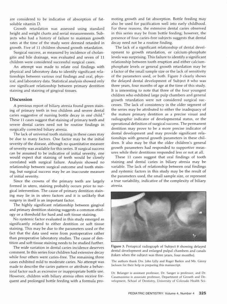
Severe dental caries and teeth staining have been noted3
(Figure 2).Treatment for multiple medical problems may be
episodic or preventive. Antibiotics are used to controlcholangitis, aminoglycoside is used for acute infections,
322 BILIARY ATRESIA: Belanger et al.
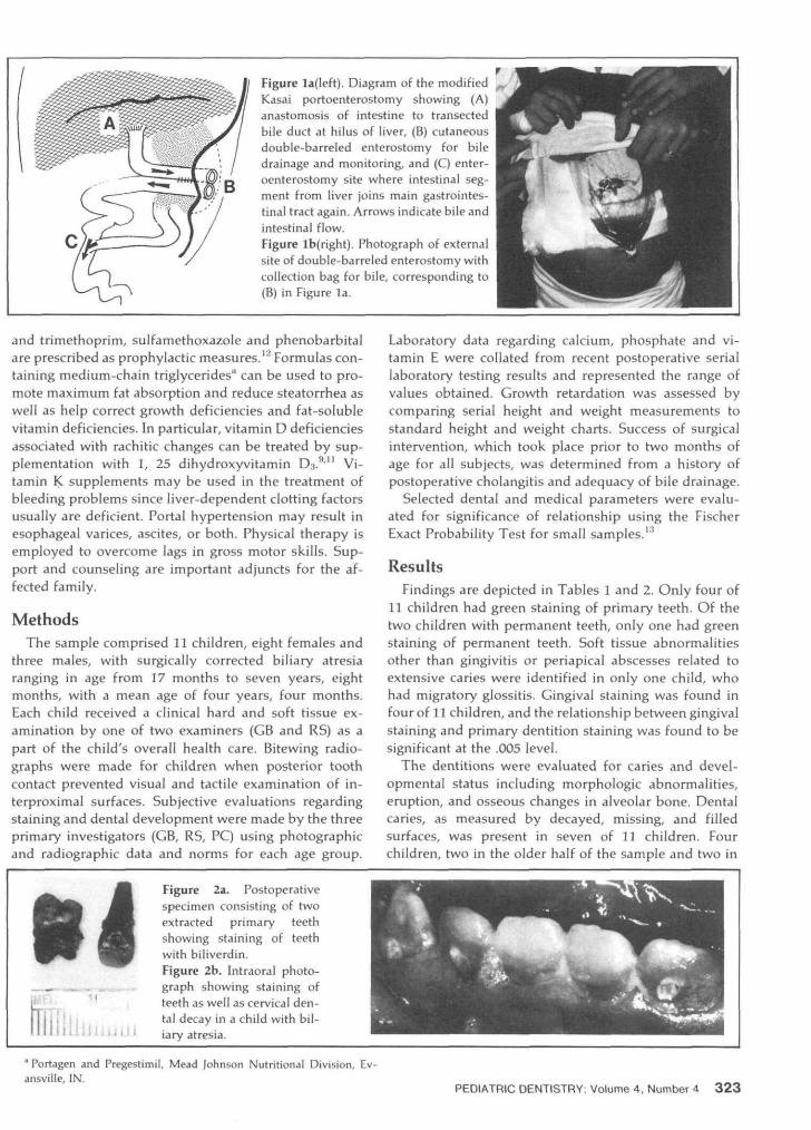
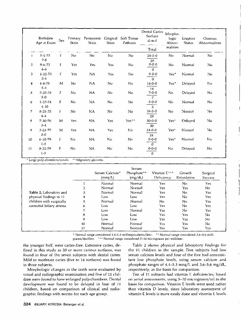
Figure la(left). Diagram of the modifiedKasai portoenterostomy showing (A)anastomosis of intestine to transectedbile duct at hilus of liver, (B) cutaneousdouble-barreled enterostomy for biledrainage and monitoring, and (C) enter-oenterostomy site where intestinal seg-ment from liver joins main gastrointes-tinal tract again. Arrows indicate bile andintestinal flow.Figure Ib(right). Photograph of externalsite of double-barreled enterostomy withcollection bag for bile, corresponding to(B) in Figure la.
and trimethoprim, sulfamethoxazole and phenobarbitalare prescribed as prophylactic measures.12 Formulas con-taining medium-chain triglycerides" can be used to pro-mote maximum fat absorption and reduce steatorrhea aswell as help correct growth deficiencies and fat-solublevitamin deficiencies. In particular, vitamin D deficienciesassociated with rachitic changes can be treated by sup-plementation with 1, 25 dihydroxyvitamin Ds.9'11 Vi-tamin K supplements may be used in the treatment ofbleeding problems since liver-dependent clotting factorsusually are deficient. Portal hypertension may result inesophageal varices, ascites, or both. Physical therapy isemployed to overcome lags in gross motor skills. Sup-port and counseling are important adjuncts for the af-fected family.
MethodsThe sample comprised 11 children, eight females and
three males, with surgically corrected biliary atresiaranging in age from 17 months to seven years, eightmonths, with a mean age of four years, four months.Each child received a clinical hard and soft tissue ex-amination by one of two examiners (GB and RS) as apart of the child's overall health care. Bitewing radio-graphs were made for children when posterior toothcontact prevented visual and tactile examination of in-terproximal surfaces. Subjective evaluations regardingstaining and dental development were made by the threeprimary investigators (GB, RS, PC) using photographicand radiographic data and norms for each age group.
Laboratory data regarding calcium, phosphate and vi-tamin E were collated from recent postoperative seriallaboratory testing results and represented the range ofvalues obtained. Growth retardation was assessed bycomparing serial height and weight measurements tostandard height and weight charts. Success of surgicalintervention, which took place prior to two months ofage for all subjects, was determined from a history ofpostoperative cholangitis and adequacy of bile drainage.
Selected dental and medical parameters were evalu-ated for significance of relationship using the FischerExact Probability Test for small samples.1'5
ResultsFindings are depicted in Tables 1 and 2. Only four of
11 children had green staining of primary teeth. Of thetwo children with permanent teeth, only one had greenstaining of permanent teeth. Soft tissue abnormalitiesother than gingivitis or periapical abscesses related toextensive caries were identified in only one child, whohad migratory glossitis. Gingival staining was found infour of 11 children, and the relationship between gingivalstaining and primary dentition staining was found to besignificant at the .005 level.
The dentitions were evaluated for caries and devel-opmental status including morphologic abnormalities,eruption, and osseous changes in alveolar bone. Dentalcaries, as measured by decayed, missing, and filledsurfaces, was present in seven of 11 children. Fourchildren, two in the older half of the sample and two in
Hi
Figure 2a. Postoperativespecimen consisting of twoextracted primary teethshowing staining of teethwith biliverdin.Figure 2b. Intraoral photo-graph showing staining ofteeth as well as cervical den-tal decay in a child with bil-iary atresia.
" Portagen and Pregestimil, Mead Johnson Nutritional Division, Ev-ansville, IN.
PEDIATRIC DENTISTRY. Volume 4, Number 4 323
Birthdate Sex Primary Permanent Gingival Soft TissueAge at Exam Stain Stain Stain Pathosis
Dental CariesSurfaced-m-f
Total
Morpho-logic
Abnor-malities
EruptiveStatus
Osseous
Abnormalities
1 5-1-73 F No No No No7-8
2 9-6-73 F Yes Yes Yes No6-6
3 1-22-75 F Yes NA Yes No5-4
4 4-8-75 M No NA No No5-4
5 7-27-75 F No NA No No5-0
6 1-27-74 E No NA No No4-10
7 8-25-75 F No NA No No4-4
8 7-30-76 M Yes NA Yes Yes**3-4
9 3-23-77 M Yes NA Yes No3-0
10 6-10-79 F No NA No Nol-5
11 8-22-79 F No NA No No1-5
24-5-0
290-0-0
00-0-0
014-0-0
147-0-0
75-0-0
524-5-0
2930-0-0
3024-0-0
240-0-0
00-0-0
0
No
Yes*
Yes*
No
No
No
Yes*
Yes*
Yes*
No
Normal
Normal
Normal
Delayed
Delayed
Normal
Normal
Delayed
Normal
Normal
Delayed
No
No
No
No
No
No
No
No
No
No
No
* Large pulp chambers/canals. ** Migratory glossitis.
Table 2. Laboratory andphysical findings in 11children with surgicallycorrected biliary atresia.
Serum
Serum Calcium* Phosphate** Vitamin E*** Growth Surgical(meq/L) (mg/dL) Deficiency Retardation Success
l Normal Normal Yes No Yes2 Normal Normal Yes Yes No3 Normal Normal Yes No Yes4 Low Low Yes No Yes5 Normal Normal No No Yes6 Low Low Yes No Yes7 Low Normal Yes No Yes8 Low Low Yes Yes No9 Low Low Yes Yes No
10 Normal Normal Yes Yes No11 Normal Normal Yes Yes Yes
* Normal range considered 4.4-5.3 milliequivalents/liter. ** Normal range considered 3.6-5.6 milli-grams/deciliter. *** Normal range considered 5-10 micrograms per milliliter.
the younger half, were caries-free. Extensive caries, de-fined in this study as 20 or more carious surfaces, wasfound in four of the seven subjects with dental caries.
Mild to moderate caries (five to 14 surfaces) was foundin three subjects.
Morphologic changes in the teeth were evaluated byvisual and radiographic examination and five of 11 chil-dren were found to have enlarged pulp chambers. Dentaldevelopment was found to be delayed in four of 11children, based on comparison of clinical and radio-graphic findings with norms for each age group.
Table 2 shows physical and laboratory findings forthe 11 children in the sample. Five subjects had lowserum calcium levels and four of the five had concomi-tant low phosphate levels, using serum calcium andphosphate ranges of 4.4-5.3 meq/L and 3.6-5.6 mg/dL,respectively, as the basis for comparison.
Ten of 11 subjects had vitamin E deficiencies, basedon serial assessments, using 5-10 micrograms/ml as thebasis for comparison. Vitamin E levels were used ratherthan vitamin D levels, since laboratory assessment ofvitamin E levels is more easily done and vitamin E levels
324 BIL~ARY ATRESIA: Belanger et al.
are considered to be indicative of absorption of fat-soluble vitamin D.
Growth retardation was assessed using standardheight and weight charts and serial measurements. Sub-jects who had a history of failure to maintain growthrates at the time of the study were deemed retarded ingrowth. Five of 11 children showed growth retardation.
Surgical success, as measured by incidence of cholan-gitis and bile drainage, was evaluated and seven of 11children were considered successful surgical cases.
An attempt was made to relate oral findings withphysical and laboratory data to identify significant rela-tionships between various oral findings and oral, phys-ical, and laboratory data. Statistical analysis showed onlyone significant relationship between primary dentitionstaining and staining of gingival tissues.
DiscussionA previous report of biliary atresia found green stain-
ing of primary teeth in two children and severe dentalcaries suggestive of nursing bottle decay in one child.''These 11 cases suggest that staining of primary teeth andsevere dental caries need not be routine findings insurgically corrected biliary atresia.
The lack of universal tooth staining in these cases maybe due to many factors. One factor may be the initialseverity of the disease, although no quantitative measureof severity was available for this series. If surgical successcan be assumed to be indicative of initial severity, onewould expect that staining of teeth would be closelycorrelated with surgical failure. Analysis showed norelationship between surgical outcome and tooth stain-ing, but surgical success may be an inaccurate measureof initial severity.
Since the crowns of the primary teeth are largelyformed in utero, staining probably occurs prior to sur-gical intervention. The cause of primary dentition stain-ing may lie in in utero factors and it is unlikely thatsurgery in itself is an important factor.
The highly significant relationship between gingivaland primary dentition staining suggests a common etiol-ogy or a threshold for hard and soft tissue staining.
No systemic factor evaluated in this study emerged assignificantly related to either dentition or soft tissuestaining. This may be due to the parameters used or thefact that the data used were from postoperative ratherthan preoperative laboratory studies. The cause of den-tition and soft tissue staining needs to be studied further.
The wide variation in dental caries incidence deservescomment. In this series four children had extensive decaywhile four others were caries-free. The remaining threecases exhibited mild to moderate caries. No attempt wasmade to describe the caries pattern or attribute a behav-ioral factor such as excessive or inappropriate bottle use.However, children with biliary atresia often receive fre-quent and prolonged bottle feeding with a formula pro-
moting growth and fat absorption. Bottle feeding mayalso be used for pacification well into early childhood.For these reasons, the extensive dental caries observedin this series may be from bottle feeding; however, thepresence of four caries-free subjects suggests that dentaldecay need not be a routine finding.
The lack of a significant relationship of dental devel-opment to growth retardation, or calcium-phosphatelevels was surprising. This failure to identify a significantrelationship between tooth eruption and either calcium-phosphate levels or general growth retardation may bea factor of the small sample size or the lack of sensitivityof the parameters used, or both. Figure 3 clearly showsthe delayed dental development of Subject 8 who wasthree years, four months of age at the time of this study.It is interesting to note that three of the four youngestchildren who exhibited large pulp chambers and generalgrowth retardation were not considered surgical suc-cesses. The lack of consistency in the older segment ofthe series may be attributed to either the inadequacy ofthe mature primary dentition as a precise visual andradiographic indicator of developmental status, or theoperational definition of surgical success. The permanentdentition may prove to be a more precise indicator ofdental development and may provide significant rela-tionships with general growth parameters in these chil-dren. It also may be that the older children's generalgrowth parameters had responded to supportive meas-ures while their dentitions responded less or not at all.
These 11 cases suggest that oral findings of toothstaining and dental caries in biliary atresia may bevariable. The lack of relationship between oral findingsand systemic factors in this study may be the result ofthe parameters used, the small sample size, or representa true variability, indicative of the complexity of biliaryatresia.
Figure 3. Periapical radiograph of Subject 8 showing delayeddental development and enlarged pulpal chambers and canals(taken when the subject was three years, four months).
The authors thank Drs. John Lilly and Roger Barkin and Ms. GinnyJackson for their help in preparing this manuscript.
Dr. Belanger is assistant professor, Dr. Sanger is professor, and Dr.Casamassimo is associate professor, Department of Growth and De-velopment, School of Dentistry, University of Colorado Health Sci-
PEDIATRIC DENTISTRY: Volume 4, Number 4 325
ences Center, 4200 E. Ninth Ave., Denver, CO 80262; and Dr. Bystromis associate professor and chairman, Department of Pediatric Dentistry,School of Dentistry, University of the Pacific, 2155 Webster St., SanFrancisco, CA 94115. Requests for reprints should be sent to Dr.Casamassimo.
1. Silverman, A., Roy, D., and Cozzetto, F. Pediatric Clinical Gastro-enterology. St. Louis, C.V. Mosby Co., 1971, p 293.
2. Kasai, M., Watanabe, I., and Ohi, R. Follow-up studies of long-term survivors after hepatic portoenterostomy for"noncorrectable" biliary atresia. J Pediatr Surg 10:173, 1975.
3. Shapiro, B.M., Gallagher, F.E., and Needleman, H.L. Dental man-agement of the patient with biliary atresia, report of two cases.Oral Surg 40:742, 1975.
4. Grosfeld, J.L. and Clathworthy, H.W., Jr. Biliary atresia, in BirthDefects Atlas and Compendium, ed. Bergsma, D. Baltimore, Wil-liams and Wilkins Co., 1973, p 210.
5. Kooh, S.W., Jones, G., Reilly, B.J., and Fraser, D. Pathogenesis ofrickets in chronic hepatobiliary disease in children. J Pediatr94:870, 1979.
6. Kasai, M., Kimura, S., Asakura, Y., and Suzuki, H. Surgicaltreatment of biliary atresia. J Pediatr Surg 3:665, 1968.
7. Lilly, J.R., Altman, R.P., Schroter, G., Shikes, R.H., and Taubman,J.O. Surgery of biliary atresia, current status. Am J Dis Child129:1429, 1975.
8. Lilly, J.R. and Altman, R.P. Hepatic portoenterostomy (the Kasaioperation) for biliary atresia. Surgery 78:76, 1975.
9. Heubi, J.E., Tsang, R.C., Steichen, J.J., Chan, G.M., Chen, I., andDeluca, H.F. 1,25-dihydroxyvitamin D;j in childhood hepatic os-teodystrophy. J Pediatr 94:977, 1979.
10. Kobayashi, A., Utsonomiya, T., Kawai, S., and Ohbe, Y. Congenitalbiliary atresia. Am J Dis Child 130:830, 1976.
11. Barkin, R.M. and Lilly, J.R. Biliary atresia and the Kasai operation:continuing care. J Pediatr 96:105, 1980.
12. Lilly, J.R. and Hitch, D.C. Postoperative ascending cholangitisfollowing portoenterostomy for biliary atresia: measures for con-trol. World J Surg 2:581, 1978.
13. Siegel, S. Nonparametric Statistics for the Behavioral Sciences.New York, McGraw-Hill, 1956, pp 96-104.
Black Forest, Western Germany, 1°74
326 BILIARY ATRESIA: Belanger et al.
![Ultrasonographic findings of type IIIa biliary atresiabiliary atresia. Further, there have been only a few reports on the US findings of biliary atresia based on its types [33,34].](https://static.fdocuments.net/doc/165x107/60a90a6926e7a533947d7637/ultrasonographic-findings-of-type-iiia-biliary-atresia-biliary-atresia-further.jpg)