Optogenetic control of nuclear protein export · 2016-08-19 · Optogenetic control of nuclear...
9
ARTICLE Received 2 Dec 2015 | Accepted 5 Jan 2016 | Published 8 Feb 2016 Optogenetic control of nuclear protein export Dominik Niopek 1,2,3 , Pierre Wehler 1 , Julia Roensch 1 , Roland Eils 1,2,3 & Barbara Di Ventura 2,3 Active nucleocytoplasmic transport is a key mechanism underlying protein regulation in eukaryotes. While nuclear protein import can be controlled in space and time with a portfolio of optogenetic tools, protein export has not been tackled so far. Here we present a light-inducible nuclear export system (LEXY) based on a single, genetically encoded tag, which enables precise spatiotemporal control over the export of tagged proteins. A constitutively nuclear, chromatin-anchored LEXY variant expands the method towards light inhibition of endogenous protein export by sequestering cellular CRM1 receptors. We showcase the utility of LEXY for cell biology applications by regulating a synthetic repressor as well as human p53 transcriptional activity with light. LEXY is a powerful addition to the optogenetic toolbox, allowing various novel applications in synthetic and cell biology. DOI: 10.1038/ncomms10624 OPEN 1 Department of Theoretical Bioinformatics, German Cancer Research Center (DKFZ), Im Neuenheimer Feld 280, 69120 Heidelberg, Germany. 2 Department of Bioinformatics and Functional Genomics, Synthetic Biology Group, Institute for Pharmacy and Biotechnology (IPMB), University of Heidelberg, Im Neuenheimer Feld 364, 69120 Heidelberg, Germany. 3 Center for Quantitative Analysis of Molecular and Cellular Biosystems (BioQuant), University of Heidelberg, Im Neuenheimer Feld 267, 69120 Heidelberg, Germany. Correspondence and requests for materials should be addressed to B.D.V. (email: [email protected]) or to R.E. (email: [email protected]). NATURE COMMUNICATIONS | 7:10624 | DOI: 10.1038/ncomms10624 | www.nature.com/naturecommunications 1
Transcript of Optogenetic control of nuclear protein export · 2016-08-19 · Optogenetic control of nuclear...

ARTICLE
Received 2 Dec 2015 | Accepted 5 Jan 2016 | Published 8 Feb 2016
Optogenetic control of nuclear protein exportDominik Niopek1,2,3, Pierre Wehler1, Julia Roensch1, Roland Eils1,2,3 & Barbara Di Ventura2,3
Active nucleocytoplasmic transport is a key mechanism underlying protein regulation in
eukaryotes. While nuclear protein import can be controlled in space and time with a
portfolio of optogenetic tools, protein export has not been tackled so far. Here we present a
light-inducible nuclear export system (LEXY) based on a single, genetically encoded tag,
which enables precise spatiotemporal control over the export of tagged proteins.
A constitutively nuclear, chromatin-anchored LEXY variant expands the method towards light
inhibition of endogenous protein export by sequestering cellular CRM1 receptors. We
showcase the utility of LEXY for cell biology applications by regulating a synthetic repressor
as well as human p53 transcriptional activity with light. LEXY is a powerful addition to the
optogenetic toolbox, allowing various novel applications in synthetic and cell biology.
DOI: 10.1038/ncomms10624 OPEN
1 Department of Theoretical Bioinformatics, German Cancer Research Center (DKFZ), Im Neuenheimer Feld 280, 69120 Heidelberg, Germany. 2 Departmentof Bioinformatics and Functional Genomics, Synthetic Biology Group, Institute for Pharmacy and Biotechnology (IPMB), University of Heidelberg, ImNeuenheimer Feld 364, 69120 Heidelberg, Germany. 3 Center for Quantitative Analysis of Molecular and Cellular Biosystems (BioQuant), University ofHeidelberg, Im Neuenheimer Feld 267, 69120 Heidelberg, Germany. Correspondence and requests for materials should be addressed to B.D.V.(email: [email protected]) or to R.E. (email: [email protected]).
NATURE COMMUNICATIONS | 7:10624 | DOI: 10.1038/ncomms10624 | www.nature.com/naturecommunications 1

Active nucleocytoplasmic transport controls the localizationand spatiotemporal dynamics of proteins in eukaryotes,thereby governing essential cellular processes including
gene expression, cell division and apoptosis. Regulation of proteinimport and export is achieved mainly by masking and unmaskingof nuclear import and nuclear export signals (NLSs and NESs)directly located within the polypeptide or by binding andunbinding to NLS- and NES-bearing partners1.
Optogenetic tools that enable controlling with light the nuclearimport of tagged proteins in mammalian cells and yeast havebeen reported2–6, but no optogenetic tools are yet available todirectly control protein export. However, such a tool would haveenormous application potential, for example, for regulatingthe activity of nuclear or cytoplasmic signalling molecules, andwould complement the existing optogenetic toolset for control ofnuclear import2–6, protein dimerization7 and oligomerization8,9,membrane recruitment10 and organelle transport and positioning11.
Here we present LEXY, a blue light-induced nuclear exportsystem enabling dynamic and spatial control over nuclear proteinexport. We show fast and fully reversible nuclear export of LEXY-tagged proteins of diverse nature and origin in various cell lines.A chromatin-anchored LEXY variant mediates light-induciblesequestration of cellular CRM1, the primary nuclear exportreceptor, thereby allowing inhibition of endogenous nuclearexport. To demonstrate the utility of LEXY for applications insynthetic and cell biology, we regulate synthetic repressors as wellas the transcriptional activity of human p53 with light.
ResultsLEXY engineering and characterization. LEXY consists of anengineered LOV2 domain from Avena sativa phototropin-1(AsLOV2), in which the C-terminal Ja helix was converted intoan artificial NES. In the dark, the NES is tightly packed againstthe AsLOV2 core and is thus inactive (Fig. 1a). On blue lightabsorption, the modified Ja helix unfolds, thereby exposing theNES to nuclear CRM1 (exportin-1) receptors mediating activenuclear export through the nuclear pores.
In contrast to previous work on AsLOV2-photocagedpeptides4,5,12–14, we applied a new approach termed ‘Ja helixtopping’, which highly preserves wild-type AsLOV2 Ja helixproperties required for successful sequence epitope caging andAsLOV2 domain photoswitching (see Supplementary Note fordetails). Using synthetic biology principles, we performed twocycles of rational design and subsequent selection, therebygradually introducing specific, known NES-like features derivedfrom literature knowledge15 into the wild-type AsLOV2 Ja helix(Supplementary Fig. 1 and Supplementary Note). The resulting33 AsLOV2-NES hybrids were screened by qualitativelyinvestigating the nucleocytoplasmic translocation of correspondingNLS-mCherry-AsLOV2-NES fusions in human embryonickidney (HEK 293T) cells on pulsatile blue light irradiation(Supplementary Fig. 2a,b). The N-terminal constitutive NLS isadded to accumulate the fusion protein in the nucleus in the dark.Unless stated otherwise, nucleocytoplasmic translocation wasanalysed using epifluorescence microscopy. To our surprise,introducing two single-point mutations (A542L and A549L) plusadding a C-terminal aspartic acid residue already converted thewild-type Ja helix into a weak photocaged NES (NES 8;Supplementary Figs 1 and 2b), for which the functionalitycould be highly improved by introducing only two additionalmutations (A543L and P547A) (NES 21; Supplementary Figs 1and 2b). A control construct bearing the wild-type Ja helix didnot show nuclear export on illumination (Supplementary Fig. 2b).Co-expression of a H2B-GFP chromatin marker confirmedthat the NLS-mCherry-AsLOV2-NES 21 fusion localizes to the
nucleus and not to a different organelle in the dark and is stronglyexported on illumination (Fig. 1b,c). ‘LEXY’ refers to theAsLOV2-NES 21 variant in all subsequent experiments.
We performed a complete induction–recovery cycle consistingof 3 min of mCherry imaging only followed by 15 min of pulsatileblue light irradiation and 20 min recovery in the dark in HEK293T expressing NLS-mCherry-LEXY. The observed mCherryexport on irradiation was very fast with half-times below 1 min(Fig. 1d and Supplementary Fig. 3). Stopping illumination led tofull recovery of the initial nuclear mCherry fluorescence withinabout 15 min due to dark state recovery of the AsLOV2 domain(Fig. 1d). We observed similar kinetics in human cervixcarcinoma (HELA) and murine hepatoma (Hepa 1–6) cells(Supplementary Fig. 4). The combination of fast export onirradiation and full recovery in the dark enabled repeated cyclesof activation and recovery without significantly compromising theperformance of LEXY over time (Fig. 1e, Supplementary Fig. 5a,band Supplementary Movies 1 and 2).
One particular advantage of optogenetics is the unmetspatiotemporal precision by which the light trigger can beapplied. We irradiated single, selected cells with a 458-nm laserbeam using a confocal laser scanning microscope. Induced cellsshowed a fivefold decrease in nuclear mCherry fluorescence whilenon-induced cells present in the same field of view remainedunaffected (Fig. 1f,g and Supplementary Movie 3).
This fold change is higher than the one seen with epifluores-cence microscopy (B2.5-fold change in nuclear fluorescence;compare Fig. 1d,g). This is not surprising considering that, inepifluorescence microscopy, there is a non-negligible contributionof fluorescent signal in the nucleus from the surroundingcytoplasm. Therefore, confocal microscopy can better reveal thedynamic range of LEXY.
To test the robustness of LEXY-mediated export when fused todifferent target proteins, we cloned two golden gate entry vectorspLEXY and pNLS-LEXY, both comprising a cytomegalovirus(CMV)-driven mCherry-LEXY expression cassette carrying abacterial toxin (ccdB16) encoding gene at its 50-terminus, flankedby BpiI (BbsI) sites (Supplementary Fig. 6a). pNLS-LEXYharbours a constitutive NLS preceding the ccdB gene, which isabsent in pLEXY. Human codon-optimized sequences of abacterial protein domain (LexA repressor DNA-binding domain),the P1 bacteriophage-derived Cre recombinase, as well as sixdifferent human proteins (Acp1, Sox2, Nxt1, Nanog, Cox17 andp21) were cloned into both entry vectors via BpiI, therebyreplacing the ccdB death gene. Note that all sequences encodedwild-type polypeptides, that is, we preserved endogenousregulatory elements including NLS/NES sequences or protein–DNA-binding interfaces. We found at least one LEXY-taggedversion for each protein that showed significant nuclear export onblue light induction (Supplementary Fig. 6b,c). LEXY was able tooutcompete endogenous NLSs, which is reflected by the efficientlight-dependent export observed for the transcription factorsSox2 and Nanog. We also found that the nuclear export kinetics isinfluenced by both the total protein size and its nature.This is exemplified by the relatively slow export kinetics of themCherry-LEXY-tagged Cre recombinase, which has not onlyabout twice the size (B85 kDa) of NLS-mCherry-LEXY alone(B45 kDa) but also binds to DNA in the nucleus, thus preventingfaster export rates (Supplementary Fig. 7).
LEXY-mediated control of protein export can be easilycombined with our previously reported LINuS method foroptogenetic control of nuclear import4. When co-expressingNLS-mCherry-LEXY and NES-EGFP-LINuS in HEK 293T, weobserved a complete inversion of the nucleocytoplasmiclocalization of the two fluorophores on blue light irradiation(Supplementary Fig. 8a–c and Supplementary Movie 4).
ARTICLE NATURE COMMUNICATIONS | DOI: 10.1038/ncomms10624
2 NATURE COMMUNICATIONS | 7:10624 | DOI: 10.1038/ncomms10624 | www.nature.com/naturecommunications

Light-dependent inhibition of endogenous nuclear export.Apart from direct light control of tagged proteins, LEXY couldalso be employed to perturb endogenous CRM1-dependentnuclear export. Anchoring LEXY to the nuclear chromatin byfusion to the histone H2B should enable light-dependentsequestration of endogenous CRM1 receptors (Fig. 2a). Thisshould lead, in turn, to the inhibition of the nuclear exportof CRM1 cargos. To verify this hypothesis we expressedH2B-GFP-LEXY alongside with a mCherry bearing a strong,constitutive NES and a weaker NLS in HEK 293T (Fig. 2b).We found that mCherry accumulated in the nucleus only inirradiated H2B-GFP-LEXY-expressing cells, but not in controlcells expressing H2B-GFP fused to the wild-type AsLOV2domain (Fig. 2c,d). The extent of nuclear mCherry translocationdepended on the abundance of H2B-GFP-LEXY (Fig. 2e). UsingH2B-GFP fused to AsLOV2-NES27, a particularly strong NESvariant from our initial library (Supplementary Figs 1 and 2b),enhanced nuclear mCherry accumulation on irradiation, but alsoresulted in considerable leakiness, that is, an increased nuclearmCherry abundance in the dark (Fig. 2c,d). Overall, this strategyrepresents a genetically encoded, fully reversible alternative tochemical methods, such as, for example, leptomycin B (LMB)treatment17–20, widely employed in cell biology to block nuclearprotein export.
Regulation of synthetic transcriptional repressors. Aninteresting application of LEXY is the spatiotemporal control oftranscriptional repressors by sequestering them into the cytosol,which could be of particular value for controlling endogenousgene activity. As proof of principle, we used the mCherry-LEXY-tagged LexA DNA-binding domain (Supplementary Fig. 6c) aspassive mammalian repressor to control transgene expressionwith light. We also tested an additional LexA repressorvariant carrying a Kruppel-associated box (KRAB), an active
transcription repression domain21. A triple transfection into HEK293T was performed with (1) a construct co-expressing theLEXY-tagged LexA repressor and a synthetic LexA-transcriptionfactor required for reporter activation, (2) a correspondingLexA-dependent firefly luciferase reporter and (3) a constitutiverenilla luciferase expression vector for normalization purposes(Fig. 3a). Following 24 h of pulsatile blue light irradiation weobserved up to 15-fold increase in firefly luciferase expression ascompared with the dark control, thereby confirming successfullight regulation of the LexA repressors (Fig. 3b). Addition of theKRAB domain strongly reduced the background reporter activityin the dark, but also significantly compromised the total level ofreporter activation on light induction (Fig. 3b).
Optical control of p53 transcriptional activity. Finally, wesought to employ LEXY for direct optogenetic control of humanp53, an important tumour suppressor frequently mutated inhuman cancers. It has been recently proposed that p53 dynamicscontrol cellular signalling and therefore cell fate22. However, dueto a lack of tools to spatiotemporally control p53 activity, ratherindirect experimental strategies had to be employed to provokespecific dynamics of p53 activation, namely a combination ofg-irradiation and small-molecule (Nutlin) treatment22. Of note,wild-type p53 harbours several NLS and NES sequencesand employs an NES masking/unmasking mechanism viatetramerization to regulate its nucleocytoplasmic localization23.The ubiquitin ligase Mdm2 furthermore regulates the overall p53abundance in a close feedback manner24. Irrespective of itscomplex regulation, ectopic expression of p53 results in strongactivation of p53 target genes25–28. Thus, we hypothesized thatectopic expression of LEXY-tagged p53 would enable directoptogenetic control of p53 activity. We fused p53 bearing anadditional, N-terminal NLS to mCherry-LEXY and expressed theconstruct in human non-small cell lung carcinoma (H1299) cells
I D E A A K E L P D A N LI D E L L K E L A D L N L D
CMV NLS mCherry AsLOV2 NESGFPH2B 2A
– Li
ght
+ L
ight
0′ 18′ 38′ 53′ 73′
0′ 10′ 30′
e
f
g
– Light + Light – Light + Light – Light
Rel
ativ
e nu
clea
rflu
ores
cenc
e
0.4
0.6
0.8
1.0
0.20 10 155 20 30 3525
+ Light – Light
LEXY
Control
Time (min)
NES
LOV2core
NES
Jα*
Blue light
Recovery
LOV2core
Jα* Wt JαNES
539 545 550
LEXY
H2B-GFP
a b
d
cNLS-mCherry-
LEXY Merge
0.20.40.60.81.0
Rel
ativ
e nu
clea
rflu
ores
cenc
e
***
30i u i u
(min)0 i u
10
NS~5-fold
Figure 1 | Engineering and characterization of LEXY, a light-inducible nuclear export system. (a) Schematic of LEXY function. Blue light irradiation
releases the photocaged NES from the AsLOV2 core, thereby inducing nuclear export. (b) Schematic of a construct encoding NLS-mCherry-LEXY and a
H2B-GFP nuclear marker. NLS, cMycP1A NLS; 2A, T2A peptide. Green boxes indicate amino-acid residues altered as compared with the wild-type AsLOV2
Ja helix (Wt Ja). Numbers show the position of the corresponding residues in the full-length AsLOV2 domain. (c) Fluorescence images of HEK 293T cells
transiently transfected with the construct in b before and after 15 min of pulsatile blue light irradiation. Scale bar, 20mm. (d) Relative nuclear fluorescence of
HEK 293T expressing NLS-mCherry-LEXY (LEXY) or NLS-mCherry-AsLOV2 (control) over time. Cells were incubated in the dark for 3 min before blue light
irradiation for 15 min followed by a 20-min dark-recovery phase (mean±s.e.m., n¼ 22 cells from 3 independent experiments). (e) Fluorescence images of
NLS-mCherry-LEXY-expressing cells undergoing repeated illumination and recovery cycles. Scale bar, 20mm. (f) Fluorescence images of cells expressing
NLS-mCherry-LEXY individually irradiated. Green circles depict regions scanned with a blue laser beam for 10 min following 20 min of dark recovery. Scale
bar, 20mm. (g) Quantification of the relative nuclear fluorescence of individually irradiated cells (i) and uninduced control cells (u) at the indicated time
points. Data represent box plots and individual data points, n¼8 induced cells and 13 uninduced cells from 3 independent experiments; ***P¼ 1.69� 10� 17
by Welch’s t-test. NS, not significant.
NATURE COMMUNICATIONS | DOI: 10.1038/ncomms10624 ARTICLE
NATURE COMMUNICATIONS | 7:10624 | DOI: 10.1038/ncomms10624 | www.nature.com/naturecommunications 3

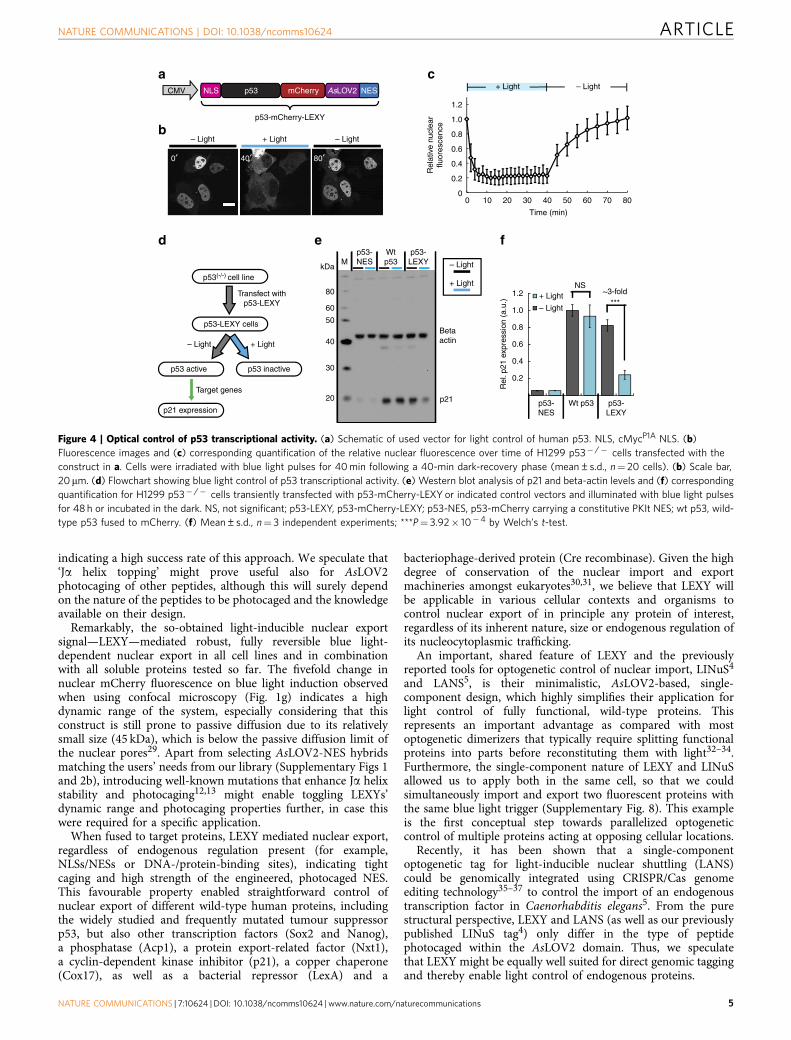
harbouring a homozygous p53 deletion (p53� /� ; Fig. 4a).Using confocal laser scanning microscopy we observed efficientand fully reversible blue light-induced nuclear export of thep53-mCherry-LEXY fusion (Fig. 4b,c). Remarkably, the dynamicrange was comparable (B5-fold change in nuclear fluorescence)to what we observed before for the NLS-mCherry-LEXYconstruct in HEK 293T (compare Fig. 4c and Fig. 1g). We theninvestigated the effect of light-mediated p53 translocation on theactivation of p21, a prominent p53 target gene25,26. On treatmentof p53-mCherry-LEXY-expressing H1299 cells with pulsatileblue light for 48 h (Fig. 4d) we observed a threefold decreaseof p21 expression compared with the dark control, therebyconfirming successful light regulation of p53 transcriptional
activity (Fig. 4e,f). Control samples expressing wild-type p53or p53 fused to a constitutive, strong NES did not showlight-dependent p21 expression (Fig. 4e,f).
DiscussionHere we reported the development of and showcased a variety ofapplications for LEXY, a light-inducible nuclear export system.
In contrast to previous studies4,5,12–14, we employed anovel approach termed ‘Ja helix topping’ for photocaging asequence epitope (here a NES) within the AsLOV2 domain(see Supplementary Note for details). Overall, about B50%of all AsLOV2-NES hybrids generated showed appreciable,light-dependent nuclear export (Supplementary Fig. 2b),
CMV
Strength:
CMV AsLOV2 NESGFPH2B
NLS mCherry NES
CR
M1
CR
M1
GFP
LOV2
H2B
Jα*
Blue light
Recovery
CR
M1 H2B
LOV2
GFP
CRM1
NES
0′ 43′23′ 23′ 23′
NLS-mCherry-NES H2B-GFP Merge– Light + Light – Light + Light + Light
Intermediate Strong
H2B-GFP-wt AsLOV2
H2B-GFP-LEXY
H2B-GFP-AsLOV2-NES27
a c
d
b
e***
***
1.0
1.5
2.0
0.5
Rel
ativ
e nu
clea
rm
Che
rry
fluor
esce
nce
0 23 43
wtAsLOV2
LEXY AsLOV2-NES27
0 23 43 0 23 43 (min)
H2B-GFP:
2.5
Rel
ativ
e nu
clea
rm
Che
rry
fluor
esce
nce
0
1.0
1.5
2.0
2.5
0.5500 1,000 1,500 2,000
Nuclear EGFP fluorescence (a.u.)
H2B-GFP-LEXYH2B-GFP-wt AsLOV2
Figure 2 | Light-dependent inhibition of endogenous nuclear export. (a) Schematic of blue light-dependent CRM1 sequestration by LEXY fused to
H2B-GFP. (b) Schematic of constructs encoding H2B-GFP fused to LEXY and a NLS-mCherry-NES reporter. (c) Fluorescence images and (d) corresponding
quantification of the relative nuclear mCherry fluorescence of cells co-transfected with the indicated constructs. Cells were incubated in the dark for 3 min
followed by blue light irradiation for 20 min and dark recovery for 20 min. AsLOV2-NES 27, a particularly strong NES variant (Supplementary Fig. 2b); wt,
wild type. Data represent box plots and individual data points, n¼ 34 cells for H2B-GFP-AsLOV2, n¼42 cells for H2B-GFP-LEXY and n¼ 28 cells for
H2B-GFP-AsLOV2-NES27, all from 3 independent experiments; ***P¼9.10� 10� 10 (wt AsLOV2 versus LEXY) and 3.45� 10� 5 (LEXY versus
AsLOV2-NES27) by Mann–Whitney test. (c) Scale bar, 20mm. (e) Scatter plot of the relative nuclear mCherry fluorescence versus nuclear EGFP
fluorescence for the indicated samples in d after blue light irradiation (23 min time point).
a b
~15-fold **
**
Rel
ativ
e lu
cife
rase
activ
ity (
a.u.
)
50
100
150
200
0
250
Rep
orte
ron
ly
Pos
itive
cont
rol
LexA
TF
+Le
xAT
R-L
EX
Y
LexA
TF
+Le
xA T
R–
KR
AB
-LE
XY
~5-fold
NS
NS
***
***+ Light
– LightCMV
LEXY
pLexA Firefly luciferase
2ALexA DBD VP64
LexA TF LexA TR-(± KRAB)-LEXY
± KRAB
TK Renilla luciferase
Constitutive Light-dependent
NLS mCherry AsLOV2 NESLexA DBD
Figure 3 | Light regulation of synthetic transcriptional repressors. (a) Schematic of used constructs for light control of transcriptional repression. DBD,
DNA binding domain; KRAB, Kruppel associated box; LexA TF, synthetic LexA transcription factor; LexA TR-LEXY, LexA repressor fused to mCherry-LEXY;
NLS, cMycP1A NLS; VP64, a strong transactivation domain; 2A, T2A peptide. (b) Quantification of relative luciferase activity in HEK 293T cells transiently
transfected with the constructs in a and irradiated with blue light pulses for 24 h or incubated in the dark. Positive control, cells co-expressing LexA TF
and NLS-mCherry-LEXY. Mean±s.d., n¼4 independent experiments; *Po0.05, **Po0.01, ***Po0.001 by Welch’s t-test. NS, not significant.
ARTICLE NATURE COMMUNICATIONS | DOI: 10.1038/ncomms10624
4 NATURE COMMUNICATIONS | 7:10624 | DOI: 10.1038/ncomms10624 | www.nature.com/naturecommunications
indicating a high success rate of this approach. We speculate that‘Ja helix topping’ might prove useful also for AsLOV2photocaging of other peptides, although this will surely dependon the nature of the peptides to be photocaged and the knowledgeavailable on their design.
Remarkably, the so-obtained light-inducible nuclear exportsignal—LEXY—mediated robust, fully reversible blue light-dependent nuclear export in all cell lines and in combinationwith all soluble proteins tested so far. The fivefold change innuclear mCherry fluorescence on blue light induction observedwhen using confocal microscopy (Fig. 1g) indicates a highdynamic range of the system, especially considering that thisconstruct is still prone to passive diffusion due to its relativelysmall size (45 kDa), which is below the passive diffusion limit ofthe nuclear pores29. Apart from selecting AsLOV2-NES hybridsmatching the users’ needs from our library (Supplementary Figs 1and 2b), introducing well-known mutations that enhance Ja helixstability and photocaging12,13 might enable toggling LEXYs’dynamic range and photocaging properties further, in case thiswere required for a specific application.
When fused to target proteins, LEXY mediated nuclear export,regardless of endogenous regulation present (for example,NLSs/NESs or DNA-/protein-binding sites), indicating tightcaging and high strength of the engineered, photocaged NES.This favourable property enabled straightforward control ofnuclear export of different wild-type human proteins, includingthe widely studied and frequently mutated tumour suppressorp53, but also other transcription factors (Sox2 and Nanog),a phosphatase (Acp1), a protein export-related factor (Nxt1),a cyclin-dependent kinase inhibitor (p21), a copper chaperone(Cox17), as well as a bacterial repressor (LexA) and a
bacteriophage-derived protein (Cre recombinase). Given the highdegree of conservation of the nuclear import and exportmachineries amongst eukaryotes30,31, we believe that LEXY willbe applicable in various cellular contexts and organisms tocontrol nuclear export of in principle any protein of interest,regardless of its inherent nature, size or endogenous regulation ofits nucleocytoplasmic trafficking.
An important, shared feature of LEXY and the previouslyreported tools for optogenetic control of nuclear import, LINuS4
and LANS5, is their minimalistic, AsLOV2-based, single-component design, which highly simplifies their application forlight control of fully functional, wild-type proteins. Thisrepresents an important advantage as compared with mostoptogenetic dimerizers that typically require splitting functionalproteins into parts before reconstituting them with light32–34.Furthermore, the single-component nature of LEXY and LINuSallowed us to apply both in the same cell, so that we couldsimultaneously import and export two fluorescent proteins withthe same blue light trigger (Supplementary Fig. 8). This exampleis the first conceptual step towards parallelized optogeneticcontrol of multiple proteins acting at opposing cellular locations.
Recently, it has been shown that a single-componentoptogenetic tag for light-inducible nuclear shuttling (LANS)could be genomically integrated using CRISPR/Cas genomeediting technology35–37 to control the import of an endogenoustranscription factor in Caenorhabditis elegans5. From the purestructural perspective, LEXY and LANS (as well as our previouslypublished LINuS tag4) only differ in the type of peptidephotocaged within the AsLOV2 domain. Thus, we speculatethat LEXY might be equally well suited for direct genomic taggingand thereby enable light control of endogenous proteins.
NS
***
p53-NES
Wt p53 p53-LEXY
+ Light
– Light
~3-fold
Rel
. p21
exp
ress
ion
(a.u
.)
1.0
0.8
0.6
0.4
0.2
1.2
p53(-/-) cell line
Transfect withp53-LEXY
p53-LEXY cells
+ Light– Light
p53 active p53 inactive
p21 expression
Target genes
0′ 40′ 80′
d
b– Light + Light – Light
f
Rel
ativ
e nu
clea
rflu
ores
cenc
e
0 20 3010
0.4
0.6
0.8
1.0
40 60 70500
80
0.2
1.2
Time (min)
amCherry AsLOV2 NESp53CMV
p53-mCherry-LEXY
NLS + Light – Lightc
Betaactin
p21
p53-NES
Wtp53
p53-LEXY – Light
+ Light
MkDa
40
50
30
20
e
60
80
Figure 4 | Optical control of p53 transcriptional activity. (a) Schematic of used vector for light control of human p53. NLS, cMycP1A NLS. (b)
Fluorescence images and (c) corresponding quantification of the relative nuclear fluorescence over time of H1299 p53� /� cells transfected with the
construct in a. Cells were irradiated with blue light pulses for 40 min following a 40-min dark-recovery phase (mean±s.d., n¼ 20 cells). (b) Scale bar,
20mm. (d) Flowchart showing blue light control of p53 transcriptional activity. (e) Western blot analysis of p21 and beta-actin levels and (f) corresponding
quantification for H1299 p53� /� cells transiently transfected with p53-mCherry-LEXY or indicated control vectors and illuminated with blue light pulses
for 48 h or incubated in the dark. NS, not significant; p53-LEXY, p53-mCherry-LEXY; p53-NES, p53-mCherry carrying a constitutive PKIt NES; wt p53, wild-
type p53 fused to mCherry. (f) Mean±s.d., n¼ 3 independent experiments; ***P¼ 3.92� 10�4 by Welch’s t-test.
NATURE COMMUNICATIONS | DOI: 10.1038/ncomms10624 ARTICLE
NATURE COMMUNICATIONS | 7:10624 | DOI: 10.1038/ncomms10624 | www.nature.com/naturecommunications 5

Since decades, LMB, a fungus-derived antibiotic19, is widelyused to block CRM1-dependent export in eukaryotes18,20—forinstance, to investigate if a protein of interest is shuttling betweenthe nucleus and the cytoplasm38–43. More recently, LMB andanalogues gained attention as potential anticancer agents44–46,likely functioning by reactivating the p53 pathway in cancers withincreased p53 export rates47–49. Unfortunately, LMB is toxicdue to its direct, irreversible binding to CRM1 receptors17,20,50,thereby compromising its application in animal models andhuman patients. Furthermore, being a small chemical inhibitor,it cannot be used for dynamic, spatiotemporal control of nuclearexport.
By using chromatin-anchored LEXY variants (H2B-GFP-LEXYand H2B-GFP-AsLOV2-NES27), we could inhibit nuclear exportof a CRM1-dependent cargo (a shuttling mCherry; Fig. 2c,d).This method can be, therefore, used to perturb the nucleocyto-plasmic distribution of endogenous proteins and could be of greatvalue for instance to dissect the role of altered nuclear exportrates during malignant transformation in space and time.Importantly, the ‘opto-LMB’ presented here has the greatadvantage of being tunable by selecting NESs of a specificstrength to be photocaged in the AsLOV2 domain, so that onlycargos with NESs of comparable or weaker strengths are affected.
A similar approach has been previously used to recruitmCherry to chromatin by triggering the interaction betweenchromatin-anchored UVR8 and NLS-mCherry-COP1 with ultra-violet light3. In this study, however, we recruit to chromatinendogenous proteins—the CRM1 receptors—thereby employingthe photocaged NES as inducible and specific ‘protein fishingrod’. It would be of great interest to further investigate thisconcept also in the context of other endogenous proteins—forinstance, chromatin modifiers. In theory, fusing AsLOV2-photocaged peptides binding such proteins to Cas9 mightenable their highly dynamic light recruitment to specificgenomic loci. However, this would require engineeringcorresponding AsLOV2-photocaged peptides that bind theendogenous proteins to be recruited with high affinity andspecificity.
In sum, LEXY is an important addition to the optogenetictoolbox for interrogating the spatiotemporal dynamics of cellularsignalling51. We believe that LEXY will provide the wide cellbiology community with robust light control of many differentproteins of interest and will enable novel studies on hownucleocytoplasmic protein dynamics impact cellular responses.
MethodsPlasmid construction. Constructs were generated using classical restrictionenzyme cloning or golden gate cloning. Oligonucleotides were obtained fromSigma-Aldrich and codon-optimized DNA sequences were obtained as gBlocksfrom Integrated DNA Technologies. Golden gate cloning has been describedpreviously by others52. A list of constructs used in this study (SupplementaryTable 1), oligonucleotide sequences (Supplementary Table 2) and protein-encodingsequences created in this study (Supplementary Data) are providedas Supplementary Information. Constructs are available via Addgene(plasmids #72655–72662) or on request.
For screening of the different AsLOV2-NES hybrid variants, vectors expressingNLS-mCherry-AsLOV2-NES fusions were cloned. As template, we used pDN34(previously reported by us4) encoding a mCherry-AsLOV2 fusion harbouring aPKIt NES N-terminally, and an artificial, bipartite NLS (biNLS2; ref. 4) photocagedwithin the AsLOV2 Ja helix C-terminally. First, we replaced the N-terminal NESwith a cMycP1A NLS (AAAKRVKLD). Therefore, the complete pDN34 vector(excluding the PKIt NES) was PCR amplified using oligos 1 and 2 followingdigestion of the amplicon with BsmBI. The cMycP1A NLS was then introduced byoligo cloning using oligos 3 and 4, thereby yielding vector pDN101. The biNLS2 inpDN101 was then replaced by different artificial Ja-NES hybrid sequences(Supplementary Fig. 1). Therefore, the whole vector pDN101 excluding the biNLS2sequence was PCR amplified with oligos 5 and 6 following digestion of theamplicon with BsmBI. Then NES 1–33 were introduced by oligo cloning usingoligos 7–72 pairwise, thereby generating vectors pDN102–pDN134 encoding 33different NLS-mCherry-AsLOV2-NES fusion variants. A control construct bearing
the wild-type AsLOV2 domain instead of a AsLOV2-NES hybrid was generatedaccordingly using oligo pair 73–74, thereby yielding construct pDN135.
Next, we generated a vector co-expressing H2B-GFP and NLS-mCherry-LEXY(harbouring the AsLOV2-NES21 hybrid) using a 2A peptide co-expressionstrategy53–55. Therefore, we PCR amplified the whole vector pDN122 using oligos2 and 75. A fragment encoding H2B-GFP was PCR amplified from vectorH2B-GFP56 (kind gift from Geoff Wahl (Addgene plasmid #11680)) using oligos76/77. Note that oligos 75 and 77 encode complementing halves of a T2A peptide54
sequence. The resulting amplicons were digested with BsmBI and ligated, therebygenerating construct pDN136.
To simplify cloning of mCherry-LEXY-tagged proteins of interest, we generatedtwo golden gate entry vectors, namely pLEXY (pDN137) and pNLS-LEXY(pDN138). These vectors encode a CMV promoter-driven mCherry-LEXYexpression cassette preceded by a bacterial toxin-encoding gene (ccdB16). The ccdBis flanked by BpiI (BbsI) sites and can thus be easily replaced by protein-encodingsequences using golden gate cloning. pNLS-LEXY furthermore contains a cMycP1A
NLS 50 of the ccdB gene, which is absent in pLEXY. We amplified a BpiI-flankedccdB-encoding sequence from pDonor (Invitrogen) using oligos 78 and 80. We alsoamplified BpiI-flanked ccdB preceded by a cMycP1A NLS using oligos 79 and 80.mCherry-LEXY was PCR amplified from pDN122 in two fragments using oligos81/82 and 83/84. Note that primers lead to mutagenesis and thereby removal of theBpiI site present within the mCherry-coding sequence. All amplicons were digestedwith BsmBI. Vector H2B-GFP56 was digested with EcoRI/NotI, thereby removingthe H2B-GFP insert. Ligation of both mCherry-LEXY fragments with either ccdBfragment (with or without 50 cMycP1A NLS) was performed into the EcoRI/NotI-linearized H2B-GFP vector, thereby yielding vectors pDN137 and pDN138. Notethat BpiI digestion of pDN137 (pLEXY) and pDN138 (pNLS-LEXY) yields thefollowing, identical overhangs: 50: CATG; 30 : GTGA. When introducing a proteinof interest by golden gate cloning, the 50 CATG overhang corresponds to the startcodon (underlined). The additional 50 cytosine is either part of the Kozakconsensus sequence (in pLEXY) or represents the last base of the last cMycP1A
NLS codon (GAC encoding the aspartic acid residue; in pNLS-LEXY).To generate mCherry-LEXY-tagged proteins, human codon-optimized
sequences encoding p21 (Uniprot ID #P38936), Sox2 (#P48431), Nanog(#Q9H9S0), Cox17 (#Q14061), Acp1 (#P24666), Nxt1 (#Q9UKK6), Crerecombinase (#P06956) and the LexA DNA-binding domain (LexA residues 1–87;#P0A7C2) were ordered as gBlocks from Integrated DNA Technologies. AllgBlocks were flanked by BpiI sites yielding 50 CATG and 30 GTGA overhangs. EachgBlock was then introduced by golden gate cloning into pNLS-LEXY and pLEXYvia BpiI, thereby replacing the ccdB gene and generating vectors pDN139–pDN154.
To test the compatibility of LEXY and LINuS4-mediated protein translocation, avector co-expressing NES-EGFP-LINuS and NLS-mCherry-LEXY was constructed.We first generated a NES-EGFP-LINuS template vector by PCR amplifying theenhanced green fluorescent protein (EGFP) insert from pEGFP-N1 (Clontech)using oligos 85 and 86. The resulting amplicon was digested with NheI/BsrGI andligated into a NheI/BsrGI-linearized pDN77 (previously reported by us4), yieldingvector pDN155. Subsequently, a fragment comprising cMycP1A-NLS-mCherry-AsLOV2-NES21 was PCR amplified from pDN122 using oligos 75 and 87. VectorpDN155 was PCR amplified completely using oligos 88 and 89. Note that oligos75 and 88 encode complementing halves of a T2A peptide54 sequence. Ampliconswere digested with BsmBI and ligated, thereby generating vector pDN156 encodingIkBa_NES-EGFP-biLINuS2-T2A-cMycP1A_NLS-mCherry-AsLOV2-NES21.
For blue light-dependent sequestration of endogenous CRM1 receptors, fusionsof AsLOV2-NES hybrids or a wild-type AsLOV2 domain (as control) to H2B-GFPwere created. Therefore, vector H2B-GFP was first digested with BsrGI/NotI,thereby linearizing the vector directly 50 of the H2B-GFP stop codon. VectorspDN122, pDN128 and pDN135 were digested with BsrGI/NotI and theAsLOV2-NES or wild-type AsLOV2-encoding fragments were purified.Subsequently, each AsLOV2-NES or wild-type AsLOV2-encoding fragmentwas ligated into the linearized H2B-GFP vector. The resulting vectorspDN157–pDN159 encode H2B-GFP N-terminally fused to the AsLOV2-NES21hybrid, AsLOV2-NES27 hybrid or the wild-type AsLOV2, respectively.
A NLS-mCherry-NES reporter was generated by PCR amplifying the wholeNLS-mCherry-AsLOV2-NES-encoding vector pDN118 with oligos 90 and 91,thereby removing the AsLOV2 domain. The resulting amplicon was digested withBsmBI and religated, yielding a NLS-mCherry-NES-encoding vector bearing acMycP1A NLS N-terminally and a strong, constitutive NES C-terminally (pDN160).
For constructing of LEXY-tagged LexA repressors, we first generated a goldengate entry vector consisting of a CMV promoter followed by a ccdB gene flankedwith BpiI sites. Therefore, a fragment comprising the ccdB toxin-encoding genewas PCR amplified from vector pDN137 using oligos 92/93. The amplicon wasdigested with EcoRI/NotI and ligated into an EcoRI/NotI-linearized vectorH2B-GFP, thereby yielding pDN161 (pANY-entry). Note that BpiI digestion ofpANY-entry generates 50 CATG (with ATG (underlined) representing the startcodon) and 30 TGAC (with TGA (underlined) representing the stop codon)overhangs.
Subsequently, constructs co-expressing a synthetic LexA-transcription factor(that is, the LexA DNA-binding domain fused to a strong VP64 transactivator) andeither NLS-mCherry-LEXY (as control) or a NLS-LexA-mCherry-LEXY repressorwith or without an additional KRAB domain (that is, amino acids 1–75 fromhuman zinc finger protein 10; Uniprot ID #P21506) were constructed. Therefore,
ARTICLE NATURE COMMUNICATIONS | DOI: 10.1038/ncomms10624
6 NATURE COMMUNICATIONS | 7:10624 | DOI: 10.1038/ncomms10624 | www.nature.com/naturecommunications

human codon-optimized sequences encoding LexA-VP64-T2A (g1) andNLS-LexA-mCherry-LEXY (g2), NLS-LexA-KRAB-mCherry-LEXY (g3) orNLS-mCherry-LEXY (g4) fusions were ordered as gBlocks from Integrated DNATechnologies. All sequences were flanked by BpiI sites. Subsequently, gBlocks wereassembled pairwise (g1–g2, g1–g3 and g1–g4) into pDN161 using golden gatecloning, thereby generating vectors pDN162–pDN164. A corresponding LexA-dependent firefly luciferase reporter (pDN100) was reported earlier by us4. Aconstitutive renilla expression vector (pRL-TK) was obtained from Promega.
For tagging p53 with mCherry-LEXY, a human p53-encoding sequence(Uniprot ID #P04637) was PCR amplified from vector p53 using oligos 94 and 95.The resulting amplicon was digested with BsmBI and ligated into a BpiI-linearizedvector pDN138 (pNLS-LEXY), thereby yielding vector pPW1. Note that theresulting NLS-p53-mCherry-LEXY fusion harbours a constitutive N-terminalcMycP1A NLS.
Subsequently, control vectors expressing p53-mCherry (pPW2) orp53-NES-mCherry bearing a constitutive PKIt NES (pPW3) were generated.Therefore, a p53-encoding sequence was PCR amplified from vector p53 witholigos 96/97 following digestion of the amplicon with HindIII/BamHI. A fragmentencoding mCherry or NES-mCherry was amplified from pmCherry-N1 (Clontech)using oligos 98/100 and 99/100, respectively, following digestion withBamHI/XhoI. The p53 fragment was ligated with either mCherry fragment (with orwithout NES) into a HindIII/XhoI-linearized pcDNA3.1 (þ ) vector (Invitrogen),thereby generating constructs pPW2 and pPW3.
Cell culture and transient transfection. Human embryonic kidney (HEK 293T;kindly provided by Dirk Grimm, Heidelberg University Clinics), human cervixcarcinoma (HELA; kindly provided by Kathleen Borner, Heidelberg UniversityClinics) and murine hepatoma (Hepa 1–6; kindly provided by Stephan Herzig,Helmholtz Diabetes Center, Munich) cells were maintained in phenol red-freeDulbecco’s Modified Eagle Medium supplemented with 10% fetal calf serum(Biochrom AG, Berlin, Germany), 2 mM L-glutamine (Invitrogen/Gibco),100 U ml� 1 penicillin and 100mg ml� 1 streptomycin (Invitrogen/Gibco). HEK293T and HELA cells were selected for LEXY characterization experiments as theycan be efficiently transformed even with very gentle transfection reagents, therebyavoiding unnecessary cellular stress or toxicity that could affect experimentaloutcomes. Human non-small cell lung carcinoma cells harbouring a homozygouspartial p53 deletion (H1299; kindly provided by Alexander Loewer, Max DelbruckCenter for Molecular Medicine, Berlin) were maintained in RPMI 1640 media(Sigma-Aldrich) supplemented with 10% fetal calf serum, 100 U ml� 1 penicillinand 100mg ml� 1 streptomycin. Cells were cultivated at 37 �C and 5% CO2 andwere passaged when reaching B90% confluency. Before usage, all cell lines wereauthenticated and tested for mycoplasma contamination using the commercialMultiplex Cell Line Authentication and Mycoplasma Test services (Multiplexion,Heidelberg, Germany; http://www.multiplexion.de/en/). Mycoplasma contamina-tion testing was repeated yearly using a PCR Mycoplasma Test Kit (PromoKine).
For microscopy experiments in HEK 293T, HELA and Hepa 1–6, cells wereseeded into 35-mm glass-bottom dishes (Greiner BIO-ONE) at densities ofB150,000 (for HELA), B300,000 (for HEK 293T) and B500,000 (for Hepa 1–6)cells per dish. The following day, cells were transfected with 500 ng (for HEK 293Tor HELA) or 2,000 ng (for Hepa 1–6) total DNA using JetPrime (Polyplus-transfection) according to the manufacturer’s instructions. For shuttlingexperiments of different LEXY fusions (including the initial screen ofNLS-mCherry-AsLOV2-NES variants), the LEXY fusion-encoding vector and apBluescript II SK stuffer were co-transfected in a ratio of 1:9. In some experiments,an H2B-GFP-expressing vector was additionally co-transfected in a ratio LEXYfusion:H2B-GFP:stuffer of 1:1:8. Microscopy analysis was performed 24 hpost transfection. For inhibition of nuclear export by light sequestration ofendogenous CRM1, H2B-GFP-LEXY and H2B-GFP-AsLOV2-NES27 vectorsor an H2B-GFP-wild-type AsLOV2 control vector were co-transfected with theNLS-mCherry-NES reporter in a ratio of 9:1 and microscopy started 48 h posttransfection to ensure high expression of the H2B fusion. For light control of p53translocation, H1299 p53� /� cells were seeded into 35-mm glass-bottom dishes ata density of 200,000 cells per dish. The next day, co-transfection was performedwith 100 ng of p53-mCherry-LEXY expression vector and 900 ng of pBluescript IISK stuffer using Lipofectamine 2000 (Invitrogen) according to the manufacturer’srecommendations. Microscopy started 24 h post transfection. Note that posttransfection and before microscopy all samples were incubated and transported inlight shielding chambers to avoid premature activation due to white light exposure.Transfections for light control of LexA repressor-dependent transgene expressionand p53 transcriptional activity are described in the corresponding paragraphsbelow.
Epifluorescence microscopy. Epifluorescence microscopy was performed usinga DeltaVision system (Applied Precision) comprising an Olympus IX invertedmicroscope (Olympus) equipped with a HBO 100 W mercury arc lamp light source(Olympus) and a CoolSnap HQ charge-coupled device camera (Photometric).Images were acquired at 5% CO2 and 37 �C using a � 63/1.40 numerical apertureoil objective (Olympus). For blue light irradiation as well as imaging of EGFP,the FITC channel (wavelengths/bandwidths (in nm): excitation 490/20, emission
528/38) was used. mCherry was imaged using the RD-TR-PE filter set-up(excitation 555/28, emission 617/73).
Cells were focused in the mCherry channel, thereby avoiding white lightirradiation before imaging. Time-lapse microscopy was performed applyingautomated imaging time courses, typically comprising a 3-min pre-inductionphase, a 15–20-min blue light induction phase and an optional 20-min dark-recovery phase. During pre-induction or dark-recovery phase, only mCherryimages were taken every 30 s. For blue light irradiation, 1-s blue light pulses wereapplied every 30 s by taking an image in the FITC channel (using 1 s exposure timeand 100% light intensity), preceded by taking an mCherry image before each bluelight pulse. For imaging of EGFP and mCherry in parallel during the blue lightinduction phase (Figs 1c and 2c,e and Supplementary Fig. 8b,c), an mCherry imagewas taken every 30 s, each following two images in the FITC channel taken with850–930 and 70–150 ms exposure times (the latter was used as image for EGFPdata analysis). Note that the sum of the FITC channel exposure times representingthe total blue light pulse length was always 1 s.
Confocal laser scanning microscopy and single-cell activation. Activation ofNLS-mCherry-LEXY translocation in single cells (Fig. 1f,g) was performed using aLeica Sp5 confocal microscope system equipped with automated temperature(37 �C) and CO2 (5%) control, a multiline argon laser and a PL Apo CS � 40 oilobjective (numerical aperture¼ 1.3). Cells were focused using the mCherry signalactivated with the 561-nm laser line and a circular region of interest (ROI;B30 mm2) was placed onto single, selected cells. The ROI was scanned with a458-nm laser beam (B2 mW intensity) for 30 ms every 10 s for 10 min following a20-min dark-recovery phase. mCherry was imaged in parallel every 10 s for 30 minusing the 561 laser line for excitation. Laser intensity was measured with a LaserPower and Energy meter (Nova). p53-mCherry-LEXY translocation (Fig. 4b,c) wasinduced by irradiating a whole field of view with a 458 nm laser beam (intensityB2 mW) every 30 s for 40 min following a 40-min dark-recovery phase. In parallel,mCherry images were taken every 30 s during the blue light induction and every5 min during the dark-recovery phase.
Image data processing and analysis. Images were processed in ImageJ (version1.46r; http://imagej.nih.gov/ij/) first by bleach-correcting the raw data using theBleach Corrector plugin (http://cmci.embl.de/downloads/bleach_corrector;developed by Kota Miura and Jens Rietdorf, European Molecular BiologyLaboratory, Heidelberg). Subsequently, nuclei were segmented manually bydrawing a circular ROI or automatically. For automated segmentation, anautomated threshold was applied to locate nuclei in the EGFP channel(H2B-EGFP), which were subsequently tracked over time using the ImageJ particleanalyser. Locations of nuclei were then used to measure the nuclear mCherryfluorescence intensity for each cell at each time point. The relative nuclearfluorescence was calculated by normalizing the resulting mCherry fluorescenceintensity values to the initial intensity values at time point 0 (that is, beginningof experiment) for each cell.
Light control of artificial repressors and luciferase assay. HEK 293T cells wereseeded into black, clear bottom 96-well plates (Corning) at a density of B12,000cells per well. Cells were co-transfected with 10 ng of a construct co-expressinga constitutive LexA-transcription factor and a mCherry-LEXY-tagged LexArepressor (or mCherry-LEXY as control), 10 ng of LexA-dependent firefly luciferasereporter (pDN100, reported previously4), 0.1 ng of a constitutive renilla expressionconstruct (pRL-TK; Promega) and 30 ng of stuffer DNA (pBluescript II SK) usingJetPrime according to the manufacturer’s instructions (DNA amounts are per well).
Fifteen hours post transfection, cells were incubated in the dark or illuminatedfor 24 h with 460 nm pulsatile blue light (5 s ON, 15 s OFF; light intensity:20 mmol s� 1 m� 2 as measured with a LI-COR LI-250A Light Meter). A custom-made LED device composed of six high-power LEDs (type CREE XP-E D5–15;LED-TECH.DE) empowered by a Switching Mode Power Supply (Manson, model:HCS-3102) served as light source. The HCS software (Manson, version 0.9) wasused for automated control of blue light intensity and generation of the pulsatileillumination regime. Subsequently, a dual luciferase assay was performed using theDual-Glo luciferase assay kit (Promega) according to the manufacturer’s protocol.In brief, cells were collected into the supplied lysis buffer, and firefly and renillaluciferase activities were quantified using a GLOMAX 96 Microplate Luminometer(Promega) with automated injectors (delay time was 2 s and integration time 10 sfor firefly and renilla). The relative luciferase activity was calculated by normalizingfirefly to renilla luciferase photon counts.
Light control of p53 activity and western blot. H1299 p53� /� cells were seededinto six-well plates at a density of B100,000 cells per well. The next day, cellswere co-transfected with 100 ng of p53-mCherry-LEXY, p53-NES-mCherry orp53-mCherry expression vectors, respectively, and 900 ng of pBluescript II SKstuffer using Lipofectamine 2000 according to the manufacturer’s recommenda-tions. Following transfection, cells were irradiated with blue light pulses asdescribed above (light intensity: 20 mmol s� 1 m� 2) for 48 h or incubated in thedark. Note that blue light irradiation started directly after transfection. Next, cellswere collected into ice-cold lysis buffer (20 mM Tris-HCl, pH 7.4, 1% Triton X-100,
NATURE COMMUNICATIONS | DOI: 10.1038/ncomms10624 ARTICLE
NATURE COMMUNICATIONS | 7:10624 | DOI: 10.1038/ncomms10624 | www.nature.com/naturecommunications 7

10% glycerol, 150 mM NaCl, 1% phenylmethylsulfonyl fluoride, 1% Benzonase(Novagen) and 1 Complete Mini Protease Inhibitor tablet (Roche)) followed byprotein separation by SDS–PAGE. Proteins were then transferred onto a poly-vinylidene difluoride membrane and the membrane was blocked using 5% milk inPBS-T. Primary antibodies were diluted in 5% milk in PBS-T and applied for 1 h todetect p21 (BD Pharmingen, #556430, diluted 1:666) and beta-actin (Abcam,#ab8226, diluted 1:1,000), followed by incubation with a secondary goat anti-mouseIgG (Hþ L)-PRPO (Dianova, 115-035-003) for 45 min. Chemiluminescence wasdetected using the SuperSignal West Pico Chemiluminescent Substrate (ThermoScientific) and the ChemoCam Imager (Intas). The relative p21 expression wascalculated by quantifying western blot band intensities using the ImageJ GelAnalyzer plugin. p21 levels were normalized to beta-actin levels for each samplefollowed by normalization to the wild-type p53 control in the dark.
Statistical analysis. Independent replicates refer to independent cell samplesseeded, transfected, treated and analysed on different days. Uncertainties in thereported mean values are indicated as the s.d. or s.e.m. as stated in the figurelegends. The differences in reported values were tested for statistical significanceusing a (two-sided) Welch’s unequal variances t-test (Welch’s t-test). For notnormally distributed data, a Mann–Whitney test was applied. P values o0.05, 0.01and 0.001 were considered statistically significant and are indicated with 1, 2 and 3asterisks, respectively. P values Z0.05 were considered statistically not significant.
Reproducibility. For each experiment, sample sizes were chosen based on aninitial pilot experiment. Similar experiments reported in previous publications werefurther used to direct sample sizes. No data were excluded from the analysis.No blinding or randomization was used in the course of the experiments.
References1. Xu, L. & Massague, J. Nucleocytoplasmic shuttling of signal transducers. Nat.
Rev. Mol. Cell Biol. 5, 209–219 (2004).2. Yang, X., Jost, A. P., Weiner, O. D. & Tang, C. A light-inducible organelle-
targeting system for dynamically activating and inactivating signaling inbudding yeast. Mol. Biol. Cell 24, 2419–2430 (2013).
3. Crefcoeur, R. P., Yin, R., Ulm, R. & Halazonetis, T. D. Ultraviolet-B-mediatedinduction of protein-protein interactions in mammalian cells. Nat. Commun. 4,1779 (2013).
4. Niopek, D. et al. Engineering light-inducible nuclear localization signals forprecise spatiotemporal control of protein dynamics in living cells. Nat.Commun. 5, 4404 (2014).
5. Yumerefendi, H. et al. Control of protein activity and cell fate specification vialight-mediated nuclear translocation. PloS ONE 10, e0128443 (2015).
6. Beyer, H. M. et al. Red light-regulated reversible nuclear localization of proteinsin mammalian cells and zebrafish. ACS Synth. Biol. 4, 951–958 (2015).
7. Pathak, G. P., Strickland, D., Vrana, J. D. & Tucker, C. L. Benchmarking ofoptical dimerizer systems. ACS Synth. Biol. 3, 832–838 (2014).
8. Zhou, X. X., Chung, H. K., Lam, A. J. & Lin, M. Z. Optical control of proteinactivity by fluorescent protein domains. Science 338, 810–814 (2012).
9. Bugaj, L. J., Choksi, A. T., Mesuda, C. K., Kane, R. S. & Schaffer, D. V.Optogenetic protein clustering and signaling activation in mammalian cells.Nat. Methods 10, 249–252 (2013).
10. Levskaya, A., Weiner, O. D., Lim, W. A. & Voigt, C. A. Spatiotemporal controlof cell signalling using a light-switchable protein interaction. Nature 461,997–1001 (2009).
11. van Bergeijk, P., Adrian, M., Hoogenraad, C. C. & Kapitein, L. C. Optogeneticcontrol of organelle transport and positioning. Nature 518, 111–114 (2015).
12. Strickland, D. et al. TULIPs: tunable, light-controlled interacting protein tagsfor cell biology. Nat. Methods 9, 379–384 (2012).
13. Lungu, O. I. et al. Designing photoswitchable peptides using the AsLOV2domain. Chem. Biol. 19, 507–517 (2012).
14. Yi, J. J., Wang, H., Vilela, M., Danuser, G. & Hahn, K. M. Manipulation ofendogenous kinase activity in living cells using photoswitchable inhibitorypeptides. ACS Synth. Biol. 3, 788–795 (2014).
15. la Cour, T. et al. NESbase version 1.0: a database of nuclear export signals.Nucleic Acids Res. 31, 393–396 (2003).
16. Bernard, P., Gabant, P., Bahassi, E. M. & Couturier, M. Positive-selectionvectors using the F plasmid ccdB killer gene. Gene 148, 71–74 (1994).
17. Kudo, N. et al. Leptomycin B inactivates CRM1/exportin 1 by covalentmodification at a cysteine residue in the central conserved region. Proc. NatlAcad. Sci. USA 96, 9112–9117 (1999).
18. Wolff, B., Sanglier, J. J. & Wang, Y. Leptomycin B is an inhibitor of nuclearexport: inhibition of nucleo-cytoplasmic translocation of the humanimmunodeficiency virus type 1 (HIV-1) Rev protein and Rev-dependentmRNA. Chem. Biol. 4, 139–147 (1997).
19. Hamamoto, T., Seto, H. & Beppu, T. Leptomycins A and B, new antifungalantibiotics. II. Structure elucidation. J. Antibiot. (Tokyo) 36, 646–650 (1983).
20. Kudo, N. et al. Leptomycin B inhibition of signal-mediated nuclear export bydirect binding to CRM1. Exp. Cell Res. 242, 540–547 (1998).
21. Margolin, J. F. et al. Kruppel-associated boxes are potent transcriptionalrepression domains. Proc. Natl Acad. Sci. USA 91, 4509–4513 (1994).
22. Purvis, J. E. et al. p53 dynamics control cell fate. Science 336, 1440–1444 (2012).23. Liang, S. H. & Clarke, M. F. Regulation of p53 localization. Eur. J. Biochem. 268,
2779–2783 (2001).24. Kubbutat, M. H., Jones, S. N. & Vousden, K. H. Regulation of p53 stability by
Mdm2. Nature 387, 299–303 (1997).25. el-Deiry, W. S. et al. WAF1, a potential mediator of p53 tumor suppression.
Cell 75, 817–825 (1993).26. Polyak, K., Xia, Y., Zweier, J. L., Kinzler, K. W. & Vogelstein, B. A model for
p53-induced apoptosis. Nature 389, 300–305 (1997).27. Wu, M., Bellas, R. E., Shen, J. & Sonenshein, G. E. Roles of the tumor
suppressor p53 and the cyclin-dependent kinase inhibitor p21WAF1/CIP1 inreceptor-mediated apoptosis of WEHI 231 B lymphoma cells. J. Exp. Med. 187,1671–1679 (1998).
28. Yamaguchi, M. et al. Ectopic expression of human p53 inhibits entry into Sphase and induces apoptosis in the Drosophila eye imaginal disc. Oncogene 18,6767–6775 (1999).
29. Wang, R. & Brattain, M. G. The maximal size of protein to diffuse through thenuclear pore is larger than 60kDa. FEBS Lett. 581, 3164–3170 (2007).
30. Mans, B. J., Anantharaman, V., Aravind, L. & Koonin, E. V. Comparativegenomics, evolution and origins of the nuclear envelope and nuclear porecomplex. Cell Cycle 3, 1612–1637 (2004).
31. DeGrasse, J. A. et al. Evidence for a shared nuclear pore complex architecturethat is conserved from the last common eukaryotic ancestor. Mol. Cell.Proteomics 8, 2119–2130 (2009).
32. Muller, K. et al. A red/far-red light-responsive bi-stable toggle switch to controlgene expression in mammalian cells. Nucleic Acids Res. 41, e77 (2013).
33. Nihongaki, Y., Kawano, F., Nakajima, T. & Sato, M. PhotoactivatableCRISPR-Cas9 for optogenetic genome editing. Nat. Biotechnol. 33, 755–760(2015).
34. Kennedy, M. J. et al. Rapid blue-light-mediated induction of proteininteractions in living cells. Nat. Methods 7, 973–975 (2010).
35. Cong, L. et al. Multiplex genome engineering using CRISPR/Cas systems.Science 339, 819–823 (2013).
36. Mali, P. et al. RNA-guided human genome engineering via Cas9. Science 339,823–826 (2013).
37. Hsu, P. D., Lander, E. S. & Zhang, F. Development and applications ofCRISPR-Cas9 for genome engineering. Cell 157, 1262–1278 (2014).
38. Stommel, J. M. et al. A leucine-rich nuclear export signal in the p53tetramerization domain: regulation of subcellular localization and p53 activityby NES masking. EMBO J. 18, 1660–1672 (1999).
39. Fukuda, M., Gotoh, Y. & Nishida, E. Interaction of MAP kinase with MAPkinase: its possible role in the control of nucleocytoplasmic transport of MAPkinase. EMBO J. 16, 1901–1908 (1997).
40. Huang, T. T., Kudo, N., Yoshida, M. & Miyamoto, S. A nuclear export signal inthe N-terminal regulatory domain of IkappaBalpha controls cytoplasmiclocalization of inactive NF-kappaB/IkappaBalpha complexes. Proc. Natl Acad.Sci. USA 97, 1014–1019 (2000).
41. Wada, A., Fukuda, M., Mishima, M. & Nishida, E. Nuclear export of actin:a novel mechanism regulating the subcellular localization of a majorcytoskeletal protein. EMBO J. 17, 1635–1641 (1998).
42. Taagepera, S. et al. Nuclear-cytoplasmic shuttling of C-ABL tyrosine kinase.Proc. Natl Acad. Sci. USA 95, 7457–7462 (1998).
43. Sachdev, S. & Hannink, M. Loss of IkappaB alpha-mediated control overnuclear import and DNA binding enables oncogenic activation of c-Rel. Mol.Cell. Biol. 18, 5445–5456 (1998).
44. Mutka, S. C. et al. Identification of nuclear export inhibitors with potentanticancer activity in vivo. Cancer Res. 69, 510–517 (2009).
45. Tai, Y. T. et al. CRM1 inhibition induces tumor cell cytotoxicity and impairsosteoclastogenesis in multiple myeloma: molecular mechanisms andtherapeutic implications. Leukemia 28, 155–165 (2014).
46. London, C. A. et al. Preclinical evaluation of the novel, orally bioavailableSelective Inhibitor of Nuclear Export (SINE) KPT-335 in spontaneous caninecancer: results of a phase I study. PloS ONE 9, e87585 (2014).
47. Hietanen, S., Lain, S., Krausz, E., Blattner, C. & Lane, D. P. Activation of p53 incervical carcinoma cells by small molecules. Proc. Natl Acad. Sci. USA 97,8501–8506 (2000).
48. Naniwa, J. et al. Leptomycin B enhances CDDP-sensitivity via nuclearaccumulation of p53 protein in HPV-positive cells. Cancer Sci. 94, 1099–1103(2003).
49. Lain, S., Midgley, C., Sparks, A., Lane, E. B. & Lane, D. P. An inhibitor ofnuclear export activates the p53 response and induces the localization of HDM2and p53 to U1A-positive nuclear bodies associated with the PODs. Exp. CellRes. 248, 457–472 (1999).
50. Sun, Q. et al. Nuclear export inhibition through covalent conjugation andhydrolysis of Leptomycin B by CRM1. Proc. Natl Acad. Sci. USA 110,1303–1308 (2013).
ARTICLE NATURE COMMUNICATIONS | DOI: 10.1038/ncomms10624
8 NATURE COMMUNICATIONS | 7:10624 | DOI: 10.1038/ncomms10624 | www.nature.com/naturecommunications

51. Toettcher, J. E., Weiner, O. D. & Lim, W. A. Using optogenetics to interrogatethe dynamic control of signal transmission by the Ras/Erk module. Cell 155,1422–1434 (2013).
52. Engler, C., Kandzia, R. & Marillonnet, S. A one pot, one step, precision cloningmethod with high throughput capability. PloS ONE 3, e3647 (2008).
53. Ryan, M. D., King, A. M. & Thomas, G. P. Cleavage of foot-and-mouth diseasevirus polyprotein is mediated by residues located within a 19 amino acidsequence. J. Gen. Virol. 72, 2727–2732 (1991).
54. Szymczak, A. L. et al. Correction of multi-gene deficiency in vivo using a single‘self-cleaving’ 2A peptide-based retroviral vector. Nat. Biotechnol. 22, 589–594(2004).
55. Kim, J. H. et al. High cleavage efficiency of a 2A peptide derived from porcineteschovirus-1 in human cell lines, zebrafish and mice. PloS ONE 6, e18556(2011).
56. Kanda, T., Sullivan, K. F. & Wahl, G. M. Histone-GFP fusion protein enablessensitive analysis of chromosome dynamics in living mammalian cells. Curr.Biol. 8, 377–385 (1998).
AcknowledgementsWe thank Kathleen Borner and Dirk Grimm (Heidelberg University Clinics), StephanHerzig (Helmholtz Diabetes Center, Munich) and Alexander Loewer (Max DellbruckCenter for molecular Medicine, Berlin) for sharing cell lines, and Geoff Wahl for sharinghis H2B-GFP vector with us. We thank members of the Di Ventura, Beaudouin and Eilslaboratories at the German Cancer Research Center (DKFZ, Heidelberg) for advice andsupport, Dirk Grimm and Thomas Hofer (DKFZ, Heidelberg) for helpful discussions, aswell as Katharina Niopek for critical reading of the manuscript. This work was supported
by the Initiative and Networking Fund of the Helmholtz Association within theHelmholtz Initiative on Synthetic Biology.
Author contributionsD.N. and B.D.V. conceived the idea; D.N. designed the constructs and performed andanalysed the experiments; P.W. cloned p53 constructs, and performed and analysed p53experiments; J.R. made practical contributions to the single-cell activation experiments;D.N. wrote the manuscript with support from B.D.V. and R.E.
Additional informationSupplementary Information accompanies this paper at http://www.nature.com/naturecommunications
Competing financial interests: The authors declare no competing financial interests.
Reprints and permission information is available online at http://npg.nature.com/reprintsandpermissions/
How to cite this article: Niopek, D. et al. Optogenetic control of nuclear protein export.Nat. Commun. 7:10624 doi: 10.1038/ncomms10624 (2016).
This work is licensed under a Creative Commons Attribution 4.0International License. The images or other third party material in this
article are included in the article’s Creative Commons license, unless indicated otherwisein the credit line; if the material is not included under the Creative Commons license,users will need to obtain permission from the license holder to reproduce the material.To view a copy of this license, visit http://creativecommons.org/licenses/by/4.0/
NATURE COMMUNICATIONS | DOI: 10.1038/ncomms10624 ARTICLE
NATURE COMMUNICATIONS | 7:10624 | DOI: 10.1038/ncomms10624 | www.nature.com/naturecommunications 9