Optical Properties and Fundus Imaging Through a PMMA Keratoprosthesis Rony R. Sayegh 1 Linda Avena...
11
Optical Properties and Fundus Imaging Through a PMMA Keratoprosthesis Rony R. Sayegh 1 Linda Avena Diaz 1 Fernando Vargas-Martín 2 Robert H. Webb 2 Claes H. Dohlman 1 Eli Peli 2 1 Massachusetts Eye & Ear Infirmary, Boston, MA 2 The Schepens Eye Research Institute, Boston, MA Correspondence: [email protected] EP and FVM were supported in part by NIH grants EY05957 and EY12890
-
Upload
elaine-atkins -
Category
Documents
-
view
215 -
download
2
Transcript of Optical Properties and Fundus Imaging Through a PMMA Keratoprosthesis Rony R. Sayegh 1 Linda Avena...

Optical Properties and Fundus Imaging Through a PMMA
Keratoprosthesis
Rony R. Sayegh1
Linda Avena Diaz1
Fernando Vargas-Martín2
Robert H. Webb2
Claes H. Dohlman1
Eli Peli2
1 Massachusetts Eye & Ear Infirmary, Boston, MA2 The Schepens Eye Research Institute, Boston, MA
Correspondence: [email protected]
EP and FVM were supported in part by NIH grants EY05957 and EY12890

Introduction
• Better understanding of the visual function of the Boston Keratoprosthesis (KPro) may lead to design and practice modifications that could further improved outcomes.
• Here, we address the– (1) effect, sources, and control of glare– (2) binocular vision benefits of implanting a KPro in a patient with
a fellow healthy eye– (3) feasibility of wide-angle fundus imaging through the KPro
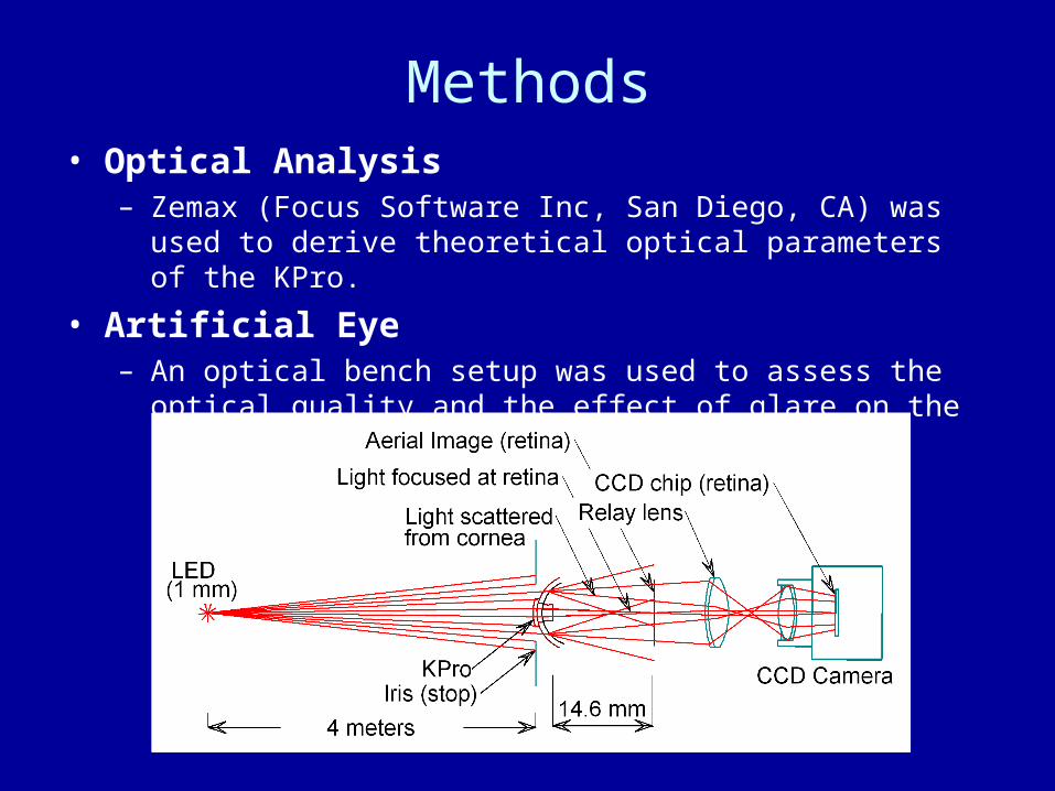
Methods• Optical Analysis
– Zemax (Focus Software Inc, San Diego, CA) was used to derive theoretical optical parameters of the KPro.
• Artificial Eye– An optical bench setup was used to assess the optical quality
and the effect of glare on the retinal image.

Methods• Glare sensitivity
– Measurements using a BAT (Mentor O&O Inc,Norwell,MA) were obtained with:
• (1) a standard transparent contact lens (CL) in place • (2) a dark iris CL, 4-mm pupil (Kontur Kontact Lens, Richmond, CA)
• Additional tests– Goldmann kinetic perimetry (V4e). – Stereo acuity measured in patients with intact fellow eye (Writ
Stereo Fly Test).
• Fundus imaging– Obtained using a Topcon TRC 50DX (Topcon, Tokyo, Japan)
fundus camera and Optos Panoramic 200MA system.

Results
• Computed MTF and PSF show that the system was close to diffraction-limited.

Results - Glare
• In the optical bench setup with the KPro installed in a metal iris, an image of a distant point-source was consistent with the computed high quality optics.
• A halo of scattered light could be eliminated by decreasing the aperture of the iris down to a 3-mm pupil.
• An opaque CL decreased the size of the scatter halo, but did not eliminate it.

Results - Glare
T II S1: African american patient implanted with type-2 Kpro
T II S2: Asian patient implanted with type-2 Kpro
T I: Patient implanted with a type-1 KPRO (n=8)
T I Dark CL: Same type-1 patients fitted with the dark contact lens

Visual Fields - 95°&90° for types I & II respectively

Results - Photography
• Stereopsis ranged from 400 to 800 sec of arc, reflecting minimal stereo vision
• Fundus Photography

Optos®Optomap

Conclusions
• The hazy cornea surrounding the KPro is a major source of glare
• A tinted soft lens can markedly reduce glare and improve vision up to about 40 percent
• The beneficial effect of a painted CL could potentially be mimicked by keeping as much of the natural iris intact during the surgery.
• KPros provide patients with a good field of view.• Little benefit to stereo vision is derived from implanting a
KPro in the presence of an undiseased contralateral eye with conserved vision.
• Adequate Optos wide-angle fundus imaging of around 150 degrees can be obtained