Optical Mapping of Impulse Propagation in Engineered ... · Optical Mapping of Impulse Propagation...
12
SPECIAL FOCUS Optical Mapping of Impulse Propagation in Engineered Cardiac Tissue Milica Radisic, Ph.D., 1 Vladimir G. Fast, Ph.D., 2 Oleg F. Sharifov, Ph.D., 2 Rohin K. Iyer, B.A.Sc., 1 Hyoungshin Park, Ph.D., 3,4 and Gordana Vunjak-Novakovic, Ph.D. 5 Cardiac tissue engineering has a potential to provide functional, synchronously contractile tissue constructs for heart repair, and for studies of development and disease using in vivo–like yet controllable in vitro settings. In both cases, the utilization of bioreactors capable of providing biomimetic culture environments is instrumental for supporting cell differentiation and functional assembly. In the present study, neonatal rat heart cells were cultured on highly porous collagen scaffolds in bioreactors with electrical field stimulation. A hallmark of excitable tissues such as myocardium is the ability to propagate electrical impulses. We utilized the method of optical mapping to measure the electrical impulse propagation. The average conduction velocity recorded for the stimulated constructs (14.4 4.1 cm=s) was significantly higher than that of the nonstimulated constructs (8.6 2.3 cm=s, p ¼ 0.003). The measured electrical propagation properties correlated to the contractile behavior and the compositions of tissue constructs. Electrical stimulation during culture significantly improved amplitude of contractions, tissue morphology, and connexin-43 expression compared to the nonsimulated controls. These data provide evidence that electrical stimulation during bioreactor cultivation can improve electrical signal propagation in engineered cardiac constructs. Introduction C ardiac tissue engineering has a potential to provide functional tissue constructs for repair of myocardial in- farction and congestive heart failure that currently affect close to 12 million people in the United States alone. 1 In an envi- sioned scenario, the scar tissue resulting from pathological remodeling after myocardial infarction can be replaced by a synchronously contracting engineered cardiac patch. In con- trast to skeletal tissues such as bone or cartilage where patients can be asked to reduce the load bearing function of these tissues for a period of time, some degree of function- ality of an engineered cardiac patch is required immediately, at the time of implantation. Importantly, engineered tissues that are being developed for applications in regenerative medicine can also find utility on a much shorter time scale in studies of development, disease progression, and drug screening, and in many other applications in biological and medical research. One paradigm of tissue engineering is that the reparative cells can be induced to form functional tissue constructs in vitro by cultivation on biomaterial scaffolds in bioreactors, with these two components jointly providing the necessary cues for cell differentiation and tissue assembly. A large num- ber of scaffolds that are biodegradable and biocompatible are available for cardiac tissue engineering, 2–8 and significant efforts in the field of stem cell biology are underway to find clinically relevant source of cardiomyocytes. 9–11 Most recen- tly, an exciting possibility of induced pluripotency (iPS) cells emerged, and it may be envisioned that iPS cells will provide an autologous source of millions of cardiomyocytes required for tissue engineering of human cardiac patches. 12–16 However, it is the cultivation in bioreactors that enables us to engineer contractile cardiac tissues in vitro and modulate their functional properties. Our work is focused on devel- oping bioreactor systems for cardiac tissue engineering using neonatal rat cardiomyocytes as a model cell source, while we anticipate that the same bioreactor systems will be translated to clinically relevant sources of human cardiomyocytes. We reported previously that cultivation of engineered cardiac tissue in the presence of electrical field stimulation (square pulses at the frequency of 1 Hz, signal amplitude of 5V=cm, and signal duration of 2 ms) remarkably improved cardiomyocyte function in engineered myocardium. 17 The surface of the stimulated tissue consisted of elongated cells ex- pressing cardiac differentiation markers, sarcomeric a-actin, 1 Institute of Biomaterials and Biomedical Engineering, University of Toronto, Toronto, Ontario, Canada. 2 Department of Biomedical Engineering, University of Alabama at Birmingham, Birmingham, Alabama. 3 Department of Surgery, Massachusetts General Hospital, Harvard Medical School, Boston, Massachusetts. 4 Department of Chemical Engineering, Massachusetts Institute of Technology, Cambridge, Massachusetts. 5 Department of Biomedical Engineering, Columbia University Vanderbilt Clinic, New York, New York. TISSUE ENGINEERING: Part A Volume 15, Number 4, 2009 ª Mary Ann Liebert, Inc. DOI: 10.1089=ten.tea.2008.0223 851
Transcript of Optical Mapping of Impulse Propagation in Engineered ... · Optical Mapping of Impulse Propagation...
SPECIAL FOCUS
Optical Mapping of Impulse Propagationin Engineered Cardiac Tissue
Milica Radisic, Ph.D.,1 Vladimir G. Fast, Ph.D.,2 Oleg F. Sharifov, Ph.D.,2 Rohin K. Iyer, B.A.Sc.,1
Hyoungshin Park, Ph.D.,3,4 and Gordana Vunjak-Novakovic, Ph.D.5
Cardiac tissue engineering has a potential to provide functional, synchronously contractile tissue constructs forheart repair, and for studies of development and disease using in vivo–like yet controllable in vitro settings. In bothcases, the utilization of bioreactors capable of providing biomimetic culture environments is instrumental forsupporting cell differentiation and functional assembly. In the present study, neonatal rat heart cells werecultured on highly porous collagen scaffolds in bioreactors with electrical field stimulation. A hallmark ofexcitable tissues such as myocardium is the ability to propagate electrical impulses. We utilized the method ofoptical mapping to measure the electrical impulse propagation. The average conduction velocity recorded for thestimulated constructs (14.4� 4.1 cm=s) was significantly higher than that of the nonstimulated constructs(8.6� 2.3 cm=s, p¼ 0.003). The measured electrical propagation properties correlated to the contractile behaviorand the compositions of tissue constructs. Electrical stimulation during culture significantly improved amplitudeof contractions, tissue morphology, and connexin-43 expression compared to the nonsimulated controls. Thesedata provide evidence that electrical stimulation during bioreactor cultivation can improve electrical signalpropagation in engineered cardiac constructs.
Introduction
Cardiac tissue engineering has a potential to providefunctional tissue constructs for repair of myocardial in-
farction and congestive heart failure that currently affect closeto 12 million people in the United States alone.1 In an envi-sioned scenario, the scar tissue resulting from pathologicalremodeling after myocardial infarction can be replaced by asynchronously contracting engineered cardiac patch. In con-trast to skeletal tissues such as bone or cartilage wherepatients can be asked to reduce the load bearing function ofthese tissues for a period of time, some degree of function-ality of an engineered cardiac patch is required immediately,at the time of implantation. Importantly, engineered tissuesthat are being developed for applications in regenerativemedicine can also find utility on a much shorter time scalein studies of development, disease progression, and drugscreening, and in many other applications in biological andmedical research.
One paradigm of tissue engineering is that the reparativecells can be induced to form functional tissue constructsin vitro by cultivation on biomaterial scaffolds in bioreactors,with these two components jointly providing the necessary
cues for cell differentiation and tissue assembly. A large num-ber of scaffolds that are biodegradable and biocompatible areavailable for cardiac tissue engineering,2–8 and significantefforts in the field of stem cell biology are underway to findclinically relevant source of cardiomyocytes.9–11 Most recen-tly, an exciting possibility of induced pluripotency (iPS) cellsemerged, and it may be envisioned that iPS cells will providean autologous source of millions of cardiomyocytes requiredfor tissue engineering of human cardiac patches.12–16
However, it is the cultivation in bioreactors that enables usto engineer contractile cardiac tissues in vitro and modulatetheir functional properties. Our work is focused on devel-oping bioreactor systems for cardiac tissue engineering usingneonatal rat cardiomyocytes as a model cell source, while weanticipate that the same bioreactor systems will be translatedto clinically relevant sources of human cardiomyocytes.
We reported previously that cultivation of engineeredcardiac tissue in the presence of electrical field stimulation(square pulses at the frequency of 1 Hz, signal amplitude of5 V=cm, and signal duration of 2 ms) remarkably improvedcardiomyocyte function in engineered myocardium.17 Thesurface of the stimulated tissue consisted of elongated cells ex-pressing cardiac differentiation markers, sarcomeric a-actin,
1Institute of Biomaterials and Biomedical Engineering, University of Toronto, Toronto, Ontario, Canada.2Department of Biomedical Engineering, University of Alabama at Birmingham, Birmingham, Alabama.3Department of Surgery, Massachusetts General Hospital, Harvard Medical School, Boston, Massachusetts.4Department of Chemical Engineering, Massachusetts Institute of Technology, Cambridge, Massachusetts.5Department of Biomedical Engineering, Columbia University Vanderbilt Clinic, New York, New York.
TISSUE ENGINEERING: Part AVolume 15, Number 4, 2009ª Mary Ann Liebert, Inc.DOI: 10.1089=ten.tea.2008.0223
851
cardiac troponin I, and a and b myosin heavy chain. Re-markably well-developed sarcomeres and gap junctions couldbe seen by transmission electron microscopy.17
In native myocardium, cardiomyocytes form a three-dimensional syncytium that enables propagation of electricalsignals across gap junctions to produce coordinated me-chanical contractions that pump blood forward. Groups ofspecialized cardiac myocytes (pace makers) generate electri-cal impulses that are propagated through the ventricles todrive periodic contractions of the heart. Excitation of eachcardiac myocyte is followed by the increase in the amountof cytoplasmic calcium that in turn triggers mechanical con-traction. Electrical propagation is therefore critical for thefunction of the heart. Engineered constructs that cannot pro-pagate electrical signals would have no utility for implan-tation because they may result in conduction blocks orgeneration of arrhythmia when implanted in the ventricles.Thus, engineered cardiac tissues need to be evaluated forimpulse propagation.18 In the present study, we focused onthe characterization of electrical impulse propagation byusing optical mapping, and the method was used to deter-mine the effects of electrical stimulation during cultivationon functional properties in the resulting tissue constructs.
Our second objective was to investigate the effects of thepresence of nonmyocytes on engineered myocardium, as ourprevious study focused exclusively on the identification andcharacterization of cardiomyocytes.17 Cardiomyocytes ac-count for only one third of all the cells found in the nativemyocardium. However, due to their large size they occupy90% of the volume and are critically responsible for con-tractile function.19,20 The remaining two thirds of the cellsare fibroblasts and endothelial cells, with smaller numbersof pericytes, smooth muscle cells, and macrophages.19,20 Fi-broblasts secrete components of the extracellular matrixand soluble factors, and transduce mechanical signals.20,21
Endothelial cells line the dense coronary vasculature andregulate the autocrine and paracrine signaling importantin angiogenesis.22 Both cell types interact with cardiomyo-cytes to coordinate tissue structure and function, and thepresence of both myocytes and nonmyocytes in the en-gineered 3D cardiac tissue may be required for appropriatefunction.23,24
Materials and Methods
Neonatal rat heart cells
Neonatal (1–2 days old) Sprague-Dawley rats were eutha-nized according to the procedure approved by the Institute’sCommittee on Animal Care. The hearts were removed andquartered, and the cells were isolated by an overnight treat-ment with trypsin (48C, 6120 units=mL in Hank’s balancedsalt solution, HBSS) followed by serial collagenase digestion(220 units=mL in HBSS) as described in previous work.25 Thesupernatants from five collagenase digests of the tissues werecollected and centrifuged at 750 rpm (94 g) for 4 min, re-suspended in culture medium, and preplated into T75 flasks(BD Biosciences, Bedford, MA) for one 1 h interval to enrichthe cell suspension for cardiomyocytes. The culture mediumconsisted of Dulbecco’s modified Eagle’s medium (Gibco,Billings, MT) with 4.5 g=L glucose, 4 mM L-glutamine, 10%certified fetal bovine serum (Gibco), 100 U=mL penicillin,100 mg=mL streptomycin, and 10 mM 4-2-hydroxyethyl-1-pi-perazineethanesulfonic acid buffer (HEPES; Gibco).
Collagen scaffold
The scaffolds were commercially available collagensponges, Ultrafoam� (Davol). Dry collagen scaffold wascross-sectioned using a sharp razor blade, sputter coated withgold, and imaged in face and cross section using HitachiS3400N Variable Pressure Scanning Electron Microscope at theMicroscopy Imaging Laboratory, University of Toronto. Forimaging of the scaffold in the hydrated state, the scaffolds werewetted in phosphate buffer saline and placed on the scanningelectron microscope stage with temperature set to 08C, andthe pressure was adjusted during imaging as required to pre-vent formation of ice crystals and to obtain clear images.
Construct preparation
Ultrafoam collagen sponge scaffolds (8�14�1.5 mm) wereprewetted with culture medium and placed into the 378C=5%CO2 humidified incubator (Napco, Winchester, VA) for 1 hourbefore inoculation. Cells (14�106) were pelleted by centrifuga-tion (1000 rpm, 10 min) and resuspended in Matrigel� (70mL,Becton-Dickinson) while working on ice to prevent prematuregelation as we described previously.2 Prewetted scaffolds weregently blotted dry and placed in six-well plates (one=well),and the Matrigel=cell suspension was delivered evenly to thetop surface of each scaffold using an automatic pipette. Ge-lation was achieved within 15 min at 378C. Subsequently,culture medium was added (4 mL=well), and the plates wereorbitally mixed (25 rpm) for 3 days. For reverse-transcriptionand polymerase chain reaction (RT-PCR), the constructs wereprepared and stimulated in an identical manner except that thethinner sponges (8�6�0.25 mm) were seeded with 1 millioncells; thus, the initial cell seeding density was identical.
Electrical stimulation
After 3 days of cultivation without electrical stimulation,the constructs were transferred to a chamber consisting ofa 100 mm glass Petri dish fitted with two 1=8-inch-diametercarbon rods (Ladd Research Industries, Williston, VT) placedat a distance of 1 cm and connected to a commercial cardiacstimulator (Nihon Kohden, Tokyo, Japan) via platinum wires(Ladd Research Industries).25 Silicone tubing (Cole-Parmer,Vernon Hills, IL) spacers were used to create six wells be-tween the two electrodes, enabling the cultivation of sixconstructs per dish (Fig. 1A). In one group (designated‘‘stimulated’’), the constructs were stimulated with squarepulses, 2 ms in duration, supra-threshold amplitude of 5 V(field strength of 5 V=cm), frequency of 1 Hz for the re-mainder of cultivation (8 days). This stimulation voltage wasselected to induce synchronous construct contractions(observable with optical microscope at 10� magnification).Construct placed in an identical chamber but without elec-trical stimulation served as control (designated ‘‘non-stimulated’’). All chambers were orbitally mixed at 25 rpm.Experiments were performed in a 378C=5% CO2 humidifiedincubator. The evaluation of construct’s functional propertiesand immunohistochemistry were performed at the end ofcultivation (8 days) as described below.
The excitation threshold of engineered cardiac constructswas evaluated at the end of cultivation before optical map-ping studies, by monitoring contractile activity upon elec-trical stimulation at 10� magnification using a microscope(Nikon Diaphot, Melville, NY).17 Each construct that wasevaluated was placed in a 100 mm Petri dish containing
852 RADISIC ET AL.
120 mL of Tyrode’s solution (140 mM NaCl, 5.4 mM KCl,0.33 mM NaH2PO4, 1.8 mM CaCl2, 1 mM MgCl2, 5 mMHEPES, 5.5 mM D-glucose, pH 7.4) between two custom-made gold electrodes connected to a cardiac stimulator(Nihon Kohden). The temperature was maintained at 298Cusing a heating tape (VWR International, Bridgeport, NJ)attached to the bottom of the Petri dish. The stimuli (squarepulses, 2 ms duration) were applied at a rate of 1 Hz startingat the amplitude of 1 V. The amplitude was increased in 0.1 Vincrements until the entire construct was observed to beatsynchronously, a voltage defined as the excitation threshold.To measure the amplitude of contractions, video-recordedbeating sequences (1–5 min in duration) were digitized at therate of 30 frames=s. The en-face area of each construct wasmeasured as function of time using image analysis (ScionImage software, Scion, Frederick, MD). Plots of the constructen-face area versus time were constructed and used to de-termine the base-line area of the construct in the relaxedstate. Deviation of the construct area greater than *0.5%from this baseline was considered to be the start of a con-traction. The time at which the construct area returned to thebase-line value was considered to be the end of a contraction.Duration of contraction was determined as the time periodbetween the start and end of the contraction. Amplitude ofcontraction was expressed as fractional area change.
Optical mapping
For optical measurements, tissue constructs were trans-ferred into a perfusion bath and superfused with Hanksbalanced salt solution having a composition of (mM): NaCl137, KCl 5.4, KH2PO4 0.4, NaH2PO4 0.4, MgSO4 0.8, CaCl21.3, NaHCO3 4.2, HEPES 5.0, and glucose 5.1. The pH of thesolution was 7.4, and the temperature was kept constantat 358C. Tissue constructs were stained with 5 mM solutionof fluorescent Vm-sensitive dye RH-237 (Molecular Probes,Eugene, OR). Dye fluorescence was excited using a 200-WHg=Xe lamp at 530–585 nm, and emitted fluorescence wasmeasured at >650 nm using a microscopic mapping systemdescribed previously.26,27 The system included a 16�16 pho-todiode array (Hamamatsu Corporation, Bridgewater, NJ) andan inverted microscope (Axiovert 135TV; Zeiss, Jena, Ger-many) with 5� and 10� objective lenses (Fluar, Zeiss, Jena,Germany) allowing optical recordings at spatial resolutions of110 and 55mm=diode. Tissue constructs were paced at 400-mscycle length using a bipolar electrode. Local activation timeswere measured at the 50% level of action potential upstrokes.Activation maps were constructed using linear interpolationand triangulation algorithms between neighboring recordinglocations. At each location, conduction velocity was calculatedusing activation times at four neighboring sites. Conductionvelocity values from all sites were used to calculate the averagemap conduction velocity.
Histology and immunostaining
At the end of cultivation the constructs were fixed in for-malin for 24 h and paraffin embedded. The constructs weresections such that the face-sectional area within the first200 mm of the construct surface was revealed. The 5-mmparaffin sections were deparaffinized and stained with he-matoxylin and eosin, Mason’s trichrome, and smooth muscleactin, at the Toronto General Hospital Pathology and His-tology Laboratory, using standard protocols. Sections were
also immunostained in-house, with antibodies to monoclonalrabbit anti-cardiac troponin I (DF 1:150; Chemicon, Teme-cula, CA), monoclonal rat anti-vimentin (Cy3 conjugated,DF 1:50; Sigma, St. Louis, MO), and polyclonal rabbitanti-von Willebrand factor (DF 1:150; Chemicon) as we de-scribed previously.28,29 Cell nuclei counterstaining with 40,6-diamidino-2-phenylindole (DAPI) was done in the mountingstep using mounting medium containing DAPI (Vector La-boratories, Burlingame, CA). Normal horse serum (10% inphosphate buffered saline; Vector Laboratories) was used forblocking prior to staining. Primary staining was done over-night at 48C in a humidified chamber. Slides were incubatedwith secondary antibody for 30 min at room temperature in ahumidified chamber using fluorescein isothiocyanate goatanti-mouse (DF 1:64; Sigma) or fluorescein isothiocyanategoat anti-rabbit (DF 1:200; Vector Laboratories) as required.Optical microscopy images were taken using a CanonPC1145 Powershot A620 Camera. Fluorescence imaging wasperformed on a Leica DMIRE2 microscope, and images werecaptured using Openlab 4.0.4 software.
RNA extraction
At the end of cultivation the constructs (n¼ 3 nonstimu-lated, 3 stimulated) were transferred into 1.5 mL RNAse-and DNAse-free tubes. The constructs were snap frozen byplacing the tubes directly into liquid nitrogen and immersingthem for 2 h. The constructs were then thawed at roomtemperature for 10 min before addition of 0.5 mL of TRIzolreagent (Invitrogen, Carlsbad, CA) to each tube for totalRNA extraction.
The samples were incubated in TRIzol at room tempera-ture for 20 min, with occasional pipetting to facilitate celldissociation. 100 mL of chloroform was added to each tube,followed by brief vortexing and centrifuging at 11000 rpm(7440 g) for 15 min to separate the aqueous and organicphases. The aqueous (RNA-containing) phase was thentransferred to a new tube, and the RNA was precipitatedby addition of 250mL of 2-propanol to each tube followedby incubation at �808C for 1 h. RNA was pelleted bycentrifuging at 11,000 rpm (7440 g) for 10 min and thenwashed by adding 500mL of 75% ethanol and briefly vor-texing each tube. The RNA was again pelleted at 8800 rpm(4762 g) for 5 min, and the supernatant discarded. The pellet,containing total RNA, was then dissolved in 50 L of DNAse-and RNAse-free distilled water and quantified using an ab-sorption, single-beam biophotometer (Eppendorf, Hamburg,Germany).
Reverse-transcription and polymerase chain reaction
Total RNA, 0.5mg, was reverse transcribed into first-strandcDNA using the SuperScript� III First Strand Synthesis System(Invitrogen) according to the manufacturer’s protocol. For eachsample, a negative control in which the reverse transcriptaseenzyme was substituted with distilled water was included toensure that genomic DNA contamination was not present.Polymerase chain reaction was used to amplify two genes ofinterest: (1) connexin-43, an important gap junctional proteininvolved in cell–cell coupling between myocytes, and (2) b-actin, a highly conserved cytoskeletal protein, used here as ahousekeeping gene. Sequence-specific primers for these genes,optimized for an annealing temperature of 528C, were asfollows:
OPTICAL MAPPING OF IMPULSE PROPAGATION IN ENGINEERED CARDIAC TISSUE 853
All PCR reactions were done in a reaction volume of 25mL,and all reagents were obtained from Invitrogen. Two mL offirst-strand cDNA was amplified by PCR using the aboveprimers at a final concentration of 0.2 mM along with 0.2 mMdNTPs, 1�PCR Buffer (20 mM Tris-HCl at pH 8.4, 50 mMKCl), 1.5 mM MgCl2, and 0.04 U=mL ‘‘Hot start’’ Platinum�
Taq DNA Polymerase. Thermal cycling was performed in aMultiGene II Thermocycler (Labnet International, Wood-bridge, NJ) with the following parameters: an initial 2-mindenaturation at 948C, followed by 35 cycles of 948C for 30 s,528C for 30 s, and 728C for 1 min were performed. A finalextension was performed at 728C for 1 min.
Gel electrophoresis and band densitometry
The PCR products, 6mL, were loaded in agarose gels (2%w=v in TAE Buffer) with a 100 base pair ladder as a standard.Gel electrophoresis was performed at 100 V for 30 min, andvisualization of gels was carried out by ethidium bromidestaining under UV light in a BioDoc-It Imaging System (UVPProducts, Upland, CA). Band densitometry was performedusing the Gel Analyzer option in ImageJ (National Institutesof Health, version 1.36b) to generate lane profile plots. Theareas under these curves were measured and expressed aspercentages of the total area to produce a semi-quantitativemeasure of mRNA expression. The resulting intensities forthe Cx43 bands and b-actin bands were plotted both as ab-solute intensities as well as normalized and averaged to giveexpression of Cx43 relative to b-actin.
Statistical analysis
The nonstimulated and stimulated groups were comparedusing T-test with Sigma Stat 3.0. Normality and equality ofvariance was tested for all data points. Differences were ac-cepted to be statistically significant at p< 0.05.
Results
The Ultrafoam collagen scaffold exhibited an isotropicmacroporous structure with large interconnected pores. Theaverage pore diameter was 190mm, and the average wallthickness was 8mm, as estimated from the dry-state images(Fig. 1B). Pores as small as 1mm in diameter and fibers assmall as 1.5 mm in diameter were present within the networkof large pores. In the hydrated state, macroporous isotropicpore structure was maintained (Fig. 1C).
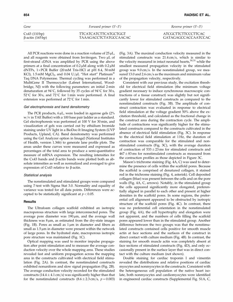
Optical mapping was used to monitor impulse propaga-tion after point stimulation and to measure the average con-duction velocity over the whole mapping area. These studiesrevealed fast and uniform propagation across the mappingarea in the constructs cultivated with electrical field stimu-lation (Fig. 2A). In contrast, the nonstimulated constructsexhibited nonuniform and slow signal propagation (Fig. 2B).The average conduction velocity recorded for the stimulatedconstructs (14.4� 4.1 cm=s) was significantly higher than thatfor the nonstimulated constructs (8.6� 2.3 cm=s, p¼ 0.003)
(Fig. 3A). The maximal conduction velocity measured in thestimulated constructs was 21.4 cm=s, which is similar tothe velocity measured in intact neonatal hearts,30,31 while thesmallest measured propagation velocity in the stimulatedgroup was 9.0 cm=s. In the nonstimulated group, we mea-sured 13.0 and 2.6 cm=s as the maximum and minimum valueof the propagation velocity, respectively.
Consistent with our previous study, the excitation thresh-old for electrical field stimulation (the minimum voltagegradient necessary to induce synchronous macroscopic con-tractions of a tissue construct) was slightly but not signifi-cantly lower for stimulated constructs as compared to thenonstimulated constructs (Fig. 3B). The amplitude of con-struct contraction was evaluated in response to electricalfield stimulation at the voltage gradient 50% above the ex-citation threshold, and calculated as the fractional change inthe construct area during the contraction cycle. The ampli-tude of contractions was significantly higher for the stimu-lated constructs compared to the constructs cultivated in theabsence of electrical field stimulation (Fig. 3C). In responseto the electrical field stimulation at 1 Hz, the duration ofcontraction was comparable for the stimulated and non-stimulated constructs (Fig. 3C), with the average durationof contraction of 533� 23 ms for stimulated constructs and467� 83 ms for nonstimulated constructs as measured fromthe contraction profiles as those depicted in Figure 3C.
Mason’s trichrome staining (Fig. 4A, C) was used to deter-mine the presence of cells within the scaffold pores. Becausethe scaffold is comprised of denatured collagen, it stainedred in the trichrome staining (Fig. 4, asterisk). Cell depositedcollagen (blue) was present between the cells and on the porewalls (Fig. 4A, C, arrows). Notably, in the stimulated groupthe cells appeared significantly more elongated, preferen-tially aligned in parallel to each other and present at higherdensities in the scaffold pores. In some regions, the prefer-ential cell alignment appeared to be obstructed by isotropicstructure of the scaffold pores (Fig. 4C). In contrast, therewas no preferential cell orientation in the nonstimulatedgroup (Fig. 4A), the cell hypertrophy and elongation werenot apparent, and the numbers of cells filling the scaffoldpores appeared lower than in the stimulated group. Anotherdifference between the two groups was that the nonstimu-lated constructs contained cells positive for smooth muscleactin at face sections and the surfaces of the construct indirect contact with culture medium (Fig. 4B). In contrast, thestaining for smooth muscle actin was completely absent atface sections of stimulated constructs (Fig. 4D), and only oc-casionally present in the surface layer that was in direct con-tact with the culture medium (not shown).
Double staining for cardiac troponin I and vimentinrevealed the distributions and relative positions of cardiacmyocytes and nonmyocytes in the constructs. Consistent withthe heterogeneous cell population of the native heart iso-late, both nonmyocytes and cardiomyocytes were identifiedin engineered cardiac constructs (Supplemental Fig. S1A, C,
Gene Forward primer (50–30) Reverse primer (50–30)
Cx43 (110 bp) TTCATCATCTTCATGCTGGT ATCGCTTCTTCCCTTCACb-actin (165 bp) TAAAGACCTCTATGCCAACAC GATAGAGCCACCAATCCAC
854 RADISIC ET AL.

available online at www.liebertonline.com=ten). In thenonstimulated group, cardiomyocytes (troponin I positive,green, Supplemental Fig. S1A) were randomly dispersedacross the construct surface with no preferential orientation.Besides being present on the scaffold surface, the non-myocytes (vimentin positive, red, Supplemental Fig. S1A) inthe nonstimulated group exhibited no preferential direc-tionality and were dispersed among the cardiomyocytes. Inthe stimulated group, cardiac troponin I–positive cells werefound to exhibit preferential alignment (SupplementalFig. S1C, green), surrounded by nonmyocytes that werelocated mainly on the construct surfaces (SupplementalFig. S1C, red). Positive staining for von Willebrand factorwas negligible in both groups, indicating the absence ofsignificant number of endothelial cells (SupplementalFig. S1B, D).
RT-PCR for Cx43 indicated significantly higher expressionof this gap junctional protein in the stimulated group than inthe nonstimulated group (Fig. 5A). The expression of house-keeping gene, b-actin, was comparable in both groups (Fig. 5).The absence of bands in negative controls, run without theenzyme reverse transcriptase, indicated the absence of ge-nomic contamination and specific amplification of Cx43transcripts.
Discussion
A hallmark of excitable tissues such as myocardium is theability to propagate electrical impulses, a property that di-rectly correlates with the tissue functionality. Thus, our goalwas to evaluate the velocity of impulse propagation in car-diac tissues cultivated in electrical stimulation bioreactors,using optical mapping. Previously, the velocity of impulsepropagation in the ventricles of 2-day-old Sprague Dawleyrats was measured to be 25.4 cm=s using a linear electrodearray,30 while the velocity in the heart ventricles of 10-day-old rats was measured to be 27 cm=s.31 The stimulatedconstructs in this study exhibited the average propagationvelocity of *55% of that in the native heart, while thehighest measured average propagation velocity was *80%of that measured in the native heart. In contrast, the averagepropagation velocity in the nonstimulated group was only*32% of that found in the native heart. The observed im-pulse propagation velocity was comparable to that repor-ted previously for neonatal rat cardiomyocytes cultivated onthe anisotropic poly(glycolic acid) scaffolds.18 In that previ-ous study, the velocity of impulse propagation in the longi-tudinal direction (i.e., parallel to the cell and scaffold fibrelong axis) was *15 cm=s at 6 days of cultivation (which isin line with the results reported for our stimulated groupcultivated for total of 8 days, Fig. 3A) and increased to*20 cm=s after 10 days of cultivation. In the transverse di-rection, the propagation velocity of *10 cm=s was measuredover the entire cultivation period,18 consistent with the valuesmeasured in the present study for the nonstimulated group(Fig. 3B).
The enhanced propagation velocity in the stimulatedgroup was most likely a result of improved cellular elonga-tion and parallel orientation in response to the electricalfield gradient, which were observed both in this study(Fig. 4C) and in our previous studies,25 as well as improvedcardiomyocyte coupling suggested by the increase in Cx43
FIG. 1. Electrical field stimulation cultivation system. (A)Set-up for electrical field stimulation consisting of a 100 mmPetri dish fitted with 1=8-inch carbon rods connected to acommercially available cardiac stimulator via platinumwires. (B) Cross section of collagen scaffold in dry state.(C) Cross section of the collagen scaffold in the hydratestate representing scaffold topography that would be expe-rienced by the cells. Color images available online at www.liebertonline.com=ten.
OPTICAL MAPPING OF IMPULSE PROPAGATION IN ENGINEERED CARDIAC TISSUE 855

expression (Fig. 5). Cx43 is predominant in the gap junctionsof the ventricular cardiomyocytes in the native heart. In ourprevious studies, using immunostaining for Cx43 and mor-phometric analysis of TEM images, we demonstrated thatthe frequency of gap junctions in the stimulated group wascomparable to that of the native heart, while in the non-stimulated group the frequency was significantly lower thanthat of the native heart. It is possible that upon implantationand maturation of the cardiac constructs in vivo, the propa-gation velocity would enhance further. For example, Zim-mermann et al.32 reported that cardiac constructs based onneonatal rat cardiomyocytes maintained for 4 weeks in theadult rat myocardium had maximum conduction velocity of55� 16 cm=s longitudinally and 16� 7 cm=s transversally,comparable to the conduction velocity in the noninfarcted
adult rat myocardium (69� 6 cm=s longitudinally and 19�5 cm=s transversally).
Porous collagen scaffold has an isotropic pore structure asillustrated by scanning electron microscopy of the scaffoldin dry as well as hydrated state (Fig. 1B, C). The field stim-ulation and excitation–contraction coupling guide parallelorientation and rearrangement of cardiomyocytes in thestimulated group that is opposed by the isotropic pores thatforce the elongating cells to ‘‘turn-corners’’ at the pore walls(Fig. 4C). To a certain extent, cells are capable of remodelingthe isotropic pore structure via action of cellular proteasesand traction forces; however, perfect alignment is hard toachieve with isotropic scaffolds. Several recent studies ofcardiomyocyte cultivations on anisotropic scaffolds clearlyindicated the importance of scaffold topography in guiding
FIG. 2. Optical mapping ofactivation spread in en-gineered cardiac tissues.(A) Isochronal activation map(top) and selected opticalrecordings of action poten-tial upstrokes (bottom) in astimulated tissue sample.(B) Activation map and opti-cal recordings in a non-stimulated control. Red circleson optical traces depict acti-vation times. The averageconduction velocity was 20.4and 5.4 cm=s in (A) and (B),respectively. Color imagesavailable online at www.liebertonline.com=ten.
856 RADISIC ET AL.
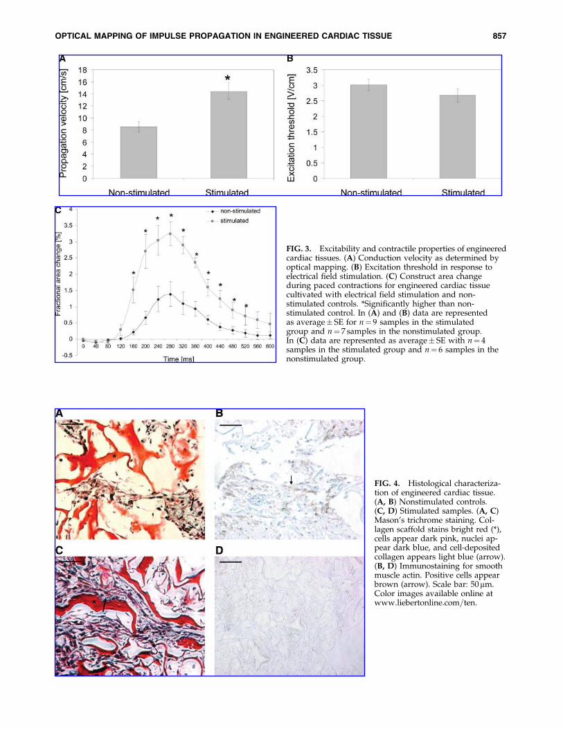
FIG. 3. Excitability and contractile properties of engineeredcardiac tissues. (A) Conduction velocity as determined byoptical mapping. (B) Excitation threshold in response toelectrical field stimulation. (C) Construct area changeduring paced contractions for engineered cardiac tissuecultivated with electrical field stimulation and non-stimulated controls. *Significantly higher than non-stimulated control. In (A) and (B) data are representedas average� SE for n¼ 9 samples in the stimulatedgroup and n¼ 7 samples in the nonstimulated group.In (C) data are represented as average� SE with n¼ 4samples in the stimulated group and n¼ 6 samples in thenonstimulated group.
FIG. 4. Histological characteriza-tion of engineered cardiac tissue.(A, B) Nonstimulated controls.(C, D) Stimulated samples. (A, C)Mason’s trichrome staining. Col-lagen scaffold stains bright red (*),cells appear dark pink, nuclei ap-pear dark blue, and cell-depositedcollagen appears light blue (arrow).(B, D) Immunostaining for smoothmuscle actin. Positive cells appearbrown (arrow). Scale bar: 50mm.Color images available online atwww.liebertonline.com=ten.
OPTICAL MAPPING OF IMPULSE PROPAGATION IN ENGINEERED CARDIAC TISSUE 857

functional properties of engineered cardiac structures.4,18 Weexpect that cultivation of neonatal rat cardiomyocytes onscaffolds with oriented (anisotropic) pores or fibers in thepresence of electrical field stimulation would further enhancethe propagation velocity and contractile properties of the en-gineered cardiac tissue.
Because cardiac fibroblasts can rapidly overgrow cardio-myocytes in monolayer cultures, the cell suspension used formonolayer studies is routinely enriched for cardiomyocytesby preplating. By analogy, early cardiac tissue engineeringstudies involved the use of cell populations enriched forcardiomyocytes by preplating,33–37 and some studies dem-onstrated that the velocity of impulse propagation increasedwith an increase in the fraction of cardiomyocytes in the cellpreparation.34 Zimmermann, Eschenhagen, and coworkers23,37
recognized the importance of the presence of multiple celltypes for the in vitro cultivation of heart tissue in their culti-vation system with cyclic stretch. Previously, we characterizedonly cardiomyocytes in the stimulated and nonstimulatedconstructs,25 but in this study we also evaluated the presenceof nonmyocytes.
In our hands, the composition of neonatal rat heart cellsuspension after one 1 h preplating step was 64� 5% cardi-omyocytes (cardiac troponin I–positive cells) and 34� 16%fibroblasts (prolyl-4-hydroxylase–positive cells),29 while aftertwo 1 h preplating steps the isolate was enriched for cardi-omyocytes (81� 14% cardiomyocytes vs. 16� 3% fibro-blasts). The fraction of other cell types remained low (e.g.,3� 3% endothelial cells).38 During cultivation, the fibroblastspersisted in the constructs but did not overgrow cardio-myocytes (Supplemental Fig. S1), possibly due to the mi-croarchitecture of the scaffold pores.39 Because endothelial
cells were mostly absent in these constructs (SupplementalFig. S1B, D), coculture or scaffold prevascularization in vitromay be required. Alternatively, scaffolds with immobilizedangiogenic factors may be utilized40 to promote rapid vas-cularization in vivo. Interestingly, in the present study therewere no smooth muscle actin–positive cells in the face sec-tions of stimulated constructs, while remarkably higher num-bers were present in the nonstimulated constructs (Fig. 4).Because smooth muscle cells account for only *3% of thenative heart cell isolate after one preplating,23 the smoothmuscle actin–positive cells in the nonstimulated constructsmay be myofibroblasts. The myofibroblasts exhibit elevatedsecretion of extracellular matrix proteins; they are implicatedin scarring upon myocardial infarction41 and the decrease inpropagation velocity when present in critical numbers (e.g.,more than 7 myofibroblasts per 100 cardiomyocytes).42
Electrical field stimulation bioreactor, such as the one usedhere, may prove to be a useful tool in engineering cardiacconstructs based on human stem=progenitor cell sources.The bioreactor we used is simple to construct and operate,thus enabling easy translation into other research laborato-ries and ultimately clinical practice. Electrical field stimula-tion is provided by a pair of parallel carbon electrodes (Fig.1A), positioned 1 cm apart within a glass chamber and con-nected to a commercially available stimulator or a custom-made pulse generator=amplifier circuit. In our previousstudies we have tested a number of electrode materials (ti-tanium, titanium nitride, and stainless steel) in additionto carbon.43 We found that carbon was indeed an optimalmaterial for this application due to high resistance to polar-ization and Faradaic reactions, high efficiency of chargetransfer to electrolyte (and hence to the tissue construct), and
FIG. 5. Expression of gapjunctional protein connexin-43. (A) Gels showingconnexin-43 and b-actin ex-pression. Lane 1, ladder;lanes 2–4, three independentnonstimulated samples sub-jected to RT-PCR withoutthe reverse transcriptaseenzyme; lanes 5–7, three in-dependent stimulated sam-ples subjected to RT-PCRwithout the reverse transcrip-tase enzyme; lanes 8–10, threeindependent nonstimulatedsamples subjected to RT-PCRwith the reverse transcriptaseenzyme; lanes 11–13, threeindependent stimulatedsamples subjected to RT-PCRwith the reverse transcriptaseenzyme. (B) Densitometricanalysis of bands representedin (A). (C) Relative expres-sion of connexin-43 in compa-rison to b-actin (connexin-43=b-actin band intensityaverage� SD).
858 RADISIC ET AL.
low impedance modulus across a large frequency range(10�2 to 106 Hz). Further, although the impedance of carbonelectrodes decreased with age when low frequencies (e.g.,1 Hz) were used, its resistance to Faradaic reactions remainedunchanged, indicating that the same setup can be re-used fornumerous experiments. The resistance measured in the bio-reactors with carbon rods spaced 1 cm apart was 20O and10O for 8- and 4-cm-long electrodes, respectively.44 Totalcharge injected with a single 5 V=cm pulse of 2 ms durationin a setup consisting of a 60 mm Petri dish with 20 mL ofphosphate buffered saline was measured to be 2.3�10�4 C.43
The collagen scaffold used in this study was a commer-cially available hemostat, Ultrafoam, which is water insolu-ble, and formed from the partial HCl salt of purified bovinedermal (corium) collagen as a sponge with interconnectedpores. Because it has already gained FDA approval as ahemostat, this scaffold may indeed be a suitable material forengineering of a clinically relevant cardiac patch. For cellseeding, we used rapid inoculation of the cells at high den-sity with a hydrogel (Matrigel).2 Because Matrigel is derivedfrom the basement membrane of a mouse sarcoma, it is notsuitable for preparation of clinically relevant constructs.Currently, we have complementary efforts underway to re-place Matrigel by biodegradable hydrogels of controllablecomposition based on chitosan45 or hyaluronic acid.46
Approaches to improve the current bioreactor involvedevelopment of second-generation bioreactors that incorpo-rate mechanical stimulation37 or culture medium17 flow alongwith electrical field stimulation. One approach to providingmechanical stimulation in such a bioreactor would includepulsatile culture medium flow, where a culture mediumpulse would be applied between the electrical pulses. To en-sure that the constructs are not moving during an experi-ment, they can be fixed by pinning into a thin layer ofpoly(dimethylsiloxane) cast on the bottom of the chamber.43
In summary, we demonstrated here that the engineeredcardiac tissue cultivated in the presence of electrical fieldstimulation exhibits impulse propagation comparable to thatof the neonatal rat ventricles. The tissues were comprised ofmultiple cell types, mostly cardiomyocytes and fibroblasts;however, no overgrowth of fibroblasts was observed. In thestimulated group, the fibroblasts were confined to the sur-face layer of the construct in the direct contact with theculture medium. Endothelial cells were largely absent in bothgroups, while smooth muscle–positive cells, most likelymyofibroblasts, were more numerous in the nonstimulatedgroup than in the stimulated group.
Acknowledgments
This study was supported by NSERC (Discovery Grant toM.R.), NIH (R01 HL076485 and P41-EB002520 to G.V.N.),and Ontario Graduate Scholarship (to R.K.I.).
References
1. Krikpatrick, J.N., Vannan, M.A., Narula, J., and Lang, R.M.Echocardiography in heart failure: applications, utility, andnew horizons. J Am Coll Cardiol 50, 381, 2007.
2. Radisic, M., Euloth, M., Yang, L., Langer, R., Freed, L.E., andVunjak-Novakovic, G. High-density seeding of myocyte cellsfor cardiac tissue engineering. Biotechnol Bioeng 82, 403, 2003.
3. Park, H., Radisic, M., Lim, J.O., Chang, B.H., and Vunjak-Novakovic, G. A novel composite scaffold for cardiac tissueengineering. In Vitro Cell Dev Biol Anim 41, 188, 2005.
4. Zong, X., Bien, H., Chung, C.Y., Yin, L., Fang, D., Hsiao, B.S.,Chu, B., and Entcheva, E. Electrospun fine-textured scaffoldsfor heart tissue constructs. Biomaterials 26, 5330, 2005.
5. McDevitt, T.C., Woodhouse, K.A., Hauschka, S.D., Murry,C.E., and Stayton, P.S. Spatially organized layers of cardio-myocytes on biodegradable polyurethanefilms for myocar-dial repair. J Biomed Mater Res A 66, 586, 2003.
6. Carrier, R.L., Rupnick, M., Langer, R., Schoen, F.J., Freed, L.E.,and Vunjak-Novakovic, G. Perfusion improves tissue archi-tecture of engineered cardiac muscle. Tissue Eng 8, 175, 2002.
7. Boublik, J., Park, H., Radisic, M., Tognana, E., Chen, F., Pei,M., Vunjak-Novakovic, G., and Freed, L.E. Mechanicalproperties and remodeling of hybrid cardiac constructsmade from heart cells, fibrin, and biodegradable, elastomericknitted fabric. Tissue Eng 11, 1122, 2005.
8. Badylak, S.F., Kochupura, P.V., Cohen, I.S., Doronin, S.V.,Saltman, A.E., Gilbert, T.W., Kelly, D.J., Ignotz, R.A., andGaudette, G.R. The use of extracellular matrix as an induc-tive scaffold for the partial replacement of functional myo-cardium. Cell Transplant 15 Suppl 1, S29, 2006.
9. Zimmermann, W.H., Didie, M., Doker, S., Melnychenko, I.,Naito, H., Rogge, C., Tiburcy, M., and Eschenhagen, T.Heart muscle engineering: an update on cardiac muscle re-placement therapy. Cardiovasc Res 71, 419, 2006.
10. Eschenhagen, T., and Zimmermann, W.H. Engineeringmyocardial tissue. Circ Res 97, 1220, 2005.
11. Laflamme, M.A., and Murry, C.E. Regenerating the heart.Nat Biotechnol 23, 845, 2005.
12. Takahashi, K., and Yamanaka, S. Induction of pluripotentstem cells from mouse embryonic and adult fibroblast cul-tures by defined factors. Cell 126, 663, 2006.
13. Takahashi, K., Tanabe, K., Ohnuki, M., Narita, M., Ichisaka,T., Tomoda, K., and Yamanaka, S. Induction of pluripotentstem cells from adult human fibroblasts by defined factors.Cell 131, 861, 2007.
14. Nakagawa, M., Koyanagi, M., Tanabe, K., Takahashi, K.,Ichisaka, T., Aoi, T., Okita, K., Mochiduki, Y., Takizawa, N.,and Yamanaka, S. Generation of induced pluripotent stemcells without Myc from mouse and human fibroblasts. NatBiotechnol 26, 101, 2008.
15. Park, I.H., Zhao, R., West, J.A., Yabuuchi, A., Huo, H., Ince,T.A., Lerou, P.H., Lensch, M.W., and Daley, G.Q. Repro-gramming of human somatic cells to pluripotency with de-fined factors. Nature 451, 141, 2008.
16. Lowry, W.E., Richter, L., Yachechko, R., Pyle, A.D., Tchieu,J., Sridharan, R., Clark, A.T., and Plath, K. Generation ofhuman induced pluripotent stem cells from dermal fibro-blasts. Proc Natl Acad Sci USA 105, 2883, 2008.
17. Radisic, M., Yang, L., Boublik, J., Cohen, R.J., Langer, R.,Freed, L.E., and Vunjak-Novakovic, G. Medium perfusionenables engineering of compact and contractile cardiac tis-sue. Am J Physiol Heart Circ Physiol 286, H507, 2004.
18. Bursac, N., Loo, Y., Leong, K., and Tung, L. Novel aniso-tropic engineered cardiac tissues: studies of electrical prop-agation. Biochem Biophys Res Commun 361, 847, 2007.
19. Nag, A.C. Study of non-muscle cells of the adult mammalianheart: a fine structural analysis and distribution. Cytobios28, 41, 1980.
20. Banerjee, I., Yekkala, K., Borg, T.K., and Baudino, T.A. Dy-namic interactions between myocytes, fibroblasts, and ex-tracellular matrix. Ann NY Acad Sci 1080, 76, 2006.
OPTICAL MAPPING OF IMPULSE PROPAGATION IN ENGINEERED CARDIAC TISSUE 859

21. Kuzuya, M., and Kinsella, J.L. Induction of endothelial celldifferentiation in vitro by fibroblast-derived soluble factors.Exp Cell Res 215, 310, 1994.
22. Seghezzi, G., Patel, S., Ren, C.J., Gualandris, A., Pintucci, G.,Robbins, E.S., Shapiro, R.L., Galloway, A.C., Rifkin, D.B.,and Mignatti, P. Fibroblast growth factor-2 (FGF-2) inducesvascular endothelial growth factor (VEGF) expression in theendothelial cells of forming capillaries: an autocrine mecha-nism contributing to angiogenesis. J Cell Biol 141, 1659, 1998.
23. Naito, H., Melnychenko, I., Didie, M., Schneiderbanger, K.,Schubert, P., Rosenkranz, S., Eschenhagen, T., and Zim-mermann, W.H. Optimizing engineered heart tissue for the-rapeutic applications as surrogate heart muscle. Circulation114, I72, 2006.
24. Radisic, M., Park, H., Martens, T.P., Salazar-Lazaro, J.E.,Geng, W., Wang, Y., Langer, R., Freed, L.E., and Vunjak-Novakovic, G. Pre-treatment of synthetic elastomeric scaf-folds by cardiac fibroblasts improves engineered heart tissue.J Biomed Mater Res A 2007.
25. Radisic, M., Park, H., Shing, H., Consi, T., Schoen, F.J.,Langer, R., Freed, L.E., and Vunjak-Novakovic, G. Func-tional assembly of engineered myocardium by electricalstimulation of cardiac myocytes cultured on scaffolds. ProcNatl Acad Sci USA 101, 18129, 2004.
26. Fast, V.G., and Cheek, E.R. Optical mapping of arrhythmiasinduced by strong electrical shocks in myocyte cultures. CircRes 90, 664, 2002.
27. Fast, V.G. Recording action potentials using voltage-sensitive dyes. In: Dhein, S., Delmar, M., and Mohr, F.W.,eds. Methods in Cardiovascular Research. Berlin: Springer-Verlag, 2005, p. 233–255.
28. Radisic, M., Park, H., Chen, F., Salazar-Lazzaro, J.E.,Wang, Y., Dennis, R., Langer, R., Freed, L.E., and Vunjak-Novakovic, G. Biomimetic approach to cardiac tissue engi-neering: oxygen carriers and channeled scaffolds. Tissue Eng12, 2077, 2006.
29. Radisic, M., Marsano, A., Maidhof, R., Wang, Y., andVunjak-Novakovic, G. Cardiac tissue engineering usingperfusion bioreactor systems. Nature Protocols 3, 719, 2008.
30. Bursac, N., Papadaki, M., Langer, R., Eisenberg, S.R., Vunjak-Novakovic, G., and Freed, L.E. Towards a functional tissueengineered cardiac muscle. 21st Annual Meeting of theBiomedical Engineering Society, Atlanta, 1999, p. 29.
31. Sun, L.S., Legato, M.J., Rosen, T.S., Steinberg, S.F., andRosen, M.R. Sympathetic innervation modulates ventricularimpulse propagation and repolarisation in the immature ratheart. Cardiovasc Res 27, 459, 1993.
32. Zimmermann, W.H., Melnychenko, I., Wasmeier, G., Didie,M., Naito, H., Nixdorff, U., Hess, A., Budinsky, L., Brune, K.,Michaelis, B., Dhein, S., Schwoerer, A., Ehmke, H., and Es-chenhagen, T. Engineered heart tissue grafts improve sys-tolic and diastolic function in infarcted rat hearts. Nat Med12, 452, 2006.
33. Carrier, R.L., Papadaki, M., Rupnick, M., Schoen, F.J., Bursac, N.,Langer, R., Freed, L.E., and Vunjak-Novakovic, G. Cardiac tis-sue engineering: cell seeding, cultivation parameters and tissueconstruct characterization. Biotechnol Bioeng 64, 580, 1999.
34. Bursac, N., Papadaki, M., Cohen, R.J., Schoen, F.J., Eisen-berg, S.R., Carrier, R., Vunjak-Novakovic, G., and Freed, L.E.Cardiac muscle tissue engineering: toward an in vitro modelfor electrophysiological studies. Am J Physiol Heart CircPhysiol 277, H433, 1999.
35. Fink, C., Ergun, S., Kralisch, D., Remmers, U., Weil, J., andEschenhagen, T. Chronic stretch of engineered heart tissue
induces hypertrophy and functional improvement. FASEB J14, 669, 2000.
36. Papadaki, M., Bursac, N., Langer, R., Merok, J., Vunjak-No-vakovic, G., and Freed, L.E. Tissue engineering of functionalcardiac muscle: molecular, structural and electrophysiologicalstudies. Am J Physiol Heart Circ Physiol 280, H168, 2001.
37. Zimmermann, W.H., Schneiderbanger, K., Schubert, P., Di-die, M., Munzel, F., Heubach, J.F., Kostin, S., Nehuber, W.L.,and Eschenhagen, T. Tissue engineering of a differentiatedcardiac muscle construct. Circ Res 90, 223, 2002.
38. Iyer, R., Chiu, L., and Radisic, M. Microfabricatedpoly(ethylene glycol) templates enable rapid screening of tri-culture conditions for cardiac tissue engineering. J BiomedMater Res Part A, 2008. [Epub ahead of print]
39. Boateng, S.Y., Hartman, T.J., Ahluwalia, N., Vidula, H.,Desai, T.A., and Russell, B. Inhibition of fibroblast prolifer-ation in cardiac myocyte cultures by surface microtopo-graphy. Am J Physiol Cell Physiol 285, C171, 2003.
40. Shen, Y.H., Shoichet, M.S., and Radisic, M. Vascular endo-thelial growth factor immobilized in collagen scaffold pro-motes penetration and proliferation of endothelial cells. ActaBiomater 4, 477, 2008.
41. Sun, Y., Kiani, M.F., Postlethwaite, A.E., and Weber, K.T.Infarct scar as living tissue. Basic Res Cardiol 97, 343, 2002.
42. Miragoli, M., Gaudesius, G., and Rohr, S. Electrotonicmodulation of cardiac impulse conduction by myofibro-blasts. Circ Res 98, 801, 2006.
43. Cannizzaro, C., Tandon, N., Figallo, E., Park, H., Gerecht, S.,Radisic, M., Elvassore, N., and Vunjak-Novakovic, G. Prac-tical aspects of cardiac tissue engineering with electricalstimulation. Methods Mol Med 140, 291, 2007.
44. Tandon, N., Cannizzaro, C., Chao, G., Maidhof, R., Marsano,A., Au, H., Radisic, M., and Vunjak-Novakovic, G. Electricalstimulation systems for cardiac tissue engineering. NatProtoc [in press].
45. Yeo, Y., Geng, W., Ito, T., Kohane, D.S., Burdick, J.A., andRadisic, M. Photocrosslinkable hydrogel for myocyte cellculture and injection. J Biomed Mater Res B Appl Biomater81, 312, 2007.
46. Gerecht, S., Burdick, J.A., Ferreira, L.S., Townsend, S.A.,Langer, R., and Vunjak-Novakovic, G. Hyaluronic acid hydro-gel for controlled self-renewal and differentiation of humanembryonic stem cells. Proc Natl Acad Sci USA 104, 11298, 2007.
Address reprint requests to:Milica Radisic, Ph.D.
Institute of Biomaterials and Biomedical EngineeringUniversity of Toronto
164 College St., Room 407Toronto, Ontario M5S 3G9
Canada
E-mail: [email protected]
Gordana Vunjak-Novakovic, Ph.D.Department of Biomedical EngineeringColumbia University Vanderbilt Clinic
12th Floor, Room 12-234 622, West 168th St.New York, NY 10032
E-mail: [email protected]
Received: April 16, 2008Accepted: July 28, 2008
Online Publication Date: October 6, 2008
860 RADISIC ET AL.

This article has been cited by:
1. Maximilian Y. Emmert, Robert W. Hitchcock, Simon P. Hoerstrup. 2013. Cell therapy, 3D culture systems and tissue engineeringfor cardiac regeneration. Advanced Drug Delivery Reviews . [CrossRef]
2. Zlata Vukadinovic-Nikolic, Birgit Andrée, Suzanne E. Dorfman, Michael Pflaum, Tibor Horvath, Marco Lux, Letizia Venturini,Antonia Bär, George Kensah, Angelica Roa Lara, Igor Tudorache, Serghei Cebotari, Denise Hilfiker-Kleiner, Axel Haverich,Andres Hilfiker. Generation of Bioartificial Heart Tissue by Combining a Three-Dimensional Gel-Based Cardiac Construct withDecellularized Small Intestinal Submucosa. Tissue Engineering Part A, ahead of print. [Abstract] [Full Text HTML] [Full TextPDF] [Full Text PDF with Links] [Supplemental Material]
3. Dr. Zlata Vukadinovic-Nikolic, Dr. Birgit Andrée, Dr. Suzanne E. Dorfman, Michael Pflaum, Tibor Horvath, Marco Lux, LetiziaVenturini, Dr. Antonia Bär, Mr. George Kensah, Dr. Angelica Roa Lara, Dr. Igor Tudorache, Dr. Serghei Cebotari, Prof. DeniseHilfiker-Kleiner, Dr. Axel Haverich, Dr. Andres Hilfiker. Generation of bioartificial heart tissue by combining a 3D gel-basedcardiac construct with decellularized small intestinal submucosa. Tissue Engineering Part A 0:ja. . [Abstract] [Full Text PDF][Full Text PDF with Links]
4. Vishal Tandon, Boyang Zhang, Milica Radisic, Shashi K. Murthy. 2013. Generation of tissue constructs for cardiovascularregenerative medicine: From cell procurement to scaffold design. Biotechnology Advances 31:5, 722-735. [CrossRef]
5. Giancarlo Forte, Stefania Pagliari, Francesca Pagliari, Mitsuhiro Ebara, Paolo Nardo, Takao Aoyagi. 2013. Towards the Generationof Patient-Specific Patches for Cardiac Repair. Stem Cell Reviews and Reports 9:3, 313-325. [CrossRef]
6. Loraine LY Chiu, Milica Radisic. 2013. Cardiac tissue engineering. Current Opinion in Chemical Engineering 2:1, 41-52.[CrossRef]
7. J Roether, H Jawad, R Rai, N Ali, S Harding, A BoccacciniPolymers for Myocardial Tissue Engineering 369-388. [CrossRef]8. Nimalan Thavandiran, Sara S Nunes, Yun Xiao, Milica Radisic. 2013. Topological and electrical control of cardiac differentiation
and assembly. Stem Cell Research & Therapy 4:1, 14. [CrossRef]9. Deborah K. Lieu, Irene C. Turnbull, Kevin D. Costa, Ronald A. Li. 2012. Engineered human pluripotent stem cell-derived
cardiac cells and tissues for electrophysiological studies. Drug Discovery Today: Disease Models 9:4, e209-e217. [CrossRef]10. Claus Svane Sondergaard, Grant Mathews, Lianguo Wang, Angela Jeffreys, Amrit Sahota, Moira Wood, Crystal M. Ripplinger,
Ming-Sing Si. 2012. Contractile and Electrophysiologic Characterization of Optimized Self-Organizing Engineered Heart Tissue.The Annals of Thoracic Surgery 94:4, 1241-1249. [CrossRef]
11. Brian Liau, Donghui Zhang, Nenad Bursac. 2012. Functional cardiac tissue engineering. Regenerative Medicine 7:2, 187-206.[CrossRef]
12. Jennifer Dawson, Olivier Schussler, Ashraf Al-Madhoun, Claudine Menard, Marc Ruel, Ilona S. Skerjanc. 2011. Collagen scaffoldswith or without the addition of RGD peptides support cardiomyogenesis after aggregation of mouse embryonic stem cells. InVitro Cellular & Developmental Biology - Animal . [CrossRef]
13. Stephen F. Badylak, Doris Taylor, Korkut Uygun. 2011. Whole-Organ Tissue Engineering: Decellularization and Recellularizationof Three-Dimensional Matrix Scaffolds. Annual Review of Biomedical Engineering 13:1, 27-53. [CrossRef]
14. Jana Dengler, Hannah Song, Nimalan Thavandiran, Stéphane Massé, Geoffrey A. Wood, Kumaraswamy Nanthakumar, Peter W.Zandstra, Milica Radisic. 2011. Engineered heart tissue enables study of residual undifferentiated embryonic stem cell activity ina cardiac environment. Biotechnology and Bioengineering 108:3, 704-719. [CrossRef]
15. P. Akhyari, M. Barth, A. LichtenbergCardiac Patch with Cells: Biological or Synthetic 367-388. [CrossRef]16. Loraine L. Y. Chiu, Milica Radisic, Gordana Vunjak-Novakovic. 2010. Bioactive Scaffolds for Engineering Vascularized Cardiac
Tissues. Macromolecular Bioscience 10:11, 1286-1301. [CrossRef]17. Peter C. Johnson, Antonios G. MikosStem Cells: State of the Art 3-11. [Citation] [Full Text PDF] [Full Text PDF with Links]18. C. Katherine Chiang, Mohammad Fahad Chowdhury, Rohin K. Iyer, William L. Stanford, Milica Radisic. 2010. Engineering
surfaces for site-specific vascular differentiation of mouse embryonic stem cells. Acta Biomaterialia 6:6, 1904-1916. [CrossRef]19. Marta Codina, Jeremy Elser, Kenneth B. Margulies. 2010. Current Status of Stem Cell Therapy in Heart Failure. Current
Cardiology Reports 12:3, 199-208. [CrossRef]20. Gordana Vunjak-Novakovic, Nina Tandon, Amandine Godier, Robert Maidhof, Anna Marsano, Timothy P. Martens, Milica
Radisic. 2010. Challenges in Cardiac Tissue Engineering. Tissue Engineering Part B: Reviews 16:2, 169-187. [Abstract] [Full TextHTML] [Full Text PDF] [Full Text PDF with Links]
21. Loraine L.Y. Chiu, Milica Radisic. 2010. Scaffolds with covalently immobilized VEGF and Angiopoietin-1 for vascularization ofengineered tissues. Biomaterials 31:2, 226-241. [CrossRef]

22. Peter C. Johnson, Antonios G. MikosAdvances in Tissue Engineering: Volume 2 . [Citation] [Full Text HTML] [Full TextPDF] [Full Text PDF with Links]