Opthalmic Preparations
103
-
Upload
shivarajendra09 -
Category
Documents
-
view
228 -
download
0
Transcript of Opthalmic Preparations
7/30/2019 Opthalmic Preparations
http://slidepdf.com/reader/full/opthalmic-preparations 1/103
7/30/2019 Opthalmic Preparations
http://slidepdf.com/reader/full/opthalmic-preparations 2/103
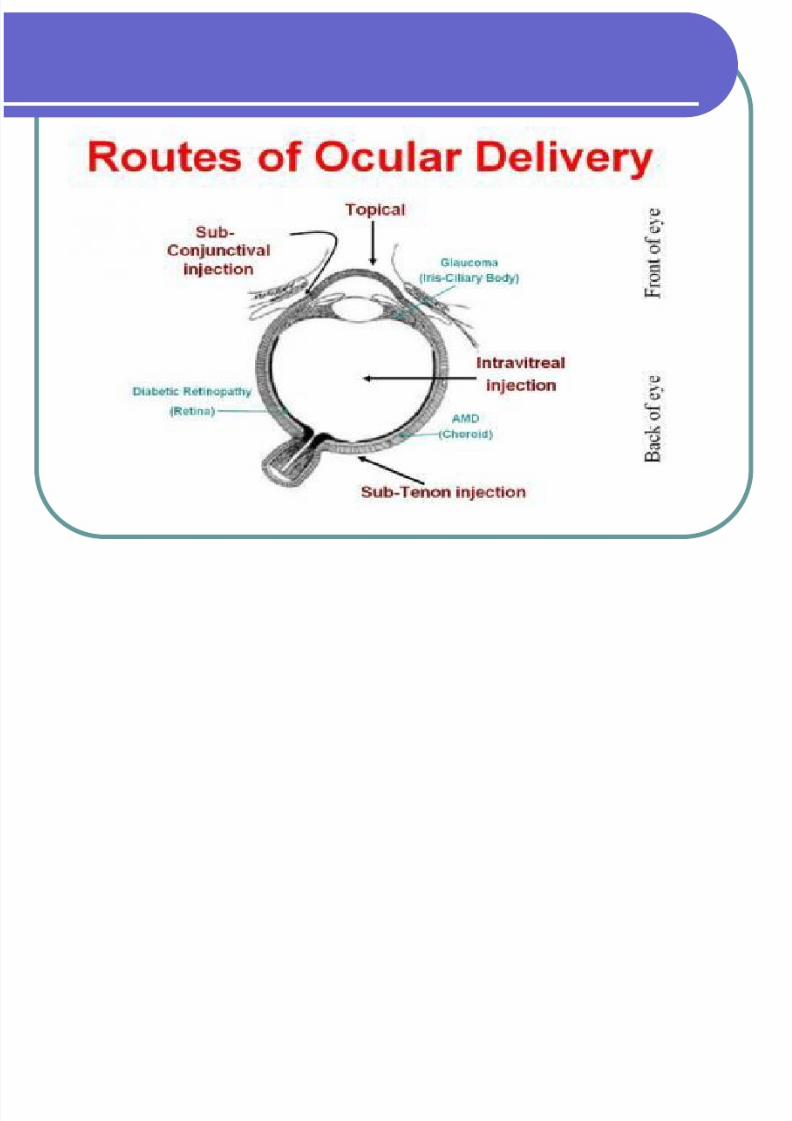
Ophthalmic preparations:
Definition: They are specialized dosage forms designed tobe instilled onto the external surface of the eye (topical),administered inside (intraocular) or adjacent (periocular) tothe eye or used in conjunction with an ophthalmic device.
The most commonly employed ophthalmic dosage forms aresolutions, suspensions, and ointments.
The newest dosage forms for ophthalmic drug delivery are:
gels, gel-forming solutions, ocular inserts , intravitrealinjections and implants.
7/30/2019 Opthalmic Preparations
http://slidepdf.com/reader/full/opthalmic-preparations 3/103
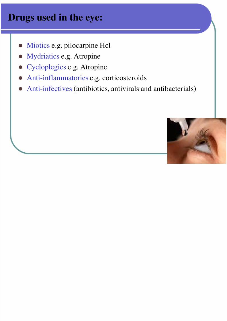
Drugs used in the eye:
Miotics e.g. pilocarpine Hcl
Mydriatics e.g. Atropine
Cycloplegics e.g. Atropine
Anti-inflammatories e.g. corticosteroids Anti-infectives (antibiotics, antivirals and antibacterials)
7/30/2019 Opthalmic Preparations
http://slidepdf.com/reader/full/opthalmic-preparations 4/103
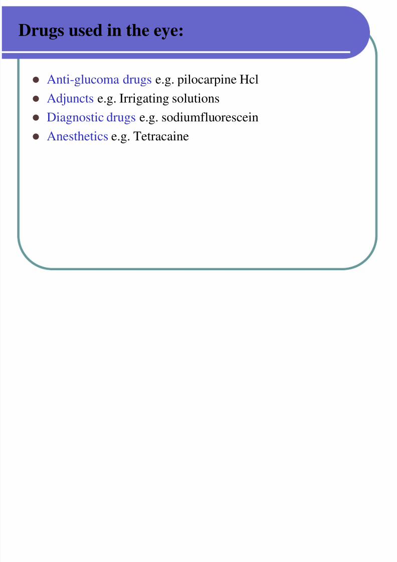
Drugs used in the eye:
Anti-glucoma drugs e.g. pilocarpine Hcl
Adjuncts e.g. Irrigating solutions
Diagnostic drugs e.g. sodiumfluorescein
Anesthetics e.g. Tetracaine
7/30/2019 Opthalmic Preparations
http://slidepdf.com/reader/full/opthalmic-preparations 5/103
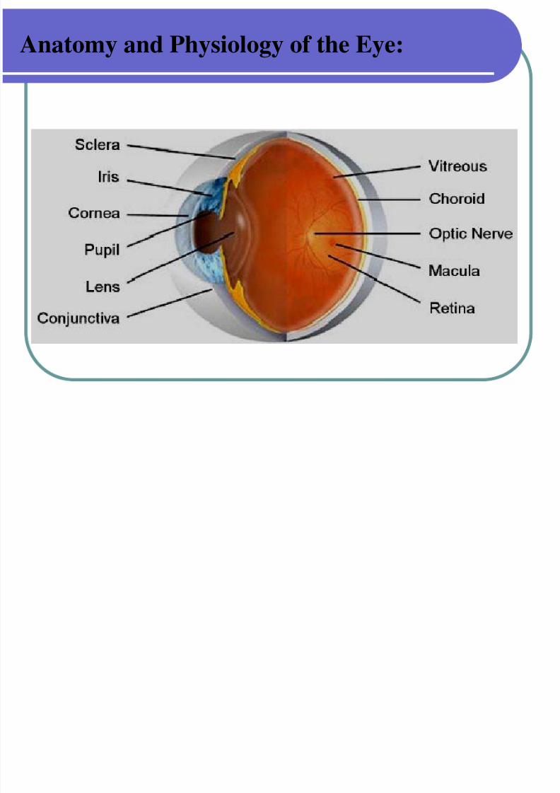
Anatomy and Physiology of the Eye:
7/30/2019 Opthalmic Preparations
http://slidepdf.com/reader/full/opthalmic-preparations 6/103
Anatomy and Physiology of the Eye (Cont.)
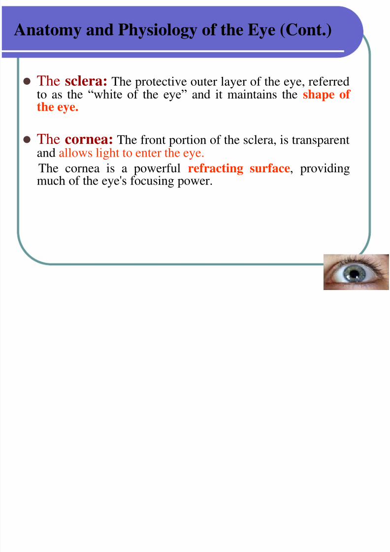
The sclera: The protective outer layer of the eye, referredto as the “white of the eye” and it maintains the shape of the eye.
The cornea: The front portion of the sclera, is transparentand allows light to enter the eye.
The cornea is a powerful refracting surface, providingmuch of the eye's focusing power.
7/30/2019 Opthalmic Preparations
http://slidepdf.com/reader/full/opthalmic-preparations 7/103
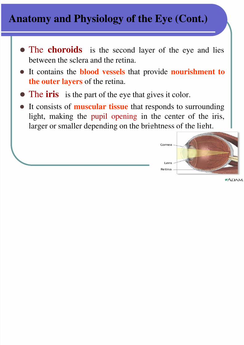
Anatomy and Physiology of the Eye (Cont.)
The choroids is the second layer of the eye and lies
between the sclera and the retina.
It contains the blood vessels that provide nourishment to
the outer layers of the retina. The iris is the part of the eye that gives it color.
It consists of muscular tissue that responds to surrounding
light, making the pupil opening in the center of the iris,
larger or smaller depending on the brightness of the light.
7/30/2019 Opthalmic Preparations
http://slidepdf.com/reader/full/opthalmic-preparations 8/103
Anatomy and Physiology of the Eye (Cont.):
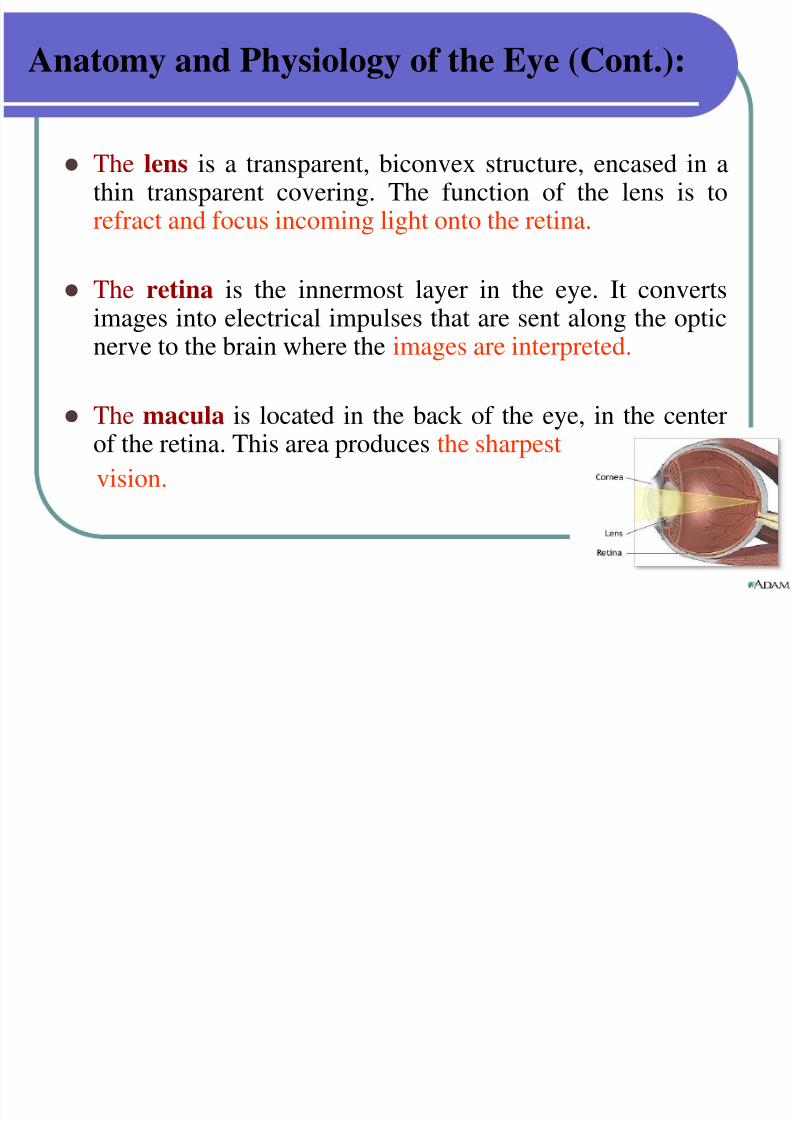
The lens is a transparent, biconvex structure, encased in athin transparent covering. The function of the lens is torefract and focus incoming light onto the retina.
The retina is the innermost layer in the eye. It convertsimages into electrical impulses that are sent along the opticnerve to the brain where the images are interpreted.
The macula is located in the back of the eye, in the centerof the retina. This area produces the sharpest
vision.
7/30/2019 Opthalmic Preparations
http://slidepdf.com/reader/full/opthalmic-preparations 9/103
Anatomy and Physiology of the Eye (Cont.):
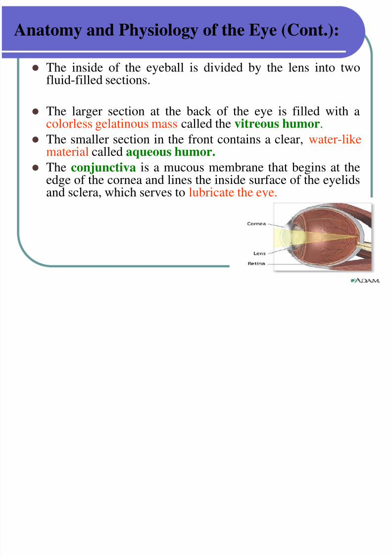
The inside of the eyeball is divided by the lens into twofluid-filled sections.
The larger section at the back of the eye is filled with acolorless gelatinous mass called the vitreous humor.
The smaller section in the front contains a clear, water-like material called aqueous humor.
The conjunctiva is a mucous membrane that begins at theedge of the cornea and lines the inside surface of the eyelidsand sclera, which serves to lubricate the eye.
7/30/2019 Opthalmic Preparations
http://slidepdf.com/reader/full/opthalmic-preparations 10/103
Absorption of drugs in the eye:
Factors affecting drug availability:
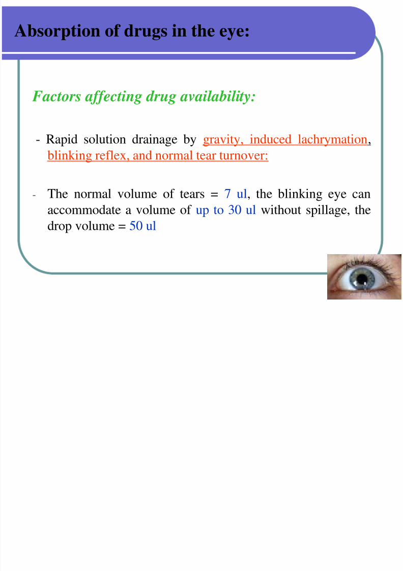
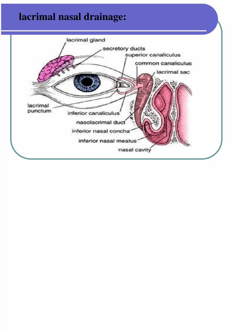
- Rapid solution drainage by gravity, induced lachrymation,blinking reflex, and normal tear turnover:
- The normal volume of tears = 7 ul, the blinking eye can
accommodate a volume of up to 30 ul without spillage, thedrop volume = 50 ul
7/30/2019 Opthalmic Preparations
http://slidepdf.com/reader/full/opthalmic-preparations 11/103
lacrimal nasal drainage:
7/30/2019 Opthalmic Preparations
http://slidepdf.com/reader/full/opthalmic-preparations 12/103
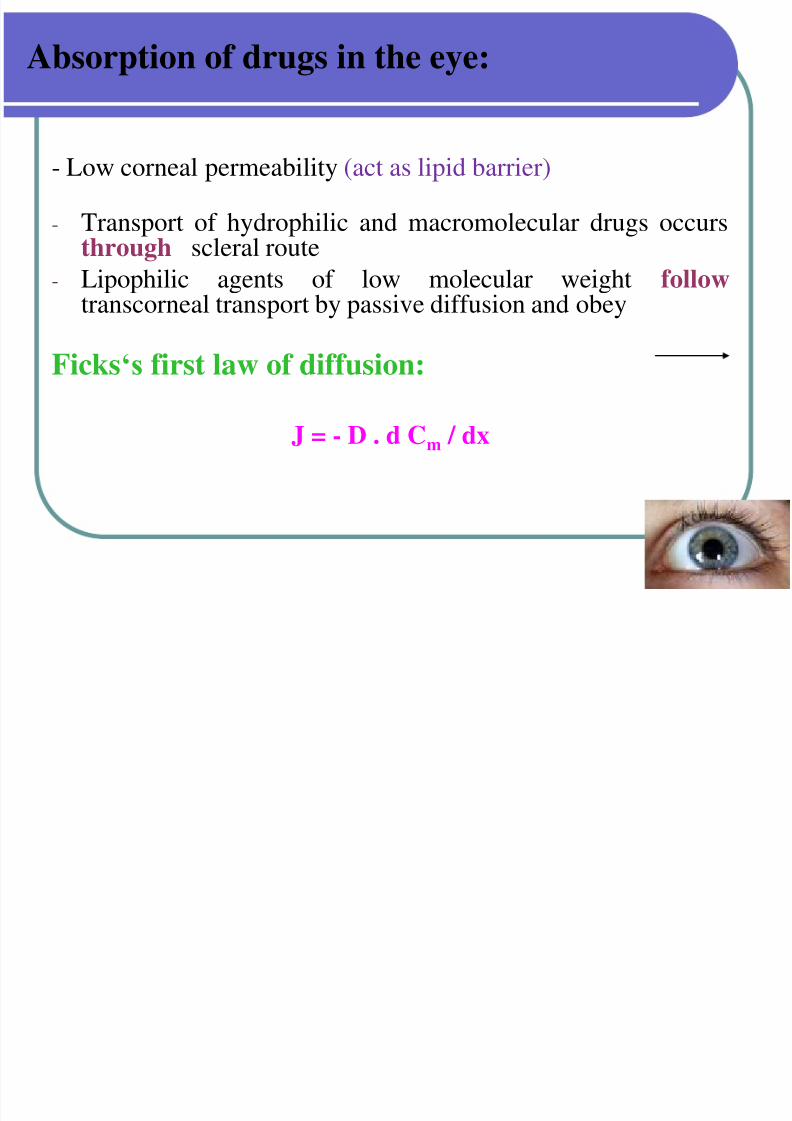
Absorption of drugs in the eye:
- Low corneal permeability (act as lipid barrier)
- Transport of hydrophilic and macromolecular drugs occursthrough scleral route
- Lipophilic agents of low molecular weight follow transcorneal transport by passive diffusion and obey
Ficks‘s first law of diffusion:
J = - D . d Cm / dx
7/30/2019 Opthalmic Preparations
http://slidepdf.com/reader/full/opthalmic-preparations 13/103
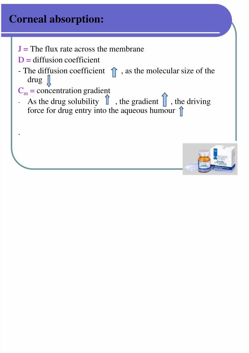
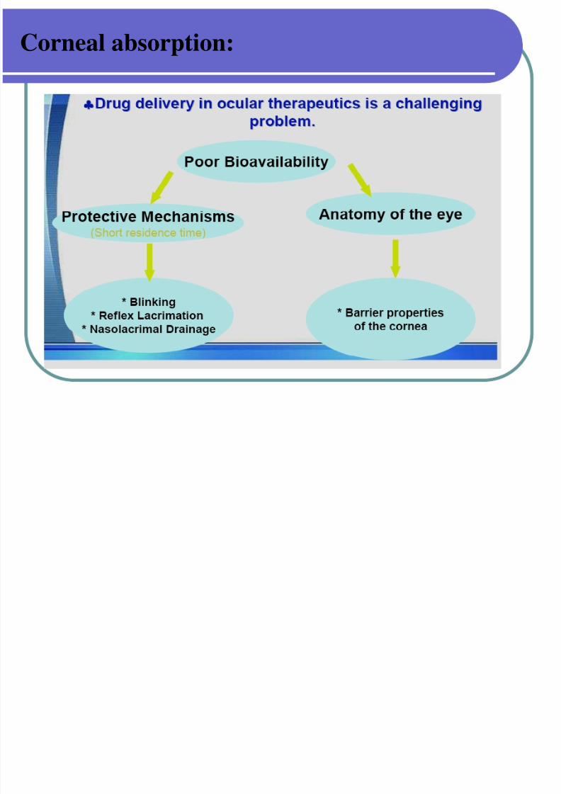
Corneal absorption:
J = The flux rate across the membrane
D = diffusion coefficient
- The diffusion coefficient , as the molecular size of thedrug
Cm = concentration gradient
- As the drug solubility , the gradient , the drivingforce for drug entry into the aqueous humour
.
7/30/2019 Opthalmic Preparations
http://slidepdf.com/reader/full/opthalmic-preparations 14/103
Corneal absorption:
7/30/2019 Opthalmic Preparations
http://slidepdf.com/reader/full/opthalmic-preparations 15/103
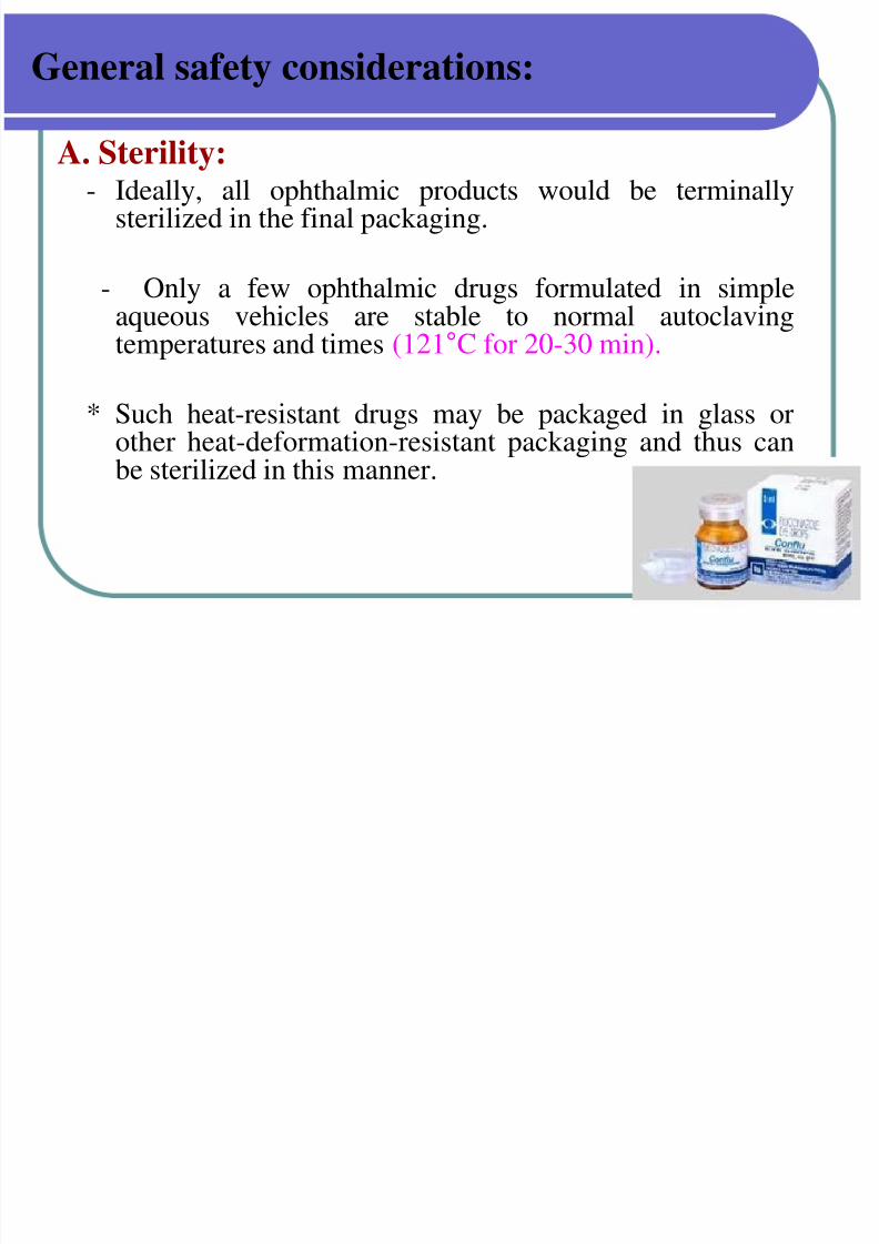
General safety considerations:
A. Sterility:- Ideally, all ophthalmic products would be terminally
sterilized in the final packaging.
- Only a few ophthalmic drugs formulated in simpleaqueous vehicles are stable to normal autoclavingtemperatures and times (121°C for 20-30 min).
* Such heat-resistant drugs may be packaged in glass orother heat-deformation-resistant packaging and thus canbe sterilized in this manner.
7/30/2019 Opthalmic Preparations
http://slidepdf.com/reader/full/opthalmic-preparations 16/103
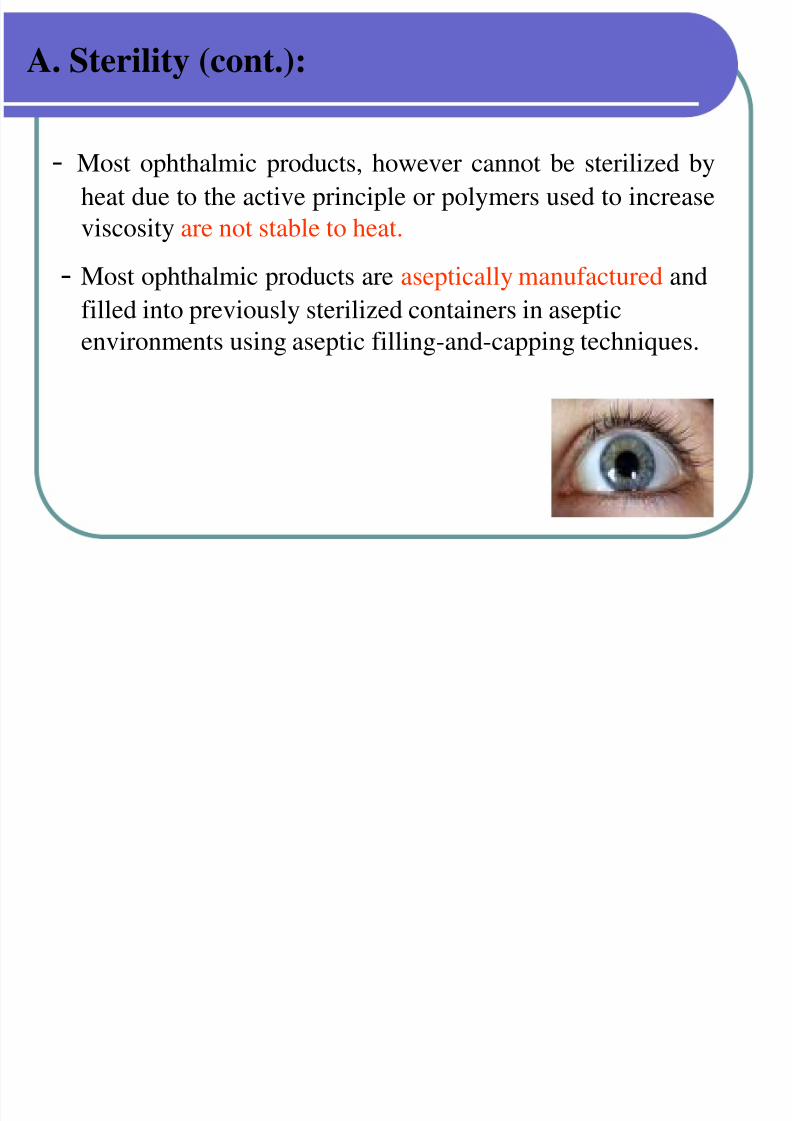
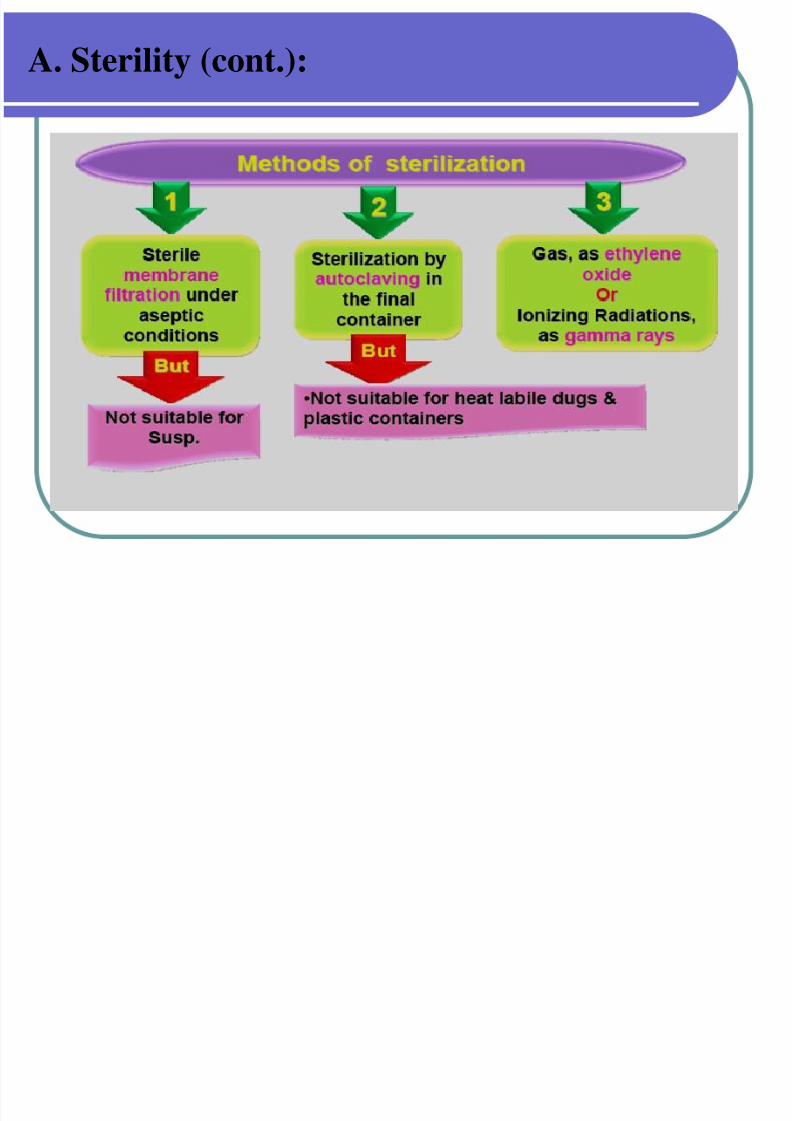
A. Sterility (cont.):
- Most ophthalmic products, however cannot be sterilized by
heat due to the active principle or polymers used to increase
viscosity are not stable to heat.
- Most ophthalmic products are aseptically manufactured andfilled into previously sterilized containers in aseptic
environments using aseptic filling-and-capping techniques.
7/30/2019 Opthalmic Preparations
http://slidepdf.com/reader/full/opthalmic-preparations 17/103
A. Sterility (cont.):
7/30/2019 Opthalmic Preparations
http://slidepdf.com/reader/full/opthalmic-preparations 18/103
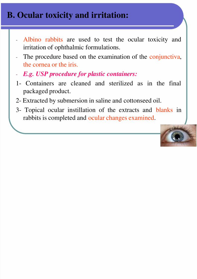
B. Ocular toxicity and irritation:
- Albino rabbits are used to test the ocular toxicity and
irritation of ophthalmic formulations.
- The procedure based on the examination of the conjunctiva,
the cornea or the iris.
- E.g. USP procedure for plastic containers:
1- Containers are cleaned and sterilized as in the final
packaged product.
2- Extracted by submersion in saline and cottonseed oil.3- Topical ocular instillation of the extracts and blanks in
rabbits is completed and ocular changes examined.
7/30/2019 Opthalmic Preparations
http://slidepdf.com/reader/full/opthalmic-preparations 19/103
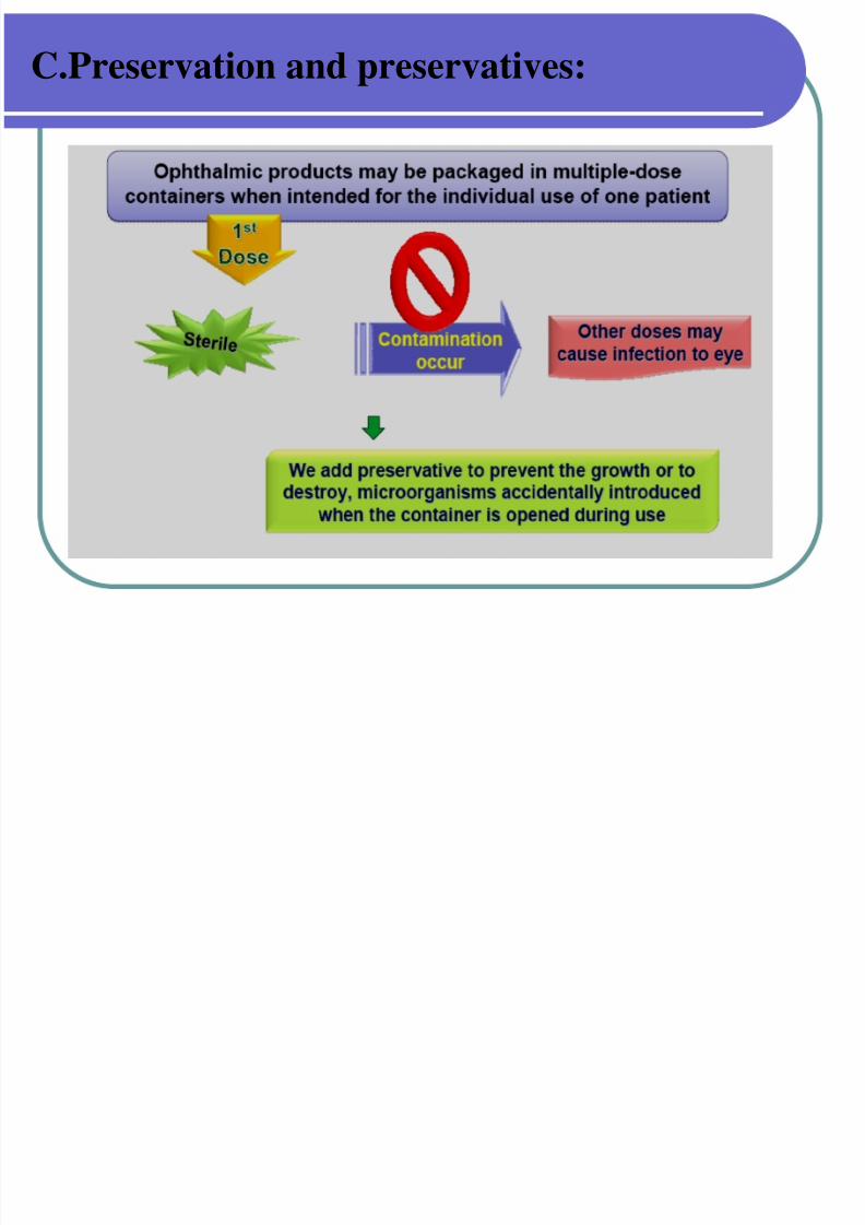
C.Preservation and preservatives:
Preservatives are included in multiple-dose eye solutions formaintaining the product sterility during use.
Preservatives not included in unit-dose package.
The use of preservatives is prohibited in ophthalmic
products that are used at the of eye surgery because, if sufficient concentration of the preservative is contacted withthe corneal endothelium, the cells can become damagedcausing clouding of the cornea and possible loss of vision.
So these products should be packaged in sterile, unit-of-use containers.
The most common organism is Pseudomonas aeruginosathat grow in the cornea and cause loss of vision.
7/30/2019 Opthalmic Preparations
http://slidepdf.com/reader/full/opthalmic-preparations 20/103
C.Preservation and preservatives:
7/30/2019 Opthalmic Preparations
http://slidepdf.com/reader/full/opthalmic-preparations 21/103
C.Preservation and preservatives:
Examples of preservatives:
1- Cationic wetting agents:
• Benzalkonium chloride (0.01%)
• It is generally used in combination with 0.01-0.1% disodium
edetate (EDTA). The chelating, EDTA has the ability to
render the resistant strains of PS aeruginosa more sensitive
to benzalkonium chloride.
2- Organic mercurials:
• Phenylmercuric nitrate 0.002-0.004%
phenylmercuric acetate 0.005-0.02%.
7/30/2019 Opthalmic Preparations
http://slidepdf.com/reader/full/opthalmic-preparations 22/103
C.Preservation and preservatives:
3-Esters of p-hydroxybenzoic acid:
• Mixture of 0.1% of both methyl and propyl hydroxybenzoate
(2 :1)
4- Alcohol Substitutes:
• Chlorobutanol(0.5%). Effective only at pH 5-6.• Phenylethanol (0.5%)
7/30/2019 Opthalmic Preparations
http://slidepdf.com/reader/full/opthalmic-preparations 23/103
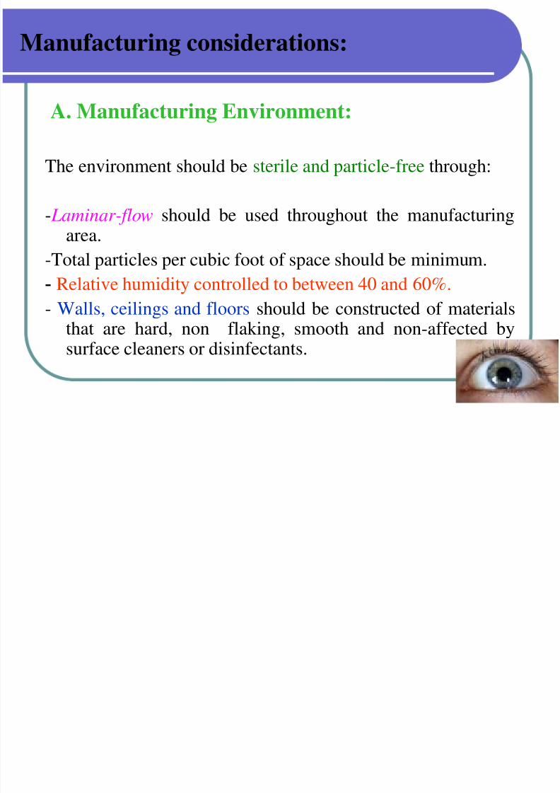
Manufacturing considerations:
A. Manufacturing Environment:
The environment should be sterile and particle-free through:
- Laminar-flow should be used throughout the manufacturingarea.
-Total particles per cubic foot of space should be minimum.
- Relative humidity controlled to between 40 and 60%.
- Walls, ceilings and floors should be constructed of materialsthat are hard, non flaking, smooth and non-affected bysurface cleaners or disinfectants.
7/30/2019 Opthalmic Preparations
http://slidepdf.com/reader/full/opthalmic-preparations 24/103
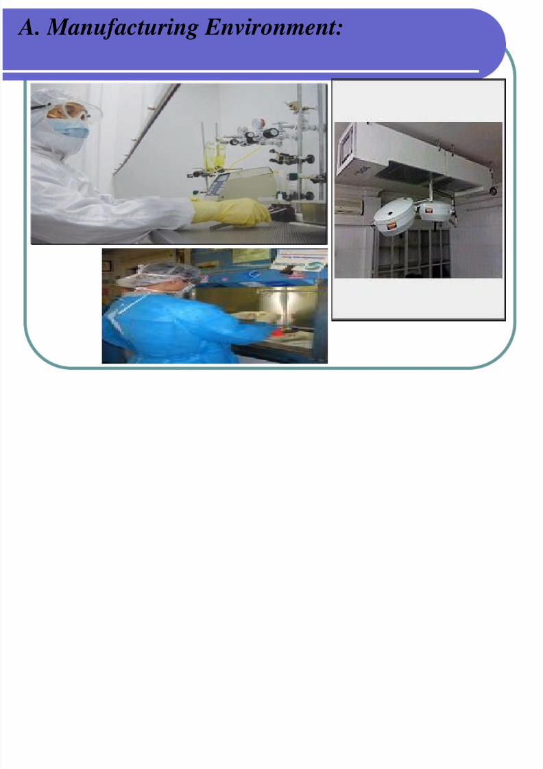
A. Manufacturing Environment:
7/30/2019 Opthalmic Preparations
http://slidepdf.com/reader/full/opthalmic-preparations 25/103
A. Manufacturing Environment:
- Ultraviolet lamps provided in flush-mounted fixtures to
maintain surface disinfection
- Separate entrance for personnel and equipment should be
provided through specially designed air locks that are
maintained at negative pressure relative to the aseptic
manufacturing area and at a positive pressure relative to the
noncontrolled area

7/30/2019 Opthalmic Preparations
http://slidepdf.com/reader/full/opthalmic-preparations 26/103
A. Manufacturing Environment:
7/30/2019 Opthalmic Preparations
http://slidepdf.com/reader/full/opthalmic-preparations 27/103
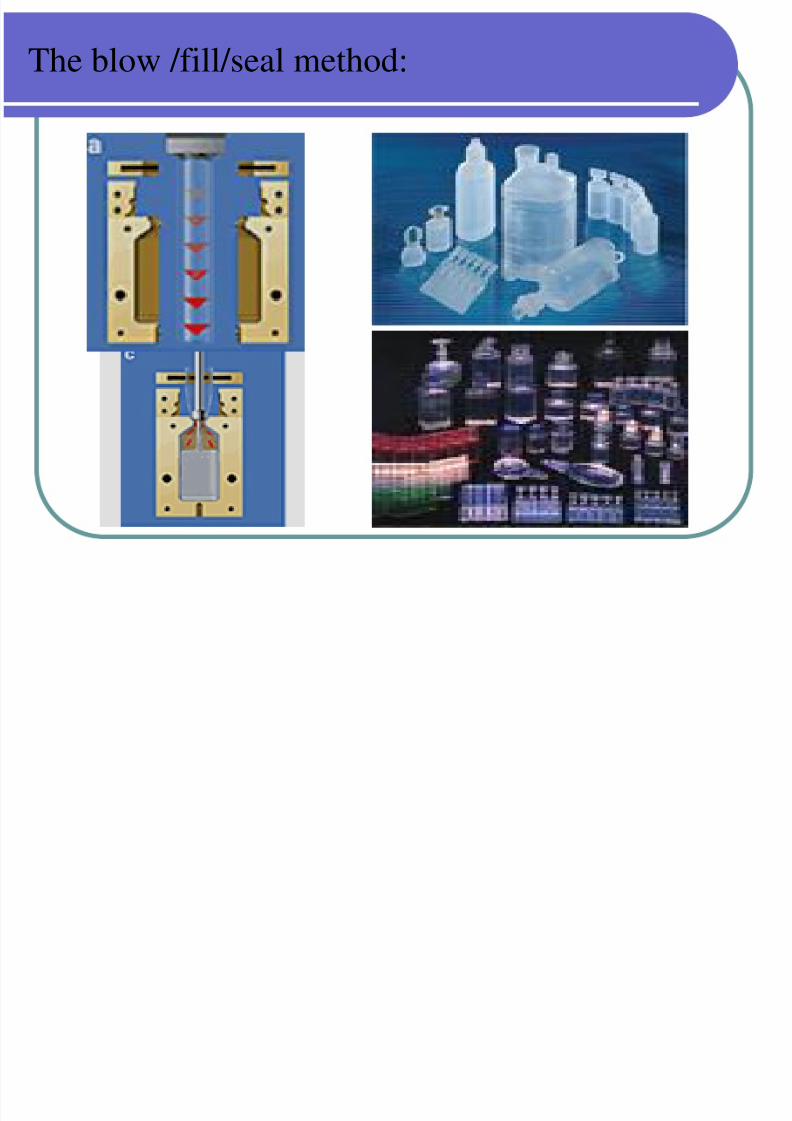
. Manufacturing Techniques:
Unpreserved formulations of active drug (s):
The blow/fill/seal method
It is used for manufacture of unpreserved ophthalmic
products , especially for artificial tear products.In this first step is : To extrude polyethylene resin at high
temperature and pressure and to form the container by
blowing the polyethylene resin into mold with compressed
air. The product is vented out, and finally the container issealed on the top.
7/30/2019 Opthalmic Preparations
http://slidepdf.com/reader/full/opthalmic-preparations 28/103
The blow /fill/seal method:
7/30/2019 Opthalmic Preparations
http://slidepdf.com/reader/full/opthalmic-preparations 29/103
C. Equipment:
All tanks, valves, pumps and piping must be of best available
Grade of corrosion – resistant stainless steel.
All products-contact surface should be polished either
mechanically or be electropolishing to provide a surface as
Free as possible from scratches or defects.
Care should be taken in the design of such equipment to
Provide adequate means of cleaning and sanitization.
7/30/2019 Opthalmic Preparations
http://slidepdf.com/reader/full/opthalmic-preparations 30/103
Ideal ophthalmic delivery system: Following characteristics are required to optimize
ocular drug delivery system:
Good corneal penetration. Prolong contact time with corneal tissue.
Simplicity of instillation for the patient.
Non irritative and comfortable form
Appropriate rheological properties
7/30/2019 Opthalmic Preparations
http://slidepdf.com/reader/full/opthalmic-preparations 31/103
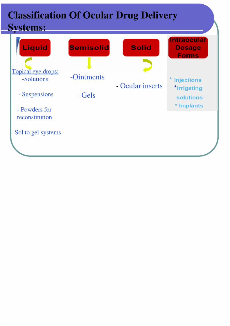
Classification Of Ocular Drug Delivery
Systems:
Topical eye drops:
-Solutions
- Suspensions
- Powders for
reconstitution
- Sol to gel systems
-Ointments
- Gels
- Ocular inserts
7/30/2019 Opthalmic Preparations
http://slidepdf.com/reader/full/opthalmic-preparations 32/103
7/30/2019 Opthalmic Preparations
http://slidepdf.com/reader/full/opthalmic-preparations 33/103
A. Topical Eye drops:
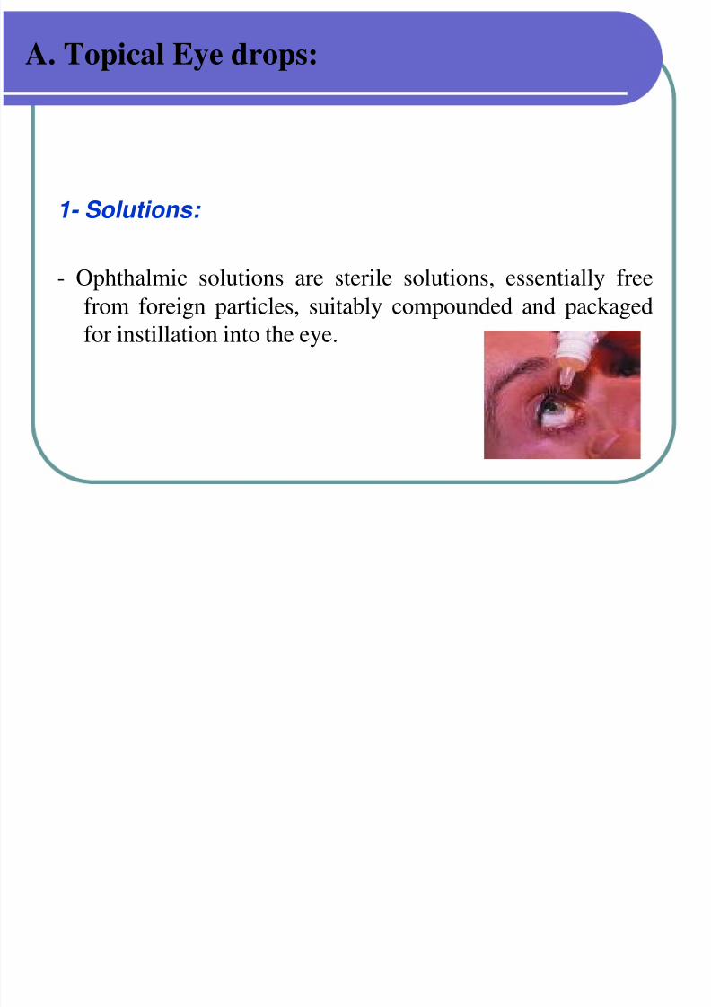
1- Solutions:
- Ophthalmic solutions are sterile solutions, essentially free
from foreign particles, suitably compounded and packaged
for instillation into the eye.
7/30/2019 Opthalmic Preparations
http://slidepdf.com/reader/full/opthalmic-preparations 34/103
A. Topical Eye drops:
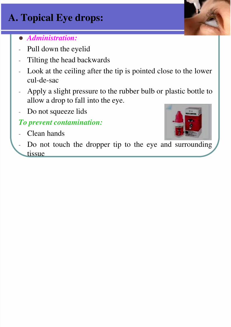
Administration:- Pull down the eyelid
- Tilting the head backwards
- Look at the ceiling after the tip is pointed close to the lower
cul-de-sac- Apply a slight pressure to the rubber bulb or plastic bottle to
allow a drop to fall into the eye.
- Do not squeeze lids
To prevent contamination:- Clean hands
- Do not touch the dropper tip to the eye and surrounding
tissue
7/30/2019 Opthalmic Preparations
http://slidepdf.com/reader/full/opthalmic-preparations 35/103
1- Solutions:
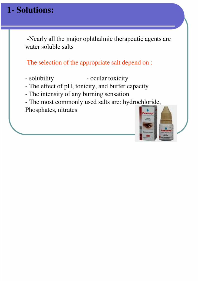
-Nearly all the major ophthalmic therapeutic agents are
water soluble salts
The selection of the appropriate salt depend on :
- solubility - ocular toxicity
- The effect of pH, tonicity, and buffer capacity
- The intensity of any burning sensation
- The most commonly used salts are: hydrochloride,Phosphates, nitrates
7/30/2019 Opthalmic Preparations
http://slidepdf.com/reader/full/opthalmic-preparations 36/103
Examples of topical eye drops:
• Atropine sulphate eye drops.
• Pilocarpine eye drops .
• Silver nitrate eye drops.
• Zinc sulphate eye drops.
7/30/2019 Opthalmic Preparations
http://slidepdf.com/reader/full/opthalmic-preparations 37/103
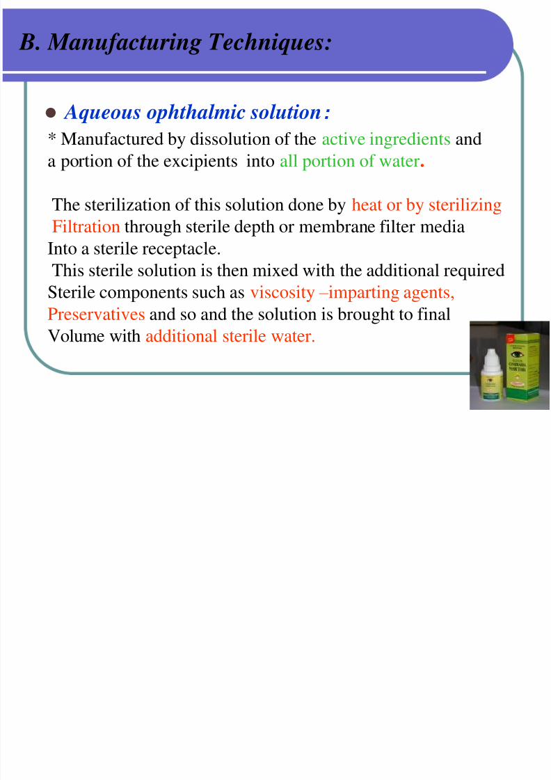
B. Manufacturing Techniques:
Aqueous ophthalmic solution:
* Manufactured by dissolution of the active ingredients and
a portion of the excipients into all portion of water.
The sterilization of this solution done by heat or by sterilizing
Filtration through sterile depth or membrane filter media
Into a sterile receptacle.
This sterile solution is then mixed with the additional required
Sterile components such as viscosity – imparting agents,Preservatives and so and the solution is brought to final
Volume with additional sterile water.
7/30/2019 Opthalmic Preparations
http://slidepdf.com/reader/full/opthalmic-preparations 38/103
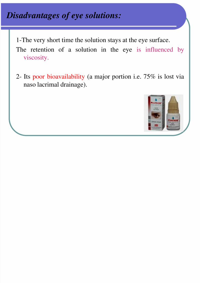
Disadvantages of eye solutions:
1-The very short time the solution stays at the eye surface.
The retention of a solution in the eye is influenced by
viscosity.
2- Its poor bioavailability (a major portion i.e. 75% is lost via
naso lacrimal drainage).
7/30/2019 Opthalmic Preparations
http://slidepdf.com/reader/full/opthalmic-preparations 39/103
2- suspensions:
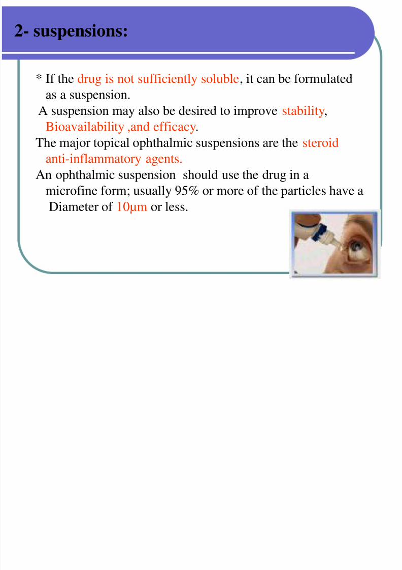
* If the drug is not sufficiently soluble, it can be formulated
as a suspension.
A suspension may also be desired to improve stability,
Bioavailability ,and efficacy.
The major topical ophthalmic suspensions are the steroidanti-inflammatory agents.
An ophthalmic suspension should use the drug in a
microfine form; usually 95% or more of the particles have a
Diameter of 10µm or less.

7/30/2019 Opthalmic Preparations
http://slidepdf.com/reader/full/opthalmic-preparations 40/103
Examples :
Prednisolone acetate suspension.
Besifloxacin suspension.
Blephamide suspension.
Fluorometholone .
7/30/2019 Opthalmic Preparations
http://slidepdf.com/reader/full/opthalmic-preparations 41/103
B. Manufacturing Techniques:
Aqueous suspensions:
Are prepared in much the same manner, except that
Before bringing to the final volume with additional
sterile water .The solid that is to be suspended is previously rendered sterile
by – heat ,exposure to ethylene oxide ,ionizing radiation
(gamma ), sterile filtration.
The particle size should be monitored.
7/30/2019 Opthalmic Preparations
http://slidepdf.com/reader/full/opthalmic-preparations 42/103
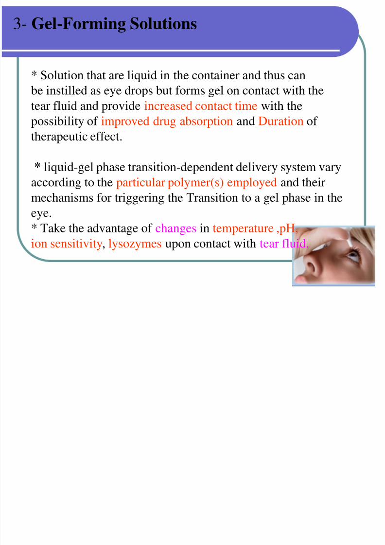
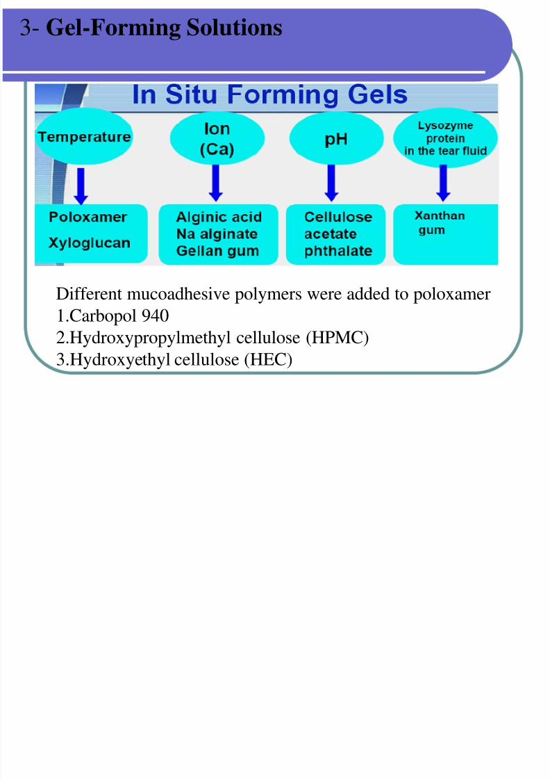
3- Gel-Forming Solutions
* Solution that are liquid in the container and thus can
be instilled as eye drops but forms gel on contact with the
tear fluid and provide increased contact time with the
possibility of improved drug absorption and Duration of
therapeutic effect.
* liquid-gel phase transition-dependent delivery system vary
according to the particular polymer(s) employed and their
mechanisms for triggering the Transition to a gel phase in the
eye.
* Take the advantage of changes in temperature ,pH,
ion sensitivity, lysozymes upon contact with tear fluid.
3 G l F i S l ti
7/30/2019 Opthalmic Preparations
http://slidepdf.com/reader/full/opthalmic-preparations 43/103
3- Gel-Forming Solutions
Different mucoadhesive polymers were added to poloxamer1.Carbopol 940
2.Hydroxypropylmethyl cellulose (HPMC)
3.Hydroxyethyl cellulose (HEC)
7/30/2019 Opthalmic Preparations
http://slidepdf.com/reader/full/opthalmic-preparations 44/103
Inactive Ingredients in Topical Drops:
The inactive ingredients in ophthalmic solution and
Suspension dosage forms are necessary to perform one or
more of the Following functions:
Adjust concentration and tonicity
1. Buffer and adjust pH,
2. Stabilize the active ingredients against decomposition ,
3. Increase solubility,4. Impart viscosity
5. And act as solvent.
7/30/2019 Opthalmic Preparations
http://slidepdf.com/reader/full/opthalmic-preparations 45/103
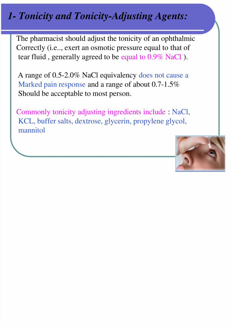
1- Tonicity and Tonicity-Adjusting Agents:
The pharmacist should adjust the tonicity of an ophthalmicCorrectly (i.e.., exert an osmotic pressure equal to that of
tear fluid , generally agreed to be equal to 0.9% NaCl ).
A range of 0.5-2.0% NaCl equivalency does not cause a
Marked pain response and a range of about 0.7-1.5%Should be acceptable to most person.
Commonly tonicity adjusting ingredients include : NaCl,
KCL, buffer salts, dextrose, glycerin, propylene glycol,mannitol
I t i it
7/30/2019 Opthalmic Preparations
http://slidepdf.com/reader/full/opthalmic-preparations 46/103
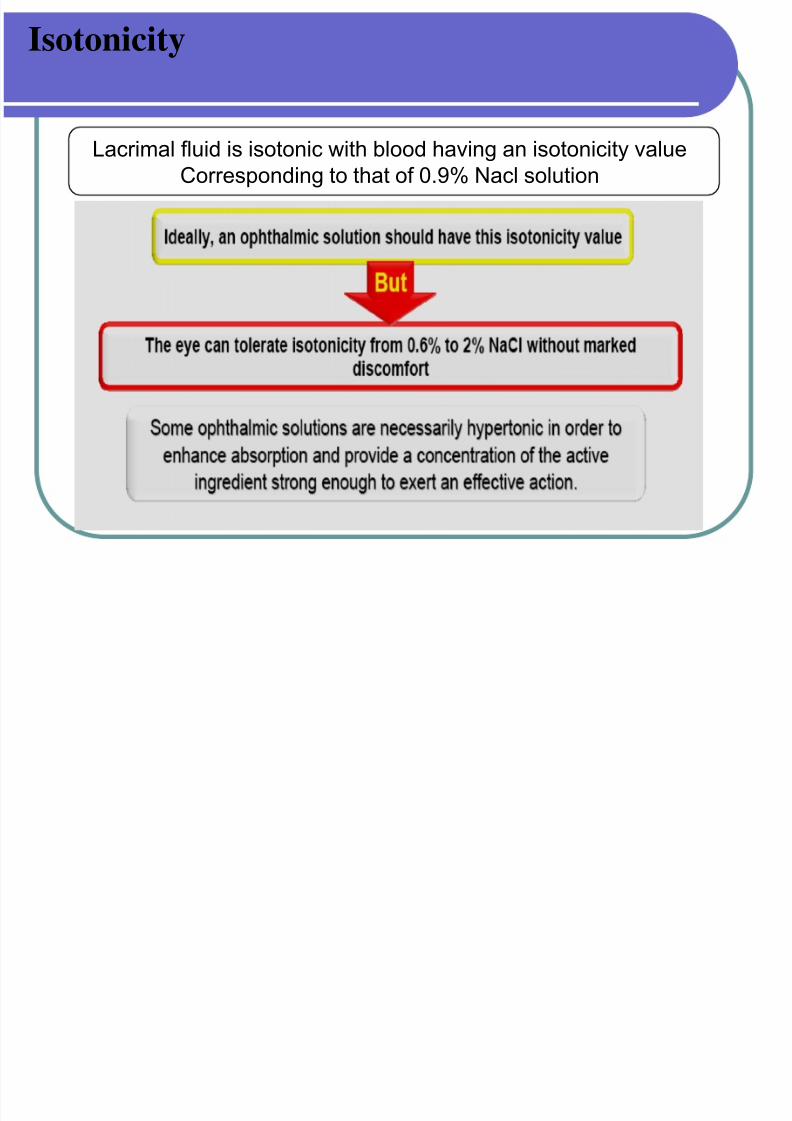
Isotonicity
Lacrimal fluid is isotonic with blood having an isotonicity valueCorresponding to that of 0.9% Nacl solution
7/30/2019 Opthalmic Preparations
http://slidepdf.com/reader/full/opthalmic-preparations 47/103
2- pH Adjustment and Buffers:
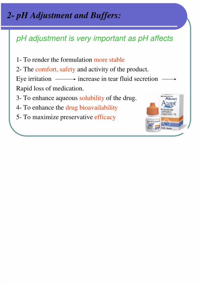
pH adjustment is very important as pH affects
1- To render the formulation more stable
2- The comfort, safety and activity of the product.Eye irritation increase in tear fluid secretion
Rapid loss of medication.
3- To enhance aqueous solubility of the drug.
4- To enhance the drug bioavailability
5- To maximize preservative efficacy
2 H Adj t t d B ff
7/30/2019 Opthalmic Preparations
http://slidepdf.com/reader/full/opthalmic-preparations 48/103
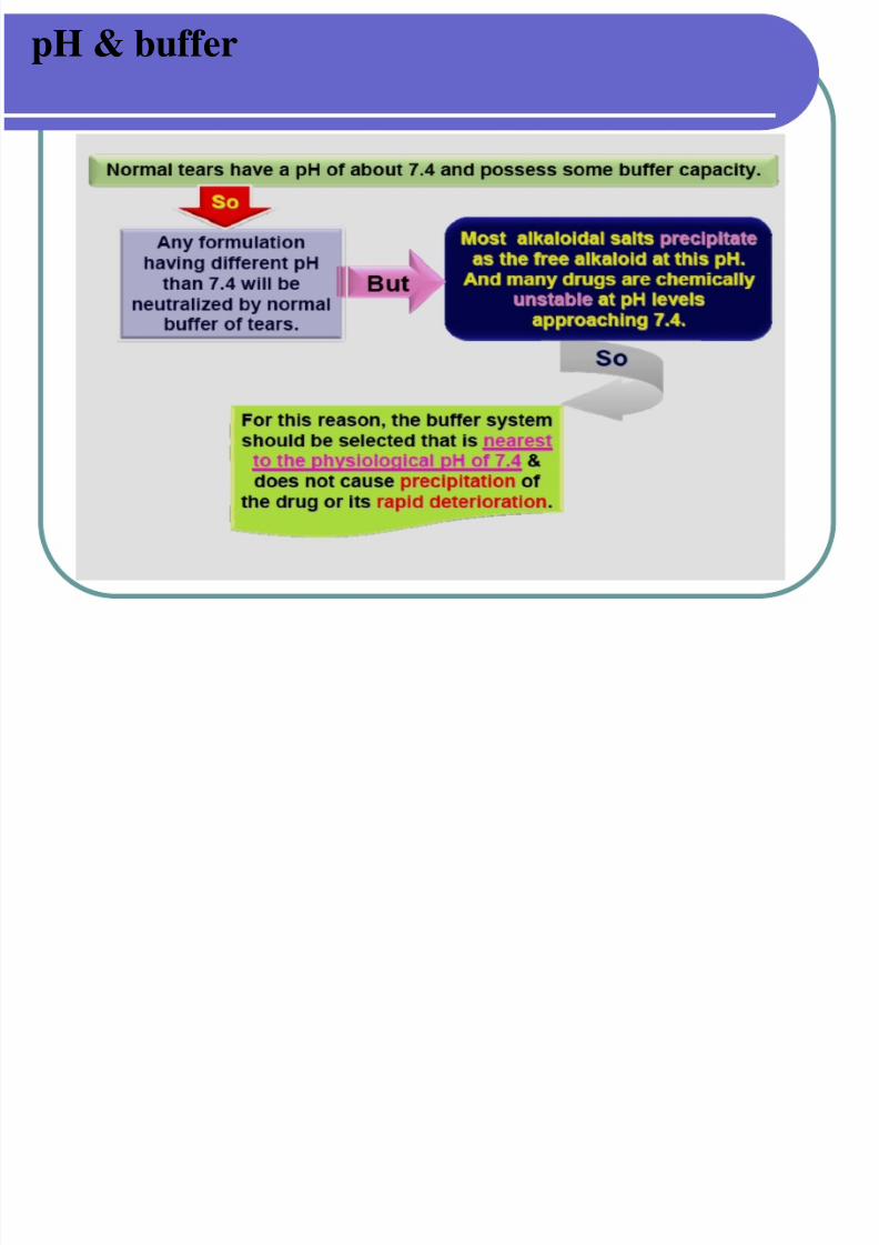
2- pH Adjustment and Buffers:
Ideally , every product would be buffered to a pH of 7.4(the normal physiological pH of tear fluid ).
When necessary they are buffered adequately to maintain
Stability within this range for at least 2 years.
If buffers are required there capacity is controlled to be
As low as possible( low buffer capacity) thus enabling the
Tear to bring the pH of the eye back to the physiological
range .
pH & b ffer
7/30/2019 Opthalmic Preparations
http://slidepdf.com/reader/full/opthalmic-preparations 49/103
pH & buffer
7/30/2019 Opthalmic Preparations
http://slidepdf.com/reader/full/opthalmic-preparations 50/103
3- Stabilizers & Antioxidants:
* Stabilizers are ingredients added to a formula to decrease the rate of decomposition of the active ingredients.
* Antioxidants are the principle stabilizers added to some
ophthalmic solutions , primarily those containing epinephrineand other oxidizable drugs.
* Sodium bisulfite or metabisulfite are used in concentration
up to 0.3% in epinephrine hydrochloride and bitartrate solutions.
The several antioxidant system have been developed :-
These consists of ascorbic acid and acetylcysteine and
sodium thiosulfate .
4 Surfactants:
7/30/2019 Opthalmic Preparations
http://slidepdf.com/reader/full/opthalmic-preparations 51/103
4- Surfactants:
The order of surfactant toxicity is :
anionic > cationic >> nonionic .
• several nonionic surfactants are used in relatively low
Concentration to aid in dispersing steroids in suspensions
and to achieve or to improve solution clarity.
• Those principally used are the sorbitan ether esters of
oleic acid ( polysorbate or tween 20 and 80 ).
7/30/2019 Opthalmic Preparations
http://slidepdf.com/reader/full/opthalmic-preparations 52/103
5- Viscosity-Imparting Agents:
Polyvinyl alcohol, methylcellulose, hydroxypropyl
methylcellulose, hydroxyethylcellulose, and carbomers,
are commonly used to increase the viscosity of solution
and suspensions (to retard the rate of setting of particles)
They increase the ocular contact time , there by decreasing
the drainage rate, increase the mucoadhesiveness and Increasingthe bioavailability .
Disadvantage : produce blurring vision as when dry, form a dry
film on the eye lids. make filteration more difficult .commercial viscous vehicles are :
1. polyvinyl alcohol (liquifilm)
2. hydroxypropyl methylcellulose (isopto )
6 V hi l
7/30/2019 Opthalmic Preparations
http://slidepdf.com/reader/full/opthalmic-preparations 53/103
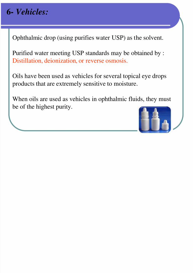
6- Vehicles:
Ophthalmic drop (using purifies water USP) as the solvent.
Purified water meeting USP standards may be obtained by :
Distillation, deionization, or reverse osmosis.
Oils have been used as vehicles for several topical eye drops
products that are extremely sensitive to moisture.
When oils are used as vehicles in ophthalmic fluids, they must
be of the highest purity.
7/30/2019 Opthalmic Preparations
http://slidepdf.com/reader/full/opthalmic-preparations 54/103
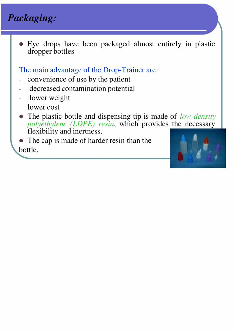
Packaging:
Eye drops have been packaged almost entirely in plasticdropper bottles
The main advantage of the Drop-Trainer are:
- convenience of use by the patient
- decreased contamination potential
- lower weight
- lower cost
The plastic bottle and dispensing tip is made of low-density
polyethylene (LDPE) resin, which provides the necessaryflexibility and inertness.
The cap is made of harder resin than the
bottle.
7/30/2019 Opthalmic Preparations
http://slidepdf.com/reader/full/opthalmic-preparations 55/103
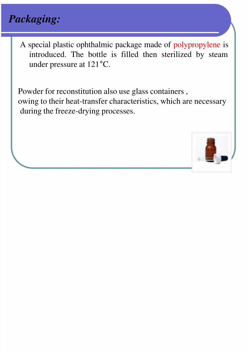
Packaging:
A special plastic ophthalmic package made of polypropylene isintroduced. The bottle is filled then sterilized by steam
under pressure at 121°C.
Powder for reconstitution also use glass containers ,owing to their heat-transfer characteristics, which are necessary
during the freeze-drying processes.
7/30/2019 Opthalmic Preparations
http://slidepdf.com/reader/full/opthalmic-preparations 56/103
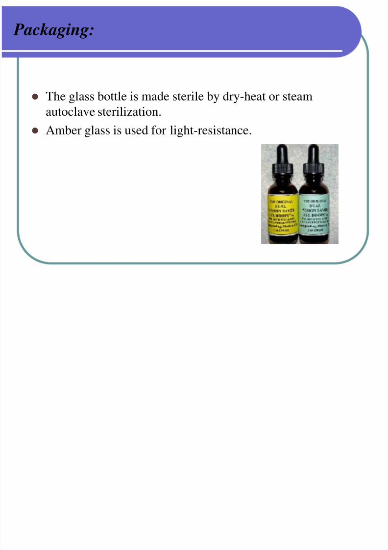
Packaging:
The glass bottle is made sterile by dry-heat or steam
autoclave sterilization.
Amber glass is used for light-resistance.
B S i lid D F O hth l i
7/30/2019 Opthalmic Preparations
http://slidepdf.com/reader/full/opthalmic-preparations 57/103
B. Semisolid Dosage Forms: Ophthalmic
Ointments and Gels:
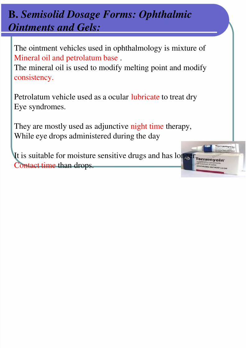
The ointment vehicles used in ophthalmology is mixture of Mineral oil and petrolatum base .
The mineral oil is used to modify melting point and modify
consistency.
Petrolatum vehicle used as a ocular lubricate to treat dry
Eye syndromes.
They are mostly used as adjunctive night time therapy,
While eye drops administered during the day
It is suitable for moisture sensitive drugs and has longer
Contact time than drops.
B. Semisolid Dosage Forms: Ophthalmic
7/30/2019 Opthalmic Preparations
http://slidepdf.com/reader/full/opthalmic-preparations 58/103
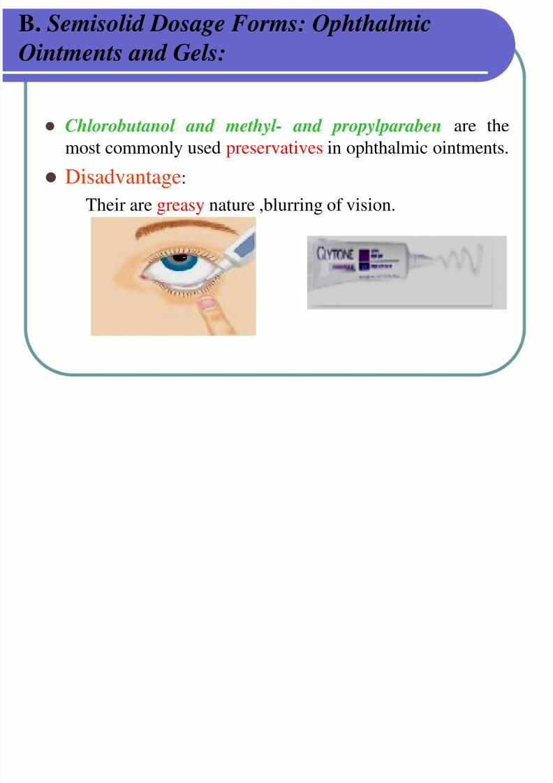
Chlorobutanol and methyl- and propylparaben are the
most commonly used preservatives in ophthalmic ointments.
Disadvantage:
Their are greasy nature ,blurring of vision.
B. Semisolid Dosage Forms: Ophthalmic
Ointments and Gels:
Manufacturing Techniques:
7/30/2019 Opthalmic Preparations
http://slidepdf.com/reader/full/opthalmic-preparations 59/103
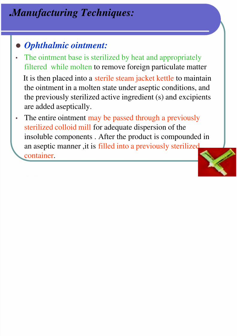
. Manufacturing Techniques:
Ophthalmic ointment: • The ointment base is sterilized by heat and appropriately
filtered while molten to remove foreign particulate matter
It is then placed into a sterile steam jacket kettle to maintain
the ointment in a molten state under aseptic conditions, andthe previously sterilized active ingredient (s) and excipients
are added aseptically.
• The entire ointment may be passed through a previously
sterilized colloid mill for adequate dispersion of theinsoluble components . After the product is compounded in
an aseptic manner ,it is filled into a previously sterilized
container.

7/30/2019 Opthalmic Preparations
http://slidepdf.com/reader/full/opthalmic-preparations 60/103
Examples :
Chloramphenicol ointment.
Tetracycline ointment.
Hydrocortisone ointment.
B Semisolid Dosage Forms: Ophthalmic
7/30/2019 Opthalmic Preparations
http://slidepdf.com/reader/full/opthalmic-preparations 61/103
B. Semisolid Dosage Forms: Ophthalmic
Ointments and Gels: Packaging:
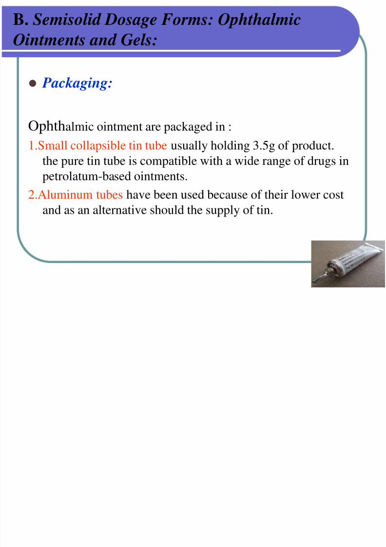
Ophthalmic ointment are packaged in :
1.Small collapsible tin tube usually holding 3.5g of product.
the pure tin tube is compatible with a wide range of drugs in
petrolatum-based ointments.
2.Aluminum tubes have been used because of their lower cost
and as an alternative should the supply of tin.
Packaging:
7/30/2019 Opthalmic Preparations
http://slidepdf.com/reader/full/opthalmic-preparations 62/103
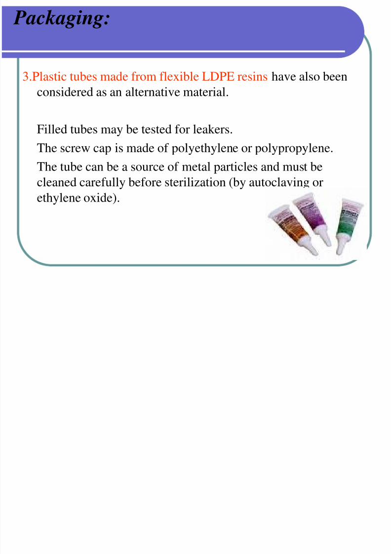
Packaging:
3.Plastic tubes made from flexible LDPE resins have also beenconsidered as an alternative material.
Filled tubes may be tested for leakers.
The screw cap is made of polyethylene or polypropylene.
The tube can be a source of metal particles and must be
cleaned carefully before sterilization (by autoclaving or
ethylene oxide).
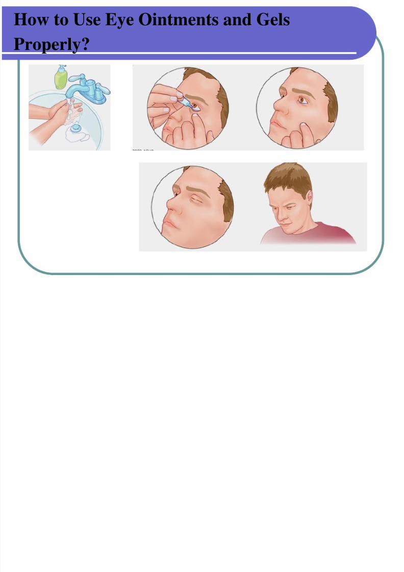
How to Use Eye Ointments and Gels
7/30/2019 Opthalmic Preparations
http://slidepdf.com/reader/full/opthalmic-preparations 63/103
How to Use Eye Ointments and Gels
Properly?
C Solid Dosage Forms
7/30/2019 Opthalmic Preparations
http://slidepdf.com/reader/full/opthalmic-preparations 64/103
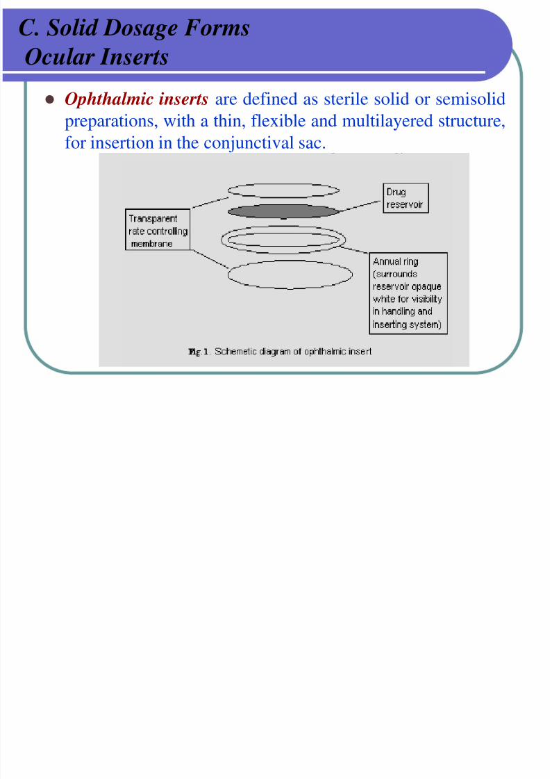
C. Solid Dosage Forms
Ocular Inserts
Ophthalmic inserts are defined as sterile solid or semisolidpreparations, with a thin, flexible and multilayered structure,
for insertion in the conjunctival sac.
C. Solid Dosage Forms
7/30/2019 Opthalmic Preparations
http://slidepdf.com/reader/full/opthalmic-preparations 65/103
g
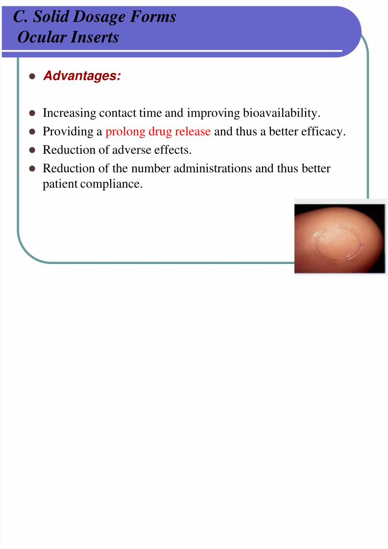
Ocular Inserts
Advantages:
Increasing contact time and improving bioavailability.
Providing a prolong drug release and thus a better efficacy.
Reduction of adverse effects.
Reduction of the number administrations and thus better
patient compliance.
C. Ocular Inserts
7/30/2019 Opthalmic Preparations
http://slidepdf.com/reader/full/opthalmic-preparations 66/103
C. Ocular Inserts
I. Insoluble inserts:
Insoluble insert is a multilayered structure consisting of a drug containing core surrounded on each side by a layer of copolymer membranes through which the drug diffuses at aconstant rate.
The rate of drug diffusion is controlled by:- The polymer composition
- The membrane thickness
- The solubility of the drug
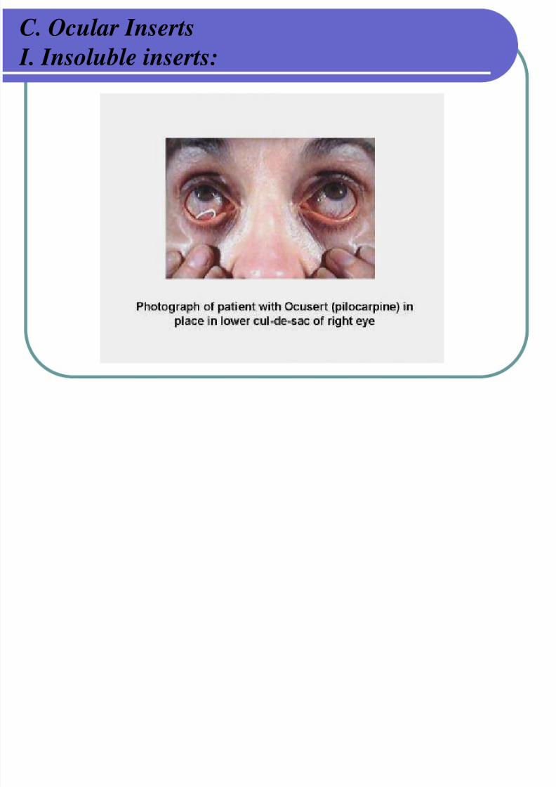
e.g. The Ocusert® Pilo-20 and Pilo-40 Ocular system
- Designed to be placed in the inferior cul-de-sac between thesclera and the eyelid and to release pilocarpine continuouslyat a steady rate for 7 days for treatment of glucoma.
C. Ocular Inserts
7/30/2019 Opthalmic Preparations
http://slidepdf.com/reader/full/opthalmic-preparations 67/103
C. Ocular Inserts
I. Insoluble inserts:
II.Soluble Ocular inserts:
7/30/2019 Opthalmic Preparations
http://slidepdf.com/reader/full/opthalmic-preparations 68/103
II.Soluble Ocular inserts:
- Soluble inserts consists of all monolytic polymeric
devices that at the end of their release, the device dissolveor erode.
Types
a) Based on natural polymers e.g. collagen.
b) Based on synthetic or semi synthetic polymers e.g. Cellulosederivatives – Hydroxypropyl cellulose, methylcellulose orPolyvinyl alcohol, ethylene vinyl acetate copolymer.
- The system soften in 10-15 sec after introduction into theupper conjunctival sac, gradually dissolves within 1h ,while releasing the drug.
- Advantage: Being entirely soluble so that they do not needto be removed from their site of application.
II.Soluble Ocular inserts:
7/30/2019 Opthalmic Preparations
http://slidepdf.com/reader/full/opthalmic-preparations 69/103
II.Soluble Ocular inserts:
Lacrisert is a sterile ophthalmic insert use in the treatment of dry
Eye syndrome and is usually recommended for patients unable
to obtain symptomatic relief with artifical tear solutions.
The insert is composed of 5 mg of Hydroxypropyl cellulosein a rod-shaped form about 1.27 mm diameter by about 3.5 mm
long.
D. Intraocular Dosage Forms
7/30/2019 Opthalmic Preparations
http://slidepdf.com/reader/full/opthalmic-preparations 70/103
D. Intraocular Dosage Forms
They are Ophthalmic products that introduced into theinterior structures of the eye primarily during ocular
surgery.
Requirements for formulation:
1- sterile and pyrogen-free
2- strict control of particulate matter
3- compatible with sensitive internal tissues
4- packaged as preservative-free single dosage
D. Intraocular Dosage Forms:
7/30/2019 Opthalmic Preparations
http://slidepdf.com/reader/full/opthalmic-preparations 71/103
g
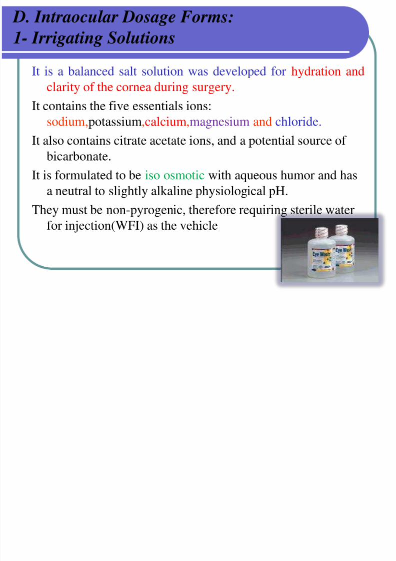
1- Irrigating Solutions
It is a balanced salt solution was developed for hydration andclarity of the cornea during surgery.
It contains the five essentials ions:
sodium,potassium,calcium,magnesium and chloride.
It also contains citrate acetate ions, and a potential source of bicarbonate.
It is formulated to be iso osmotic with aqueous humor and has
a neutral to slightly alkaline physiological pH.
They must be non-pyrogenic, therefore requiring sterile waterfor injection(WFI) as the vehicle
D. Intraocular Dosage Forms
7/30/2019 Opthalmic Preparations
http://slidepdf.com/reader/full/opthalmic-preparations 72/103
g
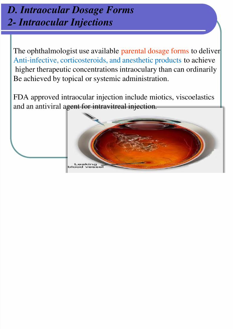
2- Intraocular Injections
The ophthalmologist use available parental dosage forms to deliver
Anti-infective, corticosteroids, and anesthetic products to achieve
higher therapeutic concentrations intraoculary than can ordinarily
Be achieved by topical or systemic administration.
FDA approved intraocular injection include miotics, viscoelastics
and an antiviral agent for intravitreal injection.
D. Intraocular Dosage Forms
7/30/2019 Opthalmic Preparations
http://slidepdf.com/reader/full/opthalmic-preparations 73/103
3- Intravitreal Implant
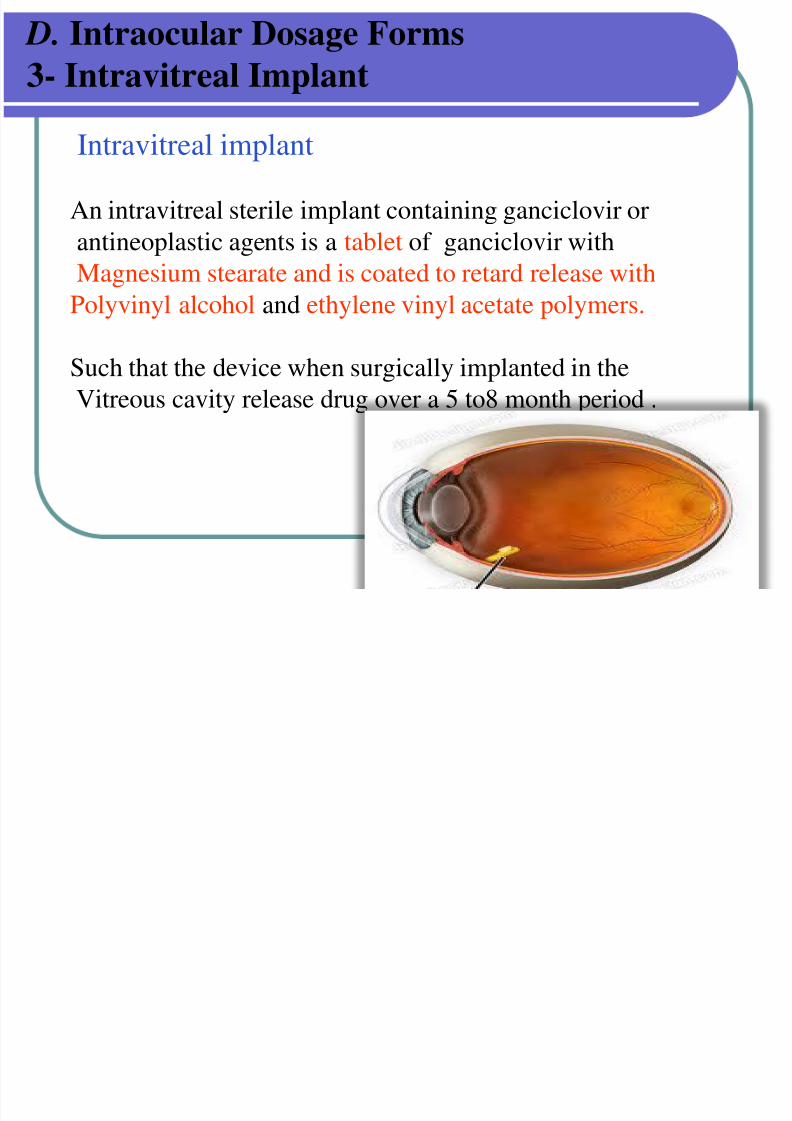
Intravitreal implant
An intravitreal sterile implant containing ganciclovir or
antineoplastic agents is a tablet of ganciclovir with
Magnesium stearate and is coated to retard release with
Polyvinyl alcohol and ethylene vinyl acetate polymers.
Such that the device when surgically implanted in the
Vitreous cavity release drug over a 5 to8 month period .
E Miscellaneous
7/30/2019 Opthalmic Preparations
http://slidepdf.com/reader/full/opthalmic-preparations 74/103
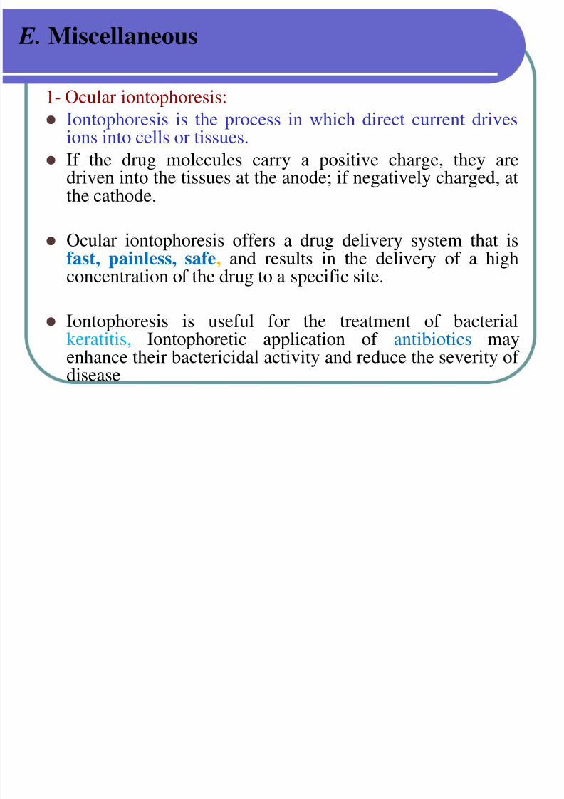
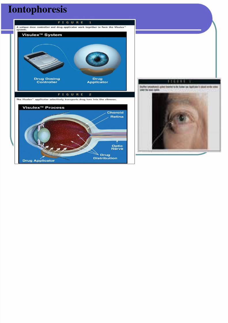
E. Miscellaneous 1- Ocular iontophoresis:
Iontophoresis is the process in which direct current drivesions into cells or tissues.
If the drug molecules carry a positive charge, they aredriven into the tissues at the anode; if negatively charged, at
the cathode.
Ocular iontophoresis offers a drug delivery system that isfast, painless, safe, and results in the delivery of a highconcentration of the drug to a specific site.
Iontophoresis is useful for the treatment of bacterialkeratitis, Iontophoretic application of antibiotics mayenhance their bactericidal activity and reduce the severity of disease
Iontophoresis
7/30/2019 Opthalmic Preparations
http://slidepdf.com/reader/full/opthalmic-preparations 75/103
E. Miscellaneous
7/30/2019 Opthalmic Preparations
http://slidepdf.com/reader/full/opthalmic-preparations 76/103
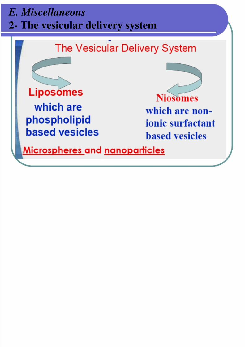
2- The vesicular delivery system
Liposomes
7/30/2019 Opthalmic Preparations
http://slidepdf.com/reader/full/opthalmic-preparations 77/103
p
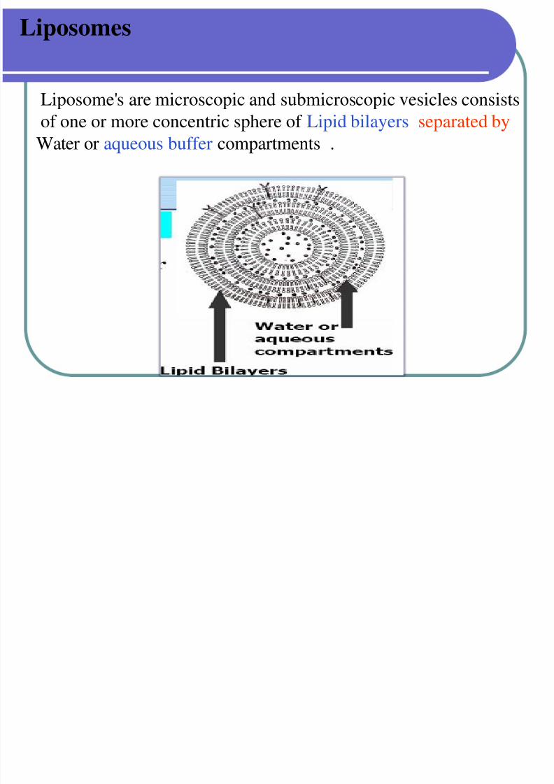
Liposome's are microscopic and submicroscopic vesicles consistsof one or more concentric sphere of Lipid bilayers separated by
Water or aqueous buffer compartments .
Niosomes
7/30/2019 Opthalmic Preparations
http://slidepdf.com/reader/full/opthalmic-preparations 78/103
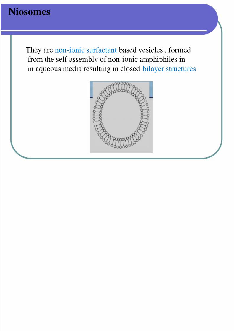
They are non-ionic surfactant based vesicles , formed
from the self assembly of non-ionic amphiphiles in
in aqueous media resulting in closed bilayer structures
Advantages of Niosomes and liposomes
7/30/2019 Opthalmic Preparations
http://slidepdf.com/reader/full/opthalmic-preparations 79/103
Advantages of Niosomes and liposomes - Sustained release of active compounds.
- Protect drugs from degradation
- Biocompatible, biodegradable and non-immunogenic.
- can entrap both hydrophilic and lipophilic drugs
- The bilayer of can efficiently penetrate the cornea of of the eye.
- They have low toxicity because of their non-ionic nature
Contact Lenses & Care Solutions:
7/30/2019 Opthalmic Preparations
http://slidepdf.com/reader/full/opthalmic-preparations 80/103
Contact Lenses & Care Solutions:
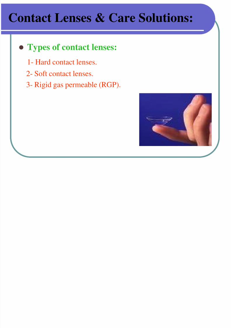
Types of contact lenses:
1- Hard contact lenses.
2- Soft contact lenses.
3- Rigid gas permeable (RGP).
Contact Lenses & Care Solutions:
7/30/2019 Opthalmic Preparations
http://slidepdf.com/reader/full/opthalmic-preparations 81/103
Contact Lenses & Care Solutions:
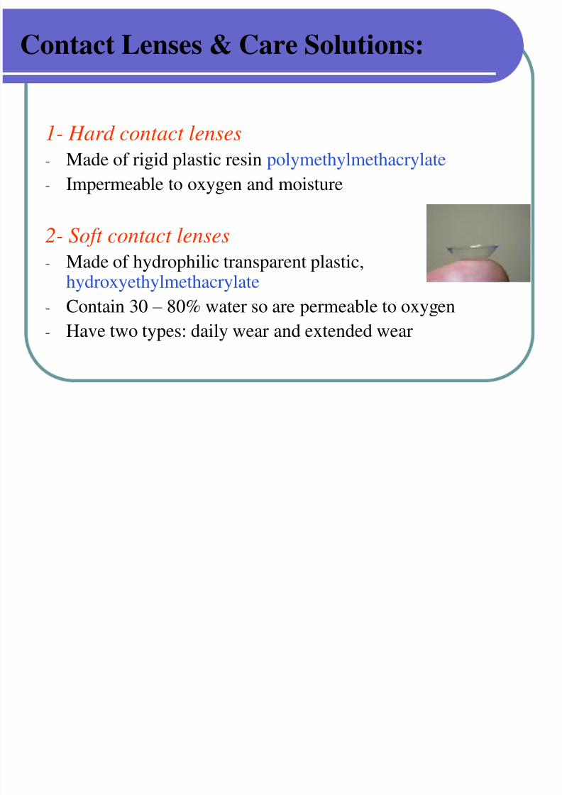
1- Hard contact lenses
- Made of rigid plastic resin polymethylmethacrylate
- Impermeable to oxygen and moisture
2- Soft contact lenses
- Made of hydrophilic transparent plastic,hydroxyethylmethacrylate
- Contain 30 – 80% water so are permeable to oxygen- Have two types: daily wear and extended wear
Contact Lenses & Care Solutions:
7/30/2019 Opthalmic Preparations
http://slidepdf.com/reader/full/opthalmic-preparations 82/103
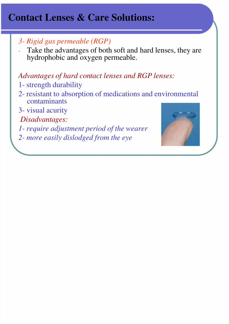
Contact Lenses & Care Solutions:
3- Rigid gas permeable (RGP)- Take the advantages of both soft and hard lenses, they are
hydrophobic and oxygen permeable.
Advantages of hard contact lenses and RGP lenses:
1- strength durability2- resistant to absorption of medications and environmental
contaminants
3- visual acurity
Disadvantages:
1- require adjustment period of the wearer
2- more easily dislodged from the eye
Contact Lenses & Care Solutions:
7/30/2019 Opthalmic Preparations
http://slidepdf.com/reader/full/opthalmic-preparations 83/103
Contact Lenses & Care Solutions:
Advantages of soft contact lenses:
1- worn for longer periods
2- do not dislodge easily
Disadvantages:
1- have a shorter life span and the wearer must ensure that the
lenses do not dry out
7/30/2019 Opthalmic Preparations
http://slidepdf.com/reader/full/opthalmic-preparations 84/103
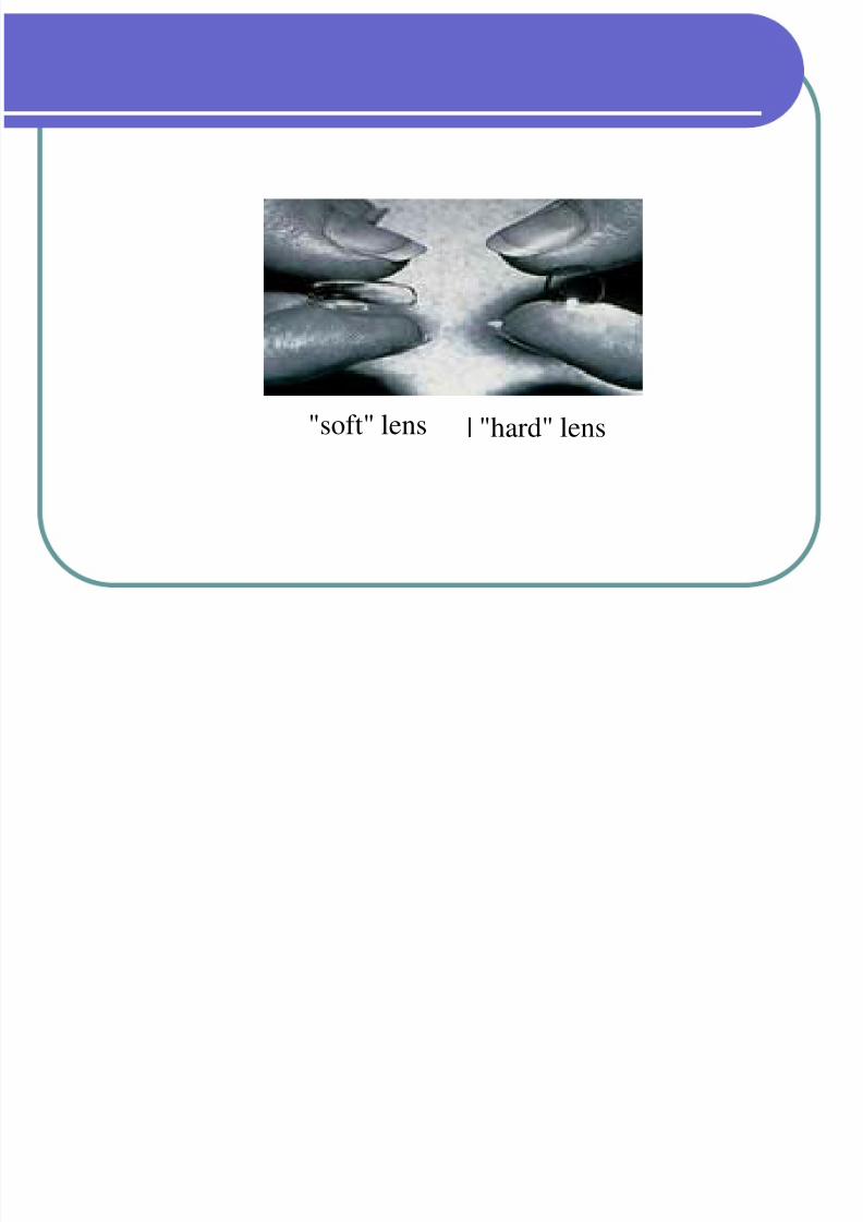
"soft" lens | "hard" lens
Care of contact lenses:
7/30/2019 Opthalmic Preparations
http://slidepdf.com/reader/full/opthalmic-preparations 85/103
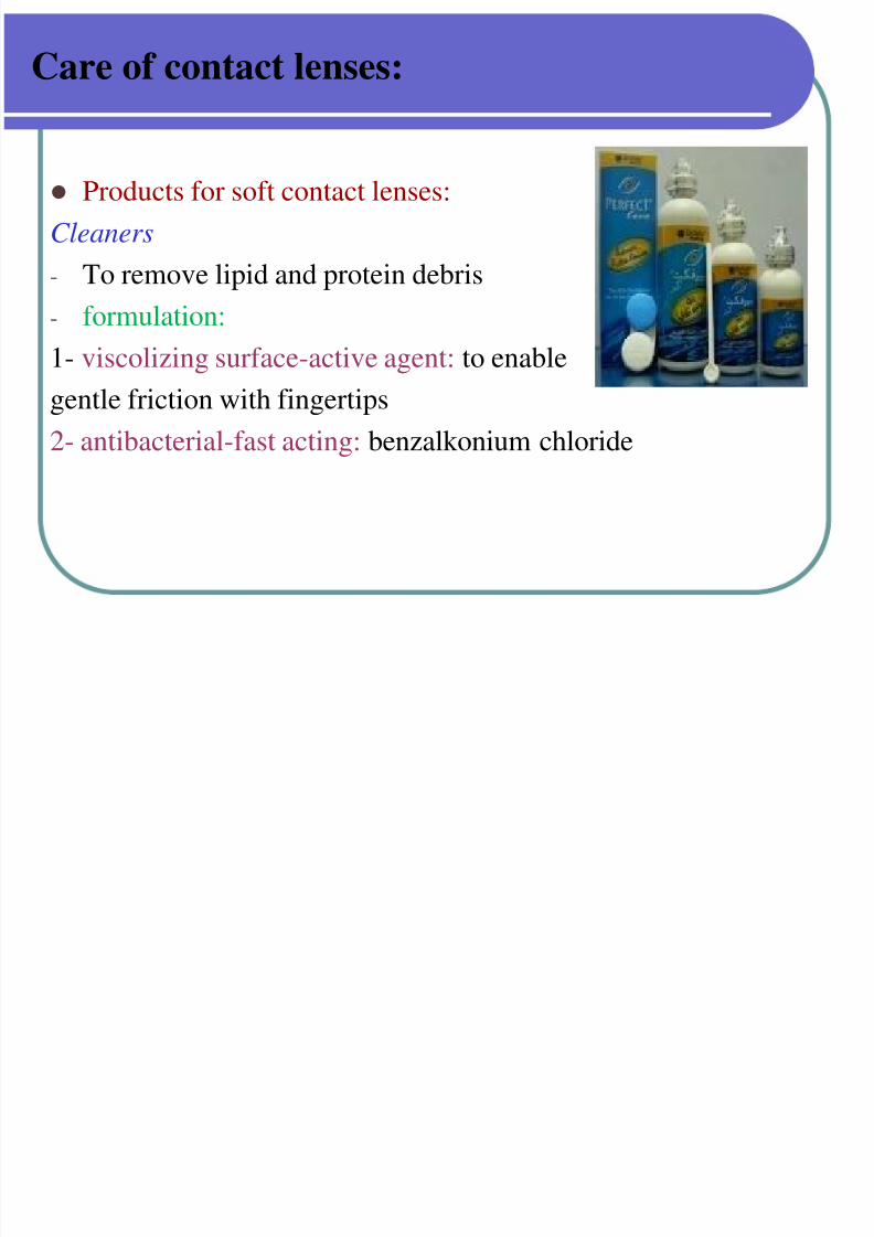
Care of contact lenses:
Products for soft contact lenses:
Cleaners
- To remove lipid and protein debris
- formulation:1- viscolizing surface-active agent: to enable
gentle friction with fingertips
2- antibacterial-fast acting: benzalkonium chloride
Products for soft contact lenses:
7/30/2019 Opthalmic Preparations
http://slidepdf.com/reader/full/opthalmic-preparations 86/103
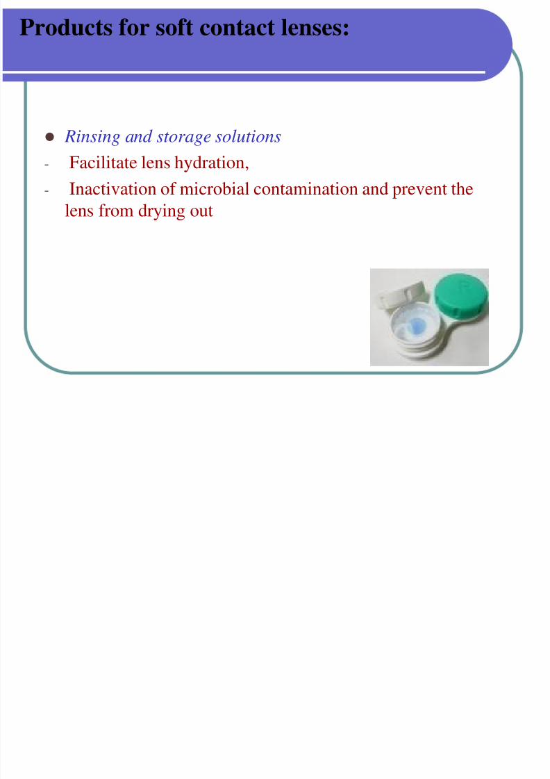
Rinsing and storage solutions
- Facilitate lens hydration,
- Inactivation of microbial contamination and prevent the
lens from drying out
Products for soft contact lenses:
7/30/2019 Opthalmic Preparations
http://slidepdf.com/reader/full/opthalmic-preparations 87/103
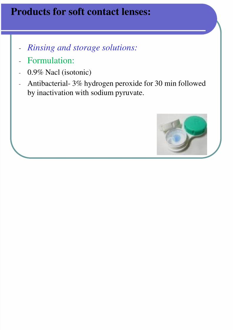
- Rinsing and storage solutions:
- Formulation:
- 0.9% Nacl (isotonic)
- Antibacterial- 3% hydrogen peroxide for 30 min followedby inactivation with sodium pyruvate.
Products for hard contact lenses:
7/30/2019 Opthalmic Preparations
http://slidepdf.com/reader/full/opthalmic-preparations 88/103
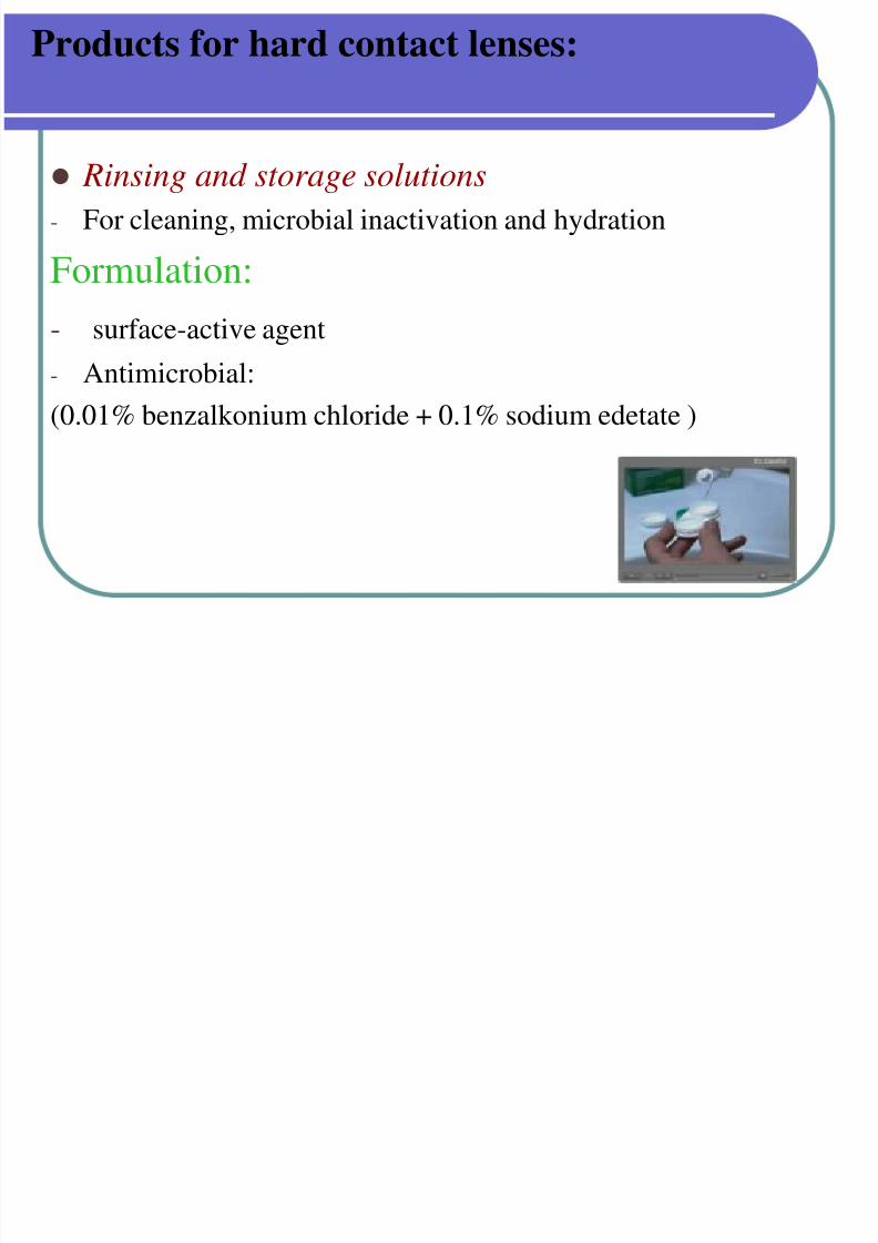
Rinsing and storage solutions- For cleaning, microbial inactivation and hydration
Formulation:
- surface-active agent
- Antimicrobial:
(0.01% benzalkonium chloride + 0.1% sodium edetate )
Products for hard contact lenses:
7/30/2019 Opthalmic Preparations
http://slidepdf.com/reader/full/opthalmic-preparations 89/103
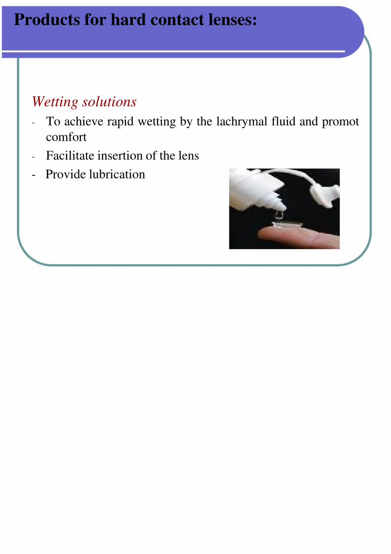
Wetting solutions
- To achieve rapid wetting by the lachrymal fluid and promot
comfort
- Facilitate insertion of the lens
- Provide lubrication
Products for hard contact lenses:
7/30/2019 Opthalmic Preparations
http://slidepdf.com/reader/full/opthalmic-preparations 90/103
Buffering solutions :
Hypromellose eye drops B.P.C:
Buffered to pH 8.4 TO 8.6 with boric acid and borax
These solutions better tolerated by eye as more alkaline or
acid preparations causes fogging effect.
Evaluation tests
7/30/2019 Opthalmic Preparations
http://slidepdf.com/reader/full/opthalmic-preparations 91/103
Evaluation tests
Metal Particles
This test is required only for ophthalmic ointments.
The presence of metal particles will irritate the corneal orconjunctival surfaces of the eye.
It is performed using 10 ointment tubes.
The content from each tube is completely removed onto a
clean 60 - mm - diameter Petri dish which possesses a flatbottom.
Metal particles :
7/30/2019 Opthalmic Preparations
http://slidepdf.com/reader/full/opthalmic-preparations 92/103
Metal particles :
The lid is closed and the product is heated at 85 ° C for 2 h.
Once the product is melted and distributed uniformly, it is
cooled to room temperature.
The lid is removed after solidification.
The bottom surface is then viewed through an optical
microscope at 30× magnification.
Metal particles :
7/30/2019 Opthalmic Preparations
http://slidepdf.com/reader/full/opthalmic-preparations 93/103
Metal particles :
The viewing surface is illuminated using an external lightsource positioned at 45 ° on the top.
The entire bottom surface of the ointment is examined,
And the number of particles 50 μm or above are countedusing a calibrated eyepiece micrometer.
The USP recommends that the number of such particles in10 tubes should not exceed 50, with not more than 8particles in any individual tube.
Metal particles :

7/30/2019 Opthalmic Preparations
http://slidepdf.com/reader/full/opthalmic-preparations 94/103
Metal particles :
limits are not met, the test is repeated with an additional 20tubes.
In this case, the total number of particles in 30 tubes should
not exceed 150, and not more than 3 tubes are allowed to
contain more than 8 particles .
Leakage test :
7/30/2019 Opthalmic Preparations
http://slidepdf.com/reader/full/opthalmic-preparations 95/103
Leakage test :
This test is mandatory for ophthalmic ointments, which
evaluates the intactness of the ointment tube and its seal.
Ten sealed containers are selected, and their exterior
surfaces are cleaned.
They are horizontally placed over absorbent blotting paper .
Maintained at 60 ± 3 ° C for 8 h.
Leakage test :
7/30/2019 Opthalmic Preparations
http://slidepdf.com/reader/full/opthalmic-preparations 96/103
Leakage test :
The test passes if leakage is not observed from any tube.
If leakage is observed, the test is repeated with an additional
20 tubes.
The test passes if not more than 1 tube shows leakage out of
30 tubes .
Sterility Tests :
7/30/2019 Opthalmic Preparations
http://slidepdf.com/reader/full/opthalmic-preparations 97/103
Sterility Tests :
Ophthalmic semisolids should be free from anaerobic andaerobic bacteria and fungi.
Sterility tests are therefore performed by the:1. Membrane filtration technique .
2. Direct - inoculation techniques.
Sterility Tests
7/30/2019 Opthalmic Preparations
http://slidepdf.com/reader/full/opthalmic-preparations 98/103
Sterility Tests
In the Membrane filtration method :
A solution of test product (1%) is prepared in isopropyl
myristate and allowed to penetrate through cellulose nitrate
filter with pore size less than 0.45 μ m.
If necessary, gradual suction or pressure is applied to aid
filtration.
Sterility Tests :
7/30/2019 Opthalmic Preparations
http://slidepdf.com/reader/full/opthalmic-preparations 99/103
Ste ty ests :
The membrane is then washed three times with 100 - mLquantities of sterile diluting and rinsing fluid and transferredaseptically into fluid thioglycolate (FTG) andsoybean – casein digest medium (SBCD) .
The membrane is finally incubated for 14 days. Growth on FTG medium indicates the presence of anaerobic
and aerobic bacteria.
Sterility Tests :
7/30/2019 Opthalmic Preparations
http://slidepdf.com/reader/full/opthalmic-preparations 100/103
Sterility Tests :
Soybean casein digest medium indicates fungi and aerobic
bacteria
Absence of any growth in both these media establishes the
sterility of the product.
Sterility Tests
7/30/2019 Opthalmic Preparations
http://slidepdf.com/reader/full/opthalmic-preparations 101/103
y
In the Direct - inoculation technique :
1 part of the product is diluted with 10 parts of sterile
diluting and rinsing fluid with the help of an emulsifying
agent
Incubated in Fluid thioglycolate (FTG) and soybean – casein digest medium (SBCD) media for 14 days .
Sterility Tests :
7/30/2019 Opthalmic Preparations
http://slidepdf.com/reader/full/opthalmic-preparations 102/103
y
In both techniques, the number of test articles is based onthe batch size of the product.
If the batch size is less than 200 the containers, either 5% of
the containers or 2 containers (whichever is greater) are
used. If the batch size is more than 200, 10 containers are used for
sterility testing .
References
7/30/2019 Opthalmic Preparations
http://slidepdf.com/reader/full/opthalmic-preparations 103/103
e e e ces
Dispensing for pharmaceutical by Cooper and gunn’s
pg: 634-661
Modern dispensing pharmacy : N K Jain pg: 13.3-14.9
Text of pharmaceutical formulation : B.M Mithal
pg: 268-278