
Opportunistic and Transplant Related Infections
77
Opportunistic and Transplant Related Infections Gonzalo Bearman MD, MPH Associate Professor of Internal Medicine and Public Health Division of Infectious Diseases Associate Hospital Epidemiologist VCU Health System 8.5.2008 For VCU Internal Medicine Residents
Transcript of Opportunistic and Transplant Related Infections
Opportunistic and Transplant Related Infections
Gonzalo Bearman MD, MPHAssociate Professor of Internal Medicine and Public Health
Division of Infectious DiseasesAssociate Hospital Epidemiologist
VCU Health System8.5.2008
For VCU Internal Medicine Residents

Disclaimer
Many physician/researchers dedicate entire careers to the study and treatment of opportunistic infections. One cannot possibly cover all ‘opportunistic’ infections in a 60 minute lecture. As such, the purpose of this lecture is to cover material deemed appropriate for an Internal Medicine Board Examination review.
Opportunistic Infection Defined
An infection by a microorganism that normally does not cause disease but pathogenic when the body's immune system is impaired and unable to fight off infection, as in AIDS, neutropenia, and congenital or iatrogenic host defense defects.

OI Following BMT
©2004 UpToDate® • www.uptodate.com
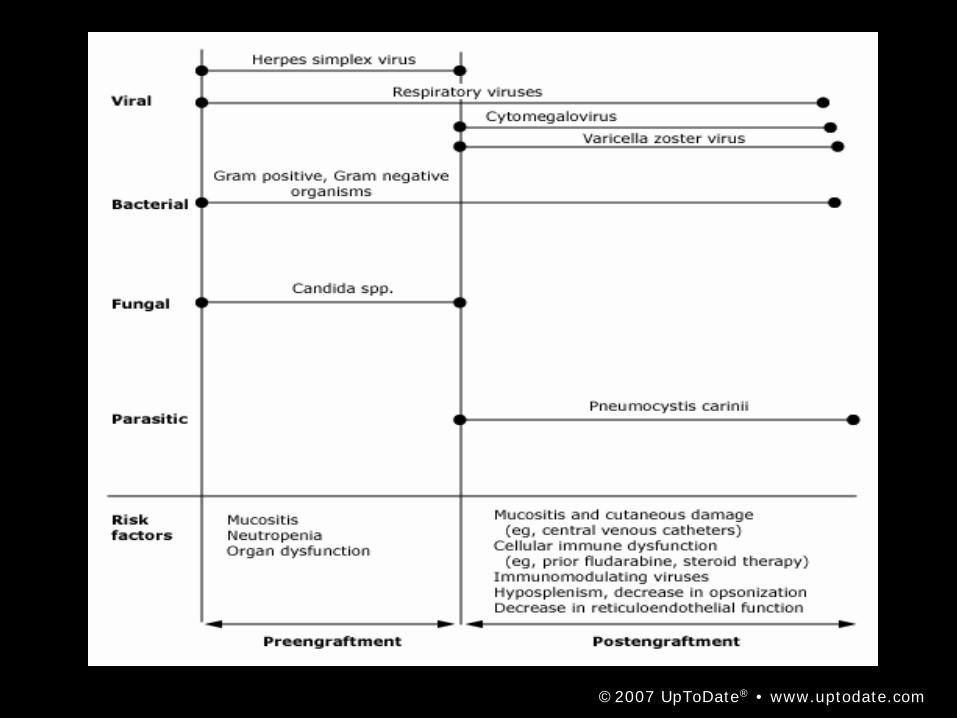
©2007 UpToDate® • www.uptodate.com

Preengraftment
• Preengraftment — less than three weeks – The major risk factors for infection during
the preengraftment period in the first three weeks after HCT are mucositis and cutaneous damage, and neutropenia with resulting loss of phagocytic abilities
Early Postengraftment
• Immediate postengraftment: three weeks to three months – Risk factors for infection during the
immediate postengraftment period three weeks to three months after HCT are mucositis and cutaneous damage along with but also cellular immune dysfunction, decrease in opsonization, and diminished reticuloendothelial function.
Late Postengraftment
• Late infectious complications are typically only seen among allogeneic recipients. The major risk factor for infection during this period is chronic GVHD and its immunosuppresive therapy
• This results in– Cellular and humoral immune dysfunction,
hyposplenism, decrease in opsonization, and diminished reticuloendothelial function
– Mucocutaneous damage

OI following Solid Organ Transplantation
©2004 UpToDate® • www.uptodate.com

Case 1
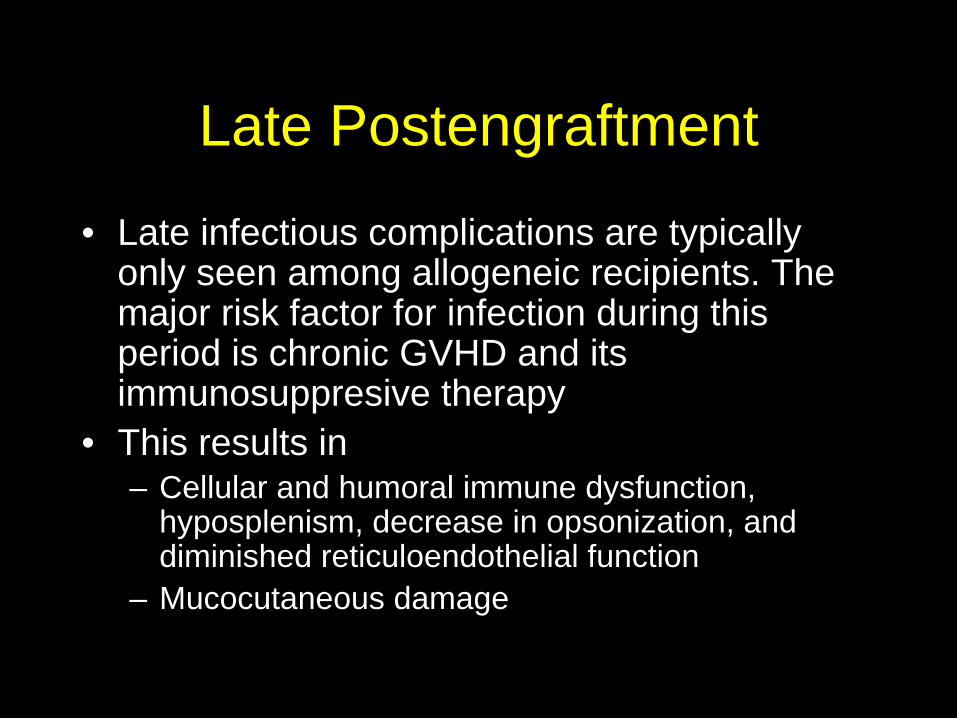
• 70 year old man with history of AML, S/P bone marrow tyransplant, hospitalized for 16 days with febrile neutropenia
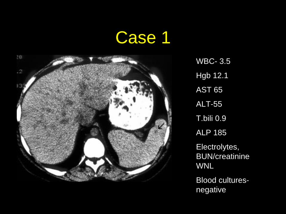
• Despite 9 days of aggressive antibiotic therapy with vancomycin, piperacillin/tazobactam and ciprofloxacin– He is febrile and is complaining of rigors and
blurred vision in the left eye.

Case 1• T-39.7, P-120,RR-16, BP 130/75• Ill appearing• PERRLA; Mouth no lesions• Chest:clear• Cardiac- tachycardic, no mumurs or gallops• Abd soft; mild tenderness RUQ, no
hepatomegaly• Mediport site: clean, no erythema, discharge
or tenderness
Case 1
•
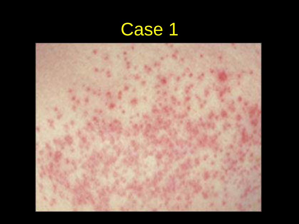
Case 1WBC- 3.5
Hgb 12.1
AST 65
ALT-55
T.bili 0.9
ALP 185
Electrolytes, BUN/creatinine WNL
Blood cultures- negative
Case 1
Candida
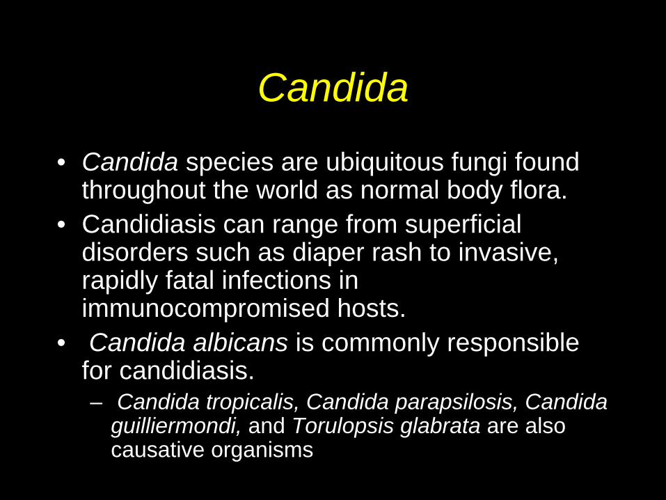
• Candida species are ubiquitous fungi found throughout the world as normal body flora.
• Candidiasis can range from superficial disorders such as diaper rash to invasive, rapidly fatal infections in immunocompromised hosts.
• Candida albicans is commonly responsible for candidiasis.– Candida tropicalis, Candida parapsilosis, Candida
guilliermondi, and Torulopsis glabrata are also causative organisms

Candida: laboratory diagnosis
• Systemic candidiasis (eg, CNS, joint, blood)• Cultures of cerebrospinal fluid (CSF), joint
fluid, urine, or surgical specimens may be obtained to identify candidal infections.
• Blood culture is useful for diagnosing endocarditis and catheter-induced sepsis.
• Urinalysis (UA) positive for Candida species may predict 38-80% of systemic candidiasis.
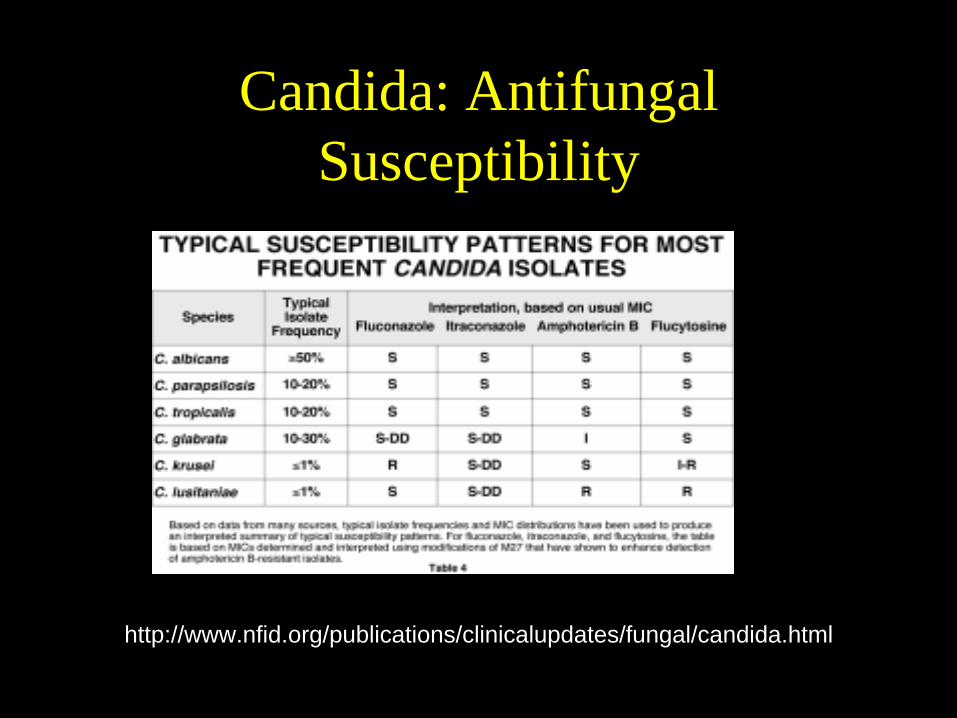
Candida: Antifungal Susceptibility
•
http://www.nfid.org/publications/clinicalupdates/fungal/candida.html
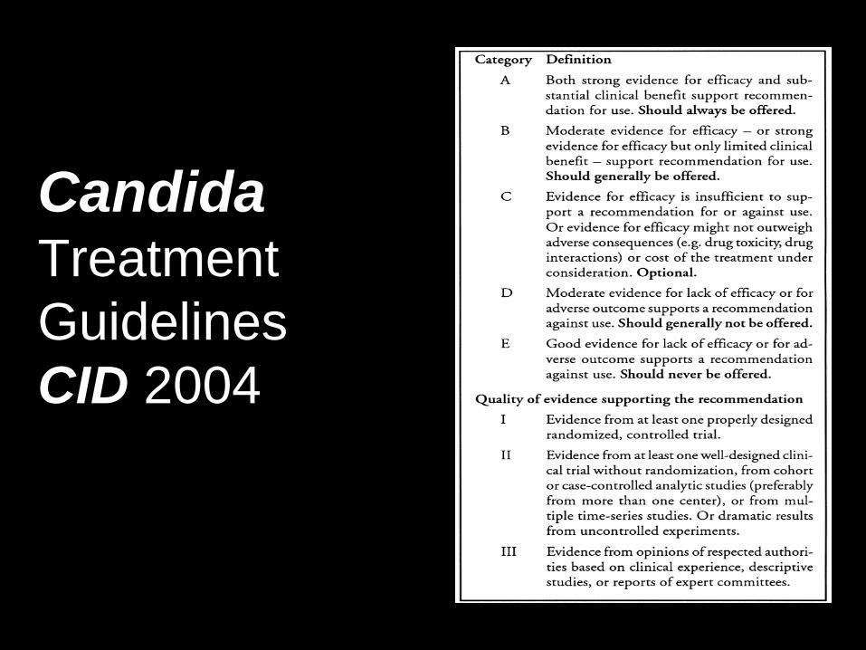
Candida Treatment Guidelines CID 2004
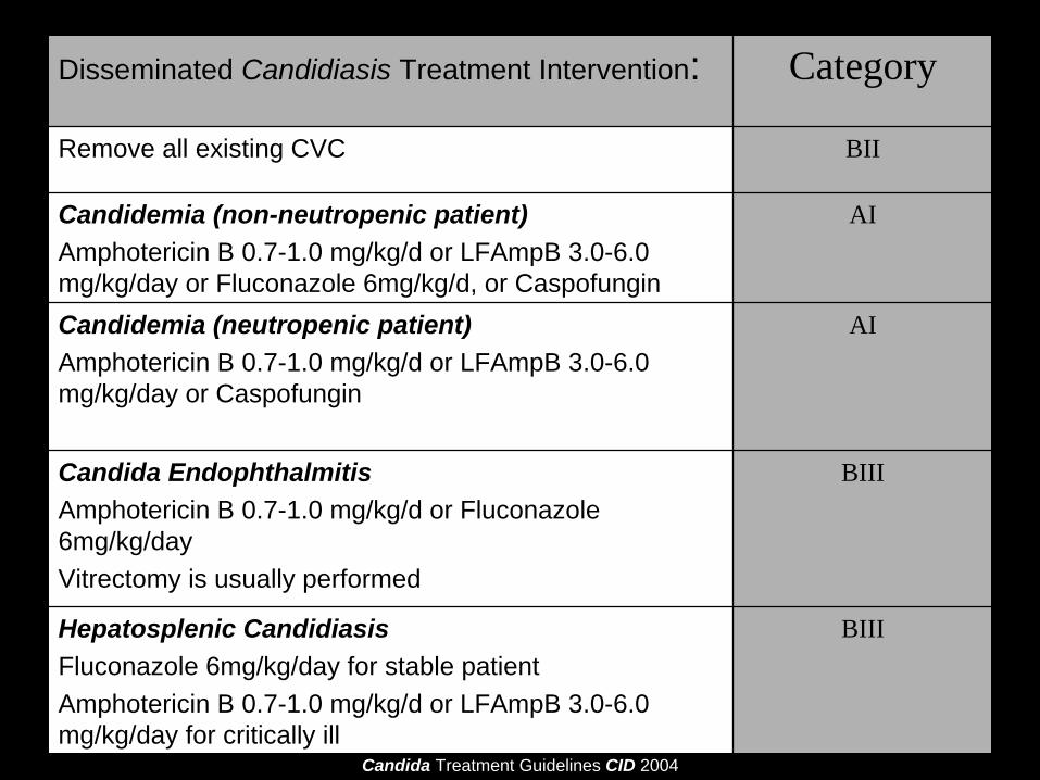
Disseminated Candidiasis Treatment Intervention: Category
Remove all existing CVC BII
Candidemia (non-neutropenic patient)Amphotericin B 0.7-1.0 mg/kg/d or LFAmpB 3.0-6.0 mg/kg/day or Fluconazole 6mg/kg/d, or Caspofungin
AI
Candidemia (neutropenic patient)Amphotericin B 0.7-1.0 mg/kg/d or LFAmpB 3.0-6.0 mg/kg/day or Caspofungin
AI
Candida EndophthalmitisAmphotericin B 0.7-1.0 mg/kg/d or Fluconazole 6mg/kg/day Vitrectomy is usually performed
BIII
Hepatosplenic CandidiasisFluconazole 6mg/kg/day for stable patientAmphotericin B 0.7-1.0 mg/kg/d or LFAmpB 3.0-6.0 mg/kg/day for critically ill
BIII
Candida Treatment Guidelines CID 2004

Echinocandins for Candidemia in adults without neutropenia
Caspofungin •Choice of an echinocandin vs. an azole for candidemia in a non-neutropenic patient is not well established•Echinocandins may be preferred preferred when C. glabrata or C. krusei is identified or suspected•Fluconazole interacts with cytochrome P450 resulting in many drug-drug interactions.
–This may dictate a preference for treatment with an echinocandin
Micafungin
Anidulafungin

Case II• 31 year old Caucasian woman with a history
of multiple ‘sinus’ infections over the last 8-9 years. Over the last 3 years she has had an episode of ‘bronchitis’ and 2 bouts of pneumonia.
• She presents to the ambulatory care clinic with a 4 days history of fever, right maxillary tenderness, and purulent nasal discharge
• She does not smoke and has no history of either seasonal or perennial allergies.
• Family history of ‘sinus’ problems and pneumonias in older sister
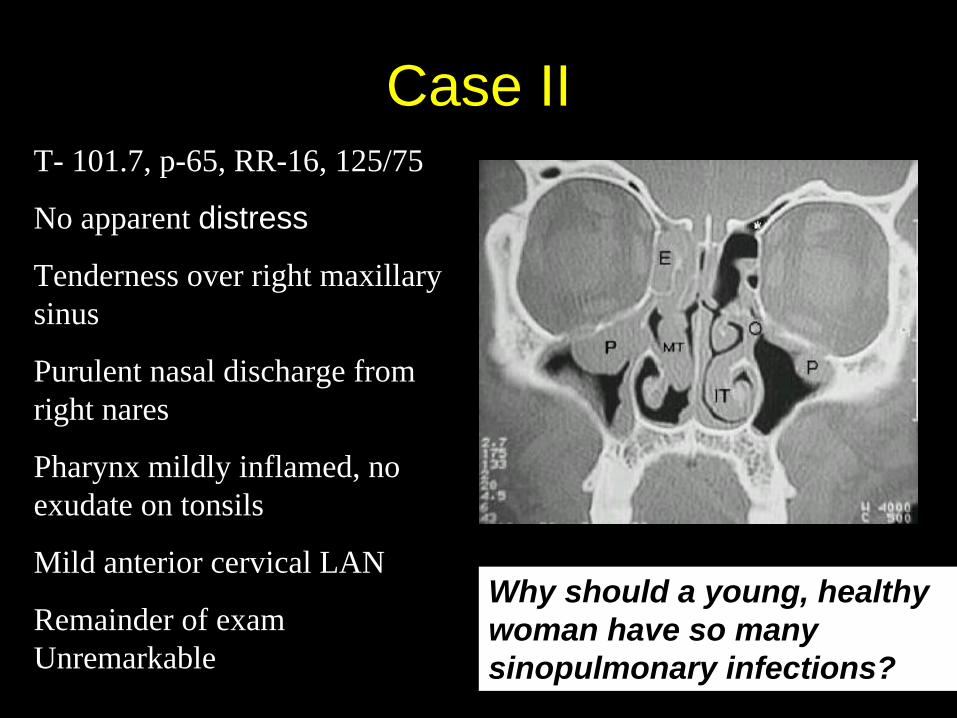
T- 101.7, p-65, RR-16, 125/75
No apparent distress
Tenderness over right maxillary sinus
Purulent nasal discharge from right nares
Pharynx mildly inflamed, no exudate on tonsils
Mild anterior cervical LAN
Remainder of exam Unremarkable
Case II
Why should a young, healthy woman have so many sinopulmonary infections?
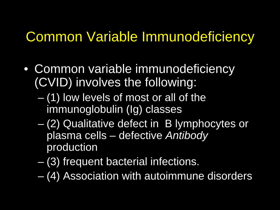
Common Variable Immunodeficiency
• Common variable immunodeficiency (CVID) involves the following: – (1) low levels of most or all of the
immunoglobulin (Ig) classes– (2) Qualitative defect in B lymphocytes or
plasma cells – defective Antibody production
– (3) frequent bacterial infections. – (4) Association with autoimmune disorders

Common Variable Immunodeficiency
More Common
Less Common
Infection Autoimmune Diseases
Other
Sinusitis, otitis media, pneumonia (encapsulated organisms)
Hemolytic Anemia
Autoimmune thyroid diseaseRheumatoid ArthritisJRA
SLESjogren’sUC/Crohn’s
LymphadenopathySplenomegaly
Bronchiectasis
Malignancy (gastric CA)
Infectious diarrhea(Giardia, Salmonella, campylobacter species)Septic arthritis(S.aureus, mycoplasma)
Meningitis(encapsulated organisms)
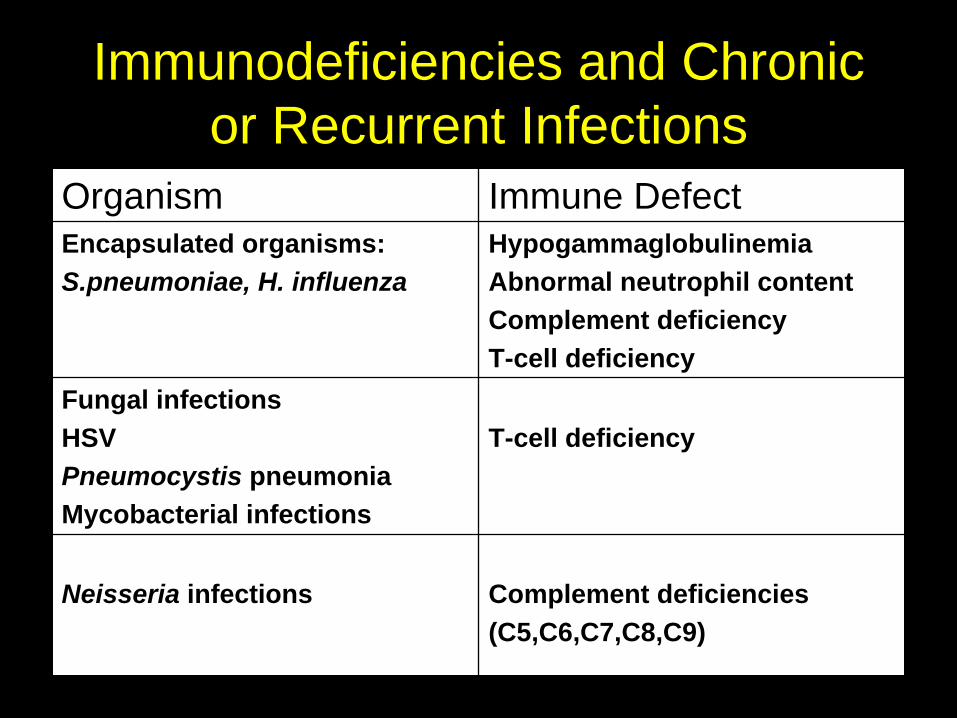
Immunodeficiencies and Chronic or Recurrent Infections
Organism Immune DefectEncapsulated organisms:S.pneumoniae, H. influenza
HypogammaglobulinemiaAbnormal neutrophil contentComplement deficiencyT-cell deficiency
Fungal infectionsHSVPneumocystis pneumoniaMycobacterial infections
T-cell deficiency
Neisseria infections Complement deficiencies(C5,C6,C7,C8,C9)
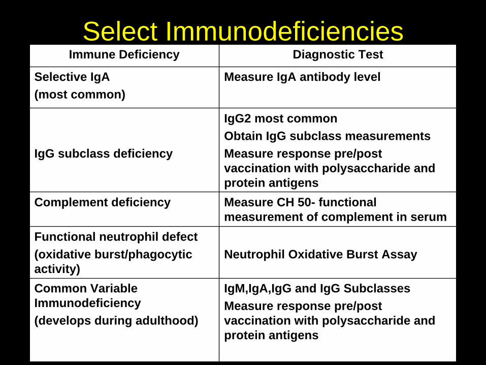
Select ImmunodeficienciesImmune Deficiency Diagnostic Test
Selective IgA(most common)
Measure IgA antibody level
IgG subclass deficiency
IgG2 most commonObtain IgG subclass measurementsMeasure response pre/post vaccination with polysaccharide and protein antigens
Complement deficiency Measure CH 50- functional measurement of complement in serum
Functional neutrophil defect(oxidative burst/phagocytic activity)
Neutrophil Oxidative Burst Assay
Common Variable Immunodeficiency(develops during adulthood)
IgM,IgA,IgG and IgG SubclassesMeasure response pre/post vaccination with polysaccharide and protein antigens

Case III
• 21-year old woman with AML is hospitalized for a febrile neutropenic episode
• She is S/P chemotherapy and had been placed on broad-spectrum antibiotic coverage that resulted in rapid defervescence and clinical improvement
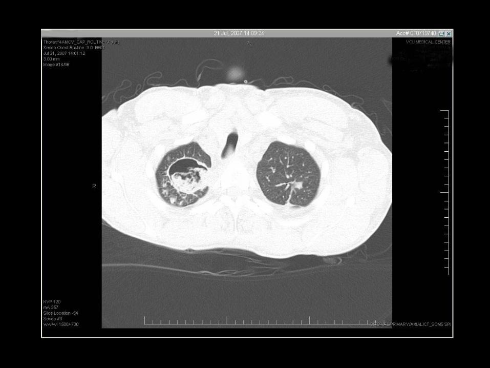
• A few days later, she develops fever to 39.6C, accompanied by fever and pleuritic chest pain.
Cunha, B. Infectious Diseases Pearls. 1999

Case III
• T 39.6, pulse 130, RR 22, BP 90/58• Ill appearing• HEENT: no abnormalities• Chest: bibasilar crackles• Cardiac: S1, S2 with II/VI SM• Abd:WNL• Ext: trace pedal edema• Skin- no rashes or dermatitis
Cunha, B. Infectious Diseases Pearls. 1999
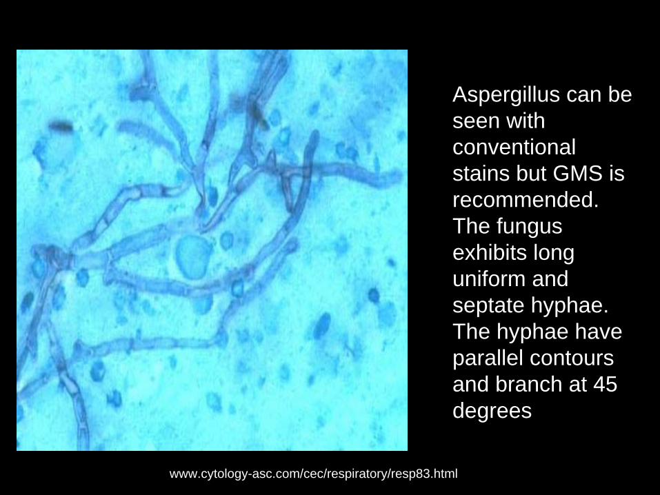
Aspergillus can be seen with conventional stains but GMS is recommended. The fungus exhibits long uniform and septate hyphae. The hyphae have parallel contours and branch at 45 degrees
www.cytology-asc.com/cec/respiratory/resp83.html
Clinical Presentations
• Invasive Aspergillosis-– Pulmonary Aspergillosis (most common)
- CNS aspergillosis - Sinonasal aspergillosis - Osteomyelitis - Endophthalmitis - Endocarditis - Renal abscesses - Cutaneous
Diagnosis• Definitive diagnosis
– Requires the demonstration of tissue invasion seen on biopsy specimen.
– Positive culture obtained from tissue obtained by invasive procedure.
– These patients are typically very sick/debilitated thereby precluding them form invasive diagnostic procedures.
Diagnosis
• Less or non-invasive tests in the setting of the appropriate clinical setting may suggest the diagnosis.– Blood cultures are typically negative.– In high risk patient, isolation of isolation of
Aspergillus from sputum or BAL is strongly suggestive of IA.
– Serologic Aspergillus precipitin assays are rarely elevated in IA and thus are of little clinical value
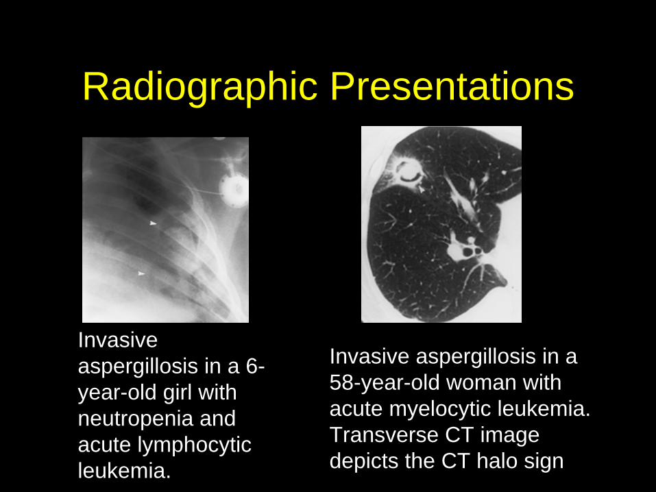
Radiographic Presentations
Invasive aspergillosis in a 6- year-old girl with neutropenia and acute lymphocytic leukemia.
Invasive aspergillosis in a 58-year-old woman with acute myelocytic leukemia. Transverse CT image depicts the CT halo sign
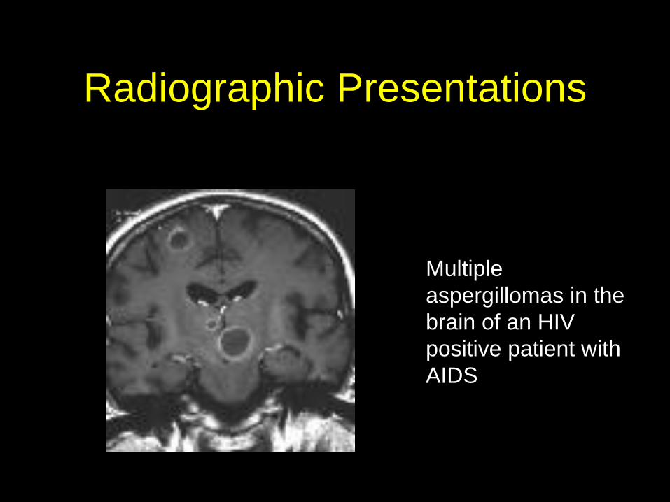
Radiographic Presentations
Multiple aspergillomas in the brain of an HIV positive patient with AIDS

Twice weekly monitoring for patients
at high risk
The Lancet Infectious Diseases Vol 4 June 2004. 349-357
Treatment of Aspergillosis: Clinical Practice Guidelines of the Infectious Diseases Society of America
Clinical Infectious Diseases 2008;46:327–360

Treatment of Aspergillosis: Clinical Practice Guidelines of the Infectious Diseases Society of America
Clinical Infectious Diseases 2008;46:327–360
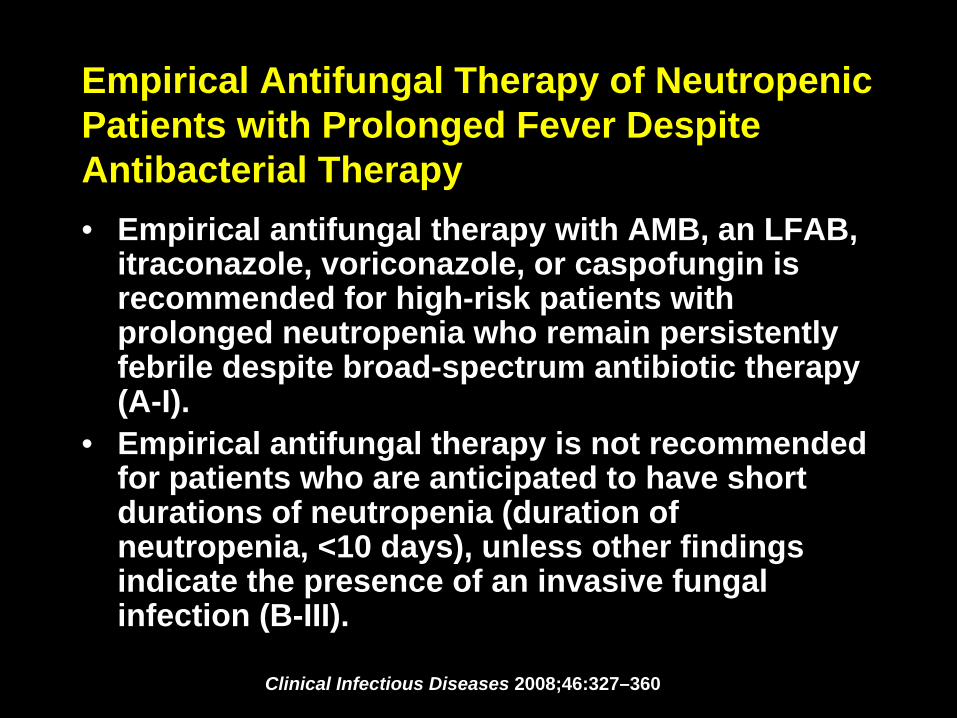
Empirical Antifungal Therapy of Neutropenic Patients with Prolonged Fever Despite Antibacterial Therapy• Empirical antifungal therapy with AMB, an LFAB,
itraconazole, voriconazole, or caspofungin is recommended for high-risk patients with prolonged neutropenia who remain persistently febrile despite broad-spectrum antibiotic therapy (A-I).
• Empirical antifungal therapy is not recommended for patients who are anticipated to have short durations of neutropenia (duration of neutropenia, <10 days), unless other findings indicate the presence of an invasive fungal infection (B-III).
Clinical Infectious Diseases 2008;46:327–360

An opportunistic infection from paradise?
Case IV• A 51-year-old Korean woman was brought to the hospital after a
close friend found her semiconscious and obtunded. • The previous day, the woman was seen at church where she
appeared healthy.On admission, she began to experience episodic chills lasting 30 to 40 minutes.
• That evening she was extremely lethargic.• The patient had a medical history of chronic active hepatitis B
virus (HBV) infection.
http://www.residentandstaff.com/article.cfm?ID=281
• The patient presented to the ED where she was lethargic and diaphoretic.
• She was tachypneic (25-32 breaths/min) and mildly tachycardic (95-105 beats/min) with a temperature of 103°F and systolic blood pressure between 90 and 100 mm Hg.
• Physical examination revealed that she was obtunded and lethargic. Her sclera was icteric, and her skin was jaundiced with mild generalized edema.
• No cardiac murmurs or a rub were heard on auscultation. An audible wheeze was heard bilaterally on expiration.
• Auscultation of her abdomen revealed decreased bowel sounds.• Palpation of the abdomen revealed diffuse tenderness, and a
liver edge was noted 2 to 3 cm below the costodiaphragmatic angle.
Case IV
http://www.residentandstaff.com/article.cfm?ID=281
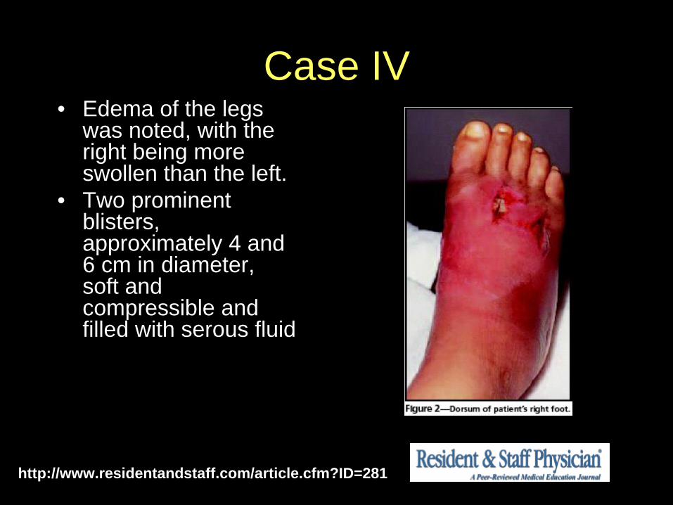
Case IV• Edema of the legs
was noted, with the right being more swollen than the left.
• Two prominent blisters, approximately 4 and 6 cm in diameter, soft and compressible and filled with serous fluid
http://www.residentandstaff.com/article.cfm?ID=281
Case IV
• The surgical specimen taken from the right ankle grew a bacillus species later identified as Vibrio vulnificus.
• It was discovered that she had purchased a can of oysters but could not recall if she consumed it.
http://www.residentandstaff.com/article.cfm?ID=281
Vibrio vulnificus
<> June 04, 1993 / 42(21);405-407
Vibrio vulnificus Infections Associated with Raw Oyster Consumption -- Florida, 1981-1992
<> July 26, 1996 / 45(29);621-624
Vibrio vulnificus Infections Associated with Eating Raw Oysters -- Los Angeles, 1996

Vibrio vulnificus
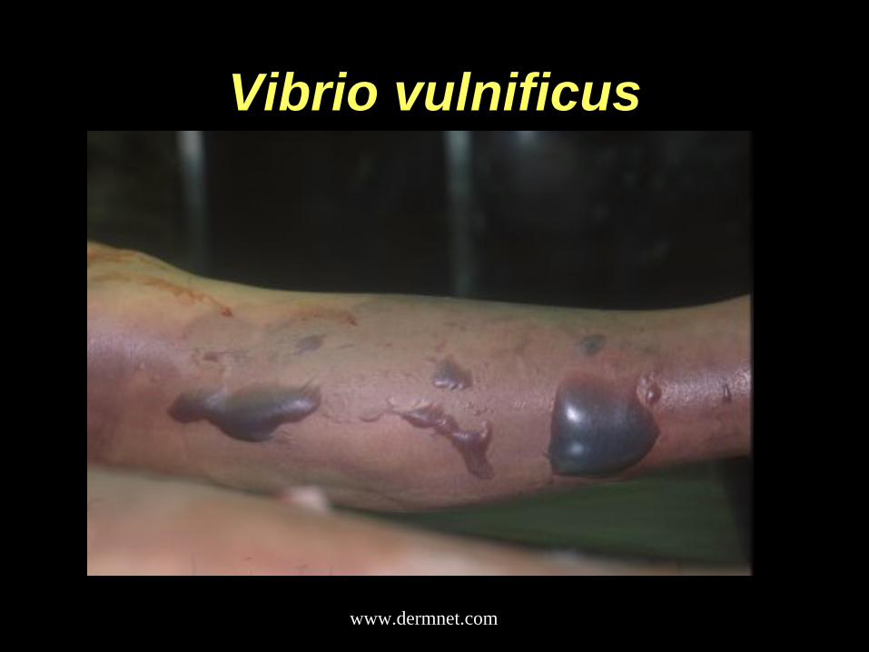
Vibrio vulnificus causes wound infections, gastroenteritis or a serious syndrome known as "primary septicema."
www.medscape.com
Vibrio vulnificusMode of Transmission Clinical Manifestations Dermatologic
Manifestations
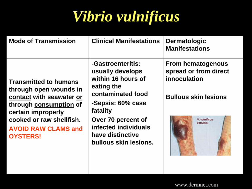
Transmitted to humans through open wounds in contact with seawater or through consumption of certain improperly cooked or raw shellfish.AVOID RAW CLAMS and OYSTERS!
-Gastroenteritis: usually develops within 16 hours of eating the contaminated food-Sepsis: 60% case fatality Over 70 percent of infected individuals have distinctive bullous skin lesions.
From hematogenous spread or from direct innoculation
Bullous skin lesions
www.dermnet.com
Vibrio vulnificus
www.dermnet.com
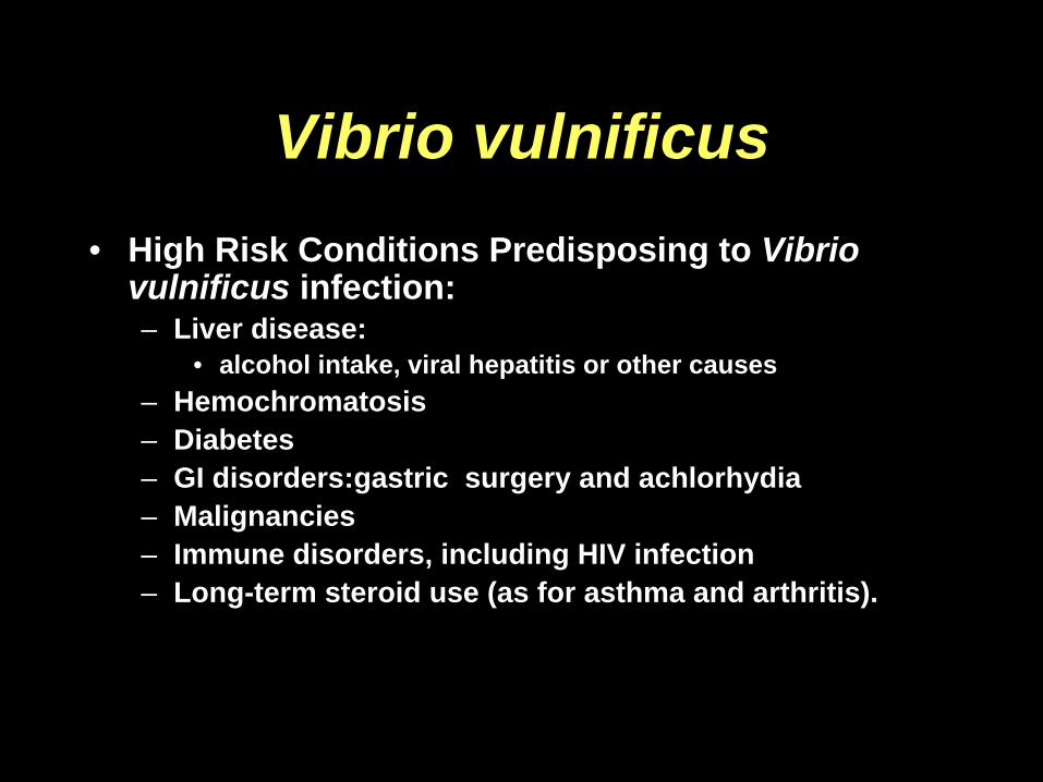
Vibrio vulnificus• High Risk Conditions Predisposing to Vibrio
vulnificus infection:– Liver disease:
• alcohol intake, viral hepatitis or other causes– Hemochromatosis– Diabetes– GI disorders:gastric surgery and achlorhydia– Malignancies– Immune disorders, including HIV infection– Long-term steroid use (as for asthma and arthritis).

Vibrio vulnificusDiagnostic Pearls Culture
-Consumption of shellfish, clams
-Exposure to seawater (bathing/swimming)-Violaceous, large bullous lesions-Sepsis -A physician should suspect V. vulnificus if a patient has watery diarrhea and has eaten raw or undercooked oysters or when a wound infection occurs after exposure to seawater
Vibrio organisms can be isolated from cultures of stool, wound, or blood. V. vulnificus infection is diagnosed by routine stool, wound, or blood cultures;
Notify the lab since a special growth medium can be used to increase the diagnostic yield
RX:Doxycycline or a third-generation cephalosporin (e.g., ceftazidime)
Case V
• 65 year old caucasian man with a history of RPGN is S/P cadaveric renal transplant 180 days ago.
• Over the last several days he has felt fatigued, with a low grade fever. His appetite has been poor.
• He is currently on prednisone and Imuran for chronic immunosuppression.
www.dermnet.com
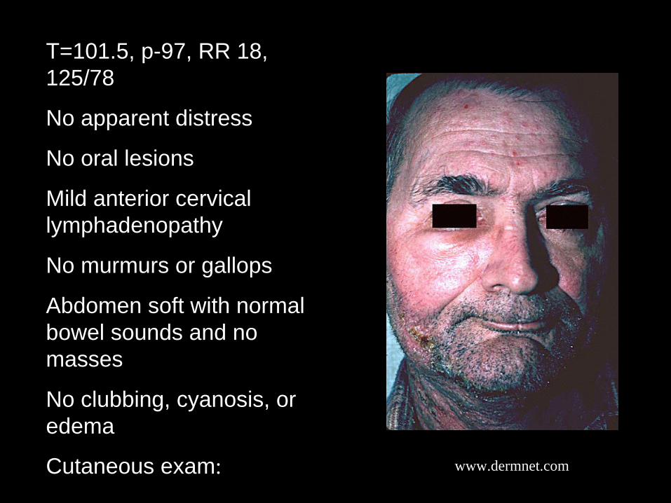
T=101.5, p-97, RR 18, 125/78
No apparent distress
No oral lesions
Mild anterior cervical lymphadenopathy
No murmurs or gallops
Abdomen soft with normal bowel sounds and no masses
No clubbing, cyanosis, or edema
Cutaneous exam:

www.dermnet.com

www.dermnet.com http://tray.dermatology.uiowa.edu
Varicella Zoster Virus
• About 95% of adults in the United States have antibodies to the varicella-zoster virus.– Herpes zoster occurs annually in 300,000-500,000
individuals– Incidence of herpes zoster increases with age.
• 80% of cases occur in persons> 20 years of age
– A minority of the cases are non-dermatomal or disseminated
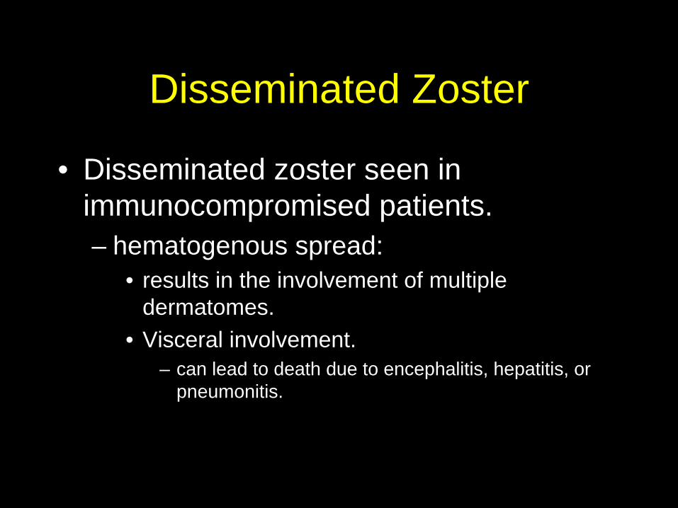
Disseminated Zoster
• Disseminated zoster seen in immunocompromised patients.– hematogenous spread:
• results in the involvement of multiple dermatomes.
• Visceral involvement. – can lead to death due to encephalitis, hepatitis, or
pneumonitis.

Disseminated ZosterDiagnosis Treatment
•Herpes zoster is based primarily on clinical findings•Varicella-zoster virus culture•Tzanck smear (vesicular lesions)•Biopsy for direct immunofluorescence
Acyclovir:Immunocompromised adults: 800 mg PO q4h (5 times/d) for 7-10 d; or 10 mg/kg/dose or 500 mg/m2/dose IV q8h

Case VI• 34 year old caucasian male, HIV positive
since 1993.• Past history significant for PCP and thrush.• Was on antiretrovirals on and off for years but
had problems with medication adherence .• Had been lost to follow up but presents to
clinic with a history of progressive weight loss, anorexia, malaise, odynophagia and subjective fever.
• Additionally, he has complained of ‘floaters’ in the right eye, but no pain or change in visual acuity

Case VI• Physical examination• T 101.8F otherwise WNL• Height 6’1’, 140 lbs• No murmurs or gallops• Lungs clear• Abdomen; soft, liver edge
2cm below costal margin• Skin warm, dry, no
significant lesions
http://www.eyemdlink.com
http://www.emedicine.com/
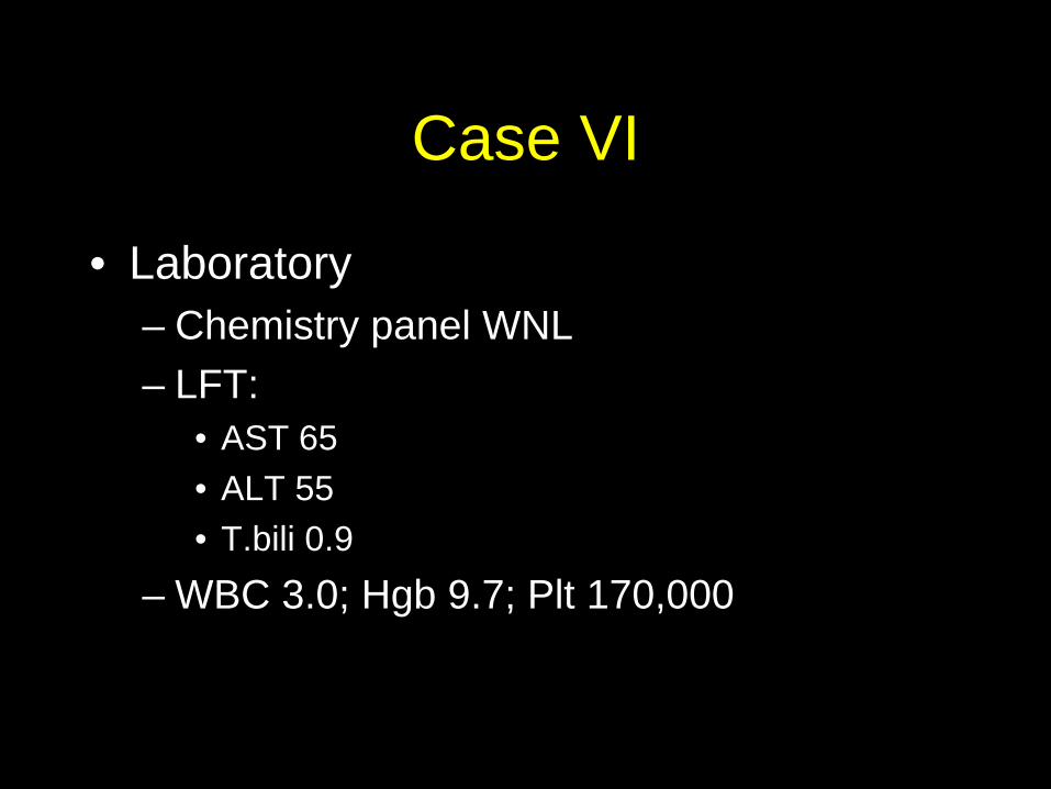
Case VI
• Laboratory– Chemistry panel WNL– LFT:
• AST 65• ALT 55• T.bili 0.9
– WBC 3.0; Hgb 9.7; Plt 170,000

CMV
• CMV:– CMV is a member of the herpesvirus group– Found universally throughout all
geographic locations and socioeconomic groups
– Infects between 50% and 85% of adults in the United States by 40 years of age
– Typically remains dormant within the body

CMV
• Transmission:– Transmission occurs from person to person.– Infection requires close, intimate contact with a
person excreting the virus:• saliva, urine, breast milk, transplanted organs, and rarely
from blood transfusions and other body fluids• Sexual transmission has been documented
– In most adults, reactivation, is the cause of symptomatic disease
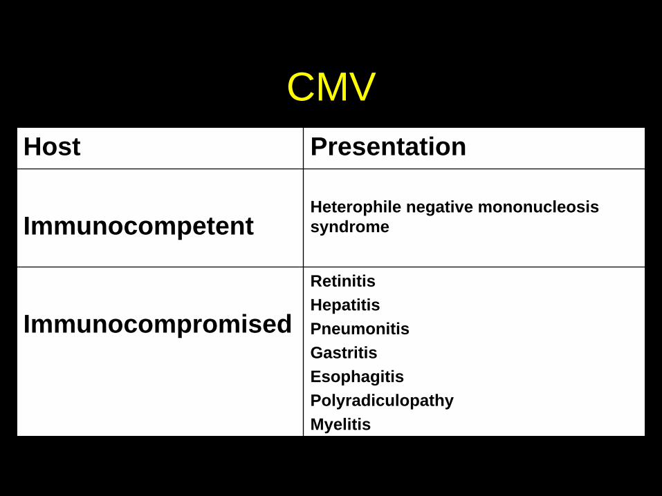
CMVHost Presentation
ImmunocompetentHeterophile negative mononucleosis syndrome
Immunocompromised
RetinitisHepatitisPneumonitisGastritisEsophagitisPolyradiculopathyMyelitis
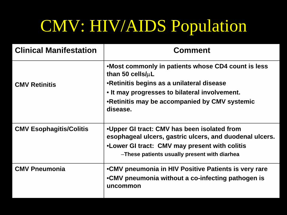
CMV: HIV/AIDS PopulationClinical Manifestation Comment
CMV Retinitis
•Most commonly in patients whose CD4 count is less than 50 cells/μL•Retinitis begins as a unilateral disease• It may progresses to bilateral involvement. •Retinitis may be accompanied by CMV systemic disease.
CMV Esophagitis/Colitis •Upper GI tract: CMV has been isolated from esophageal ulcers, gastric ulcers, and duodenal ulcers. •Lower GI tract: CMV may present with colitis
–These patients usually present with diarhea
CMV Pneumonia •CMV pneumonia in HIV Positive Patients is very rare•CMV pneumonia without a co-infecting pathogen is uncommon
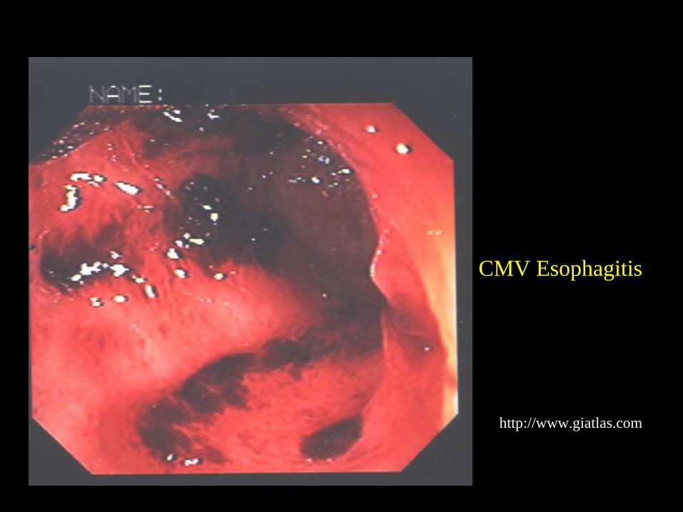
http://www.giatlas.com
CMV Esophagitis
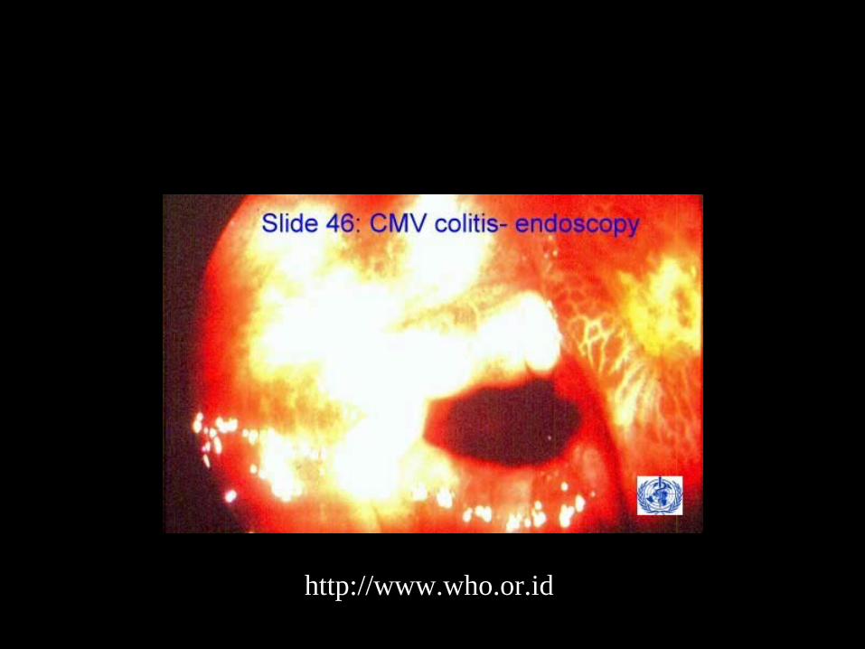
http://www.who.or.id
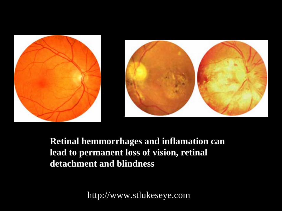
http://www.stlukeseye.com
Retinal hemmorrhages and inflamation can lead to permanent loss of vision, retinal detachment and blindness

http://www.ulb.ac.be/erasme/edu/gastrocd/Case35/C35c03.htm
•Diagnosis of CMV gastrointestinal disease by biopsy specimen demonstrating the CMV intranuclear inclusions

CMV- Organ transplantationClinical Manifestation
Comment
CMV pneumonia•Incidence varies depending on the transplant population
–Higher incidence and high mortality (86%) in allogeneic bone marrow transplant recipients–Less common and lower mortality in solid organ transplant recipients.–Major risk factor is a CMV seronegative transplant recipient receiving a CMV positive organ
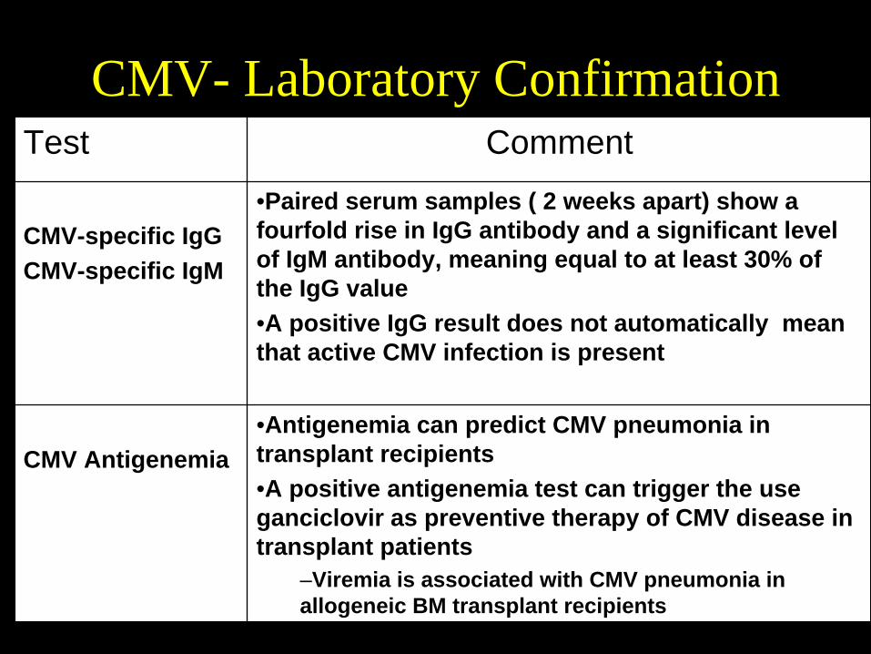
CMV- Laboratory ConfirmationTest Comment
CMV-specific IgGCMV-specific IgM
•Paired serum samples ( 2 weeks apart) show a fourfold rise in IgG antibody and a significant level of IgM antibody, meaning equal to at least 30% of the IgG value•A positive IgG result does not automatically mean that active CMV infection is present
CMV Antigenemia•Antigenemia can predict CMV pneumonia in transplant recipients•A positive antigenemia test can trigger the use ganciclovir as preventive therapy of CMV disease in transplant patients
–Viremia is associated with CMV pneumonia in allogeneic BM transplant recipients
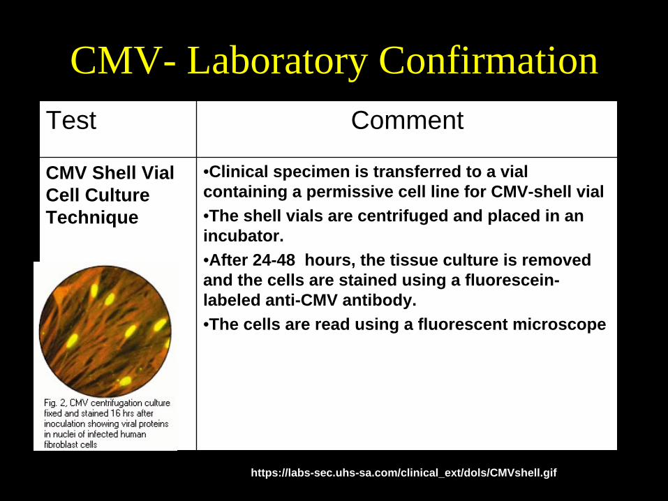
CMV- Laboratory ConfirmationTest Comment
CMV Shell Vial Cell Culture Technique
•Clinical specimen is transferred to a vial containing a permissive cell line for CMV-shell vial•The shell vials are centrifuged and placed in an incubator. •After 24-48 hours, the tissue culture is removed and the cells are stained using a fluorescein- labeled anti-CMV antibody. •The cells are read using a fluorescent microscope
https://labs-sec.uhs-sa.com/clinical_ext/dols/CMVshell.gif
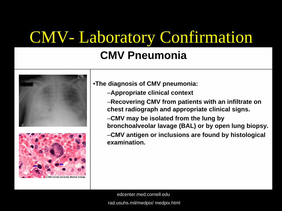
CMV- Laboratory ConfirmationCMV Pneumonia
•The diagnosis of CMV pneumonia:–Appropriate clinical context–Recovering CMV from patients with an infiltrate on chest radiograph and appropriate clinical signs. –CMV may be isolated from the lung by bronchoalveolar lavage (BAL) or by open lung biopsy.–CMV antigen or inclusions are found by histological examination.
edcenter.med.cornell.edu
rad.usuhs.mil/medpix/ medpix.html

CMV Treatment-Ganciclovir• Nucleoside analogue that
inhibits DNA synthesis • Major adverse effects of
are neutropenia and thrombocytopenia.
• Valganciclovir is the prodrug for ganciclovir– Absolute oral bioavailability
is approximately 60%– FDA approved for Rx of
CMV retinitis
Valganciclovir
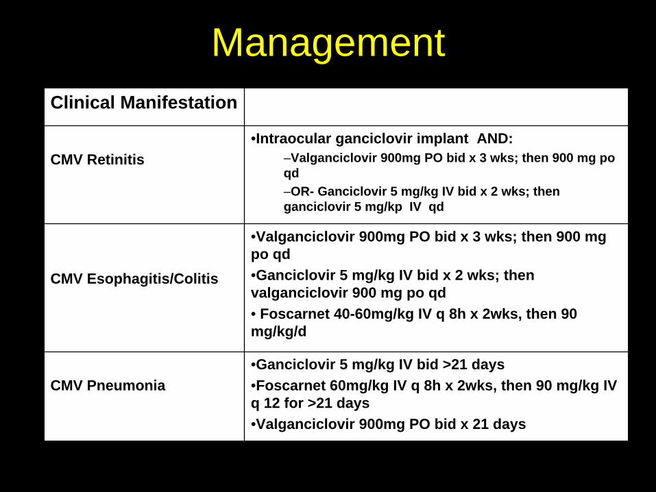
ManagementClinical Manifestation
CMV Retinitis•Intraocular ganciclovir implant AND:
–Valganciclovir 900mg PO bid x 3 wks; then 900 mg po qd–OR- Ganciclovir 5 mg/kg IV bid x 2 wks; then ganciclovir 5 mg/kp IV qd
CMV Esophagitis/Colitis
•Valganciclovir 900mg PO bid x 3 wks; then 900 mg po qd•Ganciclovir 5 mg/kg IV bid x 2 wks; then valganciclovir 900 mg po qd• Foscarnet 40-60mg/kg IV q 8h x 2wks, then 90 mg/kg/d
CMV Pneumonia•Ganciclovir 5 mg/kg IV bid >21 days•Foscarnet 60mg/kg IV q 8h x 2wks, then 90 mg/kg IV q 12 for >21 days•Valganciclovir 900mg PO bid x 21 days
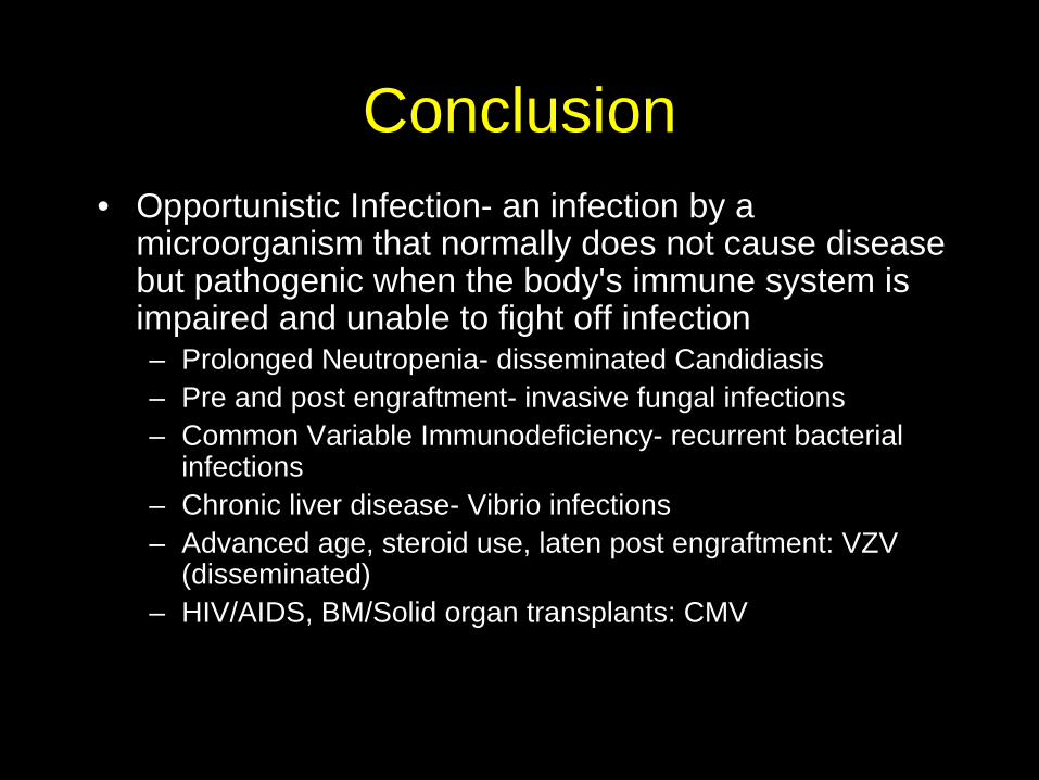
Conclusion• Opportunistic Infection- an infection by a
microorganism that normally does not cause disease but pathogenic when the body's immune system is impaired and unable to fight off infection– Prolonged Neutropenia- disseminated Candidiasis– Pre and post engraftment- invasive fungal infections– Common Variable Immunodeficiency- recurrent bacterial
infections– Chronic liver disease- Vibrio infections– Advanced age, steroid use, laten post engraftment: VZV
(disseminated)– HIV/AIDS, BM/Solid organ transplants: CMV

THE END