
Open stax biology (nonmajors) ch10
19
COLLEGE PHYSICS Chapter # Chapter Title PowerPoint Image Slideshow CONCEPTS OF BIOLOGY Chapter 10 BIOTECHNOLOGY PowerPoint Image Slideshow
-
Upload
lumen-learning -
Category
Documents
-
view
82 -
download
1
Transcript of Open stax biology (nonmajors) ch10

CONCEPTS OF BIOLOGYChapter 10 BIOTECHNOLOGY
PowerPoint Image Slideshow

FIGURE 10.1
(a) A thermal cycler, such as the one shown here, is a basic tool used to study DNA in a process called the polymerase chain reaction (PCR). The polymerase enzyme most often used with PCR comes from a strain of bacteria that lives in (b) the hot springs of Yellowstone National Park. (credit a: modification of work by Magnus Manske; credit b: modification of work by Jon Sullivan)

FIGURE 10.2
This diagram shows the basic method used for the extraction of DNA.

FIGURE 10.3
Shown are DNA fragments from six samples run on a gel, stained with a fluorescent dye and viewed under UV light. (credit: modification of work by James Jacob, Tompkins Cortland Community College)

FIGURE 10.4
Polymerase chain reaction, or PCR, is used to produce many copies of a specific sequence of DNA using a special form of DNA polymerase.
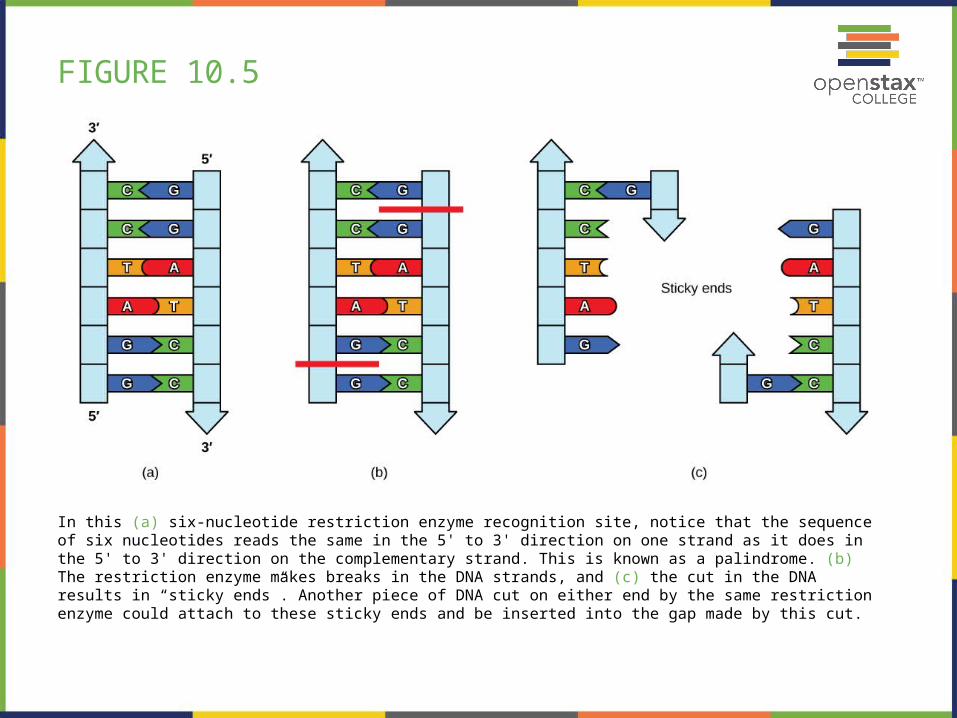
FIGURE 10.5
In this (a) six-nucleotide restriction enzyme recognition site, notice that the sequence of six nucleotides reads the same in the 5' to 3' direction on one strand as it does in the 5' to 3' direction on the complementary strand. This is known as a palindrome. (b) The restriction enzyme makes breaks in the DNA strands, and (c) the cut in the DNA results in “sticky ends”. Another piece of DNA cut on either end by the same restriction enzyme could attach to these sticky ends and be inserted into the gap made by this cut.

FIGURE 10.6
This diagram shows the steps involved in molecular cloning.

FIGURE 10.7
Dolly the sheep was the first agricultural animal to be cloned. To create Dolly, the nucleus was removed from a donor egg cell. The enucleated egg was placed next to the other cell, then they were shocked to fuse. They were shocked again to start division. The cells were allowed to divide for several days until an early embryonic stage was reached, before being implanted in a surrogate mother.

FIGURE 10.8
This diagram shows the steps involved in curing disease with gene therapy using an adenovirus vector. (credit: modification of work by NIH)

FIGURE 10.9
It can be seen that two of these mice are transgenic because they have a gene that causes them to fluoresce under a UV light. The non-transgenic mouse does not have the gene that causes fluorescence. (credit: Ingrid Moen et al.)

FIGURE 10.10
Corn, a major agricultural crop used to create products for a variety of industries, is often modified through plant biotechnology. (credit: Keith Weller, USDA)

FIGURE 10.11
This is a physical map of the human X chromosome. (credit: modification of work by NCBI, NIH)

FIGURE 10.12
Much basic research is done with model organisms, such as the mouse, Mus musculus; the fruit fly, Drosophila melanogaster; the nematode Caenorhabditis elegans; the yeast Saccharomyces cerevisiae; and the common weed, Arabidopsis thaliana. (credit “mouse”: modification of work by Florean Fortescue; credit “nematodes”: modification of work by “snickclunk”/Flickr; credit “common weed”: modification of work by Peggy Greb, USDA; scale-bar data from Matt Russell)

FIGURE 10.13
Metagenomics involves isolating DNA from multiple species within an environmental niche. The DNA is cut up and sequenced, allowing entire genome sequences of multiple species to be reconstructed from the sequences of overlapping pieces.

FIGURE 10.14
Renewable fuels were tested in Navy ships and aircraft at the first Naval Energy Forum. (credit: modification of work by John F. Williams, US Navy)

FIGURE 10.15
Bacillus anthracis is the organism that causes anthrax. (credit: modification of work by CDC; scale-bar data from Matt Russell)

FIGURE 10.16
Transgenic agricultural plants can be made to resist disease. These transgenic plums are resistant to the plum pox virus. (credit: Scott Bauer, USDA ARS)

FIGURE 10.17
This machine is preparing to do a proteomic pattern analysis to identify specific cancers so that an accurate cancer prognosis can be made. (credit: Dorie Hightower, NCI, NIH)

This PowerPoint file is copyright 2011-2013, Rice University. All Rights Reserved.