Open access Protocol Protocol for tumour-focused dose ...
13
1 Hafeez S, et al. BMJ Open 2020;10:e041005. doi:10.1136/bmjopen-2020-041005 Open access Protocol for tumour-focused dose- escalated adaptive radiotherapy for the radical treatment of bladder cancer in a multicentre phase II randomised controlled trial (RAIDER): radiotherapy planning and delivery guidance Shaista Hafeez , 1,2 Amanda Webster, 3 Vibeke N Hansen, 4 Helen A McNair, 1,2 Karole Warren-Oseni, 5 Emma Patel, 3 Ananya Choudhury, 6,7 Joanne Creswell, 8 Farshad Foroudi, 9 Ann Henry, 10,11 Tomas Kron, 12 Duncan B McLaren, 13 Anita V Mitra, 14 Hugh Mostafid, 15 Daniel Saunders, 16 Elizabeth Miles, 3 Clare Griffin, 17 Rebecca Lewis, 17 Emma Hall, 17 Robert Huddart 1,2 To cite: Hafeez S, Webster A, Hansen VN, et al. Protocol for tumour-focused dose- escalated adaptive radiotherapy for the radical treatment of bladder cancer in a multicentre phase II randomised controlled trial (RAIDER): radiotherapy planning and delivery guidance. BMJ Open 2020;10:e041005. doi:10.1136/ bmjopen-2020-041005 ► Prepublication history and additional material for this paper are available online. To view these files, please visit the journal online (http://dx.doi. org/10.1136/bmjopen-2020- 041005). Received 31 May 2020 Revised 03 November 2020 Accepted 12 November 2020 For numbered affiliations see end of article. Correspondence to Dr Shaista Hafeez; [email protected] Protocol © Author(s) (or their employer(s)) 2020. Re-use permitted under CC BY. Published by BMJ. ABSTRACT Introduction Daily radiotherapy delivered with radiosensitisation offers patients with muscle invasive bladder cancer (MIBC) comparable outcomes to cystectomy with functional organ preservation. Most recurrences following radiotherapy occur within the bladder. Increasing the delivered radiotherapy dose to the tumour may further improve local control. Developments in image-guided radiotherapy have allowed bladder tumour- focused ‘plan of the day’ radiotherapy delivery. We aim to test within a randomised multicentre phase II trial whether this technique will enable dose escalation with acceptable rates of toxicity. Methods and analysis Patients with T2-T4aN0M0 unifocal MIBC will be randomised (1:1:2) between standard/control whole bladder single plan radiotherapy, standard dose adaptive tumour-focused radiotherapy or dose-escalated adaptive tumour-focused radiotherapy (DART). Adaptive tumour-focused radiotherapy will use a library of three plans (small, medium and large) for treatment. A cone beam CT taken prior to each treatment will be used to visualise the anatomy and inform selection of the most appropriate plan for treatment. Two radiotherapy fractionation schedules (32f and 20f) are permitted. A minimum of 120 participants will be randomised in each fractionation cohort (to ensure 57 evaluable DART patients per cohort). A comprehensive radiotherapy quality assurance programme including pretrial and on-trial components is instituted to ensure standardisation of radiotherapy planning and delivery. The trial has a two-stage non-comparative design. The primary end point of stage I is the proportion of patients meeting predefined normal tissue constraints in the DART group. The primary end point of stage II is late Common Terminology Criteria for Adverse Events grade 3 or worse toxicity aiming to exclude a rate of >20% (80% power and 5% alpha, one sided) in each DART fractionation cohort. Secondary end points include locoregional MIBC control, progression-free survival overall survival and patient- reported outcomes. Ethics and dissemination This clinical trial is approved by the London-Surrey Borders Research Ethics Committee (15/LO/0539). The results when available will be disseminated via peer-reviewed scientific journals, conference presentations and submission to regulatory authorities. Trial registration number NCT02447549; Pre-results ARTICLE SUMMARY We present the first international randomised controlled trial protocol evaluating a dose- escalated tumour-focused image-guided adaptive radiotherapy technique. The study Strengths and limitations of this study ► Phase II international multicentre randomised con- trolled study evaluating a novel adaptive radiothera- py technique (strength). ► Treatment allocation favours 75% of participants receiving novel adaptive radiotherapy techniques (strength). ► Detailed guidance and training are provided for the contouring, planning and delivery of this radiother- apy technique to ensure standardisation across participating centres with robust pretrial and on- trial radiotherapy quality assurance programme (strength). ► Primary end point focus is based on determining safety of treatment based on late grade 3 toxicity scoring (strength). ► Non-comparative trial design (limitation). on February 26, 2022 by guest. Protected by copyright. http://bmjopen.bmj.com/ BMJ Open: first published as 10.1136/bmjopen-2020-041005 on 31 December 2020. Downloaded from
Transcript of Open access Protocol Protocol for tumour-focused dose ...
1Hafeez S, et al. BMJ Open 2020;10:e041005. doi:10.1136/bmjopen-2020-041005
Open access
Protocol for tumour- focused dose- escalated adaptive radiotherapy for the radical treatment of bladder cancer in a multicentre phase II randomised controlled trial (RAIDER): radiotherapy planning and delivery guidance
Shaista Hafeez ,1,2 Amanda Webster,3 Vibeke N Hansen,4 Helen A McNair,1,2 Karole Warren- Oseni,5 Emma Patel,3 Ananya Choudhury,6,7 Joanne Creswell,8 Farshad Foroudi,9 Ann Henry,10,11 Tomas Kron,12 Duncan B McLaren,13 Anita V Mitra,14 Hugh Mostafid,15 Daniel Saunders,16 Elizabeth Miles,3 Clare Griffin,17 Rebecca Lewis,17 Emma Hall,17 Robert Huddart1,2
To cite: Hafeez S, Webster A, Hansen VN, et al. Protocol for tumour- focused dose- escalated adaptive radiotherapy for the radical treatment of bladder cancer in a multicentre phase II randomised controlled trial (RAIDER): radiotherapy planning and delivery guidance. BMJ Open 2020;10:e041005. doi:10.1136/bmjopen-2020-041005
► Prepublication history and additional material for this paper are available online. To view these files, please visit the journal online (http:// dx. doi. org/ 10. 1136/ bmjopen- 2020- 041005).
Received 31 May 2020Revised 03 November 2020Accepted 12 November 2020
For numbered affiliations see end of article.
Correspondence toDr Shaista Hafeez; shaista. hafeez@ icr. ac. uk
Protocol
© Author(s) (or their employer(s)) 2020. Re- use permitted under CC BY. Published by BMJ.
ABSTRACTIntroduction Daily radiotherapy delivered with radiosensitisation offers patients with muscle invasive bladder cancer (MIBC) comparable outcomes to cystectomy with functional organ preservation. Most recurrences following radiotherapy occur within the bladder. Increasing the delivered radiotherapy dose to the tumour may further improve local control. Developments in image- guided radiotherapy have allowed bladder tumour- focused ‘plan of the day’ radiotherapy delivery. We aim to test within a randomised multicentre phase II trial whether this technique will enable dose escalation with acceptable rates of toxicity.Methods and analysis Patients with T2- T4aN0M0 unifocal MIBC will be randomised (1:1:2) between standard/control whole bladder single plan radiotherapy, standard dose adaptive tumour- focused radiotherapy or dose- escalated adaptive tumour- focused radiotherapy (DART). Adaptive tumour- focused radiotherapy will use a library of three plans (small, medium and large) for treatment. A cone beam CT taken prior to each treatment will be used to visualise the anatomy and inform selection of the most appropriate plan for treatment.Two radiotherapy fractionation schedules (32f and 20f) are permitted. A minimum of 120 participants will be randomised in each fractionation cohort (to ensure 57 evaluable DART patients per cohort).A comprehensive radiotherapy quality assurance programme including pretrial and on- trial components is instituted to ensure standardisation of radiotherapy planning and delivery.The trial has a two- stage non- comparative design. The primary end point of stage I is the proportion of patients meeting predefined normal tissue constraints in the DART group. The primary end point of stage II is late Common Terminology Criteria for Adverse Events grade 3 or worse toxicity aiming to exclude a rate of >20% (80% power and 5% alpha, one sided) in each DART fractionation cohort.
Secondary end points include locoregional MIBC control, progression- free survival overall survival and patient- reported outcomes.Ethics and dissemination This clinical trial is approved by the London- Surrey Borders Research Ethics Committee (15/LO/0539). The results when available will be disseminated via peer- reviewed scientific journals, conference presentations and submission to regulatory authorities.Trial registration number NCT02447549; Pre-results
ARTICLE SUMMARYWe present the first international randomised controlled trial protocol evaluating a dose- escalated tumour- focused image- guided adaptive radiotherapy technique. The study
Strengths and limitations of this study
► Phase II international multicentre randomised con-trolled study evaluating a novel adaptive radiothera-py technique (strength).
► Treatment allocation favours 75% of participants receiving novel adaptive radiotherapy techniques (strength).
► Detailed guidance and training are provided for the contouring, planning and delivery of this radiother-apy technique to ensure standardisation across participating centres with robust pretrial and on- trial radiotherapy quality assurance programme (strength).
► Primary end point focus is based on determining safety of treatment based on late grade 3 toxicity scoring (strength).
► Non- comparative trial design (limitation).
on February 26, 2022 by guest. P
rotected by copyright.http://bm
jopen.bmj.com
/B
MJ O
pen: first published as 10.1136/bmjopen-2020-041005 on 31 D
ecember 2020. D
ownloaded from

2 Hafeez S, et al. BMJ Open 2020;10:e041005. doi:10.1136/bmjopen-2020-041005
Open access
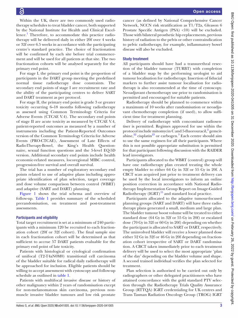
population are patients with unifocal localised muscle invasive bladder cancer. Patients will be randomised (1:1:2) between standard (control) whole bladder single plan radiotherapy (WBRT), or standard dose adaptive tumour- focused radiotherapy (SART) or dose- escalated adaptive tumour- focused radiotherapy (DART). For those randomised to adaptive tumour- focused radiotherapy groups treatment will be delivered using a library of three plans (plan of the day). If successful, the trial will demon-strate feasibly of multicentre implementation of this new radiotherapy technique and inform design of a future phase III trial to establish the optimum organ preserving treatment option for patients with MIBC.
INTRODUCTIONRadical management of localised muscle invasive bladder cancer (MIBC) involves either radical cystectomy or a course of daily radiotherapy delivered with radiosensi-tisation over 4–7 weeks.1–5 Although both have compa-rable overall survival outcomes in appropriately selected patients, radiotherapy offers opportunity for cancer cure with functional organ preservation.6
Most recurrences following radiotherapy occur within the bladder, the majority of which are believed to occur at the original MIBC tumour site, suggesting persistent occult local disease.7 The modelled dose- response rela-tionship of MIBC to radiotherapy suggests improved local control and overall survival would be expected at higher doses.8–10
The ability to safely increase dose beyond the current accepted standard has been restrained by reliable radio-therapy delivery to the bladder. The bladder is a mobile organ which is subject to marked shape and volume change during the course of treatment.11–13 This bladder motion means historically up to 57% of fractions (f) incur some element of geographical miss even when safety margins of up to 1.5 cm are applied to create the plan-ning target volume (PTV).14 The expected consequence of improving bladder radiotherapy targeting would be improved tumour control and reduced treatment- related toxicity.
Optimisation of target coverage has been enabled by technology integrated on current generation linear accelerators which allow a three- dimensional (3D) image known as a cone beam CT (CBCT) to be acquired. This is of sufficient contrast to allow soft tissue visualisation. When acquired immediately prior to treatment, it informs positional adjustment to ensure coverage of target with the radiotherapy plan.15
A solution enabled by CBCT soft tissue visualisation is ‘plan of the day’. Rather than having a single plan available for treatment, a library of plans of varying PTV bladder sizes can be created to cover the range of expected filling and positional variation of the bladder. A plan which best fits the bladder target with least normal tissue irradiation as seen on CBCT immediately prior to treatment is then selected for use each day.14 In bladder
cancer radiotherapy treatment delivery based on a library of plans has reported benefit in reducing normal tissue irradiation compared with single plan treatment delivery.16–19 It is yet to be demonstrated whether this approach translates to improved clinical outcomes.
Tumour- focused radiotherapy delivery may offer further opportunity to reduce normal tissue irradia-tion. Sparing the uninvolved bladder does not appear to compromise local control but randomised controlled studies have failed to demonstrate statistically significant improvement in toxicity.20 21 Bladder sparing is unlikely to have been optimally achieved in radiotherapy delivery predating CBCT image guidance given the positional uncertainties, the large margins applied and treatment delivery on an empty bladder.
In a single- centre phase I study (NCT01124682), feasi-bility and safety of tumour- focused dose escalation to 70 Gy delivered using plan of the day has been demon-strated. The RAIDER trial seeks to examine feasibility of this approach in a multicentre setting and to deter-mine the clinical benefit of bladder tumour- focused dose escalation.
Below, we describe the RAIDER trial protocol with particular emphasis on the radiotherapy procedural aspects, including preparatory imaging, treatment plan-ning and delivery with the aim of providing comprehen-sive description of the radiotherapy implemented for the study.
HypothesisTumour- focused dose- escalated adaptive radiotherapy using library of three plans can be translated to multiple centres. It will be well tolerated and offer the opportunity to improve local disease control for patients with bladder cancer.
MATERIALS AND ANALYSISStudy designRAIDER is an international multicentre, multi- arm, two- stage non- blinded phase II randomised controlled trial conducted in accordance with the Research Governance Framework for Health and Social Care and principles of Good Clinical Practice. The trial is registered on the clin-icaltrials. gov database (NCT02447549) and is included in the National Institute for Health Research (NIHR) Clin-ical Research Network portfolio. The final ethics approved version of the RAIDER trial protocol is provided in the supplementary files (online supplemental appendix 1).
Patients will be randomised (1:1:2) between standard (control) WBRT, SART or DART. Treatment allocation is by minimisation with a random element; balancing factors will be centre, neoadjuvant chemotherapy useand concomitant radiosensitising therapy use. Randomisation will take place centrally by the Clinical Trials and Statis-tics Unit, The Institute of Cancer Research (ICR- CTSU) within a maximum of 10 weeks prior to the planned radiotherapy start date.
on February 26, 2022 by guest. P
rotected by copyright.http://bm
jopen.bmj.com
/B
MJ O
pen: first published as 10.1136/bmjopen-2020-041005 on 31 D
ecember 2020. D
ownloaded from
3Hafeez S, et al. BMJ Open 2020;10:e041005. doi:10.1136/bmjopen-2020-041005
Open access
Within the UK, there are two commonly used radio-therapy schedules to treat bladder cancer, both supported by the National Institute for Health and Clinical Excel-lence.1 Therefore, to accommodate this practice radio-therapy will be delivered daily in either 20f over 4 weeks or 32f over 6.5 weeks in accordance with the participating centre’s standard practice. The choice of fractionation will be confirmed by each site before trial commence-ment and will be used for all patients at that site. The two fractionation cohorts will be analysed separately for the primary end point.
For stage I, the primary end point is the proportion of participants in the DART group meeting the predefined normal tissue radiotherapy dose constraints. The secondary end points of stage I are recruitment rate and the ability of the participating centres to deliver SART and DART treatment as per protocol.
For stage II, the primary end point is grade 3 or greater toxicity occurring 6–18 months following radiotherapy as assessed using Common Terminology Criteria for Adverse Events (CTCAE V.4). The secondary end points of stage II are acute toxicity as measured by CTCAE V.4, patient- reported outcomes as measured by a number of instruments including the Patient- Reported Outcomes version of the Common Terminology Criteria for Adverse Events (PRO- CTCAE), Assessment of Late Effects of RadioTherapy- Bowel, the King’s Health Question-naire, sexual function questions and the 5- level EQ- 5D version. Additional secondary end points include health economic- related measures, locoregional MIBC control, progression- free survival and overall survival.
The trial has a number of exploratory secondary end points related to use of adaptive plans including appro-priate identification of plan selection, target coverage and dose volume comparison between control (WBRT) and adaptive (SART and DART) planning.
Figure 1 shows the trial schema and overview of follow- up. Table 1 provides summary of the scheduled prerandomisation, on treatment and post- treatment assessments.
Participants and eligibilityTotal target recruitment is set at a minimum of 240 partic-ipants with a minimum 120 be recruited to each fraction-ation cohort (20f or 32f cohort). The final sample size in each fractionation cohort will be determined as that sufficient to accrue 57 DART patients evaluable for the primary end point of late toxicity.
Patients with histological or cytological confirmation of unifocal (T2- T4aN0M0) transitional cell carcinoma of the bladder suitable for radical daily radiotherapy will be approached for inclusion. Eligible patients should be willing to accept assessment with cystoscopy and follow- up schedule as outlined in table 1.
Patients with multifocal invasive disease or history of other malignancy within 2 years of randomisation except for non- melanomatous skin carcinoma, previous non- muscle invasive bladder tumours and low risk prostate
cancer (as defined by National Comprehensive Cancer Network, NCCN risk stratification as T1/T2a, Gleason 6 Prostate Specific Antigen (PSA) <10) will be excluded. Those with bilateral prosthetic hip replacements, previous history of radiation to the pelvis or other contraindication to pelvic radiotherapy, for example, inflammatory bowel disease will also be excluded.
Study treatmentAll participants should have had a transurethral resec-tion of the bladder tumour (TURBT) with completion of a bladder map by the performing urologist to aid tumour localisation for radiotherapy. Insertion of fiducial markers to further assist tumour localisation for radio-therapy is also recommended at the time of cystoscopy. Neoadjuvant chemotherapy use prior to randomisation is permitted and encouraged for suitable patients.
Radiotherapy should be planned to commence within a maximum of 10 weeks after randomisation or neoadju-vant chemotherapy completion (if used), to allow suffi-cient time for treatment planning.
Delivery of radiotherapy with concomitant radiosen-tiser is permitted. Regimes approved for use within the protocol include mitomycin C and 5- fluorouracil,2 gemcit-abine,22 cisplatin23 or carbogen.3 Each centre should aim to use the same regimen for all their participants. Where this is not possible appropriate substitution is permitted for that participant following discussion with the RAIDER lead investigators.
Participants allocated to the WBRT (control) group will have one radiotherapy plan created treating the whole empty bladder to either 64 Gy in 32f or 55 Gy in 20f. A CBCT scan acquired just prior to treatment delivery can be used by the local investigators to inform an online position correction in accordance with National Radio-therapy Implementation Group Report on Image- Guided Radiotherapy (IGRT)15 and standard local practice.
Participants allocated to the adaptive tumour- focused planning groups (SART and DART) will have three radio-therapy plans generated a small, medium and large plan. The bladder tumour boost volume will be treated to either standard dose (64 Gy in 32f or 55 Gy in 20f) or escalated dose (70 Gy in 32f or 60 Gy in 20f) depending on whether the participant is allocated to SART or DART, respectively. The uninvolved bladder will receive a lower planned dose either 52 Gy in 32f or 46 Gy in 20f depending on fraction-ation cohort irrespective of SART or DART randomisa-tion. A CBCT taken immediately prior to each treatment delivery will be used to select the most appropriate ‘plan of the day’ depending on the bladder volume and shape. A second trained individual verifies the plan selected for treatment.
Plan selection is authorised to be carried out only by radiographers or other delegated practitioners who have attained concordance with the gold standard PTV selec-tion through the Radiotherapy Trials Quality Assurance Group (RTTQA) IGRT credentialing for UK centres and Trans Tasman Radiation Oncology Group (TROG) IGRT
on February 26, 2022 by guest. P
rotected by copyright.http://bm
jopen.bmj.com
/B
MJ O
pen: first published as 10.1136/bmjopen-2020-041005 on 31 D
ecember 2020. D
ownloaded from

4 Hafeez S, et al. BMJ Open 2020;10:e041005. doi:10.1136/bmjopen-2020-041005
Open access
credentialing for Australian and New Zealand centres. This is to ensure all those participating in plan selection have the necessary advanced skill level required for the study.
A comprehensive quality assurance (QA) programme has been implemented for the RAIDER trial. This includes pretrial and on- trial components. Selection of appropriate treatment plans for the adaptive planning group will also be independently monitored during patient recruitment as part of the radiotherapy QA process.
Radiotherapy planning and deliveryRadiotherapy planning CT scanBladder preparation procedures vary depending on randomisation group. For WBRT, an empty bladder is required. Patients should be asked to abstain from drinking fluids for 30 min before the scheduled planning CT scan and are required to void their bladder immedi-ately before the planning CT scan is acquired (CT0).
For both SART and DART groups patients are instructed to void their bladder and then drink 350 mL of water. Two
Figure 1 Trial schema. f, fraction; PRO, patient- reported outcome; RTOG, Radiation Therapy Oncology Group.
on February 26, 2022 by guest. P
rotected by copyright.http://bm
jopen.bmj.com
/B
MJ O
pen: first published as 10.1136/bmjopen-2020-041005 on 31 D
ecember 2020. D
ownloaded from

5Hafeez S, et al. BMJ Open 2020;10:e041005. doi:10.1136/bmjopen-2020-041005
Open access
Tab
le 1
S
ched
ule
of a
sses
smen
ts
Vis
it/A
sses
smen
t
Pre
neo
adju
vant
ch
emo
ther
apy
(if g
iven
)P
rera
ndo
mis
atio
n*P
re- R
TO
n tr
eatm
ent
6 wee
ks
afte
r st
art
RT
†
10
wee
ks
afte
r st
art
RT
3 mo
nths
af
ter
end
R
T
6 mo
nths
af
ter
end
R
T
9 mo
nths
af
ter
end
R
T
12
mo
nths
af
ter
end
R
T
18
mo
nths
af
ter
end
RT
24
mo
nths
af
ter
end
R
TA
nnua
lly
to 5
yea
rsA
nnua
lly
ther
eaft
erA
t re
curr
ence
/d
isea
se p
rog
ress
ion
Rad
iolo
gica
l as
sess
men
t‡X
X
Dis
ease
st
atus
an
d
surv
ival
Tr
eatm
ent
acco
rdin
g to
loca
l p
ract
ice,
follo
w- u
p
for
dis
ease
sta
tus
and
sur
viva
l
TUR
BT
with
co
mp
letio
n of
b
lad
der
map
XX
Pla
cem
ent
of
fiduc
ial m
arke
rs
(op
tiona
l)
XX
X§
Ass
essm
ent
of
sym
pto
ms/
toxi
city
X¶
X¶
**X
¶X
¶X
¶X
††X
††X
††X
††X
††X
††
Full
blo
od
coun
t, u
r ea
and
el
ectr
olyt
es
XX
‡‡
X
X
PR
O q
uest
ionn
aire
(if
par
ticip
atin
g)
X
X§§
XX
¶¶
X¶
¶X
¶¶
X¶
¶
Rig
id c
ysto
scop
y an
d b
iop
sy o
f tu
mou
r b
ed
X
Che
st X
- ray
X
Flex
ible
cys
tosc
opy
XX
XX
XX
CT
of a
bd
omen
an
d p
elvi
s
X
X
X
Che
st X
- ray
or
CT
ches
t
X
XX
XX
Hea
lth r
esou
rce
utili
satio
n
X
XX
XX
X
*For
pat
ient
s w
ho h
ave
not
rece
ived
neo
adju
vant
che
mot
hera
py.
†For
pat
ient
s in
the
20f
coh
ort
only
.‡R
ecom
men
ded
imag
ing:
MR
I pel
vis,
CT
ches
t an
d a
bd
omen
. Min
imum
acc
epta
ble
is c
hest
, ab
dom
en, p
elvi
s C
T or
CT
ches
t an
d C
T ur
ogra
m.
§For
pat
ient
s w
ho r
ecei
ved
neo
adju
vant
che
mot
hera
py.
¶C
TCA
E V
.4.
**W
eekl
y on
tre
atm
ent.
††C
TCA
E V
.4 a
nd R
adia
tion
Ther
apy
Onc
olog
y G
roup
(RTO
G).
‡‡D
urin
g w
eeks
1, 4
and
6 (w
eek
6 on
ly if
rec
eivi
ng 3
2f).
§§A
t la
st fr
actio
n.¶
¶Q
uest
ionn
aire
s ad
min
iste
red
to
UK
par
ticip
ants
by
the
Clin
ical
Tria
ls a
nd S
tatis
tics
Uni
t, T
he In
stitu
te o
f Can
cer
Res
earc
h fr
om 6
mon
ths
onw
ard
s.C
TCA
E, C
omm
on T
erm
inol
ogy
Crit
eria
for
Ad
vers
e E
vent
s; f,
frac
tion;
RT,
rad
ioth
erap
y; T
UR
BT,
tra
nsur
ethr
al r
esec
tion
of t
he b
lad
der
tum
our.
on February 26, 2022 by guest. P
rotected by copyright.http://bm
jopen.bmj.com
/B
MJ O
pen: first published as 10.1136/bmjopen-2020-041005 on 31 D
ecember 2020. D
ownloaded from
6 Hafeez S, et al. BMJ Open 2020;10:e041005. doi:10.1136/bmjopen-2020-041005
Open access
planning scans are acquired, the first at 30 min following drinking (CT30) and the second 60 min following drinking (CT60). No voiding is permitted between the two scans. However, if voiding is unavoidable because of patient discomfort, then only the available CT30 scan is used for planning.
Given bladder deformation can occur with a loaded rectum, all participants should be encouraged to evac-uate their bowels of flatus and faeces prior to acquisition of the radiotherapy planning scanning. The use of micro-enemas is permitted if it is standard local practice but is not mandated.
All patients will be positioned supine with arms comfort-ably positioned out of the radiotherapy field using appro-priate immobilisation techniques for planning CT scan acquisition. CT slices of ≤3 mm thickness will be obtained from at least 4 cm above the dome of the bladder to 2 cm below the ischial tuberosities. No oral or intravenous contrast is required.
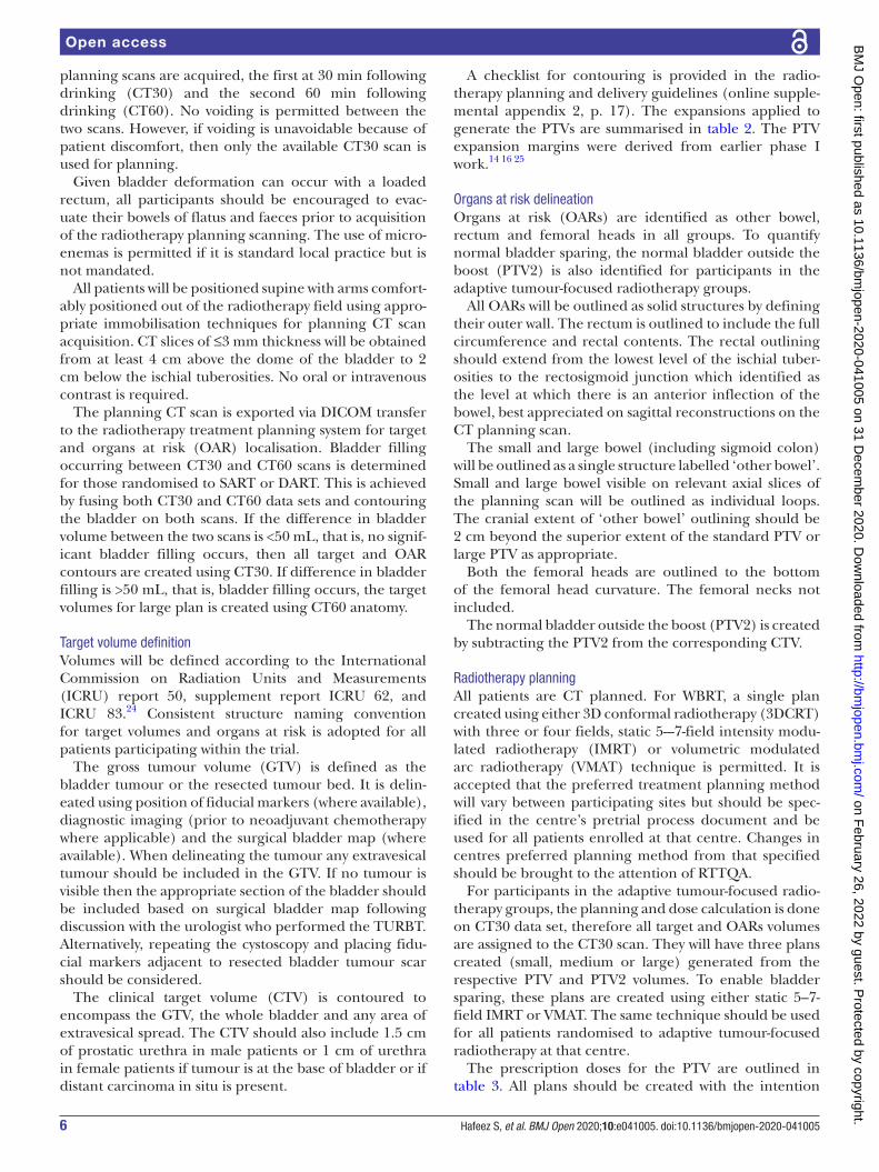
The planning CT scan is exported via DICOM transfer to the radiotherapy treatment planning system for target and organs at risk (OAR) localisation. Bladder filling occurring between CT30 and CT60 scans is determined for those randomised to SART or DART. This is achieved by fusing both CT30 and CT60 data sets and contouring the bladder on both scans. If the difference in bladder volume between the two scans is <50 mL, that is, no signif-icant bladder filling occurs, then all target and OAR contours are created using CT30. If difference in bladder filling is >50 mL, that is, bladder filling occurs, the target volumes for large plan is created using CT60 anatomy.
Target volume definitionVolumes will be defined according to the International Commission on Radiation Units and Measurements (ICRU) report 50, supplement report ICRU 62, and ICRU 83.24 Consistent structure naming convention for target volumes and organs at risk is adopted for all patients participating within the trial.
The gross tumour volume (GTV) is defined as the bladder tumour or the resected tumour bed. It is delin-eated using position of fiducial markers (where available), diagnostic imaging (prior to neoadjuvant chemotherapy where applicable) and the surgical bladder map (where available). When delineating the tumour any extravesical tumour should be included in the GTV. If no tumour is visible then the appropriate section of the bladder should be included based on surgical bladder map following discussion with the urologist who performed the TURBT. Alternatively, repeating the cystoscopy and placing fidu-cial markers adjacent to resected bladder tumour scar should be considered.
The clinical target volume (CTV) is contoured to encompass the GTV, the whole bladder and any area of extravesical spread. The CTV should also include 1.5 cm of prostatic urethra in male patients or 1 cm of urethra in female patients if tumour is at the base of bladder or if distant carcinoma in situ is present.
A checklist for contouring is provided in the radio-therapy planning and delivery guidelines (online supple-mental appendix 2, p. 17). The expansions applied to generate the PTVs are summarised in table 2. The PTV expansion margins were derived from earlier phase I work.14 16 25
Organs at risk delineationOrgans at risk (OARs) are identified as other bowel, rectum and femoral heads in all groups. To quantify normal bladder sparing, the normal bladder outside the boost (PTV2) is also identified for participants in the adaptive tumour- focused radiotherapy groups.
All OARs will be outlined as solid structures by defining their outer wall. The rectum is outlined to include the full circumference and rectal contents. The rectal outlining should extend from the lowest level of the ischial tuber-osities to the rectosigmoid junction which identified as the level at which there is an anterior inflection of the bowel, best appreciated on sagittal reconstructions on the CT planning scan.
The small and large bowel (including sigmoid colon) will be outlined as a single structure labelled ‘other bowel’. Small and large bowel visible on relevant axial slices of the planning scan will be outlined as individual loops. The cranial extent of ‘other bowel’ outlining should be 2 cm beyond the superior extent of the standard PTV or large PTV as appropriate.
Both the femoral heads are outlined to the bottom of the femoral head curvature. The femoral necks not included.
The normal bladder outside the boost (PTV2) is created by subtracting the PTV2 from the corresponding CTV.
Radiotherapy planningAll patients are CT planned. For WBRT, a single plan created using either 3D conformal radiotherapy (3DCRT) with three or four fields, static 5-–7- field intensity modu-lated radiotherapy (IMRT) or volumetric modulated arc radiotherapy (VMAT) technique is permitted. It is accepted that the preferred treatment planning method will vary between participating sites but should be spec-ified in the centre’s pretrial process document and be used for all patients enrolled at that centre. Changes in centres preferred planning method from that specified should be brought to the attention of RTTQA.
For participants in the adaptive tumour- focused radio-therapy groups, the planning and dose calculation is done on CT30 data set, therefore all target and OARs volumes are assigned to the CT30 scan. They will have three plans created (small, medium or large) generated from the respective PTV and PTV2 volumes. To enable bladder sparing, these plans are created using either static 5–7- field IMRT or VMAT. The same technique should be used for all patients randomised to adaptive tumour- focused radiotherapy at that centre.
The prescription doses for the PTV are outlined in table 3. All plans should be created with the intention
on February 26, 2022 by guest. P
rotected by copyright.http://bm
jopen.bmj.com
/B
MJ O
pen: first published as 10.1136/bmjopen-2020-041005 on 31 D
ecember 2020. D
ownloaded from

7Hafeez S, et al. BMJ Open 2020;10:e041005. doi:10.1136/bmjopen-2020-041005
Open access
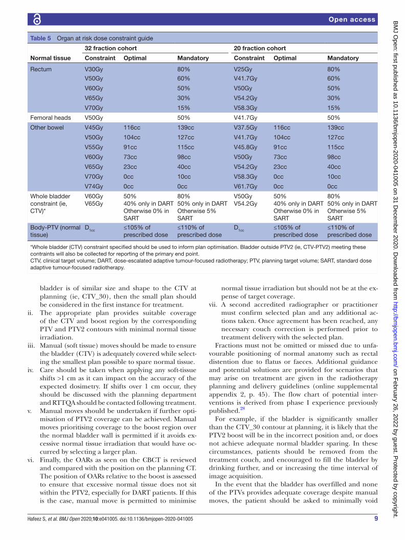
of achieving the target volume objectives as outlined in table 4. Dose to OARs should be as low possible. The OARs dose volume constraints for both fractionations are summarised in table 5.
The other bowel, rectumand femoral heads constraints for the 32f schedule were derived from previous phase III prostate (CHHiP, convential or hypofractionated high dose intensity modulated radiotherapy for pros-tate cancer; ISRCTN97182923) and bladder (BC2001; ISRCTN68324339) studies2 26 27 and from phase I work.28 The absence of previously defined OARs constraints when dose escalating in 20f meant that the OARs constraints at higher doses were marginally more conservative than if otherwise converted exactly from 32f constraint level using the linear quadratic model alone.29 The constraints used for the 20f schedule were estimated from the 32f constraint level using the linear quadratic equation assuming that all α/β of organs at risk is 3 but the dose constraint is reached in 3 Gy per fraction.
Dose objectives to the PTV should not be compromised to achieve dose to OAR constraints. The recommended hierarchy of planning priorities is providing radiotherapy planning and delivery guidelines (online supplemental appendix 2, p. 27).
For patients randomised to WBRT, it is at the local principal investigator’s (PI) discretion to accept the OAR doses. For those randomised to adaptive tumour- focused radiotherapy groups it is recommended that the predefined optimal dose constraints are met for the small plan, and the mandatory constraints for the medium plan wherever possible. It is accepted that the rectum and bowel dose constraints of the large plan may not be met despite adequate optimisation. Assessment of ‘other bowel’ dose on the large plan represents an overestima-tion of actual dose compared with ‘other bowel’ when this plan is actually used to deliver treatment. This is because when the large plan is selected for treatment, a propor-tion of bowel moves out of the field with bladder filling.
For patients allocated to DART, if the mandatory constraints are not met on the medium plan advice must be sought from the RTTQA team. Decision will be then made by the RAIDER trial team regarding the appropri-ateness of proceeding at the DART prescription dose or to lowering the prescribed dose as per SART randomi-sation. It is therefore recommended that the medium plan be optimised first. If patients are not able to receive DART (in either fractionation cohort) for any reason then details of the deviation from allocated treatment will be requested
Preradiotherapy checksTo minimise risk of error at the time of plan importing, exporting and plan selection, it is recommended that each plan, beam name and ID reflect the assigned plan, for example, Sm_Plan used for labelling the beams making up the small plan in the adaptive tumour- focused radiotherapy groups. It is also important to ensure that the local record and verify systems for Ta
ble
2
PTV
exp
ansi
on d
etai
ls
Pat
ient
ran
do
mis
atio
nC
T d
ata
set
PT
V
CT
V t
o P
TV
exp
ansi
on
(cm
)
PT
V2
GT
V t
o P
TV
2 ex
pan
sio
n (c
m)
Late
rally
Ant
erio
rly
Po
ster
iorl
yS
uper
iorl
yIn
feri
orl
yLa
tera
llyA
nter
iorl
yP
ost
erio
rly
Sup
erio
rly
Infe
rio
rly
Gro
up 1
Sta
ndar
d w
hole
b
lad
der
(WB
RT)
CT0
PTV
0.8
1.5
1.2
1.5
0.8
Not
ap
plic
able
Gro
up 2
and
3A
dap
tive
tum
our-
fo
cuse
d(S
AR
T an
d D
AR
T)
CT3
0P
TV_S
m0.
50.
50.
50.
50.
5P
TV2_
Sm
0.5
0.5
0.5
0.5
0.5
CT3
0P
TV_M
ed0.
51.
51.
01.
50.
5P
TV2_
Med
0.5
1.5
1.0
1.5
0.5
If C
T60-
CT3
0 b
lad
der
filli
ng <
50 m
L th
en a
pp
ly
CT3
0P
TV_L
ar_3
00.
82.
01.
22.
50.
8P
TV2_
Lar_
300.
82.
01.
22.
50.
8
If C
T60-
CT3
0 b
lad
der
filli
ng >
50 m
L th
en a
pp
ly
CT6
0P
TV_L
ar_6
00.
51.
51.
01.
50.
5P
TV2_
Lar_
600.
51.
51.
01.
50.
5
CTV
, clin
ical
tar
get
volu
me;
PTV
, pla
nnin
g ta
rget
vol
ume.
on February 26, 2022 by guest. P
rotected by copyright.http://bm
jopen.bmj.com
/B
MJ O
pen: first published as 10.1136/bmjopen-2020-041005 on 31 D
ecember 2020. D
ownloaded from

8 Hafeez S, et al. BMJ Open 2020;10:e041005. doi:10.1136/bmjopen-2020-041005
Open access
3DCRT and IMRT cannot mix beams from different plans at the time of exporting or deliver more than one plan at treatment.
Treatment schedulingRadiotherapy can start on any day of the week and should be delivered 5 days a week until completion. Interrup-tions during radiotherapy should be avoided as they have detrimental effect on outcome.30 All missed fractions are to be reported to the ICR- CTSU and RTTQA team.
In the event of missed fractions due to machine breakdown, bank holiday or any other logistical reason compensation for the missed fraction is advised. This is expected to be achieved by either treating at a weekend or by hyperfractionating, that is, undertaking two frac-tions a day (ideally on a Friday) with a minimum 6- hour gap between treatments. Should a treatment break occur due to toxicity, centres are advised to contact ICR- CTSU and/or RTTQA. Compensation is not expected in circumstances where missed treatment is a result of radiotherapy- related toxicity.
For those allocated to adaptive tumour- focused radio-therapy groups if plan selection capabilities are unavail-able, either because of absence of trained staff, machine breakdown and/or gap day treatment, patients may be treated for up to 5 days using the PTV medium plan without plan selection. These pretreatment CBCTs (if acquired) should be sent to RTTQA for review.
Treatment deliveryThe same patient preparation instructions used at plan-ning CT should be implemented prior to each fraction delivered.
For those patients allocated to SART or DART, CBCT of the pelvis should be acquired prior to each fraction. For those patients randomised to WBRT, pretreatment CBCT should be used in accordance with guidance provided in the NRIG IGRT report.15 It is therefore expected that this CBCT will inform appropriate corrections (either manual or automatic) to be applied prior to the delivered fraction in accordance with the centre’s local practice to ensure that treatment is accurately directed. Any changes made on the basis of the scan including exposures that do not lead to treatment because of patient factors should be reported in the case report forms (CRF) and to RTTQA.
For those randomised to adaptive tumour- focused radiotherapy groups, the pretreatment CBCT is acquired and registered to bone according to the guidance provided in the NRIG IGRT report.15 An appropriately trained radiographer or practitioner reviews the bone- matched CBCT assessing the bladder size and position in relation to the PTVs and the coverage they provide.
To assist trained radiographers or practitioners with optimal plan selection the following sequential assess-ment is advised:i. Following CBCT acquisition, the bladder filling and
shape is first checked against CTV_30 contour. If the
Table 3 Prescription doses
Patient randomisation Volume
32 fraction cohort 20 fraction cohort
Dose (Gy) Dose per fraction (Gy) Dose (Gy) Dose per fraction (Gy)
Group 1WBRT
PTV_Std 64 2 55 2.75
Group 2SART
PTV2 64 2 55 2.75
52 1.625 46 2.3
Group 3DART
PTV2 70 2.1875 60 3
52 1.625 46 2.3
DART, dose- escalated adaptive tumour- focused radiotherapy; PTV, planning target volume; SART, standard dose adaptive tumour- focused radiotherapy; WBRT, whole bladder single plan radiotherapy.
Table 4 Target volume dose objectives
Volume Dose constraints Optimal Mandatory
PTV2 D98% ≥95% of prescribed dose ≥90% of prescribed dose
D50%* – ±1% of prescribed dose
D2% ≤105% of prescribed dose ≤107% of prescribed dose
PTV(PTV–PTV2)
D98% ≥95% of prescribed dose ≥90% of prescribed dose
*Please note that D50% constraint refers only to PTV2. PTV D50% is likely to be exceeded depending on size of PTV2. Therefore, no compromise to PTV2 coverage should be made at the expense of achieving D50% PTV constraint.PTV, planning target volume.
on February 26, 2022 by guest. P
rotected by copyright.http://bm
jopen.bmj.com
/B
MJ O
pen: first published as 10.1136/bmjopen-2020-041005 on 31 D
ecember 2020. D
ownloaded from
9Hafeez S, et al. BMJ Open 2020;10:e041005. doi:10.1136/bmjopen-2020-041005
Open access
bladder is of similar size and shape to the CTV at planning (ie, CTV_30), then the small plan should be considered in the first instance for treatment.
ii. The appropriate plan provides suitable coverage of the CTV and boost region by the corresponding PTV and PTV2 contours with minimal normal tissue irradiation.
iii. Manual (soft tissue) moves should be made to ensure the bladder (CTV) is adequately covered while select-ing the smallest plan possible to spare normal tissue.
iv. Care should be taken when applying any soft- tissue shifts >1 cm as it can impact on the accuracy of the expected dosimetry. If shifts over 1 cm occur, they should be discussed with the planning department and RTTQA should be contacted following treatment.
v. Manual moves should be undertaken if further opti-misation of PTV2 coverage can be achieved. Manual moves prioritising coverage to the boost region over the normal bladder wall is permitted if it avoids ex-cessive normal tissue irradiation that would have oc-curred by selecting a larger plan.
vi. Finally, the OARs as seen on the CBCT is reviewed and compared with the position on the planning CT. The position of OARs relative to the boost is assessed to ensure that excessive normal tissue does not sit within the PTV2, especially for DART patients. If this is the case, manual move is permitted to minimise
normal tissue irradiation but should not be at the ex-pense of target coverage.
vii. A second accredited radiographer or practitioner must confirm selected plan and any additional ac-tions taken. Once agreement has been reached, any necessary couch correction is performed prior to treatment delivery with the selected plan.
Fractions must not be omitted or missed due to unfa-vourable positioning of normal anatomy such as rectal distention due to flatus or faeces. Additional guidance and potential solutions are provided for scenarios that may arise on treatment are given in the radiotherapy planning and delivery guidelines (online supplemental appendix 2, p. 45). The flow chart of potential inter-ventions is derived from phase I experience previously published.28
For example, if the bladder is significantly smaller than the CTV_30 contour at planning, it is likely that the PTV2 boost will be in the incorrect position and, or does not achieve adequate normal bladder sparing. In these circumstances, patients should be removed from the treatment couch, and encouraged to fill the bladder by drinking further, and or increasing the time interval of image acquisition.
In the event that the bladder has overfilled and none of the PTVs provides adequate coverage despite manual moves, the patient should be asked to minimally void
Table 5 Organ at risk dose constraint guide
Normal tissue
32 fraction cohort 20 fraction cohort
Constraint Optimal Mandatory Constraint Optimal Mandatory
Rectum V30Gy 80% V25Gy 80%
V50Gy 60% V41.7Gy 60%
V60Gy 50% V50Gy 50%
V65Gy 30% V54.2Gy 30%
V70Gy 15% V58.3Gy 15%
Femoral heads V50Gy 50% V41.7Gy 50%
Other bowel V45Gy 116cc 139cc V37.5Gy 116cc 139cc
V50Gy 104cc 127cc V41.7Gy 104cc 127cc
V55Gy 91cc 115cc V45.8Gy 91cc 115cc
V60Gy 73cc 98cc V50Gy 73cc 98cc
V65Gy 23cc 40cc V54.2Gy 23cc 40cc
V70Gy 0cc 10cc V58.3Gy 0cc 10cc
V74Gy 0cc 0cc V61.7Gy 0cc 0cc
Whole bladder constraint (ie, CTV)*
V60GyV65Gy
50%40% only in DARTOtherwise 0% in SART
80%50% only in DARTOtherwise 5% SART
V50GyV54.2Gy
50%40% only in DARTOtherwise 0% in SART
80%50% only in DARTOtherwise 5% SART
Body- PTV (normal tissue)
D1cc ≤105% of prescribed dose
≤110% of prescribed dose
D1cc ≤105% of prescribed dose
≤110% of prescribed dose
*Whole bladder (CTV) constraint specified should be used to inform plan optimisation. Bladder outside PTV2 (ie, CTV- PTV2) meeting these contraints will also be collected for reporting of the primary end point.CTV, clinical target volume; DART, dose- escalated adaptive tumour- focused radiotherapy; PTV, planning target volume; SART, standard dose adaptive tumour- focused radiotherapy.
on February 26, 2022 by guest. P
rotected by copyright.http://bm
jopen.bmj.com
/B
MJ O
pen: first published as 10.1136/bmjopen-2020-041005 on 31 D
ecember 2020. D
ownloaded from

10 Hafeez S, et al. BMJ Open 2020;10:e041005. doi:10.1136/bmjopen-2020-041005
Open access
and the CBCT is repeated. If this is not possible, patient should void completely and restart drinking protocol with a reduction in the time interval for CBCT acquisi-tion. In these circumstances, a member of the clinical team should also be notified to ensure the patient is not in urinary retention.
When amending the drinking protocol to optimise patient’s anatomy to fit the existing PTV contours, it is advised that one aspect is changed at a time, that is, interval for CBCT acquisition timing or the amount of water that is drunk. This is so the impact of the interven-tion can be determined and altered for subsequent frac-tions as required.
If no PTV contours are suitable to cover the target because of rectal gas, then the patient should be removed from the bed and ask to void. The CBCT image acquisi-tion is then repeated. If the PTV contours still are not optimal, it is recommended that the most suitable plan is selected which optimises coverage of PTV2 and mini-mises the inclusion of OARs is chosen for treatment. If this occurs repeatedly (eg, more than twice in five frac-tions) RTTQA should be contacted for advice.
All CBCT exposures including those not resulting in treatment should be recorded on the CRF and plan selec-tion form.
In all randomised groups, a post- treatment CBCT should be taken during the first week and once a week thereafter. This CBCT should be reviewed locally to ensure intrafraction filling has been accommodated for at the time of plan selection.
Radiotherapy protocol compliance programmeThe RAIDER trial is subject to radiotherapy QA programme that aims to standardise contouring, plan-ning and delivery of image- guided and adaptive bladder radiotherapy in participating centres. The RTTQA group coordinates the UK QA programme for the study. For Australian and New Zealand participants, this is coordi-nated by the TROG QA Team.
The QA programme has a pretrial and on- trial compo-nent. Each centre will be required to complete the pre- trial QA prior to commencing recruitment.
Prior to trial entry, participating centres will be asked to complete a facility questionnaire in order to gauge current local IGRT experience. A separate process docu-ment is used to collect task details of all aspects of a complete patient pathway.
The PI at each participating site is asked to contour two benchmark clinical cases as per protocol. One case includes tumour bed GTV as defined by placement of fiducial markers (radio- opaque contrast agent, lipiodol). UK PIs who completed outlining benchmark cases for the preceding phase II adaptive bladder radiotherapy trial (HYBRID Trial, NCT01810757) will be asked to contour only the target volumes as the OARs contouring is unchanged for the RAIDER protocol.31 Structured feedback to the PI will be provided via RTTQA team.
All participating trial centres will also be required to complete a planning benchmark case. Centres will be provided with access to CT DICOM data and preout-lined structure set. They will be requested to the plan this patient in their own treatment planning system as if randomised to the DART arm. It is the responsibility of the local investigator to ensure that appropriate plan checking QA process is in place at their local institu-tion. Once the three plans of the benchmark case have been created, reviewed and accepted by the local PI, the DICOM CT, dose cubes, RTplan and structure sets are returned to the RTTQA team and structured feedback is provided.
It is a pretrial requirement that all participating centres have both an established IGRT training programme in place for their radiographers and be using CBCT to assess bladder treatment delivery. Trial- specific bladder IGRT competency will be completed through an online plan selection training package, and practical workshop.32
The online plan selection training consists of two practice cases each with six CBCTs to work through. Step- by- step instructions with correct plan selections is provided. Following this, a credentialing assessment consisting of 12 plan selections will be carried out. The plan selections and matched reviews will be assessed by RTTQA and structured feedback provided. Only those who meet minimum threshold of concordance of plan selection as predefined by the trial team will be approved for performing RAIDER plan selection. Those who were accredited for plan selection in the HYBRID study31 or in the TROG 10.1 BOLART trial training (NCT01142102)33 will not be asked to repeat this assessment.
As part of the on- trial QA, the contouring and planning of at least the first adaptive patient and the first DART will be subject to prospective review by the RTTQA group.
All planning data and treatment delivery data including paired weekly pretreatment and post- treatment CBCTs, registration objects and treatment forms will be collected and reviewed retrospectively by the RTTQA group to ensure adherence to the RAIDER planning and delivery protocol is maintained. Remote retrospective plan selec-tion review will take place for adaptive radiotherapy patients during the trial.
Statistical considerationsThe primary aim of the study is to evaluate the feasibility (stage I) and safety (stage II) of DART. Control (WBRT) and SART treatment groups are included to enable SART to be carried forward to stage II if dose constraints cannot be met in the DART group and to assess equipoise and feasibility of recruitment for any subsequent phase III trial. Prospectively collected contemporaneous toxicity data for WBRT and SART will also allow benchmarking of DART results. Patients are randomised 1:1:2 to maxi-mise information on DART. Recruitment to stage II will continue seamlessly while stage I is evaluated, unless advised otherwise by the Independent Data Monitoring
on February 26, 2022 by guest. P
rotected by copyright.http://bm
jopen.bmj.com
/B
MJ O
pen: first published as 10.1136/bmjopen-2020-041005 on 31 D
ecember 2020. D
ownloaded from

11Hafeez S, et al. BMJ Open 2020;10:e041005. doi:10.1136/bmjopen-2020-041005
Open access
Committee (IDMC). Patients recruited in stage I will contribute to analysis of stage II.
The sample size of stage I is based on proportion of patients allocated to DART meeting the predefined dose constraints of bladder, bowel and rectum on the medium plan. A patient in the 32f cohort will be defined as meeting the dose constraints if all mandatory constraints of the following are met for the medium plan: rectum constraints at 50 Gy, 60 Gy, 65 Gy and 70 Gy; bladder outside PTV2 at 60 Gy and 65 Gy and small bowel at V55, V60, V65, V70 and V74. A patient in the 20f cohort will be defined as meeting the dose constraints if all mandatory constraints of the following are met for the medium plan: rectum constraints at 41.7 Gy, 50 Gy, 54.2 Gy and 58.3 Gy; bladder outside PTV2 at 50 Gy and 54.2 Gy and bowel at V45.8, V50, V54.2, V58.3 and V61.7.
It is expected that in 80% of DART patients the predefined dose constraints of the medium plan to the normal bladder, bowel and rectum will be met. If this proportion is <50%, it will be concluded that DART delivery is not feasible. Using an A’Hern single stage design (p0=0.5, p1=0.8, 5% alpha and 80% power), 18 patients are required in each DART fractionation cohort. If at least 13/18 meet dose constraints, it will be concluded that DART treatment is feasible; if dose constraints are not met for six or more patients in either fractionation cohort, the IDMC will advise on continuation of the trial with the option of dropping the DART arm in one or both fractionation cohorts and continuing to stage II with randomisation to WBRT versus SART. Stage I will there-fore require a total of 72 patients (36 in each fraction-ation cohort) randomised 1:1:2 between WBRT, SART and DART.
There are no formal early stopping rules for acute toxicity or efficacy but if after six patients have been treated per fractionation cohort, >50% of patients experience acute >grade 3 treatment- related toxicity, the IDMC would be asked to advise on suitability of continuation.
Stage II has a non- comparative design aiming to rule out an upper limit of any late ≥grade 3 CTCAE toxicity in each DART fractionation cohort. To be considered eval-uable for the primary end point of late toxicity, a patient must receive at least one fraction of allocated treatment and have at least one toxicity assessment performed between 6 and 18 months after completing radiotherapy. It is expected that the proportion of patients in the control group reporting >grade 3 toxicity CTCAE toxicity between 6 and 18 months postradiotherapy will be 8%.20 Again using an A’Hern single stage design (p0 (toxicity free)=0.80, p1=0.92, 5% alpha and 80% power), 57 patients in each DART fractionation cohort will allow a >20% >grade 3 toxicity CTCAE toxicity to be excluded. If more than >6/57 evaluable DART patients experience ≥grade 3 toxicity in either fractionation cohort, then the late toxicity threshold will be exceeded and on the IDMC’s recommendation the trial could either be stopped or the DART arm dropped.
Allowing for 5% non- evaluability for late toxicity by 18 months gives a sample size of 120 patients (30 WBRT, 30 SART, 60 DART) for each fractionation cohort, that is, a total target sample size of 240. The non- evaluability rate will be monitored and, with IDMC endorsement, cohort recruitment will continue until there are 57 evaluable DART patients per cohort. During stage II, following IDMC review, consideration would be given to dropping the WBRT or SART arms, if it was felt sufficient data had accrued for these arms and it would expedite meeting the aims of the trial. The IDMC will also monitor recurrence rates. If an absolute excess of locoregional recurrence is seen, early termination of the trial would be considered.
For stage I, the primary end point will be presented as the frequency and percentage of randomised patients able to meet the trial dose constraints in the DART group. For stage II, the primary end point will be based on the evaluable population. The proportion of patients with any >grade 3 CTCAE toxicity occurring within 6–18 months postradiotherapy will be presented for each randomised treatment group together with the 90% one- sided bino-mial CI (the 90% two- sided CI will also be presented). A sensitivity analysis will be conducted using a per- protocol population. The per- protocol population will include evaluable patients who received their complete fraction-ation schedule (either 32f or 20f) according to their randomised allocation group.
The local control rate at 2 years will be presented by treatment group with a 95% CI. Acute and late toxicity will be summarised by frequencies and proportions at each time point by treatment group. Kaplan- Meier methods will also be used to analyse time to local disease progression and overall survival with data presented by randomised group.
ETHICS AND DISSEMINATIONThe trial is approved by the London- Surrey Borders Research Ethics Committee (15/LO/0539).
The first participant was enrolled in October 2015. The study recruitment is scheduled to complete in Spring 2020. It is expected that the trial will report in 2022, following which the results will be disseminated via peer- reviewed scientific journals, conference presentations and submission to regulatory authorities.
Safety reportingData are collected at each trial visit regarding any adverse events graded according to CTCAE V.4 criteria on the CRF. The highest grade observed since the last visit should be reported. All serious adverse events (SAEs) occurring from the start of radiotherapy up to 30 days following the last fraction and any radiotherapy- related >grade 3 events occurring between 6 and 18 months are reported to the ICR- CTSU within 24 hours of the PI becoming aware of the event. SAEs should be followed up until clinical recovery is complete or until the condition has stabilised. Any safety concerns will be reported to the main research
on February 26, 2022 by guest. P
rotected by copyright.http://bm
jopen.bmj.com
/B
MJ O
pen: first published as 10.1136/bmjopen-2020-041005 on 31 D
ecember 2020. D
ownloaded from
12 Hafeez S, et al. BMJ Open 2020;10:e041005. doi:10.1136/bmjopen-2020-041005
Open access
and ethics committee by ICR- CTSU as part of the annual progress report.
Trial monitoring and oversightA Trial Management Group (TMG) will be set up and will include the Chief Investigator, ICR- CTSU Methodology Lead, co- investigators, identified collaborators, the trial statistician, trial manager and patient representative.
The ICR- CTSU Urology Radiotherapy Trials Steering Committee (TSC) includes a chairperson not directly involved in the trial, and at least two other independent members who will oversee the RAIDER trial. The TSC will meet annually.
An IDMC will be set up to monitor the progress of the trial and will include at least three independent members, one of whom will be a medical statistician. The Commit-tee’s terms of reference, roles and responsibilities will be defined in a charter issued by ICR- CTSU. The IDMC will meet in confidence at regular intervals, and at least annu-ally. A summary of findings and any recommendations will be produced following each meeting. This summary will be submitted to the TMG and TSC, and if required, the main REC.
Patient and public involvementThe RAIDER trial has been reviewed and endorsed by patient and carer representatives from the National Cancer Research Institute (NCRI) Consumer Liaison Group and the NCRI Clinical and Translational Radio-therapy Research Group (CTRad) working group. The CTRad consumer group also approved the proposal for randomisation ratio to be weighted towards partici-pants receiving advanced radiotherapy techniques.
Patient and public involvement began at the protocol design and development stage via national and local consumer oversight committee review. This included the NIHR Biomedical Research Centre radiotherapy studies consumer panel at The Institute of Cancer Research and The Royal Marsden NHS Foundation Trust, and the NCRI Bladder Clinical Studies Group, which includes consumer representation.
Patients who had participated in the phase I bladder radiotherapy studies25 28 were asked to assess if the burden of involvement required for participation was appropriate. This included review of the patient- reported outcomes questionnaires.
The trial patient information sheet and consent form were reviewed by the South West London Cancer Research Network consumer group. Their feedback was adopted and incorporated into the final version of both documents. Copy of the ethics approved final version of the patient information sheet and consent form are provided in the online supplemental appendix 3.
Patient representation on the TMG advises on day- to- day management of the trial including patient recruitment, and it is expected that they will also
participate in dissemination of results via bladder cancer patient groups.
CONCLUSIONSRAIDER represents the first randomised trial of dose- escalated adaptive tumour- focused ‘plan of the day’ radiotherapy and provides a framework for multicentre implementation of this technique. It seeks to investigate whether this approach will allow an increase of radia-tion dose to be delivered to the tumour with acceptable toxicity. Results will inform the design of a future phase III trial to establish the optimum organ preserving treat-ment option for patients with MIBC.
Author affiliations1Radiotherapy and Imaging, The Institute of Cancer Research, London, UK2Radiotherapy Department, The Royal Marsden NHS Foundation Trust, London, UK3National Radiotherapy Trials Quality Assurance Group (RTTQA), Mount Vernon Hospital, Northwood, UK4Laboratory of Radiation Physics, Odense University Hospital, Odense, Denmark5Joint Department of Physics, The Institute of Cancer Research and The Royal Marsden NHS Foundation Trust, London, UK6Division of Cancer Studies, The University of Manchester, Manchester, UK7Department of Clinical Oncology, Christie NHS Foundation Trust, Manchester, UK8Department of Urology, James Cook University Hospital, Middlesbrough, UK9Department of Radiation Oncology, Austin Health, Heidelberg, Victoria, Australia10Leeds Institute of Medical Research, University of Leeds, Leeds, UK11Department of Clinical Oncology, Leeds Teaching Hospitals NHS Trust, Leeds, UK12Department of Physical Sciences, Peter MacCallum Cancer Centre, Melbourne, Victoria, Australia13Edinburgh Cancer Centre, Western General Hospital, Edinburgh, UK14Cancer Services, University College London Hospitals NHS Foundation Trust, London, UK15The Stokes Centre for Urology, Royal Surrey Hospital NHS Foundation Trust, Guildford, UK16Department of Clinical Oncology, Nottingham University Hospitals NHS Trust, Nottingham, UK17Clinical Trials and Statistics Unit, The Institute of Cancer Research, London, UK
Twitter Shaista Hafeez @Shaista_Hafeez
Acknowledgements SH, KW- O, HMcN, RL, EH and RH acknowledge this study represents independent research supported by the National Institute for Health Research (NIHR) Biomedical Research Centre at The Royal Marsden NHS Foundation Trust and The Institute of Cancer Research, London. AC is supported by the NIHR Manchester Biomedical Research Centre. The trial was supported by the National Radiotherapy Trials Quality Assurance Team (RTTQA).
Contributors All authors met at least one of the criteria recommended by the ICMJE. RH and EH conceived the study design. SH wrote the first draft of the radiotherapy protocol and manuscript. All authors were involved in protocol development and contributed to subsequent revisions of the protocol and manuscript.
Funding The RAIDER trial is funded by Cancer Research UK (CRUK/14/016) with programme grants to support the work of the Clinical Trials and Statistics Unit at The Institute of Cancer Research (ICR- CTSU) (grant number C1491/A15955).
Disclaimer The views expressed are those of the author(s) and not necessarily those of the NIHR or the Department of Health and Social Care.
Competing interests SH reports non- financial support from Elekta (Elekta AB, Stockholm, Sweden), non- financial support from Merck Sharp & Dohme (MSD), personal fees and non- financial support from Roche outside the submitted work; AC reports grants from National Institute of Health Research Manchester Biomedical Research Centre, grants from Cancer Research, UK, grants from Medical Research Council, UK, grants from Prostate Cancer, UK, grants from Bayer, UK, personal fees from Janssen Pharmaceutical, non- financial support from ASCO, grants and non- financial support from Elekta AB, outside the submitted work; AH reports grants
on February 26, 2022 by guest. P
rotected by copyright.http://bm
jopen.bmj.com
/B
MJ O
pen: first published as 10.1136/bmjopen-2020-041005 on 31 D
ecember 2020. D
ownloaded from
13Hafeez S, et al. BMJ Open 2020;10:e041005. doi:10.1136/bmjopen-2020-041005
Open access
from CRUK, grants from MRC, grants from NIHR, outside the submitted work; TK reports reports grants from Cancer Australia, during the conduct of the study; and his group has a research collaborative agreement with Varian Medical System not related to this project; EH reports grants from Cancer Research UK during the conduct of the study; grants from Accuray Inc., grants from Varian Medical Systems Inc., outside the submitted work; RH reports non- financial support from Janssen, grants and personal fees from MSD, personal fees from Bristol Myers Squibb, grants from Cancer Research UK, other from Nektar Therapeutics, personal fees and non- financial support from Roche outside the submitted work.
Patient consent for publication Not required.
Provenance and peer review Not commissioned; externally peer reviewed.
Supplemental material This content has been supplied by the author(s). It has not been vetted by BMJ Publishing Group Limited (BMJ) and may not have been peer- reviewed. Any opinions or recommendations discussed are solely those of the author(s) and are not endorsed by BMJ. BMJ disclaims all liability and responsibility arising from any reliance placed on the content. Where the content includes any translated material, BMJ does not warrant the accuracy and reliability of the translations (including but not limited to local regulations, clinical guidelines, terminology, drug names and drug dosages), and is not responsible for any error and/or omissions arising from translation and adaptation or otherwise.
Open access This is an open access article distributed in accordance with the Creative Commons Attribution 4.0 Unported (CC BY 4.0) license, which permits others to copy, redistribute, remix, transform and build upon this work for any purpose, provided the original work is properly cited, a link to the licence is given, and indication of whether changes were made. See: https:// creativecommons. org/ licenses/ by/ 4. 0/.
ORCID iDShaista Hafeez http:// orcid. org/ 0000- 0002- 2057- 0946
REFERENCES 1 National Institute for Health and Clinical Excellence (NICE) Guidance.
Bladder cancer: diagnosis and management, 2015. Available: http://wwwniceorguk/guidance/ng2/evidence/full-guideline-3744109
2 James ND, Hussain SA, Hall E, et al. Radiotherapy with or without chemotherapy in muscle- invasive bladder cancer. N Engl J Med 2012;366:1477–88.
3 Hoskin PJ, Rojas AM, Bentzen SM, et al. Radiotherapy with concurrent carbogen and nicotinamide in bladder carcinoma. J Clin Oncol 2010;28:4912–8.
4 Bellmunt J, Orsola A, Leow JJ, et al. Bladder cancer: ESMO practice guidelines for diagnosis, treatment and follow- up. Ann Oncol 2014;25 Suppl 3:iii40–8.
5 Alfred Witjes J, Lebret T, Compérat EM, et al. Updated 2016 EAU guidelines on muscle- invasive and metastatic bladder cancer. Eur Urol 2017;71:462–75.
6 Hafeez S, Horwich A, Omar O, et al. Selective organ preservation with neo- adjuvant chemotherapy for the treatment of muscle invasive transitional cell carcinoma of the bladder. Br J Cancer 2015;112:1626–35.
7 Zietman AL, Grocela J, Zehr E, et al. Selective bladder conservation using transurethral resection, chemotherapy, and radiation: management and consequences of TA, T1, and tis recurrence within the retained bladder. Urology 2001;58:380–5.
8 Majewski W, Maciejewski B, Majewski S, et al. Clinical radiobiology of stage T2- T3 bladder cancer. Int J Radiat Oncol Biol Phys 2004;60:60–70.
9 Steel G. Basic clinical radiobiology. 3rd edn. Oxford University Press, 2002.
10 Pos FJ, Hart G, Schneider C, et al. Radical radiotherapy for invasive bladder cancer: what dose and fractionation schedule to choose? Int J Radiat Oncol Biol Phys 2006;64:1168–73.
11 Fokdal L, Honoré H, Høyer M, et al. Impact of changes in bladder and rectal filling volume on organ motion and dose distribution of the bladder in radiotherapy for urinary bladder cancer. Int J Radiat Oncol Biol Phys 2004;59:436–44.
12 Lotz HT, Pos FJ, Hulshof MCCM, et al. Tumor motion and deformation during external radiotherapy of bladder cancer. Int J Radiat Oncol Biol Phys 2006;64:1551–8.
13 Meijer GJ, Rasch C, Remeijer P, et al. Three- Dimensional analysis of delineation errors, setup errors, and organ motion during
radiotherapy of bladder cancer. Int J Radiat Oncol Biol Phys 2003;55:1277–87.
14 Lalondrelle S, Huddart R, Warren- Oseni K, et al. Adaptive- predictive organ localization using cone- beam computed tomography for improved accuracy in external beam radiotherapy for bladder cancer. Int J Radiat Oncol Biol Phys 2011;79:705–12.
15 National radiotherapy implementation group report image guided radiotherapy (IGRT) guidance for implementation and use, 2012. Available: https://wwwsororg/sites/default/files/document-versions/National%20Radiotherapy%20Implementation%20Group%20Report%20IGRT%20Finalpdf
16 McDonald F, Lalondrelle S, Taylor H, et al. Clinical implementation of adaptive hypofractionated bladder radiotherapy for improvement in normal tissue irradiation. Clin Oncol 2013;25:549–56.
17 Vestergaard A, Muren LP, Lindberg H, et al. Normal tissue sparing in a phase II trial on daily adaptive plan selection in radiotherapy for urinary bladder cancer. Acta Oncol 2014;53:997–1004.
18 Foroudi F, Wong J, Kron T, et al. Online adaptive radiotherapy for muscle- invasive bladder cancer: results of a pilot study. Int J Radiat Oncol Biol Phys 2011;81:765–71.
19 Tuomikoski L, Collan J, Keyriläinen J, et al. Adaptive radiotherapy in muscle invasive urinary bladder cancer--an effective method to reduce the irradiated bowel volume. Radiother Oncol 2011;99:61–6.
20 Huddart RA, Hall E, Hussain SA, et al. Randomized noninferiority trial of reduced high- dose volume versus standard volume radiation therapy for muscle- invasive bladder cancer: results of the BC2001 trial (CRUK/01/004). Int J Radiat Oncol Biol Phys 2013;87:261–9.
21 Cowan RA, McBain CA, Ryder WDJ, et al. Radiotherapy for muscle- invasive carcinoma of the bladder: results of a randomized trial comparing conventional whole bladder with dose- escalated partial bladder radiotherapy. Int J Radiat Oncol Biol Phys 2004;59:197–207.
22 Caffo O, Thompson C, De Santis M, et al. Concurrent gemcitabine and radiotherapy for the treatment of muscle- invasive bladder cancer: a pooled individual data analysis of eight phase I- II trials. Radiother Oncol 2016;121:193–8.
23 Coppin CM, Gospodarowicz MK, James K, et al. Improved local control of invasive bladder cancer by concurrent cisplatin and preoperative or definitive radiation. The National cancer Institute of Canada clinical Trials Group. J Clin Oncol 1996;14:2901–7.
24 International Committe on Radiation Units and Measurements. ICRU report 83 prescribing, recording, and reporting intensity- modulated Photon- Beam therapy (IMRT), 2010.
25 Hafeez S, McDonald F, Lalondrelle S, et al. Clinical outcomes of image guided adaptive Hypofractionated Weekly radiation therapy for bladder cancer in patients unsuitable for radical treatment. Int J Radiat Oncol Biol Phys 2017;98:115–22.
26 Dearnaley D, Syndikus I, Sumo G, et al. Conventional versus hypofractionated high- dose intensity- modulated radiotherapy for prostate cancer: preliminary safety results from the CHHiP randomised controlled trial. Lancet Oncol 2012;13:43–54.
27 McDonald F, Waters R, Gulliford S, et al. Defining bowel dose volume constraints for bladder radiotherapy treatment planning. Clin Oncol 2015;27:22–9.
28 Hafeez S, Warren- Oseni K, McNair HA, et al. Prospective study delivering simultaneous integrated high- dose tumor boost (≤70 Gy) with image guided adaptive radiation therapy for radical treatment of localized muscle- invasive bladder cancer. Int J Radiat Oncol Biol Phys 2016;94:1022–30.
29 Dale RG. The application of the linear- quadratic dose- effect equation to fractionated and protracted radiotherapy. Br J Radiol 1985;58:515–28.
30 Bese NS, Hendry J, Jeremic B. Effects of prolongation of overall treatment time due to unplanned interruptions during radiotherapy of different tumor sites and practical methods for compensation. Int J Radiat Oncol Biol Phys 2007;68:654–61.
31 Hafeez S, Patel E, Webster A, et al. Protocol for hypofractionated adaptive radiotherapy to the bladder within a multicentre phase II randomised trial: radiotherapy planning and delivery guidance. BMJ Open 2020;10:e037134.
32 Patel E, Tsang Y, Baker A, et al. Quality assuring "Plan of the day" selection in a multicentre adaptive bladder trial: Implementation of a pre- accrual IGRT guidance and assessment module. Clin Transl Radiat Oncol 2019;19:27–32.
33 Foroudi F, Pham D, Rolfo A, et al. The outcome of a multi- centre feasibility study of online adaptive radiotherapy for muscle- invasive bladder cancer TROG 10.01 BOLART. Radiother Oncol 2014;111:316–20.
on February 26, 2022 by guest. P
rotected by copyright.http://bm
jopen.bmj.com
/B
MJ O
pen: first published as 10.1136/bmjopen-2020-041005 on 31 D
ecember 2020. D
ownloaded from