Open Access Original Article Effect of Facilitated Tucking...
6
* Corresponding author: Zohre Karjoo, Neonatal Intensive Care, Faculty of Nursing and Midwifery, Shahid Sadoughi University of Medical Sciences, Yazd, Iran. Tel: 09030934983; Email: [email protected] Please cite this paper as: Salmani N, Karjoo Z, Dehghani Kh, Sadeghnia A. Effect of Facilitated Tucking Created with Simulated Hands on Physiological Pain Indicators during Venipuncture in Premature Infants. Iranian Journal of Neonatology. 2017 Dec: 8(4). DOI: 10.22038/ijn.2017.21562.1247 Original Article Open Access Effect of Facilitated Tucking Created with Simulated Hands on Physiological Pain Indicators during Venipuncture in Premature Infants Nayer Salmani 1 , Zohre Karjoo 2* , Khadige Dehghani 1 , Alireza Sadeghnia 3 1. Faculty of Nursing and Midwifery, Shahid Sadoughi University of Medical Sciences, Yazd, Iran 2. Neonatal Intensive Care, Faculty of Nursing and Midwifery, Shahid Sadoughi University of Medical Sciences, Yazd, Iran 3. Faculty of Medicine, Isfahan University of Medical Sciences, Isfahan, Iran ABSTRACT Background: There is a low threshold of pain in newborns, especially premature infants, who are extremely sensitive to pain and painful procedures by showing a strong response. This study aimed to determine the effect of facilitated tucking with simulated hands on physiological factors of pain during venipuncture in premature infants. Methods: This experiment was conducted on preterm infants admitted to the neonatal intensive care unit of Amin and Shahid Beheshti hospitals in Isfahan, Iran in 2016. In total, 66 patients were selected through convenience sampling and were randomly assigned to the intervention (N=33) and control (N=33) groups. In the intervention group, venipuncture procedure was performed as infants were placed in the facilitated tucking position using simulated hands. Data were collected applying the new Iranian S1800 monitors for hemodynamic control. Data analysis was performed in SPSS version 18 using Kolmogorov-Smirnov, Chi-square, independent t-test and repeated measures ANOVA. Results: In this research, a statistically significant difference was observed between the mean of arterial oxygen saturation and respiratory rate of the intervention and control groups (P<0.05), which confirmed the effectiveness of the intervention. Conclusion: According to the results of the research, placing premature infants in the facilitated tucking position using simulated hands can reduce physiological changes during venipuncture process. Keywords: Facilitated tucking, Pain, Physiological response, Premature infant Introduction Every year, 15 million premature infants are born worldwide (1). However, their chance of survival has significantly increased with recent achievements in medical care. Meanwhile, all these medical interventions expose the premature newborns to many stress factors due to being transferred from a prenatal safe environment to a chaotic, stressful and painful postnatal condition (2). At the same time, the functional and automatic responses of the infants have not fully developed yet. The stressful and painful postnatal conditions can lead to delay in development of the brain and possible psycho-behavioral problems later in the childhood life of neonates (3). Every premature newborn experience an average of 14 painful procedures per day in his first two weeks of life. While the majority of the procedures in neonatal intensive care unit (NICU) are considered painful, only one third of premature newborns receive sufficient pain management interventions (4). Historically, it has been reckoned that infants would experience less pain due to the undeveloped nervous system, compared to adults. Nevertheless, it is scientifically proven that pain nerve fibers are developed in the fetus. As a result, post-term newborns have the same level of pain sensitivity as infants and children. Compared to post-term newborns, pre-term newborns might even be more sensitive to pain (5). In fact, the pain nerve fibers are formed and functional in early
Transcript of Open Access Original Article Effect of Facilitated Tucking...
* Corresponding author: Zohre Karjoo, Neonatal Intensive Care, Faculty of Nursing and Midwifery, Shahid Sadoughi University of Medical Sciences, Yazd, Iran. Tel: 09030934983; Email: [email protected]
Please cite this paper as: Salmani N, Karjoo Z, Dehghani Kh, Sadeghnia A. Effect of Facilitated Tucking Created with Simulated Hands on Physiological Pain Indicators during Venipuncture in Premature Infants. Iranian Journal of Neonatology. 2017 Dec: 8(4). DOI: 10.22038/ijn.2017.21562.1247
Original Article Open Access Effect of Facilitated Tucking Created with Simulated Hands on Physiological Pain Indicators during Venipuncture in Premature Infants Nayer Salmani1, Zohre Karjoo2*, Khadige Dehghani1, Alireza Sadeghnia3
1. Faculty of Nursing and Midwifery, Shahid Sadoughi University of Medical Sciences, Yazd, Iran 2. Neonatal Intensive Care, Faculty of Nursing and Midwifery, Shahid Sadoughi University of Medical Sciences, Yazd, Iran 3. Faculty of Medicine, Isfahan University of Medical Sciences, Isfahan, Iran
ABSTRACT
Background: There is a low threshold of pain in newborns, especially premature infants, who are extremely sensitive to pain and painful procedures by showing a strong response. This study aimed to determine the effect of facilitated tucking with simulated hands on physiological factors of pain during venipuncture in premature infants. Methods: This experiment was conducted on preterm infants admitted to the neonatal intensive care unit of Amin and Shahid Beheshti hospitals in Isfahan, Iran in 2016. In total, 66 patients were selected through convenience sampling and were randomly assigned to the intervention (N=33) and control (N=33) groups. In the intervention group, venipuncture procedure was performed as infants were placed in the facilitated tucking position using simulated hands. Data were collected applying the new Iranian S1800 monitors for hemodynamic control. Data analysis was performed in SPSS version 18 using Kolmogorov-Smirnov, Chi-square, independent t-test and repeated measures ANOVA. Results: In this research, a statistically significant difference was observed between the mean of arterial oxygen saturation and respiratory rate of the intervention and control groups (P<0.05), which confirmed the effectiveness of the intervention. Conclusion: According to the results of the research, placing premature infants in the facilitated tucking position using simulated hands can reduce physiological changes during venipuncture process.
Keywords: Facilitated tucking, Pain, Physiological response, Premature infant
Introduction Every year, 15 million premature infants are
born worldwide (1). However, their chance of survival has significantly increased with recent achievements in medical care. Meanwhile, all these medical interventions expose the premature newborns to many stress factors due to being transferred from a prenatal safe environment to a chaotic, stressful and painful postnatal condition (2). At the same time, the functional and automatic responses of the infants have not fully developed yet. The stressful and painful postnatal conditions can lead to delay in development of the brain and possible psycho-behavioral problems later in the childhood life of neonates (3). Every premature newborn experience an average of 14 painful
procedures per day in his first two weeks of life. While the majority of the procedures in neonatal intensive care unit (NICU) are considered painful, only one third of premature newborns receive sufficient pain management interventions (4).
Historically, it has been reckoned that infants would experience less pain due to the undeveloped nervous system, compared to adults. Nevertheless, it is scientifically proven that pain nerve fibers are developed in the fetus. As a result, post-term newborns have the same level of pain sensitivity as infants and children. Compared to post-term newborns, pre-term newborns might even be more sensitive to pain (5). In fact, the pain nerve fibers are formed and functional in early
Salmani N et al Effect of Facilitated Tucking Created with Simulated Hands on Physiological Pain
8 Iranian Journal of Neonatology 2017; 8(4)
stages of fetus development. The afferent nerve fibers to the cerebral cortex are formed in weeks 20-26 of pregnancy (6). Evidently, low pain thresholds are observed in infants, particularly pre-term infants. This group of patients is extremely sensitive to pain stimuli and shows a strong reaction to painful procedures. In fact, the physiological reactions of these infants can change in response to pain. Painful and stressful stimuli can increase catecholamine level, heart rate, blood pressure, and intracranial pressure. Other possible reactions observed in infants in response to pain include decreased blood oxygen level and acidosis, increased blood sugar, respiratory arrhythmia, and pneumothorax (7). Therefore, use of medical and non-medical techniques in infants to manage pain during invasive procedures is of paramount importance (8). In this regard, one of the pain management methods is “flexed fetal positioning” (9). Flexed fetal positioning includes swaddling the newborns and providing touch and body heat, which can decrease the procedural pain (10). Moreover, positioning can be used to sooth the newborns and decrease their stress level. Physical boundaries and flexed fetal positioning are gentle stimuli for proprioceptive, thermal, and tactile sensory systems, which may modify gate control mechanisms resulting in change of pain transmission (11). Nonetheless, these techniques require the assistance of medical staff, which might not be possible due to the heavy workload of neonatal intensive care unit (NICU) nurses (12).
New breakthrough technologies in the field of NICU have revolutionized the quality of care offered to newborns. In this regard, one of the recent inventions is Zaky® gloves, ergonomically designed as a supportive pillow, which simulates the shape, weight (500 gr), heat and touch of human’s hands and arms. These gloves have been designed by a mother, who had a premature infant in the hospital. It was argued that parental care could be provided by these gloves in the absence of parents. To date, Zaky® gloves have been used in one study to evaluate the physiological and behavioral effect of parental simulation on newborns (13). According to the results of this study, there was a higher number of self-relaxation incidents and lower number of apnea and bradycardia in the evaluated newborns (13). So far, no study has been conducted to evaluate the impact of the Zaky® gloves on pain control. Given the limited available data on the efficiency of Zaky® intervention, this study aimed to evaluate the effect of facilitated tucking created
with simulated hands on physiological pain indicators during venipuncture in premature infants.
Methods This clinical trial was conducted on
hospitalized newborns in NICUs of Shahid Beheshti and Amin hospitals in Isfahan, Iran. The main reason for selection of these hospitals is the use of similar pain management routines and equivalent conditions. Sample population was determined based on the confidence interval of 95% and test power of 80%. In total, 33 subjects were allocated to each study group using the following equation:
Inclusion criteria were age of 28-37 weeks, no
history of using analgesics or opioids, no symptom of withdrawal syndrome, no history of hypothermia, hypoglycemia or hyperglycemia and surgery, no mechanical ventilation requirement, and five-minute Apgar score of seven or higher. However, all newborns with any of the mentioned conditions were excluded from the research. On the other hand, the research would be terminated for newborns receiving more than one try for venipuncture or experiencing apnea during venipuncture.
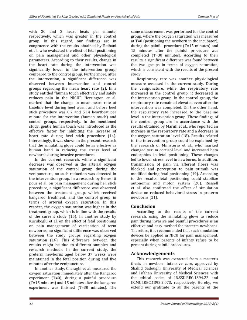
In total, 66 patients were selected through convenience sampling and were randomly assigned to the intervention (N=33) and control (N=33) groups using two similar envelops, which contained the name of the methods to be used. After selecting the eligible newborns, one of the nurses picked up an envelope to determine the method. For the intervention group, Zaky-mediated flexed fetal positioning was applied before venipuncture (Figure 1). On the other hand, no interventional positioning was used in the control group, and venipuncture was performed according to the routine NICU procedure. Data were collected using demographic characteristics questionnaire and a sheet to record physiological measures (i.e., heart rate, oxygen saturation and respiratory rate). These items represent the response of the body to the pain. Heart rate and oxygen saturation were measured by previously calibrated Saadat Novin monitor. To determine the respiratory rate, six qualified nurses counted respiratory rates at the same time for ten

Effect of Facilitated Tucking Created with Simulated Hands on Physiological Pain Salmani N et al
9 Iranian Journal of Neonatology 2017; 8(4)
Figure 1. Premature baby in the facilitated tucking position with hand simulation (Zaky® gloves)
neonates in two hospitals. The ICC obtained from this data was 0.987 for Amin and 0.995 for Shahid Beheshti hospitals, which confirmed the reliability of respiratory rate count by nurses.
After receiving the approval of the ethics committee, the objectives of the research were explained to the parents. Participants were allowed to withdraw from the research at any time, which had no impact on the routine pain management of newborns. In addition, parents were assured of the confidentiality terms regarding their personal information. Before venipuncture, all routine preparations were performed and the body temperature of the infants was recorded. The normal body temperature was assessed by placing the infants wearing only diapers under the radiant warmer for 30 minutes with no intervention. Moreover, a monitoring probe was used for recording the heart rate and arterial oxygen level. In addition, a Saadat Novin model S1800 (made in Iran) was exploited for hemodynamic monitoring.
After preparing the venipuncture set, the process was carried out by experienced nurses of each shift with a minimum of five years of experience in the NICU. Venipuncture was performed by Meditex catheter gauge 24 for all evaluated newborns. In both groups, the heart and respiration rates and arterial oxygen level were recorded as the baseline (t=0) 30 minutes (required time for self-regulation) after no intervention. Following that, the subjects of the intervention group were placed in flexed fetal position and were stabilized with the palm of one Zaky glove cupping the head and the palm of the other Zaky glove cupping the lower part of body and legs. As a result, the newborn was positioned in a C shape prior to the initiation of the
venipuncture procedure by the nurse. As soon as the catheter entered the artery, the second set of data was recorded. After the stabilization of the catheter at the end of venipuncture, the newborn was maintained in fetal position for another five minutes. At the end, the third set of physiological measures was recorded. In the control group, the venipuncture was started on the newborns with no prior intervention. Physiological measures were monitored before, during and after the venipuncture. Data analysis was performed in SPSS version 18 using Kolmogorov-Smirnov, Chi-square, independent t-test and repeated measures ANOVA. In addition, P-value less than 0.05 was considered statistically significant.
Results In this research, it was possible to use
parametric tests due to the confirmation of the normal distribution of the data by Kolmogorov–Smirnov. According to the independent t-test, no significant difference was observed between mean age, body temperature and weight in the intervention and control groups (P>0.05). In addition, the Chi-square test demonstrated no significant difference between the study groups in terms of gender (P>0.05) (Table 1).
On the other hand, the repeated measures ANOVA showed no significant difference between the intervention and control groups before, during and after the intervention regarding mean heart rate of the infants (P>0.05, Table 2). In addition, no significant difference was observed in mean heart rate of the subjects in the intervention group before, during and after the intervention (P>0.05). However, a significant difference was found in the control group before, during and after the intervention (P<0.05). While the least significant difference (LSD) test indicated no difference in mean heart rate before, during and after the intervention (P=0.842), a significant difference was observed in heart rate at periods of before and during the intervention (p=0.001) and during and after the intervention (P=0.001). These results were indicative of increased heart rate of newborns during venipuncture in the control group (Table 2).
Similarly, no significant difference was observed between the intervention and control group in terms of the arterial oxygen saturation level before the intervention (P=0.327). However, a significant difference was found between the two groups regarding the arterial oxygen saturation during (P =0.001) and after the intervention (P =0.02, Table 3).

Salmani N et al Effect of Facilitated Tucking Created with Simulated Hands on Physiological Pain
10 Iranian Journal of Neonatology 2017; 8(4)
Table 1. Distribution of mean and standard deviation of demographic variables P-value Standard deviation Mean Numbers Groups Demographic variables
0.07 2.95 33.02 33 Control
Gestational age (weeks) 2.00 31.28 33 Intervention
0.08 733.558 1994.67 33 Control
Weight (kg) 455.819 1542.36 33 Intervention
0.06 0.3066 36.682 33 Control
Temperature (°C) 0.3289 36.276 33 Intervention
Table 2. Comparison of mean and standard deviation of heart rate in intervention and control groups during three periods of before, during and after the intervention
P-value Standard deviation Mean Numbers Groups Levels
0.585 19.30 146.30 33 Control
Before 15.90 150.58 33 Intervention
0.158 25.08 164.94 33 Control
During 20.41 154.97 33 Intervention
0.233 14.14 146.97 33 Control
After 13.60 153.61 28 Intervention
Table 3. Comparison of mean and standard deviation of oxygen saturation in intervention and control groups during three period of before, during and after the intervention
P-value Standard deviation Mean Numbers Groups Levels
0.327 9.34 94.30 33 Control
Before 2.41 95.27 33 Intervention
0.001 6.44 88.70 33 Control
During 2.11 95.21 33 Intervention
0.005 6.17 91.30 33 Control
After 2.86 94.50 28 Intervention
Moreover, results of repeated measures
ANOVA demonstrated no significant difference in the intervention group before, during and after the venipuncture (P=0.05). Nonetheless, the same statistical test showed a significant difference in mean arterial oxygen saturation of the control group before, during and after the intervention (P<0.05). Furthermore, the LSD test revealed a significant difference in mean arterial oxygen saturation between the periods of before and during the intervention (P=0.001) and during and after the intervention (P=0.016). According to the results, mean arterial oxygen saturation decreased during the venipuncture process in the control group. It is noteworthy that mean arterial oxygen saturation did not reach the baseline level even after the completion of the intervention. According to ANOVA results, no significant difference was observed between the study groups in terms of mean respiratory rate before the intervention (P=0.103). However, there was a significant difference between the two groups during and after the intervention in this regard (P=0.011).
Similarly, a significant difference was found in mean respiratory rate at all periods in each group (P<0.05). In the intervention group, LSD test detected a significant difference in mean respiratory rate at periods of before and during
the intervention (P=0.027) and during and after the intervention (P=0.000). Nevertheless, the difference between before and after the intervention was not significant (P=0.579). These results indicated that mean respiratory rate decreased during venipuncture in the intervention group, returning to its baseline after the fixing of the catheter.
Moreover, a significant difference was observed in mean respiratory rate of the control group between the periods of before and during the intervention (P=0.01) and during and after the intervention (P=0.04). This could be interpreted as increased respiratory rate during venipuncture in the control group and lack of returning to the baseline level after the completion of the intervention.
Discussion According to the results of the current
research, the simulating gloves had positive effects on the physiological measures, such as heart and respiratory rates and arterial oxygen saturation during venipuncture. No significant difference was observed in mean heart rate of the subjects in the control and intervention groups before the venipuncture. In both groups, there was an increase in the heart rate of the control and intervention groups during the intervention
Effect of Facilitated Tucking Created with Simulated Hands on Physiological Pain Salmani N et al
11 Iranian Journal of Neonatology 2017; 8(4)
with 20 and 3 heart beats per minute, respectively, which was greater in the control group. In this regard, our findings are in congruence with the results obtained by Reihani et al., who evaluated the effect of fetal positioning on pain management and other physiological parameters. According to their results, change in the heart rate during the intervention was significantly lower in the intervention group, compared to the control group. Furthermore, after the intervention, a significant difference was observed between intervention and control groups regarding the mean heart rate (2). In a study entitled “human touch effectively and safely reduces pain in the NICU”, Herrington et al. marked that the change in mean heart rate at baseline level during heel warm and before heel stick procedure was 0.7 and 5.14 heartbeat per minute for the intervention (human touch) and control groups, respectively. In the mentioned study, gentle human touch was introduced as the effective factor for inhibiting the increase of heart rate during heel stick procedure (14). Interestingly, it was shown in the present research that the simulating glove could be as effective as human hand in reducing the stress level of newborns during invasive procedures.
In the current research, while a significant decrease was observed in the arterial oxygen saturation of the control group during the venipuncture, no such reduction was detected in the intervention group. In a research by Beheshti pour et al. on pain management during hell stick procedure, a significant difference was observed between the treatment group, which received kangaroo treatment, and the control group in terms of arterial oxygen saturation. In this respect, the oxygen saturation was higher in the treatment group, which is in line with the results of the current study (15). In another study by Kucukoglu et al. on the effect of fetal positioning on pain management of vaccination of term newborns, no significant difference was observed between the study groups regarding oxygen saturation (16). This difference between the results might be due to different samples and research methods. In the current study, the preterm newborns aged below 37 weeks were maintained in the fetal position during and five minutes after the venipuncture.
In another study, Cheraghi et al. measured the oxygen saturation immediately after the Kangaroo experiment (T=0), during a painful procedure (T=15 minutes) and 15 minutes after the kangaroo experiment was finished (T=30 minutes). The
same measurement was performed for the control group, where the oxygen saturation was measured at T=0 (positioning the newborn in the incubator), during the painful procedure (T=15 minutes) and 15 minutes after the painful procedure was completed (T=30 minutes). According to their results, a significant difference was found between the two groups in terms of oxygen saturation, which is consistent with the results of the present study.
Respiratory rate was another physiological measure assessed in the current study. During the venipuncture, while the respiratory rate increased in the control group, it decreased in the intervention group. In the control group, the respiratory rate remained elevated even after the intervention was completed. On the other hand, the respiratory rate increased to the baseline level in the intervention group. These findings of the control group are in accordance with the results obtained by Marufi et al., who reported an increase in the respiratory rate and a decrease in the oxygen saturation level (18). Results related to the intervention group could be explained by the research of Ministerio et al., who marked changed serum cortisol level and increased beta endorphins in fetal positioning. These changes led to lower stress level in newborns. In addition, transmission of pain via afferent fibers was blocked and perception to pain stimuli was modified during fetal positioning (19). According to the results, fetal positioning could stabilize autonomic and motor system (20). Russell et al. also confirmed the effect of simulation device on reduced behavioral stress in preterm newborns (21).
Conclusion According to the results of the current
research, using the simulating glove to reduce pain during invasive and painful procedures is an effective and easy method for preterm newborns. Therefore, it is recommended that such simulation devices be applied in NICU for pain management, especially when parents of infants refuse to be present during painful procedures.
Acknowledgements This research was extracted from a master’s
thesis in newborn intensive care, approved by Shahid Sadoughi University of Medical Sciences and Isfahan University of Medical Sciences with the ethical codes of IR.SSU.REC.1394.22 and IR.MUI.REC.1395.2.073, respectively. Hereby, we extend our gratitude to all the parents of the
Salmani N et al Effect of Facilitated Tucking Created with Simulated Hands on Physiological Pain
12 Iranian Journal of Neonatology 2017; 8(4)
subjects for their cooperation with the research.
Conflicts of interests None declared.
References 1. Preterm birth. World Health Organization. Available at:
URL: http://www.who.int/mediacentre/factsheets/ fs363/en/; 2015.
2. Reyhani T, Mohebi T, Boskabadi H, Gholami H. The effect of facilitated tucking during venipuncture on pain and physiological parameters in preterm infants. Evid Based Care. 2012; 2(2):47-56 )Persian).
3. Badr LK, Abdallah B, Hawari M, Sidani S, Kassar M, Nakad P, et al. Determinants of premature infant pain responses to heel sticks. Pediatr Nurs. 2010; 36(3):129-36.
4. Axelin A, Salanterä S, Lehtonen L. ‘Facilitated tucking by parents’ in pain management of preterm infants-a randomized crossover trial. Early Hum Dev. 2006; 82(4):241-7.
5. Sheikhbahaeddinzadeh E. Examination, diagnosis & nursing care in the NICU. Tehran: Boshra; 2012 (Persian).
6. Hartley KA, Miller CS, Gephart SM. Facilitated tucking to reduce pain in neonates: evidence for best practice. Adv Neonatal Care. 2015; 15(3):201-8.
7. Nicholson JM, Berthelsen D, Abad V, Williams K, Bradley J. Impact of music therapy to promote positive parenting and child development. J Health Psychol. 2008; 13(2):226-38.
8. Obeidat H, Kahalaf I, Callister LC, Froelicher ES. Use of facilitated tucking for nonpharmacological pain management in preterm infants: a systematic review. J Perinat Neonatal Nurs. 2009; 23(4):372-7.
9. Cignacco E, Hamers JP, Stoffel L, Lingen RA, Gessler P, McDougall J, et al. The efficacy of non‐pharmacological interventions in the management of procedural pain in preterm and term neonates. Eur J Pain. 2007; 11(2): 139-52.
10. Liaw JJ, Zeng WP, Yang L, Yuh YS, Yin T, Yang MH. Nonnutritive sucking and oral sucrose relieve neonatal pain during intramuscular injection of hepatitis vaccine. J Pain Symptom Manage. 2011; 42(6):918-30.
11. Grunau RE, Linhares MB, Holsti L, Oberlander TF, Whitfield MF. Does prone or supine position
influence pain responses in preterm infants at 32 weeks gestational age? Clin J Pain. 2004; 20(2): 76-82.
12. Stevens B, Yamada J, Lee GY, Ohlsson A. Sucrose for analgesia in newborn infants undergoing painful procedures. Cochrane Database Syst Rev. 2013; 1:CD001069.
13. Russell K, Weaver B, Vogel RL. Neuroprotective core measure 2: partnering with families-effects of a weighted maternally-scented parental simulation device on premature infants in neonatal intensive care. Newborn Infant Nurs Rev. 2015; 15(3):97-103.
14. Herrington CJ, Chiodo LM. Human touch effectively and safely reduces pain in the newborn intensive care unit. Pain Manag Nurs. 2014; 15(1):107-15.
15. Beheshtipoor N, Memarizadeh A, Hashemi F, Porarian S, Rambod M. The effect of kangaroo care on pain and physiological parameters in preterm infants on heel-stick procedure: a randomized controlled, cross-over study. Galen Med J. 2013; 2(4):157-68.
16. Kucukoglu S, Kurt S, Aytekin A. The effect of the facilitated tucking position in reducing vaccination-induced pain in newborns. Ital J Pediatr. 2015; 41(1):61.
17. Cheraghi F, Pakseresht M, Parsa P, Basiri B. Effect of kangaroo mother care on premature newborns’ pain due to invasive procedures in neonatal intensive care unit of hospital Fatemieh, Hamadan. J Ilam Univ Med Sci. 2014; 22(1):31-40 (Persian).
18. Marofi M, Nikobakht F, Ali MN, Badiei Z. Comparing the effect of listening to melody vs. breast-feeding on neonates’pain intensity during heel-blood sampling in neonatal intensive care unit. J Anesthesiol Pain. 2015; 5(3):45-54 (Persian).
19. Lamy ZC, Gomes MA, Gianini NO, Hennig MA. Atenção humanizada ao recém-nascido de baixo peso-Método Canguru: a proposta brasileira. Ciênc Saúde Coletiva. 2005; 10(3):659-8.
20. Hill S, Engle S, Jorgensen J, Kralik A, Whitman K. Effects of facilitated tucking during routine care of infants born preterm. Pediatr Phys Ther. 2005; 17(2):158-63.
21. Russell K, Weaver B, Vogel RL. Neuroprotective core measure 2: partnering with families-effects of a weighted maternally-scented parental simulation device on premature infants in neonatal intensive care. Newborn Infant Nurs Rev. 2015; 15(3):97-103.