Open Access Full Text Article Intracellular CXCR4 cell ...
12
© 2012 Unzueta et al, publisher and licensee Dove Medical Press Ltd. This is an Open Access article which permits unrestricted noncommercial use, provided the original work is properly cited. International Journal of Nanomedicine 2012:7 4533–4544 International Journal of Nanomedicine Intracellular CXCR4 + cell targeting with T22-empowered protein-only nanoparticles Ugutz Unzueta 1–3 María Virtudes Céspedes 3,4 Neus Ferrer-Miralles 1–3 Isolda Casanova 3,4 Juan Cedano 5 José Luis Corchero 1–3 Joan Domingo-Espín 1–3 Antonio Villaverde 1–3 Ramón Mangues 3,4 Esther Vázquez 1–3 1 Institut de Biotecnologia i de Biomedicina, 2 Departamento de Genètica i de Microbiologia, Universitat Autònoma de Barcelona, Bellaterra, Barcelona, 3 CIBER en Bioingeniería, Biomateriales y Nanomedicina, Bellaterra, Barcelona, 4 Oncogenesis and Antitumor Drug Group, Biomedical Research Institute Sant Pau, Hospital de la Santa Creu i Sant Pau, Barcelona, Spain; 5 Laboratory of Immunology, Regional Norte, Universidad de la Republica, Salto, Uruguay Correspondence: Antonio Villaverde Institut de Biotecnologia i de Biomedicina, Universitat Autònoma de Barcelona, Bellaterra, 08193 Barcelona, Spain Tel +349 3581 3086 Fax +349 3581 2011 Email [email protected] Background: Cell-targeting peptides or proteins are appealing tools in nanomedicine and innovative medicines because they increase the local drug concentration and reduce potential side effects. CXC chemokine receptor 4 (CXCR4) is a cell surface marker associated with several severe human pathologies, including colorectal cancer, for which intracellular targeting agents are currently missing. Results: Four different peptides that bind CXCR4 were tested for their ability to internalize a green fluorescent protein-based reporter nanoparticle into CXCR4 + cells. Among them, only the 18 mer peptide T22, an engineered segment derivative of polyphemusin II from the horseshoe crab, efficiently penetrated target cells via a rapid, receptor-specific endosomal route. This resulted in accumulation of the reporter nanoparticle in a fully fluorescent and stable form in the perinuclear region of the target cells, without toxicity either in cell culture or in an in vivo model of metastatic colorectal cancer. Conclusion: Given the urgent demand for targeting agents in the research, diagnosis, and treatment of CXCR4-linked diseases, including colorectal cancer and human immunodeficiency virus infection, T22 appears to be a promising tag for the intracellular delivery of protein drugs, nanoparticles, and imaging agents. Keywords: peptide tag, CXCR4, intracellular targeting, self-assembling, nanoparticles, colorectal cancer Introduction Unlike conventional therapies, nanomedicine and innovative medicines in general pursue targeted intracellular delivery of chemotherapy and imaging agents, and are expected to result in significantly lower effective therapeutic doses, production costs, and toxicity. 1 Cell-penetrating peptides offer a broad potential for efficient internalization of attached cargo because of their affinity for and associated abil- ity to cross cell membranes. 2,3 However, because these activities are not dependent on specific cell surface receptors, appropriate cell targeting and biodistribution of therapeutic complexes cannot be achieved using cell-penetrating peptides. The discovery of disease-linked cell surface markers enables subsequent identification of specific ligands for receptor-mediated endocytosis. These entities should be capable of driving the uptake of large macromolecular complexes such as nano- particles, which are useful in a therapeutic context as drug carriers and stabilizers. The CXC chemokine receptor 4 (CXCR4) plays a role in inflammation, autoim- munity, ischemia, and stem cell mobilization. 4 In addition, CXCR4 is a coreceptor for the human immunodeficiency virus (HIV) 5 and an important stem cell marker in several common human cancers, 6,7 including metastatic colorectal cancer. 8,9 Dovepress submit your manuscript | www.dovepress.com Dovepress 4533 ORIGINAL RESEARCH open access to scientific and medical research Open Access Full Text Article http://dx.doi.org/10.2147/IJN.S34450 Video abstract Point your SmartPhone at the code above. If you have a QR code reader the video abstract will appear. Or use: http://dvpr.es/NKs8C4
Transcript of Open Access Full Text Article Intracellular CXCR4 cell ...
© 2012 Unzueta et al, publisher and licensee Dove Medical Press Ltd. This is an Open Access article which permits unrestricted noncommercial use, provided the original work is properly cited.
International Journal of Nanomedicine 2012:7 4533–4544
International Journal of Nanomedicine
Intracellular CXCR4+ cell targeting with T22-empowered protein-only nanoparticles
Ugutz Unzueta1–3
María Virtudes Céspedes3,4
Neus Ferrer-Miralles1–3
Isolda Casanova3,4
Juan Cedano5
José Luis Corchero1–3
Joan Domingo-Espín1–3
Antonio Villaverde1–3
Ramón Mangues3,4
Esther Vázquez1–3
1Institut de Biotecnologia i de Biomedicina, 2Departamento de Genètica i de Microbiologia, Universitat Autònoma de Barcelona, Bellaterra, Barcelona, 3CIBER en Bioingeniería, Biomateriales y Nanomedicina, Bellaterra, Barcelona, 4Oncogenesis and Antitumor Drug Group, Biomedical Research Institute Sant Pau, Hospital de la Santa Creu i Sant Pau, Barcelona, Spain; 5Laboratory of Immunology, Regional Norte, Universidad de la Republica, Salto, Uruguay
Correspondence: Antonio Villaverde Institut de Biotecnologia i de Biomedicina, Universitat Autònoma de Barcelona, Bellaterra, 08193 Barcelona, Spain Tel +349 3581 3086 Fax +349 3581 2011 Email [email protected]
Background: Cell-targeting peptides or proteins are appealing tools in nanomedicine and
innovative medicines because they increase the local drug concentration and reduce potential
side effects. CXC chemokine receptor 4 (CXCR4) is a cell surface marker associated with
several severe human pathologies, including colorectal cancer, for which intracellular targeting
agents are currently missing.
Results: Four different peptides that bind CXCR4 were tested for their ability to internalize a
green fluorescent protein-based reporter nanoparticle into CXCR4+ cells. Among them, only the
18 mer peptide T22, an engineered segment derivative of polyphemusin II from the horseshoe
crab, efficiently penetrated target cells via a rapid, receptor-specific endosomal route. This
resulted in accumulation of the reporter nanoparticle in a fully fluorescent and stable form in
the perinuclear region of the target cells, without toxicity either in cell culture or in an in vivo
model of metastatic colorectal cancer.
Conclusion: Given the urgent demand for targeting agents in the research, diagnosis, and
treatment of CXCR4-linked diseases, including colorectal cancer and human immunodeficiency
virus infection, T22 appears to be a promising tag for the intracellular delivery of protein drugs,
nanoparticles, and imaging agents.
Keywords: peptide tag, CXCR4, intracellular targeting, self-assembling, nanoparticles,
colorectal cancer
IntroductionUnlike conventional therapies, nanomedicine and innovative medicines in general
pursue targeted intracellular delivery of chemotherapy and imaging agents, and
are expected to result in significantly lower effective therapeutic doses, production
costs, and toxicity.1 Cell-penetrating peptides offer a broad potential for efficient
internalization of attached cargo because of their affinity for and associated abil-
ity to cross cell membranes.2,3 However, because these activities are not dependent
on specific cell surface receptors, appropriate cell targeting and biodistribution
of therapeutic complexes cannot be achieved using cell-penetrating peptides. The
discovery of disease-linked cell surface markers enables subsequent identification
of specific ligands for receptor-mediated endocytosis. These entities should be
capable of driving the uptake of large macromolecular complexes such as nano-
particles, which are useful in a therapeutic context as drug carriers and stabilizers.
The CXC chemokine receptor 4 (CXCR4) plays a role in inflammation, autoim-
munity, ischemia, and stem cell mobilization.4 In addition, CXCR4 is a coreceptor
for the human immunodeficiency virus (HIV)5 and an important stem cell marker
in several common human cancers,6,7 including metastatic colorectal cancer.8,9
Dovepress
submit your manuscript | www.dovepress.com
Dovepress 4533
O R I G I N A L R E S E A R C H
open access to scientific and medical research
Open Access Full Text Article
http://dx.doi.org/10.2147/IJN.S34450
Video abstract
Point your SmartPhone at the code above. If you have a QR code reader the video abstract will appear. Or use:
http://dvpr.es/NKs8C4

International Journal of Nanomedicine 2012:7
Therefore, developing new therapeutic agents targeted to
CXCR4 is a recognized priority in emerging medicines,4
but because CXCR4 ligands remain poorly studied and
peptides suitable for CXCR4-mediated endocytosis are
not available, intracellular targeting to CXCR4+ cells is
still not feasible. In the present study, we describe peptide
T22, a known antagonist of CXCR4, that binds to and
penetrates CXCR4+ cells efficiently via CXCR4-specific
endocytosis. T22 is an engineered version polyphemusin
II peptide from the horseshoe crab, in which three sub-
stitutions at residues Tyr5, Lys7, and Tyr12 dramatically
increase the natural affinity of this peptide for CXCR4.10,11
When fused to a self-assembling green fluorescent protein
(GFP)-based building block, T22 promotes fast perinuclear
accumulation of stable and highly fluorescent nanoparticles
without cytotoxicity, both in cell culture and in vivo. Thus,
we propose T22 as a novel cell-targeting peptide suitable
for functionalization of nanoparticles and appropriate for
intracellular delivery in CXCR4-associated medicines.
Materials and methodsProtein design, production, purification, and characterizationFour chimeric genes were designed inhouse and provided
by Geneart (Regensburg, Germany). Using NdeI/HindIII
restriction sites, these genes were introduced into pET22b
(Novagen 69744-3). All the encoded proteins were produced
in Escherichia coli Origami B (BL21, OmpT-, Lon-, TrxB-,
Gor-, Novagen) overnight at 20°C upon addition of 0.1 mM
isopropyl-β-D-thiogalactopyronaside. Bacterial cells were
then centrifuged for 45 minutes (5000 g at 4°C) and resus-
pended in Tris buffer (Tris 20 mM, pH 8.0, NaCl 500 mM,
imidazole 10 mM) in the presence of ethylenediamine
tetra-acetic acid-free protease inhibitor (Complete EDTA-
Free, Roche, Basel, Switzerland). The cells were disrupted
at 1100 psi in a French press (Thermo FA-078A) and their
proteins were purified by 6 × His tag affinity chromatography
using HiTrap Chelating HP 1 mL (GE Healthcare, Piscataway,
NJ) columns with an AKTA purifier FPLC (GE Healthcare).
Elution was achieved by a linear gradient of Tris 20 mM,
pH 8.0, 500 mM NaCl, and 500 mM imidazole, and the pro-
teins eluted were finally dialyzed against phosphate-buffered
solution (140 mM NaCl, 7.5 mM Na2HPO
4, 2.5 mM NaH
2PO
4)
plus 10% glycerol, pH 7.4, against carbonate buffer (166 mM
NaCO3H + 333 mM NaCl, pH 7.4) or against Tris 20 mM +
NaCl 500 mM, pH 7.5. The integrity of the resulting proteins
was checked by both mass spectrometry and N-terminal
sequencing using the Edman degradation method, and
their amounts were determined by Bradford’s assay.12
In addition, all products were analyzed by Coomassie-stained
sodium dodecyl sulfate polyacrylamide gel electrophoresis
and anti-His Western blot analysis. The fusion proteins
were named according to N→C modular organization by
the name of the CXCR4 ligand, followed by GFP and H6
(hexahistidine tail).
TEM, fluorescence determination, and dynamic light scatteringPurified proteins were diluted to 0.2 mg/mL and contrasted
by evaporation of 1 nm platinum layer in carbon-coated grids.
Samples were visualized in a Hitachi H-7000 transmission
electron microscope. Fluorescence of the nanoparticles was
determined in a Cary Eclipse fluorescence spectrophotometer
(Varian Inc, Palo Alto, CA) at 510 nm using an excitation
wavelength of 450 nm. The volume and size distribution of
the nanoparticles was determined by dynamic light scattering
at 633 nm (Zetasizer Nano ZS, Malvern Instruments Limited,
Malvern, Worcestershire, UK).
Protein stability analysisStability of the T22-GFP-H6 (amino terminus of a His-tagged
enhanced GFP) was analyzed in triplicate in human serum
(S2257-5ML, Sigma, St Louis, MO) at 37°C, with agitation
and at a final concentration of 0.23 µg/µL. Fluorescence
was determined as described earlier, and the integrity of
the T22-GFP-H6 was confirmed by sodium dodecyl sulfate
polyacrylamide gel electrophoresis and further Western
blotting. Nitrocellulose membranes were developed using
an anti-GFP rabbit polyclonal serum.
Cell culture and confocal laser scanning microscopyThe cells were cultured in modified Eagle’s medium (Gibco,
Rockville, MD) supplemented with 10% fetal calf serum
(Gibco), and incubated at 37°C and 5% CO2 in a humidified
atmosphere. Nanoparticles were added to the cell culture in
the presence of Optipro medium (Gibco) 20 hours before
confocal analysis, except for the time-course and internaliza-
tion studies in the presence of serum (complete medium). For
confocal analysis, the cells were grown on MatTek culture
dishes (MatTek Corporation, Ashland, MA). The nuclei were
labeled with 0.2 µg/mL Hoechst 33342 (Molecular Probes,
Eugene, OR) and the plasma membranes with 2.5 µg/mL
CellMaskTM Deep Red (Molecular Probes) for 10 minutes
in the dark. The cells were washed in phosphate-buffered
saline (Sigma-Aldrich Chemie GmbH, Steinheim, Germany).
submit your manuscript | www.dovepress.com
Dovepress
Dovepress
4534
Unzueta et al
International Journal of Nanomedicine 2012:7
Live cells were recorded by TCS-SP5 confocal laser scanning
microscopy (Leica Microsystems, Heidelberg, Germany)
using a Plan Apo 63 × /1.4 (oil HC × PL APO lambda blue)
objective as described elsewhere.13 To determine particle
localization inside the cell, stacks of 10–20 sections for
every 0.5 µm of cell thickness were collected and three-
dimensional models were generated using Imaris version
6.1.0 software (Bitplane, Zürich, Switzerland) as reported
previously.14 Cell samples were analyzed after treatment
with 1 mg/mL trypsin (Gibco) for 15 minutes on a FACS-
Canto system (Becton Dickinson, Franklin Lakes, NJ) using
a 15 mW air-cooled argon ion laser at 488 nm excitation.
Fluorescence emission was measured with a D detector
(530/30 nm band pass filter). Cell viability was determined
by 3-(4,5-dimethylthiazol-2-yl)-2,5-diphenyltetrazolium
bromide (MTT) assay as described elsewhere.15 An HeLa
cell line was obtained from the American Type Culture
Collection (reference CCL-2, Manassas, VA) and SW1417
was a generous gift from Xavier Mayol (Institut Municipal
D’Investigacio Médica, Barcelona, Spain).16
Molecular modelingProtein homology models were generated using Modeller,
Phyre, and Swiss-PdbViewer as reported earlier.17 Also,
different Haddock models were obtained, in which the bind-
ing residues were established using crystallographic data
from multimeric forms of GFP (1GFL, 1JC0, 3GJ2, 2QLE,
1EMC) and the higher interaction energy solutions resulting
from protein-protein docking calculation.
Biodistribution analysisFive-week-old female Swiss nu/nu mice, weighing 18–20 g
(Charles River, France) maintained in specific pathogen-free
conditions, were used for the in vivo experiments. All pro-
cedures were approved by the Hospital de Sant Pau animal
ethics committee. To generate a metastatic colorectal cancer
model, the mice were injected with 2 million SW-1417 cells
via the cecal wall, using an orthotopic cell microinjection
technique.18 Two months after microinjection, when local
tumor and metastases had appeared, each experimental
animal received a single intravenous bolus of T22-GFP-H6
nanoparticles resuspended in a 20 mM Tris, 500 mM NaCl,
pH 7.4 buffer, at a dose of 20 µg (n = 3 mice) or 500 µg
(n = 3 mice). Control animals received a single bolus of
empty buffer. After euthanizing the mice at 5, 24, or 48 hours
post-administration, we measured ex vivo the amount of
nanoparticles in normal and tumor-bearing organs from the
experimental and control mice, quantifying the fluorescence
emitted by each organ. To this end, primary tumors, organs
bearing metastatic foci, and several samples of normal tissue
(kidney, liver, lung, heart) were obtained at necropsy, cut into
slices, and placed in separate wells to detect the emitted signal
using IVIS® Spectrum equipment (Xenogen Biosciences,
Waltham, MA). The amount of nanoparticles distributed
in each tissue was calculated as the increased fluorescent
(FLI) ratio. The fluorescence signal was first digitalized,
displayed as a pseudocolor overlay, and expressed as radi-
ant efficiency. Thereafter, the FLI ratio was calculated for
each organ, dose, and post-treatment time, dividing the FLI
signal from the nanoparticle-treated mice by the FLI signal
from the control mice. Finally, all organs were collected and
fixed with 4% formaldehyde in phosphate-buffered solution
for 24 hours, and then embedded in paraffin for histological
and immunohistochemical evaluation.
ImmunohistochemistrySections of normal and tumor tissues 4 µm thick were stained
with hematoxylin and eosin. The sections were examined
histopathologically to analyze the primary tumor and to
search for metastatic foci in organs with no macroscopic
metastases. Paraffin-embedded tissue sections were depar-
affinized, rehydrated, and washed in phosphate-buffered
solution with Tween-20. Antigen retrieval was performed
using citrate buffer at 120°C. After quenching peroxidase
activity by incubating the slides in 3% H2O
2 for 10 minutes,
the slides were washed in phosphate-buffered solution with
Tween-20. The slides were incubated for 30 minutes with the
primary antibody against CXCR4 (1:20, Biotrend, Destin,
FL) to detect expression of this receptor in normal tissue
and tumor tissue. A primary anti-His antibody (1:1000;
Abcam, Cambridge, UK) was used to detect nanoparticle
accumulation and localization in normal and tumor (primary
or metastatic) tissue. After incubation, the samples were
washed in phosphate-buffered solution with Tween-20
and incubated with the biotinylated secondary antibody
for 30 minutes at room temperature. Finally, the sections
were counterstained with hematoxylin and mounted using
DPX mounting medium. Representative pictures were taken
using Cell^B software (Olympus Soft Imaging) at 200× and
1000× magnifications.
Statistical analysisThe data were evaluated by one-way Anova analysis of
variance with a confidence level of 99.9% (P , 0.001).
Dose-response plots were analyzed by nonlinear regression
analysis using SigmaPlot 10. The data for both the HeLa and
submit your manuscript | www.dovepress.com
Dovepress
Dovepress
4535
Peptide-assisted intracellular targeting in CXCR4+ cells

International Journal of Nanomedicine 2012:7
SW1417 cells fitted into a double rectangular hyperbolic
function, with a significance level of 99.9% (P , 0.001).
All data were expressed as the mean ± standard error of
the mean.
ResultsScreening CXCR4 peptidic ligands for cell-targeted internalizationTo identify peptides suitable for use as tags for CXCR4-
mediated cell internalization of large macromolecular com-
plexes, four known molecular ligands of CXCR4, namely
peptide T22, the protein domains CXCL2 and vCCL2, and
V1, an amino-terminal peptide of vCCL2 (Figure 1A), were
tested for their ability to promote receptor-mediated delivery
of attached macromolecular entities into CXCR4-expressing
cells. All these protein segments were fused to GFP-H6.
This fluorescent protein, when containing cationic peptides
at its amino terminus, shows a tendency to self-assemble
as regular-sized nanoparticles, presumably by electrostatic
interaction between monomers.13 Because of their emission
of fluorescence, these nanoparticles are very convenient
reporters for use in internalization and trafficking studies.14
Four equivalent modular constructs differing only by the
CXCR4 ligand (Figure 1B) were designed according to this
strategy, produced in bacteria, and purified as full-length
forms of the expected molecular mass and N-terminal
amino acid sequence (Figure 2). Their reactivity in anti-His
Western blot analysis indicated protein integrity also at the
C-terminus. Only vCCL12 showed partial degradation at the
N-terminus, but retained most of the expected molecular mass
(Figure 2). This proteolytic instability was observed under
all tested production conditions and in several E. coli strains
tested as hosts (data not shown). In addition, the single major
peak in mass spectroscopy and the unambiguous N-terminal
sequence, coincident with a few dominant bands in Western
blot analysis, indicated the existence of conformational
isoforms of vCCL12.
When these proteins were added to the culture medium,
HeLa (CXCR4+) cells exposed to T22-GFP-H6 were ten-
fold more fluorescent than those exposed to V1, CXCL12,
and vCCL2 fusion (Figure 3). Interestingly, cell uptake of
CXCL12 and V1 has been reported previously,19,20 but the
excellent performance of T22 was completely unexpected
because its ability to internalize cells has not been previously
suggested or described, despite its well known properties as
an CXCR4 antagonist.21 As expected, the untagged parental
GFP-H6 building block did not label the cells.
CXCR4-dependent cell uptake of T22-empowered constructsThe high cell penetrability of T22-activated GFP was
further confirmed by confocal microscopy of treated HeLa
cell cultures (Figure 4A). In full agreement with data from
Figure 3, no penetration of GFP-H6 was observed, while
some uptake of CXCL12 and to a minor extent some vCCL2-
GFP-H6 was seen. V1-GFP-H6 was observed inside cells as
low in abundance and poorly fluorescent punctuate entities.
Three-dimensional reconstructions of individual cells exposed
to T22-GFP-H6 indicated perinuclear localization of fluo-
rescence in the form of nanoparticles (Figure 4B). Discrete
yellow merging signals (green particles incorporated into
red membranous vesicles) were observed close to the plasma
membrane (Figure 4C), indicative of uptake by endosomes.
The finding of nanoparticles within the endosomes was fully
confirmed by three-dimensional confocal reconstructions as
yellow spots, some of them fully internalized in the cytoplasm
and separate from the plasma membrane (Figure 4C, inset).
However, the relative low proportion of yellow signals and
N
A B
L eGFP CH6
GGSSRSS KHHHHHH
T22-GFP-H6T22 RRWCYRKCYKGYCYRKCR
KPVSLSYRCPCRFFESHVARANVKHLKILNTPNCALQIVARLKNNNRQVCIDPKLKWIQEYLEKALN
LGASWHRPDKCCLGYQKRPLPLGASWHRPDKCCLGYQKRPLPQVLLSSWYPTSQLCSKPGVIFLTKRGRQVCADKDWVKKLMQQLPVTA
V1vCCL2CXCL12
V1-GFP-H6 vCCL2-GFP-H6 CXCL12-GFP-H6
Figure 1 Features of protein constructs containing peptidic CXCR4 ligands. (A) Schematic representation of CXCR4-binding constructs indicating their modular composition. A linker (yellow box) commonly used in phage display was inserted between the protein ligand (L, blue) and eGFP (green). The amino acid sequences of the four ligands are shown. In all cases, an additional amino terminal methionine, derived from the cloning strategy adapted to Escherichia coli was expected. (B) Predicted structure of the different GFP-derived constructs. The color code of panel A is maintained here for both ligand- (blue) and H6- (red) overhanging ends.Abbreviation: GFP, green fluorescent protein.
submit your manuscript | www.dovepress.com
Dovepress
Dovepress
4536
Unzueta et al

International Journal of Nanomedicine 2012:7
rapid accumulation of nanoparticles close to the nuclear
region through fast cytoplasmic trafficking (Figure 4D) were
indicative of early endosomal escape. This was probably
promoted by the accompanying hexahistidine tag, that has
powerful endosomolytic properties,22 inducing early endo-
somal escape in related protein-only nanoparticles.14 Ten
minutes after exposure to these T22 constructs, the uptake
of nanoparticles was already evident, and the amount of
intracellular fluorescence progressively increased for up to
24 hours at least (Figure 4E).
To exclude the possibility that T22-mediated penetration
of GFP was limited to a particular cell type, we also
evaluated the intracellular fluorescence in exposed CXCR4+
SW1417 cells, a human cell line used to generate a mice
model of metastatic colorectal cancer for preclinical
studies through orthotopic implantation.18 Again, strong
penetration (Figure 5A) and perinuclear fluorescence
labeling (Figure 5B) were seen in this model, indicative
of efficient penetration of the nanoparticles. No changes
in cell morphology (which would have been indicative of
toxicity) were observed (Figures 4A and 5A), or when com-
pared with cells exposed to parental GFP-H6 (Figures 4A
and 5C). Furthermore, viability of T22-GFP-H6-exposed
SW1417 cells remained unaffected up to 72 hours after
exposure (Figure 5D), even when high protein concentrations
were added. On the other hand, penetration of T22-GFP-H6
was observed to be dose-dependent (Figure 5E) in both
human HeLa and SW1417 cells, and efficiently inhibited
by increasing amounts of SDF1α (Figure 5F), the natural
ligand of CXCR4.23 These data confirm the specificity of
intracellular delivery driven by the T22 peptide.
Stability and architecture of T22-empowered GFP nanoparticlesThe confocal microscopy analyses shown in Figures 4 and 5
indicate that T22-GFP-H6 has a nanostructure, as has been
1000
T22
MRRWCY30.67 kDa
MLGAS36.30 kDa
MLGAS30.7 kDa
MKPVS36.1 kDa
CXCL12
V1
vCCL2
500
0
800
20 30
Molecular weight (kDa)
Inte
nsi
ty (
Ab
s u
nit
s)
40
20 30 40 20 30 40
20 30 40
400
0
1000
400
0
500
0
Co WB
Figure 2 Biochemical characterization of protein constructs upon protein purification. Notes: Mass spectrometry of the purified constructs indicating the experimental molecular weight. The obtained N-terminal sequence is also shown, always coincident with the predicted sequence (Figure 1A). Protein integrity is also shown through Coomassie blue-stained sodium dodecyl sulfate polyacrylamide gel electrophoresis gels (Co) and by H6 immunodetection in Western blot (WB).
No protein
GFP
vCCL2
V1
T22
CXCL12
10
0
101 102
FITC-A
Eve
nts
103
Figure 3 Differential internalization of CXCR4 ligands. Notes: Internalization of T22-GFP-H6 and alternative constructs in HeLa cells, monitored by flow cytometry 24 hours after exposure.Abbreviation: GFP, green fluorescent protein.
submit your manuscript | www.dovepress.com
Dovepress
Dovepress
4537
Peptide-assisted intracellular targeting in CXCR4+ cells

International Journal of Nanomedicine 2012:7
previously demonstrated for the related construct, R9-GFP-H6.13
Although precise characterization of these assemblies
is beyond the scope of this study, we were interested in
confirming that T22-GFP-H6 is organized in nanoparticle
form and that T22 can achieve targeted internalization
of large macromolecular complexes. If so, T22 would
be of broad interest for the functionalization of other
categories of nanoparticles (eg, nonproteins) in emerg-
ing medicine. First, we determined that the construct
was highly stable in human serum (Figure 6A) and able
to internalize target cells fully in the presence of fetal
bovine serum (Figure 6B). These features are suggestive
of proteolytic stability, tight architecture, and regular
organization of T22-GFP-H6 building blocks, in contrast
with the random particle size and morphologies observed
during amorphous protein aggregation, even in the form of
soluble aggregates.24–27 Indeed, the mean size of T22-GFP-
H6, measured by dynamic light scattering, was 13.45 nm
(Figure 6C), a value fully compatible with images of these
nanoparticles under transmission electron microscopy
(Figure 6D) showing relatively monodispersed entities.
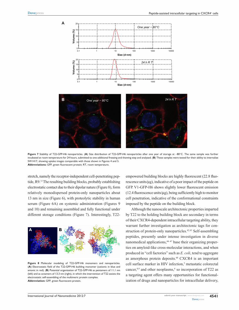
To assess further the structural and functional stability of
T22-empowered nanoparticles, we determined their size
distribution after storage for one year at -80°C, and also at
room temperature for an additional 24 hours, followed by
one additional step of freezing and thawing. As observed
(Figure 7A), no important variations in nanoparticle size
were observed, in agreement with their high stability in
human serum (Figure 6A). After 24 hours of incubation at
room temperature, we observed a slight tendency for the
nanoparticles to become more compact entities, although
the reduction in size was very moderate (around 1 nm on
average). These nanoparticles were indistinguishable from
the original material in terms of their ability to internalize
cultured cells (Figure 7B). All these data confirm that the
presence of T22, as it occurs with the cationic peptide R9,
imparts self-organizing properties to His-tagged GFP, that
cannot form multimeric complexes on its own.13
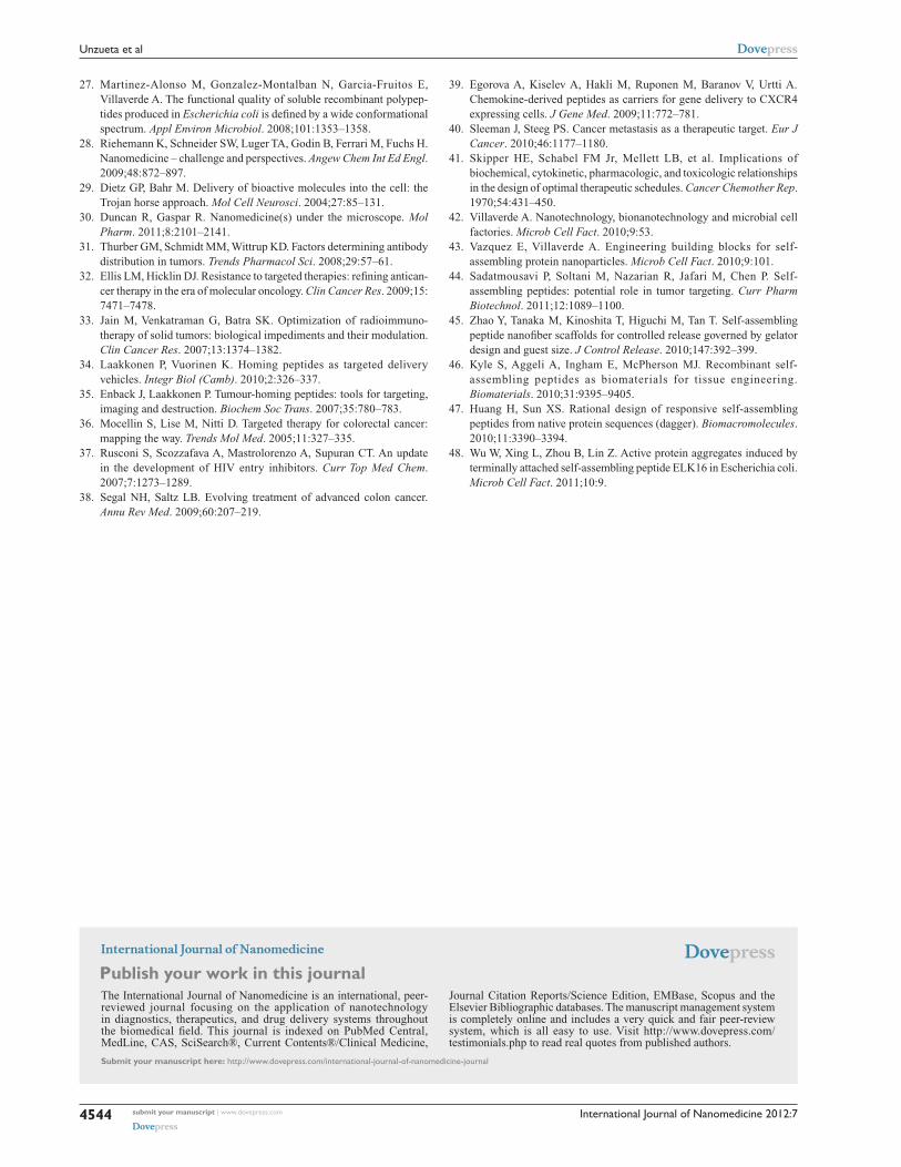
Given that T22 is also highly cationic (note its primary
sequence in Figure 1A), we wondered if this peptide could
promote electrostatic interaction between monomers to
achieve stable nanoparticulate entities. To explore this
possibility, we generated a charge map of T22-GFP-H6
(Figure 8A). The highly dipolar charge distribution of the
multifunctional protein did indeed enable tight electrostatic
contact between the charged sides of the GFP beta barrel
and the consequent generation of regular oligomers. Two
stable multimeric assemblies of T22-GFP-H6 monomers into
approximately 13 nm nanoparticles are shown in Figure 8B,
but alternative arrangements of the building blocks were
also thermodynamically feasible (data not shown). Further
structural analyses are in progress to elucidate the nature of
the architectonic properties of T22, beyond those associated
with its cell-targeting ability.
T22-mediated intracellular targeting in a model of metastatic colorectal cancerThe excellent in vitro performance of T22 in receptor-
specif ic intracellular targeting and the architectonic
A
B C
D
E010 min20306012024024 h
100 101 102
FITC-A
FIT
C-A
Figure 4 Differential internalization and intracellular trafficking of CXCR4 ligands. (A) Confocal images of HeLa cells exposed to differently tagged proteins for 24 hours. Nuclei are labeled in blue and cell membranes in red. Bar indicates 20 µm. (B) Detail of a HeLa cell exposed to T22-GFP-H6, showing the intracellular localization of nanostructured, fluorescent entities, in an isosurface representation within a three-dimensional volumetric x-y-z data field. (C) Yellow spots in the cell membrane, marked with an arrow, indicate early endosomal localization of green fluorescent particles (merging of red and green signals). In the insets, details of endosome-embedded fluorescent particles dissected by three-dimensional reconstruction. (D) Intracellular tracking of individual fluorescent particles monitored by confocal microscopy. (E) Time course monitoring of T22-GFP-H6 internalization in HeLa cells by flow cytometry.Abbreviation: GFP, green fluorescent protein.
submit your manuscript | www.dovepress.com
Dovepress
Dovepress
4538
Unzueta et al
International Journal of Nanomedicine 2012:7
2500HeLa
HeLa
* * **
SW1417
SW14172000
1500
1000
500
0
120
100
80
60
40
20
0
120
120
100
80
60
40
2020 30 40 50
BufferT22-GFP-H60.51
42
8 µM
60
Time (h)
Via
ble
cel
ls (
%)
Peptide concentration (nM)
Flu
ore
scen
ce u
nit
s
Flu
ore
scen
ce (
%)
Noprotein
GFP GLA 0/1 1/1 10/1 Noprotein
GFP GLA 0/1 1/1 10/1
SDF1α/protein SDF1α/protein
70 80
100
80
60
40
20
00 0.5 10 25 600 2000
A B C D
E F
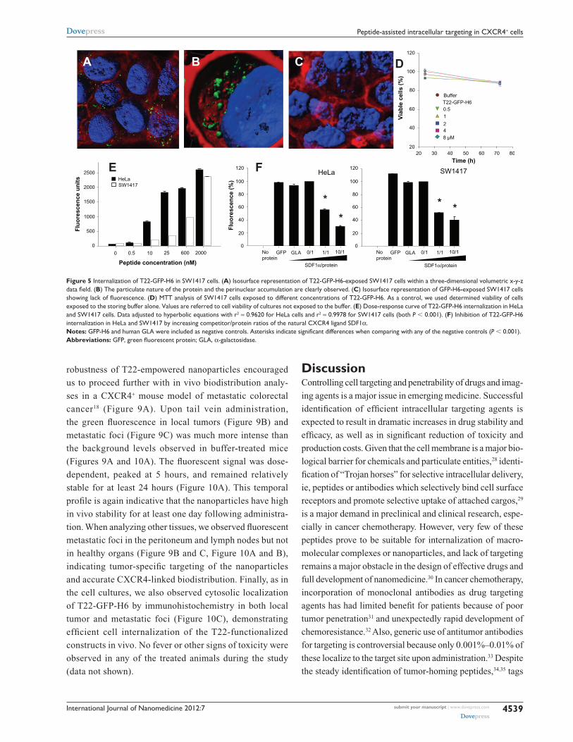
Figure 5 Internalization of T22-GFP-H6 in SW1417 cells. (A) Isosurface representation of T22-GFP-H6-exposed SW1417 cells within a three-dimensional volumetric x-y-z data field. (B) The particulate nature of the protein and the perinuclear accumulation are clearly observed. (C) Isosurface representation of GFP-H6-exposed SW1417 cells showing lack of fluorescence. (D) MTT analysis of SW1417 cells exposed to different concentrations of T22-GFP-H6. As a control, we used determined viability of cells exposed to the storing buffer alone. Values are referred to cell viability of cultures not exposed to the buffer. (E) Dose-response curve of T22-GFP-H6 internalization in HeLa and SW1417 cells. Data adjusted to hyperbolic equations with r2 = 0.9620 for HeLa cells and r2 = 0.9978 for SW1417 cells (both P , 0.001). (F) Inhibition of T22-GFP-H6 internalization in HeLa and SW1417 by increasing competitor/protein ratios of the natural CXCR4 ligand SDF1α. Notes: GFP-H6 and human GLA were included as negative controls. Asterisks indicate significant differences when comparing with any of the negative controls (P , 0.001).Abbreviations: GFP, green fluorescent protein; GLA, α-galactosidase.
robustness of T22-empowered nanoparticles encouraged
us to proceed further with in vivo biodistribution analy-
ses in a CXCR4+ mouse model of metastatic colorectal
cancer18 (Figure 9A). Upon tail vein administration,
the green fluorescence in local tumors (Figure 9B) and
metastatic foci (Figure 9C) was much more intense than
the background levels observed in buffer-treated mice
(Figures 9A and 10A). The fluorescent signal was dose-
dependent, peaked at 5 hours, and remained relatively
stable for at least 24 hours (Figure 10A). This temporal
profile is again indicative that the nanoparticles have high
in vivo stability for at least one day following administra-
tion. When analyzing other tissues, we observed fluorescent
metastatic foci in the peritoneum and lymph nodes but not
in healthy organs (Figure 9B and C, Figure 10A and B),
indicating tumor-specific targeting of the nanoparticles
and accurate CXCR4-linked biodistribution. Finally, as in
the cell cultures, we also observed cytosolic localization
of T22-GFP-H6 by immunohistochemistry in both local
tumor and metastatic foci (Figure 10C), demonstrating
efficient cell internalization of the T22-functionalized
constructs in vivo. No fever or other signs of toxicity were
observed in any of the treated animals during the study
(data not shown).
DiscussionControlling cell targeting and penetrability of drugs and imag-
ing agents is a major issue in emerging medicine. Successful
identification of efficient intracellular targeting agents is
expected to result in dramatic increases in drug stability and
efficacy, as well as in significant reduction of toxicity and
production costs. Given that the cell membrane is a major bio-
logical barrier for chemicals and particulate entities,28 identi-
fication of “Trojan horses” for selective intracellular delivery,
ie, peptides or antibodies which selectively bind cell surface
receptors and promote selective uptake of attached cargos,29
is a major demand in preclinical and clinical research, espe-
cially in cancer chemotherapy. However, very few of these
peptides prove to be suitable for internalization of macro-
molecular complexes or nanoparticles, and lack of targeting
remains a major obstacle in the design of effective drugs and
full development of nanomedicine.30 In cancer chemotherapy,
incorporation of monoclonal antibodies as drug targeting
agents has had limited benefit for patients because of poor
tumor penetration31 and unexpectedly rapid development of
chemoresistance.32 Also, generic use of antitumor antibodies
for targeting is controversial because only 0.001%–0.01% of
these localize to the target site upon administration.33 Despite
the steady identification of tumor-homing peptides,34,35 tags
submit your manuscript | www.dovepress.com
Dovepress
Dovepress
4539
Peptide-assisted intracellular targeting in CXCR4+ cells

International Journal of Nanomedicine 2012:7
for receptor-dependent internalization of macromolecular
complexes and nanoparticles are still unavailable.36 In this
study, we identified that T22, a short amino acid segment
(Figure 1A), is an unusually strong agent for intracellular
targeting in CXCR4+ cells, the selectivity, stability and effi-
cacy of penetration of which have been fully demonstrated
in cell culture (Figures 3–5) and in vivo (Figures 9 and 10).
100
0 10
Min h
20 30 1 3 5 2280
60
20
No protein
No serumSerum
15
10
5
00.1 1 10
102
FITC-A
Time (h)
A
B
C
D
101100
Eve
nts
Flu
ore
scen
ce (
%)
100Size (d·nm)
Vo
lum
e (%
)
0 5 10 15 20
50 nm
Figure 6 Characterization of T22-empowered nanoparticles. (A) Remaining fluorescence during incubation of T22-GFP-H6 in human serum. In the inset, integrity of T22-GFP-H6 monomers monitored by Western blot. (B) Internalization of T22-GFP-H6 in HeLa cells in the presence of 10% fetal calf serum, monitored by the number of fluorescent cells. (C) Dynamic light scattering size analysis of T22-GFP-H6 nanoparticles in NaCO3H buffer. (D) Transmission electron microscopy of T22-GFP-H6 nanoparticles.Abbreviation: GFP, green fluorescent protein.
In addition, we produced T22 at high yields in a recombi-
nant form as a domain of a highly stable modular protein
(Figure 2), demonstrating lack of toxicity of this peptide in
E. coli and pointing to the feasibility of production of further
(improved or adapted) engineered versions.
Interestingly, T22 had been previously identified as a
CXCR4 ligand and explored in the context of antiretroviral
therapies,37 because CXCR4 is a coreceptor for HIV and
T22 inhibits viral attachment. However, the ability of T22
to penetrate cells was not suspected. Importantly, internal-
ization of T22 does not depend on mere interaction with
CXCR4, because other ligands tested in this study failed
to promote efficient uptake (Figures 3 and 4), even show-
ing that affinity for the receptor was higher than for T22.10
Dissociation between affinity for the receptor and endocytosis
might account for the limited penetrability and poor uptake
of antibody-empowered drugs.31,38 Other CXCR4 ligands
previously investigated for targeted drug delivery showed
very low or null penetration, even revealing themselves
as agonists of CXCR4 and stimulating cell division.39 In
contrast, in our hands, T22-exposed cells never showed
significant proliferation compared with controls (data not
shown). The efficient endosomal escape of the constructs
generated might be due to the proton-sponge activity of the
accompanying polyhistidines that, while useful for one-step
protein purification, act also as a proton sponge, permitting
endosomal disruption and delivery of the functionalized
materials into the cytoplasm.22
On the other hand, when tested in an in vivo animal
model of colorectal cancer, in which CXCR4+ cells were
associated with aggressiveness, T22-empowered nanopar-
ticles selectively localized not only in the primary tumor but
also in metastatic foci (Figures 9C, 10B and C) confirming
good stability of the protein-only nanoparticles generated
in this study and suggesting that these nanoparticles might
eventually be able to be attached to drugs to control tumor
spread. This would be especially promising if used in the
early stages of disease, because current treatment strategies
for colorectal cancer are targeted to the primary tumor rather
than to disseminated disease.40 Moreover, achieving higher
intracellular concentrations of anticancer agents is expected
to lead to a better antitumor effect, given that most of the
chemotherapeutic agents used have a steep dose-response
relationship.41 In the same context, precise targeting of
imaging agents to metastatic foci would create additional
diagnostic strategies.
Finally, T22 was able to impart self-organizing properties
to GFP-H6, as has been previously shown for another cationic
submit your manuscript | www.dovepress.com
Dovepress
Dovepress
4540
Unzueta et al
International Journal of Nanomedicine 2012:7
Figure 8 Molecular modeling of T22-GFP-H6 monomers and nanoparticles. (A) Electrostatic field of the T22-GFP-H6 building monomer (cationic in blue and anionic in red). (B) Potential organization of T22-GFP-H6 as pentamers of 11.1 nm (left) and as octamers of 13.3 nm (right), in which the intervention of T22 assists the electrostatic self-assembling of the multimeric protein complex.Abbreviation: GFP, green fluorescent protein.
20
15
10
5
0
20
15
10
5
0
0.1 1 10 100 1000 10000
0.1 1 10 100 1000 10000
Size (d·nm)
Size (d·nm)
24 h R T
24 h R T
One year − 80°C
One year − 80°CB
A
Vo
lum
e (%
)V
olu
me
(%)
Figure 7 Stability of T22-GFP-H6 nanoparticles. (A) Size distribution of T22-GFP-H6 nanoparticles after one year of storage at -80°C. The same sample was further incubated at room temperature for 24 hours, submitted to one additional freezing and thawing step and analyzed. (B) These samples were tested for their ability to internalize SW1417, showing uptake images comparable with those shown in Figures 4 and 5.Abbreviations: GFP, green fluorescent protein; RT, room temperature.
stretch, namely the receptor-independent cell-penetrating pep-
tide, R9.13 The resulting building blocks, probably establishing
electrostatic contact due to their dipolar nature (Figure 8), form
relatively monodispersed protein-only nanoparticles about
13 nm in size (Figure 6), with proteolytic stability in human
serum (Figure 6A) on systemic administration (Figures 9
and 10) and remaining assembled and fully functional under
different storage conditions (Figure 7). Interestingly, T22-
empowered building blocks are highly fluorescent (22.8 fluo-
rescence units/µg), indicative of a poor impact of the peptide on
GFP. V1-GFP-H6 shows slightly lower fluorescent emission
(12.4 fluorescence units/µg), being sufficiently high to monitor
cell penetration, indicative of the conformational constraints
imposed by the peptide on the building block.
Although the nanoscale architectonic properties imparted
by T22 to the holding building block are secondary in terms
of their CXCR4-dependent intracellular targeting ability, they
warrant further investigation as architectonic tags for con-
struction of protein-only nanoparticles.42,43 Self-assembling
peptides, presently under intense investigation in diverse
nanomedical applications,44–47 base their organizing proper-
ties on amyloid-like cross-molecular interactions, and when
produced in “cell factories” such as E. coli, tend to aggregate
as amorphous protein deposits.48 CXCR4 is an important
cell surface marker in HIV infection,5 metastatic colorectal
cancer,8,9 and other neoplasms,6 so incorporation of T22 as
a targeting agent offers many opportunities for functional-
ization of drugs and nanoparticles for intracellular delivery,
submit your manuscript | www.dovepress.com
Dovepress
Dovepress
4541
Peptide-assisted intracellular targeting in CXCR4+ cells

International Journal of Nanomedicine 2012:7
60
A C
B
50
40
30
20
10
0
60Peritoneal tumor foci
Incr
ease
d F
LI r
atio
(NP
/bu
ffer
)In
crea
sed
FL
I rat
io(N
P/b
uff
er)
Local tumor
Local tumor
Buffer
400X 400X
400X 400X
1000X 1000X
T22-GFP-H6
T22-GFP-H6
Peritoneal mets
Buffer20 µg T22-GFP-H6
100 µg T22-GFP-H6500 µg T22-GFP-H6
Buffer500 µg T22-GFP-H6
50
40
30
20
10
0
5 h 24 h
5 h 24 h 48 h
Figure 10 Accumulation of T22-empowered nanoparticles in colorectal cancer metastatic foci. Enhanced green fluorescence associated with nanoparticle accumulation in local tumors (A) and peritoneal metastases (B) in experimental mice, as compared with buffer-treated controls. (C) Anti-His tag inmunostaining showing cytosolic localization of T22-GFP-H6 in local tumor tissue and peritoneal metastases in mice injected with T22-NP, which was absent in control animals injected with buffer.Abbreviations: FLI, increased fluorescence; GFP, green fluorescent protein.
Buffer
A
B
C
400x
CXCR4
200x
Lo
cal t
um
or
sect
ion
500 µg, 5 h 20 µg, 24 h 20 µg, 48 h
Figure 9 Biodistribution of T22-empowered nanoparticles in an animal model of colorectal cancer. (A) Nude mouse bearing a local tumor (black asterisk), mesenteric lymph node (black arrow), and peritoneal metastases (empty arrow) after microinjecting 2 × 10⁶ SW1417 human colorectal cancer cells into the cecal wall. The local tumor and mesenteric lymph node metastases overexpress CXCR4 in this model, as assessed by immunohistochemistry. (B) Selective biodistribution of T22-GFP-H6 in local tumor tissues 5, 24, or 48 hours after intravenous administration of 500 µg or 20 µg of nanoparticles as measured ex vivo. Fluorescence was undetectable in tumors from buffer-treated animals. (C) Accumulation of nanoparticles in peritoneal and lymph node metastases. No fluorescence was observed in any normal (liver, kidney, lung, heart) tissue, except for the biliary vesicle which showed fluorescence both in control and experimental animals.Abbreviation: GFP, green fluorescent protein.
submit your manuscript | www.dovepress.com
Dovepress
Dovepress
4542
Unzueta et al

International Journal of Nanomedicine 2012:7
especially for disorders in which CXCR4 expression plays
a pathophysiological role, including cancer, inflammation,
autoimmunity, and ischemic lesions.4
ConclusionThe peptide T22, a known ligand of CXCR4, has been shown
to be an unusually powerful tag for intracellular targeting in
CXCR4+ cells, both in cell culture and in vivo. T22 is able
to mediate the internalization of self-assembling protein-only
nanoparticles 13 nm in mean diameter, keeping the stability
and fluorescence emission of GFP-based building blocks.
Rapid endosomal uptake and perinuclear accumulation of
T22-empowered nanoparticles without cytotoxicity offer a
wide spectrum of diagnostic and therapeutic opportunities
for use of T22 in emerging nanomedicine to treat CXCR4-
linked diseases, for which intracellular targeting agents are
currently missing.
AcknowledgmentsWe appreciate the technical support of Fran Cortés from the
Cell Culture Unit of the Servei de Cultius Cellulars, Produc-
ció d’Anticossos i Citometria, of the Servei de Microscòpia,
and of the Protein Production Platform (CIBER-BBN). We
also acknowledge the financial support received for the design
and production of artificial viruses for gene therapy to EV,
RM, and AV from FIS (PS0900165, PS0900965), MICINN
(ACI2009-0919), AGAUR (2009SGR-108), and CIBER de
Bioingeniería, Biomateriales y Nanomedicina, an initiative
funded by the VI National R&D&i Plan 2008–2011, Inicia-
tiva Ingenio 2010, Consolider Program, CIBER Actions and
financed by the Instituto de Salud Carlos III with assistance
from the European Regional Development Fund. UU and
JDE have received predoctoral fellowships from ISCIII and
MICINN, respectively, and AV has received an Institució
Catalana de Recerca i Estudis Avançats Academia award.
DisclosuresUU, EV, NFM, AV, RM, IC, and MVC are cited as inven-
tors in a patent application (EP11382005.4) covering the
therapeutic use of T22. All other authors report no conflicts
of interest in this work.
References1. Pautler M, Brenner S. Nanomedicine: promises and challenges for the
future of public health. Int J Nanomedicine. 2010;5:803–809.2. Ferrer-Miralles N, Vazquez E, Villaverde A. Membrane-active peptides
for non-viral gene therapy: making the safest easier. Trends Biotechnol. 2008;26:267–275.
3. Milletti F. Cell-penetrating peptides: classes, origin, and current landscape. Drug Discov Today. March 23, 2012. [Epub ahead of print.]
4. Peled A, Wald O, Burger J. Development of novel CXCR4-based therapeutics. Expert Opin Investig Drugs. 2012;21:341–353.
5. Wilen CB, Tilton JC, Doms RW. Molecular mechanisms of HIV entry. Adv Exp Med Biol. 2012;726:223–242.
6. Klonisch T, Wiechec E, Hombach-Klonisch S, et al. Cancer stem cell markers in common cancers – therapeutic implications. Trends Mol Med. 2008;14:450–460.
7. Sun X, Cheng G, Hao M, et al. CXCL12/CXCR4/CXCR7 chemokine axis and cancer progression. Cancer Metastasis Rev. 2010;29:709–722.
8. Kim J, Mori T, Chen SL, et al. Chemokine receptor CXCR4 expression in patients with melanoma and colorectal cancer liver metastases and the association with disease outcome. Ann Surg. 2006;244:113–120.
9. Liang Z, Yoon Y, Votaw J, Goodman MM, Williams L, Shim H. Silencing of CXCR4 blocks breast cancer metastasis. Cancer Res. 2005;65:967–971.
10. Liang X. CXCR4, inhibitors and mechanisms of action. Chem Biol Drug Des. 2008;72:97–110.
11. Murakami T, Zhang TY, Koyanagi Y, et al. Inhibitory mechanism of the CXCR4 antagonist t22 against human immunodeficiency virus type 1 infection. J Virol. 1999;73:7489–7496.
12. Bradford MM. A rapid and sensitive method for the quantitation of microgram quantities of protein utilizing the principle of protein-dye binding. Anal Biochem. 1976;72:248–254.
13. Vazquez E, Roldan M, Díez-Gil C, et al. Protein nanodisk assembling and intracellular trafficking powered by an arginine-rich (R9) peptide. Nanomedicine (Lond). 2010;5:259–268.
14. Vazquez E, Cubarsi R, Unzueta U, et al. Internalization and kinetics of nuclear migration of protein-only, arginine-rich nanoparticles. Biomaterials. 2010;31:9333–9339.
15. Seras-Franzoso J, Díez-Gil C, Vazquez E, et al. Bioadhesiveness and efficient mechanotransduction stimuli synergistically provided by bacterial inclusion bodies as scaffolds for tissue engineering. Nanomedicine (Lond). 2012;7:79–93.
16. Barbera VM, Martin M, Marinoso L, et al. The 18q21 region in colorectal and pancreatic cancer: independent loss of DCC and DPC4 expression. Biochim Biophys Acta. 2000;1502:283–296.
17. Baig MS, Manickam N. Homology modeling and docking studies of Comamonas testosteroni B-356 biphenyl-2,3-dioxygenase involved in degradation of polychlorinated biphenyls. Int J Biol Macromol. 2010; 46:47–53.
18. Cespedes MV, Espina C, Garcia-Cabezas MA, et al. Orthotopic micro-injection of human colon cancer cells in nude mice induces tumor foci in all clinically relevant metastatic sites. Am J Pathol. 2007;170: 1077–1085.
19. Amara A, Gall SL, Schwartz O, et al. HIV coreceptor downregulation as antiviral principle: SDF-1alpha-dependent internalization of the chemokine receptor CXCR4 contributes to inhibition of HIV replication. J Exp Med. 1997;186:139–146.
20. Zhou NM, Luo ZW, Luo JS, Hall JW, Huang ZW. A novel peptide antagonist of CXCR4 derived from the N-terminus of viral chemokine vMIP-II. Biochemistry. 2000;39:3782–3787.
21. Fujii N, Nakashima H, Tamamura H. The therapeutic potential of CXCR4 antagonists in the treatment of HIV. Expert Opin Investig Drugs. 2003;12:185–195.
22. Ferrer-Miralles N, Corchero JL, Kumar P, et al. Biological activities of histidine-rich peptides; merging biotechnology and nanomedicine. Microb Cell Fact. 2011;10:101.
23. Kucia M, Jankowski K, Reca R, et al. CXCR4-SDF-1 signalling, locomotion, chemotaxis and adhesion. J Mol Histol. 2004;35: 233–245.
24. Toledo-Rubio V, Vazquez E, Platas G, et al. Protein aggregation and soluble aggregate formation screened by a fast microdialysis assay. J Biomol Screen. 2010;15:453–457.
25. Domingo-Espin J, Vazquez E, Ganz J, et al. The nanoparticulate architecture of protein-based artificial viruses is supported by protein-DNA interactions. Nanomedicine (Lond). 2011;6:1047–1061.
26. Vazquez E, Corchero JL, Villaverde A. Post-production protein stability: trouble beyond the cell factory. Microb Cell Fact. 2011;10:60.
submit your manuscript | www.dovepress.com
Dovepress
Dovepress
4543
Peptide-assisted intracellular targeting in CXCR4+ cells
International Journal of Nanomedicine
Publish your work in this journal
Submit your manuscript here: http://www.dovepress.com/international-journal-of-nanomedicine-journal
The International Journal of Nanomedicine is an international, peer-reviewed journal focusing on the application of nanotechnology in diagnostics, therapeutics, and drug delivery systems throughout the biomedical field. This journal is indexed on PubMed Central, MedLine, CAS, SciSearch®, Current Contents®/Clinical Medicine,
Journal Citation Reports/Science Edition, EMBase, Scopus and the Elsevier Bibliographic databases. The manuscript management system is completely online and includes a very quick and fair peer-review system, which is all easy to use. Visit http://www.dovepress.com/ testimonials.php to read real quotes from published authors.
International Journal of Nanomedicine 2012:7
27. Martinez-Alonso M, Gonzalez-Montalban N, Garcia-Fruitos E, Villaverde A. The functional quality of soluble recombinant polypep-tides produced in Escherichia coli is defined by a wide conformational spectrum. Appl Environ Microbiol. 2008;101:1353–1358.
28. Riehemann K, Schneider SW, Luger TA, Godin B, Ferrari M, Fuchs H. Nanomedicine – challenge and perspectives. Angew Chem Int Ed Engl. 2009;48:872–897.
29. Dietz GP, Bahr M. Delivery of bioactive molecules into the cell: the Trojan horse approach. Mol Cell Neurosci. 2004;27:85–131.
30. Duncan R, Gaspar R. Nanomedicine(s) under the microscope. Mol Pharm. 2011;8:2101–2141.
31. Thurber GM, Schmidt MM, Wittrup KD. Factors determining antibody distribution in tumors. Trends Pharmacol Sci. 2008;29:57–61.
32. Ellis LM, Hicklin DJ. Resistance to targeted therapies: refining antican-cer therapy in the era of molecular oncology. Clin Cancer Res. 2009;15: 7471–7478.
33. Jain M, Venkatraman G, Batra SK. Optimization of radioimmuno-therapy of solid tumors: biological impediments and their modulation. Clin Cancer Res. 2007;13:1374–1382.
34. Laakkonen P, Vuorinen K. Homing peptides as targeted delivery vehicles. Integr Biol (Camb). 2010;2:326–337.
35. Enback J, Laakkonen P. Tumour-homing peptides: tools for targeting, imaging and destruction. Biochem Soc Trans. 2007;35:780–783.
36. Mocellin S, Lise M, Nitti D. Targeted therapy for colorectal cancer: mapping the way. Trends Mol Med. 2005;11:327–335.
37. Rusconi S, Scozzafava A, Mastrolorenzo A, Supuran CT. An update in the development of HIV entry inhibitors. Curr Top Med Chem. 2007;7:1273–1289.
38. Segal NH, Saltz LB. Evolving treatment of advanced colon cancer. Annu Rev Med. 2009;60:207–219.
39. Egorova A, Kiselev A, Hakli M, Ruponen M, Baranov V, Urtti A. Chemokine-derived peptides as carriers for gene delivery to CXCR4 expressing cells. J Gene Med. 2009;11:772–781.
40. Sleeman J, Steeg PS. Cancer metastasis as a therapeutic target. Eur J Cancer. 2010;46:1177–1180.
41. Skipper HE, Schabel FM Jr, Mellett LB, et al. Implications of biochemical, cytokinetic, pharmacologic, and toxicologic relationships in the design of optimal therapeutic schedules. Cancer Chemother Rep. 1970;54:431–450.
42. Villaverde A. Nanotechnology, bionanotechnology and microbial cell factories. Microb Cell Fact. 2010;9:53.
43. Vazquez E, Villaverde A. Engineering building blocks for self- assembling protein nanoparticles. Microb Cell Fact. 2010;9:101.
44. Sadatmousavi P, Soltani M, Nazarian R, Jafari M, Chen P. Self-assembling peptides: potential role in tumor targeting. Curr Pharm Biotechnol. 2011;12:1089–1100.
45. Zhao Y, Tanaka M, Kinoshita T, Higuchi M, Tan T. Self-assembling peptide nanofiber scaffolds for controlled release governed by gelator design and guest size. J Control Release. 2010;147:392–399.
46. Kyle S, Aggeli A, Ingham E, McPherson MJ. Recombinant self- assembling peptides as biomaterials for tissue engineering. Biomaterials. 2010;31:9395–9405.
47. Huang H, Sun XS. Rational design of responsive self-assembling peptides from native protein sequences (dagger). Biomacromolecules. 2010;11:3390–3394.
48. Wu W, Xing L, Zhou B, Lin Z. Active protein aggregates induced by terminally attached self-assembling peptide ELK16 in Escherichia coli. Microb Cell Fact. 2011;10:9.
submit your manuscript | www.dovepress.com
Dovepress
Dovepress
Dovepress
4544
Unzueta et al