Oneida Lake Cyanobacteria Guide
47
Oneida Lake Cyanobacteria Guide Heather Lee Cornell Biological Field Station Summer 2011
-
Upload
phungthuan -
Category
Documents
-
view
225 -
download
4
Transcript of Oneida Lake Cyanobacteria Guide

Oneida Lake Cyanobacteria Guide
Heather Lee
Cornell Biological Field Station
Summer 2011


Oneida Lake Cyanobacteria Guide
ii
Acknowledgements
This research was funded by the Cornell Biological Field Station and the Andrew W. Mellon
Foundation. Special thanks to Dr. Lars Rudstam1, Dr. Rebecca Schneider2, Amy Hetherington3,
Chris Hotaling4, and Dr. Bill Schaffer5.
1 Professor, Cornell Biological Field Station, 900 Shackelton Point Road, Bridgeport, NY 13030, [email protected] 2 Assistant Professor, Cornell University, Department of Natural Resources, Bruckner Hall, Ithaca, NY 14853, [email protected] 3 Graduate Student, Cornell University, Department of Natural Resources, Bruckner Hall, Ithaca, NY 14853, [email protected] 4 Technician, Cornell Biological Field Station, 900 Shackelton Point Road, Bridgeport, NY 13030, [email protected] 5 Professor Emeritus, Cornell University, Department of Ecology & Evolutionary Biology, Corson-Mudd Hall Ithaca, NY 14853, [email protected]


Oneida Lake Cyanobacteria Guide
iii
Table of Contents
Introduction . . . . . . . . . . . . . . . . . . . . . . . . . . . . . . . . . . . . . . . . . . . . . . . . . . . . . . . . . . . . . . . . . . . 1
Anabaena . . . . . . . . . . . . . . . . . . . . . . . . . . . . . . . . . . . . . . . . . . . . . . . . . . . . . . . . . . . . . . . . . . . . 2
Aphanizomenon . . . . . . . . . . . . . . . . . . . . . . . . . . . . . . . . . . . . . . . . . . . . . . . . . . . . . . . . . . . . . . . 4
Aphanocapsa . . . . . . . . . . . . . . . . . . . . . . . . . . . . . . . . . . . . . . . . . . . . . . . . . . . . . . . . . . . . . . . . . . 6
Aphanothece . . . . . . . . . . . . . . . . . . . . . . . . . . . . . . . . . . . . . . . . . . . . . . . . . . . . . . . . . . . . . . . . . . 8
Chroococcus . . . . . . . . . . . . . . . . . . . . . . . . . . . . . . . . . . . . . . . . . . . . . . . . . . . . . . . . . . . . . . . . . . 10
Gloeotrichia . . . . . . . . . . . . . . . . . . . . . . . . . . . . . . . . . . . . . . . . . . . . . . . . . . . . . . . . . . . . . . . . . . 12
Gomphosphaeria . . . . . . . . . . . . . . . . . . . . . . . . . . . . . . . . . . . . . . . . . . . . . . . . . . . . . . . . . . . . . . 16
Lyngbya . . . . . . . . . . . . . . . . . . . . . . . . . . . . . . . . . . . . . . . . . . . . . . . . . . . . . . . . . . . . . . . . . . . . . 18
Merismopedia . . . . . . . . . . . . . . . . . . . . . . . . . . . . . . . . . . . . . . . . . . . . . . . . . . . . . . . . . . . . . . . . 20
Microcystis . . . . . . . . . . . . . . . . . . . . . . . . . . . . . . . . . . . . . . . . . . . . . . . . . . . . . . . . . . . . . . . . . . . 22
Myxobaktron [Keratococcus, Dactylococcus] . . . . . . . . . . . . . . . . . . . . . . . . . . . . . . . . . . . . . . . 24
Oscillatoria . . . . . . . . . . . . . . . . . . . . . . . . . . . . . . . . . . . . . . . . . . . . . . . . . . . . . . . . . . . . . . . . . . 26
Phormidium . . . . . . . . . . . . . . . . . . . . . . . . . . . . . . . . . . . . . . . . . . . . . . . . . . . . . . . . . . . . . . . . . . 28
Rhabdoderma . . . . . . . . . . . . . . . . . . . . . . . . . . . . . . . . . . . . . . . . . . . . . . . . . . . . . . . . . . . . . . . . 30
Synechococcus . . . . . . . . . . . . . . . . . . . . . . . . . . . . . . . . . . . . . . . . . . . . . . . . . . . . . . . . . . . . . . . . 32
Synechocystis . . . . . . . . . . . . . . . . . . . . . . . . . . . . . . . . . . . . . . . . . . . . . . . . . . . . . . . . . . . . . . . . . 34
Glossary . . . . . . . . . . . . . . . . . . . . . . . . . . . . . . . . . . . . . . . . . . . . . . . . . . . . . . . . . . . . . . . . . . . . . 36
References . . . . . . . . . . . . . . . . . . . . . . . . . . . . . . . . . . . . . . . . . . . . . . . . . . . . . . . . . . . . . . . . . . . 39


Oneida Lake Cyanobacteria Guide
1
Introduction Overview This guide provides a reference to commonly identified cyanobacteria in Oneida Lake. Cyanobacteria, also known as blue-green algae, are a very ancient group of photosynthetic bacteria. As probably the first photosynthetic organisms releasing oxygen, they altered the primitive, virtually oxygen-free atmosphere leading to the evolution of today’s living world. They are of universal occurrence in fresh and salt water, from the Antarctic to the arctic, in permanently frozen lakes and in hot springs, in soil, deserts and tundra. Although small in size, cyanobacteria can have a tremendous impact on ecosystems due to their numbers. Aquatic cyanobacteria are probably best known for the extensive and highly visible blooms that can have the appearance of blue-green paint or scum on the water’s surface. The association of toxicity with such blooms has frequently led to the closure of recreational waters when blooms are observed. Cyanobacteria thrive in warm, shallow, nutrient-rich waters which makes Oneida Lake a perfect habitat during the summer months. In Oneida Lake, cyanobacteria are the main components of the algal bloom that historically begin in early July. These blooms decrease water quality and have closed beaches in recent years due to potential toxicity of the water. Methodology Identification: Cyanobacteria are distinguishable from algae due to their homogenous cell contents and lack of a definitive cell wall. The unique blue-green color of cyanobacteria can also be a good identifier, but not always reliable since aerotopes and secondary metabolites can mask the color. Cyanobacteria can be identified by using this guide and other taxonomic keys available (Baker et al. 2011; Wehr et al. 2003). The detailed descriptions in this guide were taken directly from Freshwater Algae of North America: Ecology and Classification (Wehr et al. 2003). Enumeration: A standard methodology for cyanobacteria enumeration was used at the Cornell Biological Field Station for Summer 2011. Samples were taken from a plankton tow with a mesh size of 53 microns. A milliliter of sample was taken and diluted, if needed, so that cyanobacteria cell counts were approximately 400 per field of view at 200x on a standard light microscope. Contents of 25 fields of view were enumerated for evaluation. Cyanobacteria were identified to genus to determine the concentration. To determine the biovolume of each genus of cyanobacteria, the average dimensions of each cell were taken and calculations were done according to the Biovolume Calculation for Pelagic and Benthic Microalgae (Hillebrand et al. 1999)

Oneida Lake Cyanobacteria Guide - Anabaena
2
Anabaena—(Nostocaceae) Overview: About 110 species have been described; five species have been identified in Oneida Lake. Some species can be toxic. They’re filamentous and regular (no tapering at ends like Aphanizomenon). Trichomes with spherical cells and intercalary heterocysts (frequency varies inversely with available dissolved nitrogen mainly as nitrate, nitrite or ammonium ions). Occasionally a "watery" sheath forms, although in this key it is classified as having no visible sheath. Staining is needed to see the thin sheaths when present. Detailed Description:
The thallus is with solitary filaments, or arranged in free clusters, or in macroscopic mats, or in the tissues of aquatic plants. Trichomes are straight, curved, or regularly coiled, rarely in bundles with parallel-oriented filaments. Sheaths are never firm, in form of mucilaginous, hyaline, diffuse envelopes, occurring in only some species. Trichomes are constricted or unconstructed at the cross walls, uniseriate, isopolar (both poles with the same morphology). Cells are spherical, ellipsoidal, short or long cylindrical, sometimes bent (reniform), pale to bright blue-green or yellow-green; the planktonic species have aerotopes, occasionally with granular contents. Hereocytes are intercalary, solitary at regular intervals along filament (metameric), up to nine (rarely more) per filament. Akinetes are spherical, cylindrical, curved, intercalary, solitary or in groups of two to five, sometimes with a yellowish colored epispore in some species adjacent to heterocytes. Cells divide perpendicularly to trichome axis; meristematic zones are not noted. Reproduction occurs via trichome fragmentation and akinete production.
http://www.ohio.edu/plantbio/vislab/algaeimage/pages/anabaena.html

Oneida Lake Cyanobacteria Guide - Anabaena
3
http://cfb.unh.edu/phycokey/Choices/Cyanobacteria/cyano_filaments/cyano_unbranched_fil/untapered_filaments/heterocysts/no_visible_sh
eath/ANABAENA/Anabaena_Image_page.html
http://publicfiles.dep.state.fl.us/dear/labs/biology/hab/images/micrographs%20of%20cyanobacteria/anabaena.sp.jpg

Oneida Lake Cyanobacteria Guide - Aphanizomenon
4
Aphanizomenon – (Nostocaceae) Overview:
Similar to Anabaena, but it tapers at the ends and the cells are not a uniform length (the end cells are usually longer). Straight, always unbranched, filaments, that in some forms, aggregate in parallel forming colonial "rafts." The primary morphology is trichomes (filaments without sheaths), secondary is colonial. Heterocysts are intercalary (between vegetative cells). Trichomes are tapered at both ends, with end cells usually longer than the central cells. Also forms akinetes. Detailed Description:
The thallus is filamentous. Filaments are free floating, solitary, or gathered in small or large fasciculated colonies with filaments in characteristically parallel arrangements (may appear as macroscopic flakes or like grass clippings on the lake surface). Trichomes are isopolar, subsymmetrical, straight, sometimes slightly curved or bent ends, cylindrical or narrowing toward the ends with or without constrictions at the cross walls. Firm sheaths are absent, but the trichomes of several species are covered with fine, diffuse, mucilaginous, colorless envelopes. Cells are cylindrical or long barrel shaped, with quite variable width/length ratio, pale blue-green with aerotopes (sometimes facultative). The end cells are often much longer than the central cells, and hyaline. In some species, they are cylindrical and rounded at the end without visible cell contents; in others, narrowed and pointed. Heterocytes are almost spherical, ellipsoidal, or cylindrical with two pores, always intercalary and solitary, usually only a few (1-3) per trichome (rarely more). Akinetes are long or short cylindrical with rounded ends, or elliptical, rarely almost spherical, solitary or groups of two or three adjacent to heterocytes or distant. Cell division occurs along the whole trichome with the exception of the end cells. Reproduction occurs via trichome disintegration and akinete germination.
http://cfb.unh.edu/phycokey/Choices/Cyanobacteria/cyano_filaments/cyano_unbranched_fil/tapered_filaments/APHANIZOMENON/Aphanizo
menon_Image_page.html#pic02

Oneida Lake Cyanobacteria Guide - Aphanizomenon
5
http://biodidac.bio.uottawa.ca/thumbnails/filedet.htm?File_name=BRIE003P&File_type=jpg

Oneida Lake Cyanobacteria Guide - Aphanocapsa
6
Aphanocapsa – (Merismopediaceae) Overview:
Similar to Microcystis, but Aphanocapsa does not have gas vacuoles. Spherical cells irregularly distributed within mucilage, forming irregularly shaped microscopic or macroscopic colonies. Detailed Description:
Microscopic to macroscopic, mucilaginous colonies with irregularly arranged cells of various densities that are spherical or irregular. Colonial mucilage is mainly homogenous and colorless, and usually has a diffuse, rarely delimited margin. Cells have no individual envelopes or, rarely, have a fine, slimy surrounding layer that is distinct from the common mucilate. Cells are spherical, but after division, they are hemispherical, pale or bright blue-green or olive green, rarely (in marine species) red or pinkish in color, with no gas vesicles, and 0.4-6(12) um in diameter. Cells divide by binary fission in two perpendicular planes in successive generation (cleavage). Reproduction is by disintegration of colonies.
http://protist.i.hosei.ac.jp/PDB5/PCD1052/htmls/10.html

Oneida Lake Cyanobacteria Guide - Aphanocapsa
7
http://www.keweenawalgae.mtu.edu/ALGAL_IMAGES/cyanobacteria/Aphanocapsa_n14_ricetow24_4016_c.jpg

Oneida Lake Cyanobacteria Guide - Aphanothece
8
Aphanothece—(Synechococcaeae) Overview:
Very similar to Aphanocapsa, but the cells are elongated rather than spherical. Many oblong, flattened cells unevenly distributed within mucilage, forming amorphous colonies. Detailed Description:
Colonies are microscopic or macroscopic (up to several centimeters in diameter), multicellular, more or less spherical or mostly amorphous, grayish, greenish, blue-green or brownish colored, with irregularly, sometimes densely arranged cells, which are embedded by colonial mucilage. Two subgenera are defined: (i) Anathece—cells in diffuse mucilage, without individual envelopes—and (ii) Aphhanothece—mucilage usually distinctly delimited, and cells (especially marginal) enveloped facultatively within individual sheaths or envelopes. Cells are widely oval to cylindrical, straight or slightly arcuate, pale grayish blue-green, green to bright blue-green (rarely reddish), usually with apparent chromatoplasm in larger cells. Gas vesicles and aerotopes are present in some planktonic species. Reproduction is by transversal binary fission of cells, which is perpendicular to the longer cell axis.
http://cfb.unh.edu/phycokey/Choices/Cyanobacteria/cyano_colonies/APHANOTHECE/Aphanothece_Image_page.html#pic03

Oneida Lake Cyanobacteria Guide - Aphanothece
9
http://www.glerl.noaa.gov/seagrant/GLWL/Algae/Cyanophyta/Cards/Aphanothece.html
Oneida Lake Cyanobacteria Guide - Chroococcus
10
Chroococcus –(Chroococcaceae) Overview:
Single cells or cells in groups of a usually even number of cells (2 up to 32 - more frequently 2 - 4) inside mucilaginous envelope. Single cells are spherical, but when in groups they are often hemispherical due to the fact that daughter cells do not fully separate after division. Detailed Description:
Cells or groups of cells (mainly 2 - 4 cells), are surrounded by mucilaginous envelopes, and usually occur in microscopic, spherical or composed colonies. Rarely form agglomerations. Mucilaginous envelops are colorless or yellowish, usually copying the cell form, sometimes lamellated, distinct or diffuse at the margin (subgenus Chroococcus) or fine, homogenous, and diffuse, in which the cells or cell groups are irregularly arranged (subgenus Limnococcus). Cells at first are spherical or oval, and later are hemispherical or in the form of a segment of the sphere. The cells are 0.7-50 um in diameter. The cell content is grey, blue-green, olive green, orange, or reddish violet and granular. Only in a few planktonic species are there gas vesicles. Cell division is by binary fission in three or more planes, or irregular (in old colonies). Reproduction is by fragmentation of colonies.
http://cfb.unh.edu/phycokey/Choices/Cyanobacteria/cyano_colonies/CHROOCOCCUS/Chroococcus_Image_page.html#pic01

Oneida Lake Cyanobacteria Guide - Chroococcus
11
http://cfb.unh.edu/phycokey/Choices/Cyanobacteria/cyano_colonies/CHROOCOCCUS/Chroococcus_Image_page.html#pic01

Oneida Lake Cyanobacteria Guide – Gloeotrichia
12
Gloeotrichia – (Nostocales:Rivulariaceae)
Overview:
Spherical colonies of radiating straight trichomes (filaments without sheaths). Each
trichome has an akinete as the basal cell near the center of the colony. Akinetes, if present, are
adjacent the heterocyst. The primary morphology is trichomous (filamentous without sheaths),
the secondary is colonial.
Detailed Description:
The thallus is filamentous, attached to substratum basally forming bristle-like groups or
thin mats. Filaments are heteropolar, with a wider basal part (with heterocytes and occasionally
an associated akinete and/or with enlarged basal vegetative cells) and an apical portion forming
an elongated, tapering, hairlike form. Heterocytes develop basally; false branching occurs
occasionally with the formation of a separated trichome inside of its own sheath. Thrichomes
are constricted or unconstructed at the cross walls and always taper terminally. Sheaths are
always present, firm, in some species lamellated or enlarged at the end, forming funnel formed
collars yellow to brownish in color or colorless. Depending on their position in the trichome,
cells may be barrel shaped, cylindrical or narrowly elongated toward the ends (hairs), especially
with nutrient limitation. Aerotopes are absent from vegetative cells but may be present in
hormogonia. Meristematic zones are known in several species. Heterocytes are ellipsoidal,
spherical to hemi-spherical, mainly basal, sometimes intercalary (especially near false branches).
Akinetes are ellipsoidal to cylindrical, appearing above basal heterocytes and developing from a
vegetative portion of the trichome. Reproduction occurs via hormogonia (sometimes with
aerotopes), which are released from the end of the filament after the hair has separated.

Oneida Lake Cyanobacteria Guide – Gloeotrichia
13
http://www.algaebase.org/search/species/detail/?species_id=132337
http://www.keweenawalgae.mtu.edu/ALGAL_PAGES/cyanobacteria2.htm

Oneida Lake Cyanobacteria Guide – Gloeotrichia
14
http://www.algaebase.org/search/species/detail/?species_id=132337

15

Oneida Lake Cyanobacteria Guide – Gomphosphaeria
16
Gomphosphaeria – (Merismopediaceae) Overview:
Spherical colonies of club-shaped cells, or heart-shaped when dividing, that are joined in the middle of the colony by a system of mucilaginous stalks that widen at the periphery of the colony to enclose individual cells. The colonies are within a mucilaginous envelope. Detailed Description:
Colonies are free-living, spherical or irregularly oval, and sometimes composed of subcolonies. They have a central system of thick mucilaginous stalks that are nearly pseudodichotomously divided and may be diffuse within the colony. Stalks widen at the ends and envelope individual cells with a thin mucilage layer. Cells are slightly elongate, obviate, or club-shaped, and radially oriented more or less at the colony periphery which is sometimes enveloped by a fine, colorless, and diffuse mucilage. Cells have a homogenous pale or bright blue-green, olive-green, or red content and are (4.2)6-12(15) x 2-8(13.2) um. After division, the cells may remain joined together and form a characteristic cordiform shape. Cells in colonies are slightly distant and sometimes slightly radially displaced from one another. Cell division occurs in two planes in successive generations, perpendicular to one another and to the colony surface. Reproduction is by colony disintegration.
http://cfb.unh.edu/phycokey/Choices/Cyanobacteria/cyano_colonies/GOMPHOSPHAERIA/Gomphosphaeria_Image_page.html#pic01

Oneida Lake Cyanobacteria Guide – Gomphosphaeria
17
http://protist.i.hosei.ac.jp/PDB/Images/prokaryotes/Chroococcaceae/Gomphosphaeria/index.html
Oneida Lake Cyanobacteria Guide – Lyngbya
18
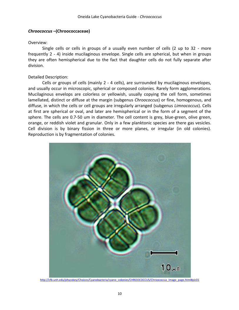
Lyngbya—(Oscillatoriaceae) Overview: Similar to Oscillatoria, but have a defined sheath that extends far past the filament. Thick and straight trichomes enclosed in a firm sheath that usually occur in mats. Filaments are usually unbranched, or sometimes with false branching. Cells are distinctively shorter than wide (discoid). Apical cells usually have a calyptra (thickened outer wall). Form motile hormogonia. No heterocysts. Detailed Description: Filaments are straight or slightly undulating (several species are finely screwlike or coiled), rarely (a few free-floating species) solitary, mainly arranged in thin or thick, flat, compact, large, layered, leathery prostrate mats on the substrate, and very rarely false branched; they are usually wider than 6 micrometers. Sheaths are always present; only hormogonia and trichomes under extreme conditions leave the sheaths. Sheaths are attached to the trichome or slightly distant, firm, thin or thick, colorless or slightly yellow-brown or reddish (very rarely bluish), sometimes slightly lamellated, and containing one motile trichome. Trichomes are cylindrical and may or may not be constricted at the cross walls. Cells are short and discoid, always shorter than wide and most without aerotopes (a few planktonic species have aerotopes). Apical cells usually have a thickened outer wall or calyptras. Reproduction occurs via trichome disintegration into usually short motile hormogonia, which often separate necridia formation.
http://www.rbgsyd.nsw.gov.au/science/Plant_Diversity_Research/australian_freshwater_algae/algpic/c
yanobacteria?SQ_DESIGN_NAME=printer_friendly

Oneida Lake Cyanobacteria Guide – Lyngbya
19
http://www.aecos.com/CPIE/algae1.html

Oneida Lake Cyanobacteria Guide - Merismopedia
20
Merismopedia—(Merismopediaceae)
Overview:
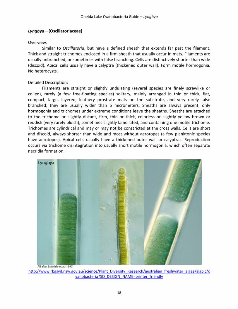
Spherical to oval cells densely arranged in rows forming flat and rectangular colonies in mucilage not extending outside a colony’s margins. No distinct sheath around individual cells. Detailed Description:
Flattened, free-living, platelike (rectangular), more or less rectangular colonies that have one layer of cells, arranged loosely or densely in perpendicular rows and enveloped by fine, colorless, usually indistinct, and marginally diffuse mucilage. Colonies are flat or slightly wavy, usually microscopic (except for a few species that are macroscopically visible), and sometimes composed of subcolonies. Cells are spherical or widely oval before division, pale or bright blue-green, (rarely reddish), and sometimes have visible centro- and chromatoplasm (parietal thylakoids). Several planktonic species have gas vesicles (few or solitary in cell centers). Occasionally the cells have slimy envelopes. After division, the cells are hemispherical and (0.4)1.2-6.5(17) micrometers in diameter. When elongated, the longer axis is situated in the plane of the colony. Cell division (binary fission) occurs regularly in two planes perpendicular to the plane of colony; the daughter cells do not move from their position after division. Reproduction is by fragmentation of colonies.
http://www.aslo.org/photopost/showphoto.php/photo/141/title/merismopedia/cat/516
Oneida Lake Cyanobacteria Guide - Merismopedia
21
http://www.keweenawalgae.mtu.edu/ALGAL_PAGES/cyanobacteria1.htm

Oneida Lake Cyanobacteria Guide - Microcystis
22
Microcystis -- (Microcystaceae) Overview:
Colonial in nature, unicellular in fast-growing cultures in the absence of flagellate, ciliate, and zooplankton predators. Detailed Description:
This genus has irregular micro or macroscopic colonies that are free-floating, compact or clathrate, may be composed of clustered subcolonies, and has sparsely or densely, irregularly arranged cells. The mucilage is fine, colorless, and diffuse or distinctly delimited, sometimes forming a wide margin around the cells (rarely with indistinct structures), or delimited along cell agglomerations. Cells are spherical or hemispherical after division and pale blue-green, but they appear brownish due to aerotopes that mask the blue-green color of the protoplast. Cells are 0.8-6(9.4) micrometers in diameter and have no individual mucilaginous envelopes. Cell division is by binary fission in three perpendicular planes in successive generations. The daughter cells grow to the original shape and size before the next division.
http://protist.i.hosei.ac.jp/PDB/Images/Prokaryotes/Chroococcaceae/Microcystis/

Oneida Lake Cyanobacteria Guide - Microcystis
23
http://cfb.unh.edu/phycokey/Choices/Cyanobacteria/cyano_colonies/MICROCYSTIS/Microcystis_Image_page.html#pic01
Oneida Lake Cyanobacteria Guide – Myxobaktron [Keratococcus, Dactylococcus]
24
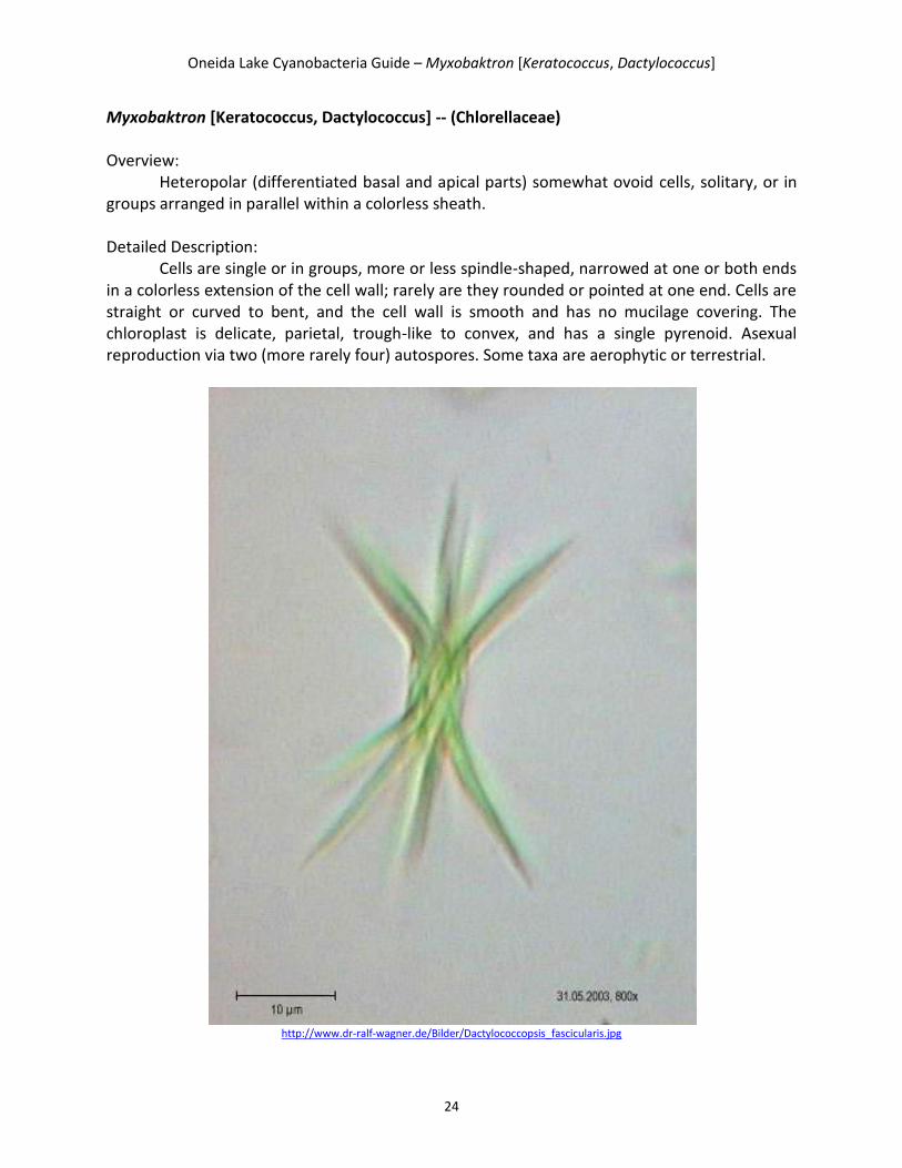
Myxobaktron [Keratococcus, Dactylococcus] -- (Chlorellaceae) Overview:
Heteropolar (differentiated basal and apical parts) somewhat ovoid cells, solitary, or in groups arranged in parallel within a colorless sheath. Detailed Description:
Cells are single or in groups, more or less spindle-shaped, narrowed at one or both ends in a colorless extension of the cell wall; rarely are they rounded or pointed at one end. Cells are straight or curved to bent, and the cell wall is smooth and has no mucilage covering. The chloroplast is delicate, parietal, trough-like to convex, and has a single pyrenoid. Asexual reproduction via two (more rarely four) autospores. Some taxa are aerophytic or terrestrial.
http://www.dr-ralf-wagner.de/Bilder/Dactylococcopsis_fascicularis.jpg

Oneida Lake Cyanobacteria Guide – Myxobaktron [Keratococcus, Dactylococcus]
25
http://uwf.edu/jcaffrey/aquaticbotany/Freshwater%20Phytoplankton/Cyanobacteria/Dactylococcopsis%201.jpg
Oneida Lake Cyanobacteria Guide - Oscillatoria
26
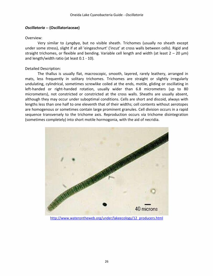
Oscillatoria -- (Oscillatoriaceae) Overview: Very similar to Lyngbya, but no visible sheath. Trichomes (usually no sheath except under some stress), slight if at all 'eingeschnurt' ('incut' at cross walls between cells). Rigid and straight trichomes, or flexible and bending. Variable cell length and width (at least 2 – 20 µm) and length/width ratio (at least 0.1 - 10). Detailed Description: The thallus is usually flat, macroscopic, smooth, layered, rarely leathery, arranged in mats, less frequently in solitary trichomes. Trichomes are straight or slightly irregularly undulating, cylindrical, sometimes screwlike coiled at the ends, motile, gliding or oscillating in left-handed or right-handed rotation, usually wider than 6.8 micrometers (up to 80 micrometers), not constricted or constricted at the cross walls. Sheaths are usually absent, although they may occur under suboptimal conditions. Cells are short and discoid, always with lengths less than one half to one eleventh that of their widths; cell contents without aerotopes are homogenous or sometimes contain large prominent granules. Cell division occurs in a rapid sequence transversely to the trichome axis. Reproduction occurs via trichome disintegration (sometimes completely) into short motile hormogonia, with the aid of necridia.
http://www.waterontheweb.org/under/lakeecology/12_producers.html

Oneida Lake Cyanobacteria Guide - Oscillatoria
27
http://protist.i.hosei.ac.jp/PDB/Images/Prokaryotes/Oscillatoriaceae/Oscillatoria/
Oneida Lake Cyanobacteria Guide – Phormidium
28
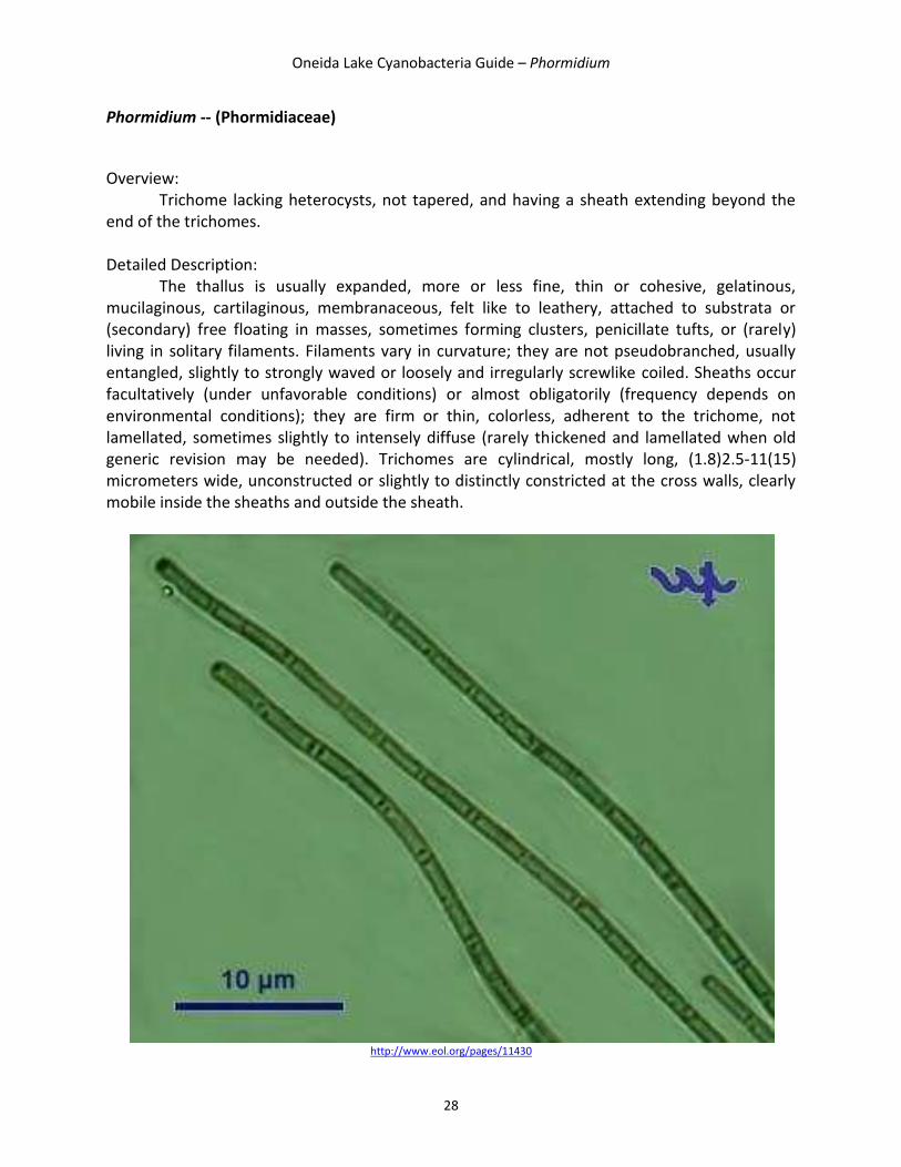
Phormidium -- (Phormidiaceae)
Overview:
Trichome lacking heterocysts, not tapered, and having a sheath extending beyond the end of the trichomes. Detailed Description:
The thallus is usually expanded, more or less fine, thin or cohesive, gelatinous, mucilaginous, cartilaginous, membranaceous, felt like to leathery, attached to substrata or (secondary) free floating in masses, sometimes forming clusters, penicillate tufts, or (rarely) living in solitary filaments. Filaments vary in curvature; they are not pseudobranched, usually entangled, slightly to strongly waved or loosely and irregularly screwlike coiled. Sheaths occur facultatively (under unfavorable conditions) or almost obligatorily (frequency depends on environmental conditions); they are firm or thin, colorless, adherent to the trichome, not lamellated, sometimes slightly to intensely diffuse (rarely thickened and lamellated when old generic revision may be needed). Trichomes are cylindrical, mostly long, (1.8)2.5-11(15) micrometers wide, unconstructed or slightly to distinctly constricted at the cross walls, clearly mobile inside the sheaths and outside the sheath.
http://www.eol.org/pages/11430

29

Oneida Lake Cyanobacteria Guide – Rhabdoderma
30
Rhabdoderma -- (Synechococcaceae)
Overview:
Cylindrical and rod-shaped cells arranged in colonies within fine mucilage Detailed Description:
Cylindrical cells are arranged more or less in the same direction, but usually not chainlike, or irregularly distant, in microscopic, irregularly oval or elongated mucilaginous colonies. The mucilage is fine, homogeneous, colorless, diffuse or delimited at the margin, and sometimes indistinct. Cells are always are cylindrical, rod-shaped, or slightly curved, rounded at the ends, sometimes several times longer than wide, (2)4-12(33) x (0.5)1-3 micrometers, with pale blue-green, grayish, or olive-green cell content and no obvious gas vesicles. Cell division is perpendicular to the longer axis and sometimes asymmetrical (particularly under suboptimal conditions). Daughter cells sometimes remain joined together in short pseudofilaments; long filamentous involution cells are known.
http://cfb.unh.edu/phycokey/Choices/Cyanobacteria/cyano_colonies/RHABDODERMA/Rhabdoderma_Image_page.htm#pic02

Oneida Lake Cyanobacteria Guide – Rhabdoderma
31
http://planktonnet.awi.de/index.php?contenttype=image_details&itemid=16684#content

Oneida Lake Cyanobacteria Guide – Synechococcus
32
Synechococcus – (Synechococcaeae)
Overview: Very small (1µm diameter) cylindrical or rod-shaped unicell without mucilaginous
sheath, cell longer than wide. Occurs singly or in groups.
Detailed Description: Cells are solitary or in irregular groups on agglomerations. They do not form distinct
colonies, have no slimy envelopes or only a very fine, diffuse, and narrow gelatinous layer. Cells are cylindrical to long rod-shaped, sometimes slightly arcuate after division in pairs. Cell content is homogeneous, occasionally has a slightly recognizable centro and chromatoplasm (few to several parietal thylakoids), have pale blue-green, olive green, or reddish color, no gas vesicles, and sometimes prominent granules. Cells are (1.5)3-15(40) x 0.4-3(6) micrometers. Cell division is by perpendicular binary fission (usually cleavage), sometimes asymmetrically. Genus specific filamentous involution cells occur under stress conditions.
http://www.ibvf.csic.es/Cultivos/Seccion_I.htm

Oneida Lake Cyanobacteria Guide – Synechococcus
33
http://www.glerl.noaa.gov/seagrant/GLWL/Algae/Cyanophyta/Cards/Synechococcus.html

Oneida Lake Cyanobacteria Guide – Synechocystis
34
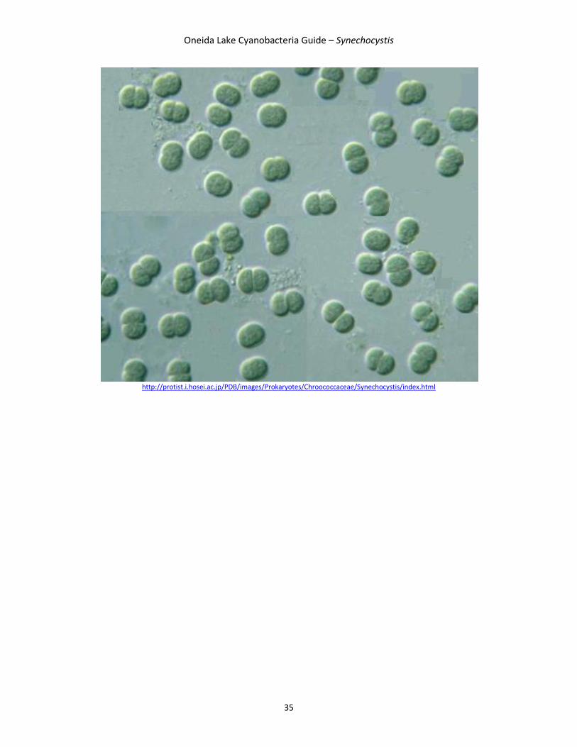
Synechocystis -- (Merismopediaceae) Detailed Description:
Cells are solitary or in pairs a short time after division, have no mucilage or a fine, narrow, colorless, indistinct, mucilage layer around the cells that is diffuse at the margins. Cells are spherical, rarely to widely oval before division, usually pale or rarely, bright blue-green or olive green, in a few species, reddish violet or red. Cells are 0.7-15(30) micrometers in diameter. Cell content is more or less homogeneous, sometimes has solitary granules and a distinguishable centro and chromatoplasm (parietal position of thylakoids). The species with irregularly distributed thylakoids and larger dimensions probably belong to another taxonomic type. Cell division is by binary fission (cleavage), regularly in two perpendicular planes in successive generations. Cells grow to the original size and shape before the next division.
http://www.ibvf.csic.es/Cultivos/Seccion_I.htm
Oneida Lake Cyanobacteria Guide – Synechocystis
35
http://protist.i.hosei.ac.jp/PDB/images/Prokaryotes/Chroococcaceae/Synechocystis/index.html

Oneida Lake Cyanobacteria Guide
36
Glossary Aerotopes – clusters of gas vesicles; the older term is gas vacuole. Akinete- thick-walled cell produced by members of several algal classes; may be released by vegetative cells or attached to filaments; functions as an asexual resting stage and typically is resistant to harsh conditions (e.g., low temperatures). Arcuate – arched or cresent shaped. Calyptra – thickened or enlarged tip; occurs at the tip of some trichomes. Clathrate – with irregular perforations or openings; lattice-like. Cordiform – heart-shaped. Eingeschnurt – incut at cross walls between cells. Facultative – ability in cells to use photosynthesis and also to obtain external sources of organic matter, such as under low light conditions. Fasticulate – occurring in bundles, as clusters of filamentous cyanobacteria. Heterocyst – thick-walled, multilayered (apparently gas-tight, anaerobic), and weakly pigmented cell; contains the nitrogenase enzyme, which enables fixation of gaseous nitrogen to ammonium also termed heterocyte. Heteropolar – asymmetric to the transverse (longitudinal) axis in a filament, cell, or other structure. Morphologically differentiated in basal to apical parts. Hormogonia – in filamentous cyanobacteria, a means of vegetative reproduction (and dispersal) formed via fragmentation of the trichome, forming distinct segments that are often motile (via gliding). Hylaine – transparent or colorless. Intercalary growth – growth in the middle of the thallus. Isopolar – symmetric to the transverse (longitudinal) axis of a filament, cell, or other structure. Lamellate – layered or arranged in layers. Metameric – at regular intervals on the filament.
Oneida Lake Cyanobacteria Guide
37
Necridia – dead cells that function to aid separation of filaments of pseudofilaments for vegetative reproduction (e.g., hormogonia) or the formation of false branches. Obovate – Ovate with a narrower end. Pyrenoid – distinct, proteinaceous structure (often spherical), embedded in or associated with chloroplasts of algae; in some, contains the enzyme RuBisCO; associated with starch in some green algae. Reniform – bean or kidney shaped. Thallus (thalli) – general form or body of an alga. Thylakoid – flattened membranous vesicles (or sacs) that form the photosynthetic membranes and contain photosynthetic pigments arranged in various patterns in cyanobacterial cells or within plastids in eukaryotic algae. Trichome – a terminal cell that is produced into an elongate hair-like structure.

38

Oneida Lake Cyanobacteria Guide
39
References Baker, J.C., Murby, A.L., Wyatt, L., Beagen, W.R., Elliott, C.S., Morison, F., Chase, L., Skoolicas, J., Baldwin, R., Phytoplankton key – Phycoplankton – An image-based key of microscopic aquatic objects, http://cfb.unh.edu/phycokey/phycokey.htm (August 5, 2011). Hillebrand, H., Duselen, C., Kirschtel, D., Pollinger, U., and Zohary T., 1999, Biovolume calculation for pelagic and benthic microalgae, Journal of Phycology, v. 35, p. 403-424. Wehr, J. D., Sheath, R.G., 2003, Freshwater Algae of North America: Ecology and Classification, Academic Press, San Diego, CA, 918.