On the Structure and Mechanics of the Protozoan Flagellum
45
ON THE STRUCTURE AND MECHANICS OF THE PROTOZOAN FLAGELLUM 1 HARLEY P. BROWN Department of Zoology and Entomology, The Ohio State University 2 The flagellum is an organelle of exceedingly wide distribution among both animals and plants. Among the Protozoa, it occurs not only in all of the Mastigo- phora, but also in many Sarcodina and Sporozoa during part of their life cycle. The swarm spores and gametes of certain algae and fungi possessflagella,as do the sperm cells of bryophytes, pteridophytes, and cycads. The tail of the typical animal spermatozoon is probably nothing more than a flagellum. The cilia of the ciliate Protozoa and of metazoan ciliated epithelium in all probability represent modified flagella. There is reason to believe that the bacterial flagellum is perhaps the simplest and most primitive manifestation of this organelle. In spite of the apparent diversity of form and function represented in these numerous examples, it seems highly probable that the organization and mode of functioning are fundamentally similar in all. During the past century numerous investigators have studied the structure and mode of action of flagella, and their conclusions have varied widely. Certain facts have been established, but many points have remained open to question. It has been evident for some time that new techniques would be necessary for the settlement of such questions. Today the electron microscope, with resolution and accompanying magnification far beyond the limits of the ordinary compound microscope, provides such a new technique for the study of minute structures. Having access to an electron microscope and to pure clone cultures of several flagellate Protozoa, I undertook an intensive study of flagellar structure, the major results of which are embodied in this paper. Flagellar action and flagellate locomotion I investigated by direct observation with the aid of a special technique, by the construction of working models, and by actual underwater swimming experiments. The work was done at the suggestion and under the supervision and encourage- ment of Professor W. J. Kostir, to whom I am especially indebted. The electron microscope employed was one constructed by Professor A. F. Prebus, of The Ohio State University, and operated by him and his assistant, Mr. John Dankworth, to both of whom I here express my appreciation for invaluable assistance. REVIEW OF PREVIOUS WORK ON THE SUBJECT STRUCTURE OF THE FLAGELLUM Until the 1880's or later, the flagellum was generally considered simply an elongate, homogeneous fiber, bearing no appendages, and characterized by weak staining (Butschli, 1883-7; Klebs, 1892; Dangeard, 1901 b). The flagella were usually depicted as tapering to a point, but Butschli considered them commonly of equal diameter throughout their entire length, or tapering only slightly at the end. In opposition to such views appeared the papers of Kunstler (1882, 1889), Loeffler (1889, 1890), and Fischer (1894, 1895). The former described (Kunstler, 1889, p. 408, translation) " . . . a dark axial line resembling a slit or canal, partic- 'From a dissertation submitted to the Graduate School of The Ohio State University in partial fulfillment of the requirements for the degree of Doctor of Philosophy. 2 The present address of the author is: Department of Zoology, University of Idaho, Moscow, Idaho. 247
Transcript of On the Structure and Mechanics of the Protozoan Flagellum

ON THE STRUCTURE AND MECHANICS OF THEPROTOZOAN FLAGELLUM1
HARLEY P. BROWN
Department of Zoology and Entomology, The Ohio State University2
The flagellum is an organelle of exceedingly wide distribution among bothanimals and plants. Among the Protozoa, it occurs not only in all of the Mastigo-phora, but also in many Sarcodina and Sporozoa during part of their life cycle.The swarm spores and gametes of certain algae and fungi possess flagella, as do thesperm cells of bryophytes, pteridophytes, and cycads. The tail of the typicalanimal spermatozoon is probably nothing more than a flagellum. The cilia of theciliate Protozoa and of metazoan ciliated epithelium in all probability representmodified flagella. There is reason to believe that the bacterial flagellum is perhapsthe simplest and most primitive manifestation of this organelle. In spite of theapparent diversity of form and function represented in these numerous examples,it seems highly probable that the organization and mode of functioning arefundamentally similar in all.
During the past century numerous investigators have studied the structureand mode of action of flagella, and their conclusions have varied widely. Certainfacts have been established, but many points have remained open to question. Ithas been evident for some time that new techniques would be necessary for thesettlement of such questions. Today the electron microscope, with resolution andaccompanying magnification far beyond the limits of the ordinary compoundmicroscope, provides such a new technique for the study of minute structures.
Having access to an electron microscope and to pure clone cultures of severalflagellate Protozoa, I undertook an intensive study of flagellar structure, themajor results of which are embodied in this paper. Flagellar action and flagellatelocomotion I investigated by direct observation with the aid of a special technique,by the construction of working models, and by actual underwater swimmingexperiments.
The work was done at the suggestion and under the supervision and encourage-ment of Professor W. J. Kostir, to whom I am especially indebted. The electronmicroscope employed was one constructed by Professor A. F. Prebus, of The OhioState University, and operated by him and his assistant, Mr. John Dankworth, toboth of whom I here express my appreciation for invaluable assistance.
REVIEW OF PREVIOUS WORK ON THE SUBJECT
STRUCTURE OF THE FLAGELLUM
Until the 1880's or later, the flagellum was generally considered simply anelongate, homogeneous fiber, bearing no appendages, and characterized by weakstaining (Butschli, 1883-7; Klebs, 1892; Dangeard, 1901 b). The flagella wereusually depicted as tapering to a point, but Butschli considered them commonlyof equal diameter throughout their entire length, or tapering only slightly atthe end.
In opposition to such views appeared the papers of Kunstler (1882, 1889),Loeffler (1889, 1890), and Fischer (1894, 1895). The former described (Kunstler,1889, p. 408, translation) " . . . a dark axial line resembling a slit or canal, partic-
'From a dissertation submitted to the Graduate School of The Ohio State University inpartial fulfillment of the requirements for the degree of Doctor of Philosophy.
2The present address of the author is: Department of Zoology, University of Idaho,Moscow, Idaho.
247

248 HARLEY P. BROWN Vol. XLV
ularly when the filaments observed are well extended horizontally; in these casesone may often distinguish delicate transverse septa, dividing the axial cleft intorather short sections." (Fig. 1 a). The dark sections he considered to representvacuoles which are separated from one another by septa of the transparent groundmass which surrounds them. He also described a cortical layer or sheath (Fig.1 b, c) which was often separated from the ground substance and its enclosed slit.
FIG. 1. a. "Oxyrrhis marina. Portion of flagellum showing the central fissure sufficiently-enlarged to be quite clearly visible, and appearing like a slit or axial groove divided into aseries of hollows by delicate transverse septa." b. Oxyrrhis marina. "The same flagellum(as c) seen at a place where the outer layer is detached only on one side, and only incompletely,in such a way as to form a rather regular sort of corrugation. The cleft which separates it fromthe internal axis becomes increasingly narrower, and, farther along, even disappears."c. "Oxyrrhis marina. Portion of flagellum greatly enlarged and treated with chromic hematox-ylin. The undulated outer coat is completely detached from the axial substance." (Redrawnfrom Kunstler, 1889, Plate XIX.)
To the presence of this enveloping membrane he attributed the weak stainabilityof the flagellum. The axial substance he considered the more contractile portion.Kunstler also first described an interesting external structure in the flagella ofOxyrrhis and Cryptomonas: " . . . the attenuated extremity often seems prolongedin a pale filament, thinner than the flagellum and of a different appearance (Fig. 2).The limits of the flagellum proper are distinguished clearly; suddenly, at its tip,
FIG. 2. ''Oxyrrhis marina. Theoretical figure representing certain flagella. One sees atthe end of the true flagellum the pale filament which may be observed in many cases. Thesame may be seen in Cryptomonas ovata." (From Kunstler, 1889, Plate XX.)
the diameter and appearance change; there begins a transparent filament, hardlyvisible, very slender, sometimes ending in a slight swelling."
Loeffler (1889) independently described this last-mentioned structure, employ-ing a new technique he was using for bacterial flagella. He suggested that the thickbasal part might be a sheath, from which extends the delicate protoplasmic fiberin the manner of a pseudopod, slightly swollen terminally. The apical swelling,he postulated, might function as a tactile organ. Loeffler also discovered on the

No. 6 THE PROTOZOAN FLAGELLUM 249
fiagellum of a monoflagellate (perhaps Oikomonas) two dense rows of extremely-delicate hairs, projecting almost at right angles to the axis of the fiagellum, onealong each side. (See Fig. 3). He did not investigate the possibility that in life thehairs actually might cover the filament instead of being arranged in two rows, andin drying be flattened down laterally, producing the impression of two rows ofhairs. At any rate, he had described and figured photomicrographs of two pre-viously unknown appendages, and outlined his method in detail.
FIG. 3. Monas sp. ? The first published figure of a flimmer-flagellum, from a mordant-stain preparation designed for bacterial flagella. (Redrawn from Loeffler, 1889, Plate II.)
FIG. 4. Euglena viridis. a. "Cast-off fiagellum . . . still completely stretched out;untwisted, with the hairs on one side . . . " b. "A cast-off flimmer-flagellum rolling up, par-tially swollen and therefore with an apparent axial fiber." c. "A cast-off, rolling-up fiagellumwhich had previously become uniformly swollen and consequently shows no such apparentstructure" (as Fig. 4 b). From preparations of undiluted culture dried upon cover glasses inabout one-half hour, and treated with a modification of Loeffler's mordant-stain technique.(Redrawn from Fischer, 1894, Plate XI.)
Fischer (1894), using a modification of Loeffler's technique, made a carefulstudy of the flagella of Euglena viridis, Monas guttula, and species of Bodo, Chloro-gonium, and Polytoma. He confirmed Loeffler's findings and discovered that inEuglena the fiagellum bears but a single row of hairs (Fig. 4), while that of Monashas two opposite rows. He termed these flagella Flimmergeisseln, or ciliatedflagella. Since "flimmer" appears to be a convenient word for the structures, anddoes not carry any obvious implications in the English language, I shall hereafteremploy the term "flimmer-flagellum " in reference to any fiagellum bearing hair-likeprojections along one, two, or all sides.
Whip-flagella, or Peitschengeisseln, Fischer found in Bodo, Chlorogonium, andPolytoma, and described as follows (p. 230, translation): "The whip-flagellumconsists of a thick homogeneous stalk, previously thought to be the entire fiag-ellum (the only part visible in unstained specimens) and, arising from its tip, avery delicate whip-thread 2-3 times as long as the stalk. This thread is slungabout like a coach-whip by the strokes of the basal stalk."
Fischer observed in Euglena what he concluded to be an artifact arising fromincomplete swelling: " . . . the central, not yet swollen part of the fiagellum appearsas a denser, more strongly stained axial fiber, while the outer, swollen part, seemsa less dense, more weakly stained ground substance." (See Fig. 4 b). Further,"In Polytoma and Bodo, a relatively commonly seen granular structure of the basalpart of the whip is likewise only a result of the technique. This flagellar structure,

250 HARLEY P. BROWN Vol. XLV
which agrees with that described by Kunstler, is to be regarded in this light" (i. e.,as an artifact). (For further discussion of the granular appearance, see Gelei, 1926.)
Fischer studied in detail the processes of flagellar disintegration, whereby hemade his observations on the artificial nature of the granular or alveolar appearanceof certain flagella. He figured in detail many examples of all the above-mentionedtypes, and discussed each matter thoroughly. In spite of the fact that he coulddemonstrate no natural inner structure in either whip- or flimmer-flagellum, hestated (p. 204): " . . . yet there is no doubt that the apparently homogeneous fibermust possess a very fine structure made up of definitely arranged particles(micelles), toward which point the twisting of the flagellum, its rolling up, and thearrangement of the flimmer-hairs."
. Regarding the arrangement of the flimmer, Fischer assumed that they mustbeat in unison, since, on any given flagellum, all the flimmer project in the samedirection.
For years flimmer-flagella were generally considered artifacts, perhaps theresult of a final wriggle and smear, or of chemical action (Plenge, 1898; Dangeard,1901b; Butschli, 1902; Schuberg, 1905; Korschikov, 1923; Giinther, 1928). On
FIG. 5. a. "Synura uvella Ehrenb. A cell from the colony with flimmer-' and whip-flagella." b. "Salpingoeca sp." (Redrawn from Petersen, 1929.)
FIG. 6. Schematic flimmer-flagellum showing appearance which would be expected tooccur in a twisted flagellum if the flimmer existed in two lateral rows, rather than all overthe surface of the flagellum. This sort of appearance has not been observed in specimens,although many of the observed flagella have probably been twisted. (From discussion anddiagram_of Vlk, 1938.)
the other hand, most of these workers confirmed the existence of whip-flagella, andin many cases observed such structures in living specimens. During the latterpart of the period covered by the above references, several investigators were,however, confirming the reports of Fischer and Loeifler. Petersen (1918) dem-onstrated the two flagella of Synura uvella to be of different types (Fig. 5a): " . . .the one of whiplike form, the other a pinnate flagellum showing two opposite rowsof little secondary filaments, and resembling a feather; it is to be presumed, how-ever, that these [filaments] extend from all sides of the primary filament." (Trans-1 ation from summary in French.) Petersen also found flimmer-flagella in Uroglenaand Dinobryon. In a later paper (1929) he expanded his observations to a largenumber of forms, some possessing only whip-flagella, some only flimmer-flagella,and some having one of each. He also described a combination whip-flimmer-flagellum in Craspedomonads. (Fig. 5 b). The distribution among the Protozoa ofconsistent flagellar types fits well into the accepted scheme of classification.

No. 6 THE PROTOZOAN FLAGELLUM 251
Mainx (1928), though failing to demonstrate flimmer-flagella in several speciesreported to possess them, confirmed their presence in Euglena viridis and figuredexcellent photomicrographs of the flimmer-flagellum of Phacus pleuronectes. Hismost exacting attempts to observe flimmer on the living flagellum failed. Inconsideration of their extreme regularity along one side of the flagellum, theirpresence with or without fixation, etc., he concluded that they could hardly beartifacts. He considered them, however, not motile, as Fischer had assumed, butrather simply as devices serving to increase the flagellar surface.
Petrova (1931) employed flagellar types in the solution of a taxonomic problem.Deflandre (1934), using a very different method of preparation, confirmed the
presence of whip- and flimmer-flagella in many organisms, and considerablyexpanded the list of species studied.
Vlk (1931, 1938) has perhaps treated the problem most thoroughly, expandingand organizing the described types, and tabulating his results. For a morethorough treatment of this phase of flagellar structure, the reader is referred par-ticularly to Vlk, 1938, or to Deflandre, 1934. Vlk even succeeded in observingflimmer (in two distinct rows) on the flagellum of active, living specimens of
FIG. 7. a. Whip-flagella of Chlamydomonas moewusii. b. Whip-flagella of Chlamydomonassestinensis. (Redrawn from Gerloff, 1940, to show variations in length of whip.)
FIG. 8. Cyclidium glaucoma. Silver preparation. Greatly enlarged cilium showing theaxial fiber extending from the basal granule through and beyond the "contractile" sheath, andending in an enlargement. The dark areas in the sheath are irregularly spaced and even lackingin some of the cilia nearby. A silver line connects adjacent basal granules. (Redrawn from partof Text Figure 34, Klein, 1929.)
Mallomonas acaroides, a species having an unusually large flagellum. Theseflimmer are described as having a length about 6 times the diameter of the flagellumproper. He was able to demonstrate these living flimmer-flagella to members ofthe institute staff, including Professor A. Pascher.
Unfortunately, Kudo (1939), in his brief treatment of the flimmer-flagellum,gave all credit to Vlk but misrepresented Vlk's diagram of a two-row flimmer-flagellum (Fig. 6). The error is certainly unintentional, but misleading. This dia-gram, which Kudo used to illustrate a two-rowed flimmer-flagellum, was originallyintended by Vlk (1938, p. 486) to represent a situation which should occur intwisting if the flagellum actually bears two distinct flimmer rows. But such asituation had never been observed. Instead, the flimmer of these forms usuallyappear somewhat as in Fig. 3 or Fig. 5 a. In other words, Vlk used it as a probableindication that the flimmer, in most organisms at least, are distributed on thesurface of the flagellum like the hairs on a dog's tail, and not just in twolateral rows.

252 HARLEY P. BROWN Vol. XLV
Recognition of the whip-flagella has been rather general, and they seem almostuniversally accepted today. GerlofT (1940) figures the whip-flagella in manyspecies of Chlamydomonas. A considerable variation in relative length of lash andstalk may be noted (Fig. 7). Many authors (e. g. Awerinzew, 1907; Minchin,1922) consider the lash or end-piece as a portion of an axial fiber extending beyondthe enveloping sheath of protoplasm and membrane. Cilia are now commonlyconsidered to have a similar structure (Klein, 1929). Klein seems to agree withLoeffler's hypothesis that the terminal swelling of the lash (Fig. 8) may have asensory function.
Figure 8 (or 10 a) will also serve for a discussion of the generally acceptedconcept of the internal structure of a flagellum. Most textbooks and articles onthe subject agree to the presence of an axial fiber surrounded by a protoplasmic
FIG. 9. Flagellum of Peranema in which the axial fiber and outer membrane are intact,although part of the intermediate substance has swollen and formed a knot or coil of the axialfiber. (Redrawn digrammatically from Korschikov, 1923, Fig. 1.)
FIG. 10. a-c. Flagellum of Chilomonas paramecium undergoing disintegration (half-schematic). The protoplasmic portion flows together into droplets, exposing the axial fiberand allowing it to straighten, d. Ceratium tripos. Part of contracted longitudinal flagellum.(Redrawn from Awerinzew, 1907.)
sheath. Beyond that there is much less agreement. A few consider the axialcore as the major contractile or vibratile portion, the sheath serving as a support-ing structure (Rosskin, 1922; Calkins, 1933). The vast majority think differently.(Butschli, 1902; Schuberg, 1905; Awerinzew, 1907; Goldschmidt, 1907; Williams,1907; Erhard, 1910; Hamburger, 1911; Minchin, 1922; Korschikov, 1923; Doflein,1929; Klein, 1929; Kudo, 1939; Hyman, 1940).
The flagellum (or its axoneme) usually arises from a basal granule (blepharo-plast), apparently essential to the continued function of the organelle. In certainforms the flagellum has two roots and basal granules (e. g. Euglena; Wager, 1899;Hall and Jahn, 1929). For information on other modes of attachment, seeProwazek, 1903; Goldschmidt, 1907; Schouteden, 1907.
Regarding the interrelationships among axoneme, surrounding plasm, andouter membrane, there are interesting reports, based upon studies of disintegratingflagella, which throw light upon the subject. Fischer (1894) made extensive

No. 6 THE PR6T0Z0AN FLAGELLUM 253
observations on the coiling or rolling-up of flagella, noting that the process mightbegin at either end of the fiagellum or at any point in between. Korschikov(1923) confirms Fischer's description of fiagellar "aggregation" (Figs. 4 b, c;Fig. 9), summarizing his observations as follows (translation from German sum-mary) : '' The axial fiber and the outer membrane are the most resistant structuralcomponents of the fiagellum. In the 'aggregation' of the fiagellum, the inter-mediate substance is first destroyed. In the corresponding region the membranebecomes swollen. In the course of this process, the axial fiber is drawn into theswelling and forms there a ball or coil (Fig. 9). Afterward, however, when thisblister of the fiagellar membrane is destroyed, the axial fiber is freed and stretchesagain in length."
Awerinzew (1907) and Hamburger (1911), describing flagella undergoingdestruction, state that the "protoplasmic" portion of the fiagellum rounds intoglobules which proceed to flow or withdraw toward the base of the fiagellum,either leaving the axoneme exposed and straightened out (Awerinzew) or telescop-ing it (Hamburger). (See Fig. 10.) All of these facts tend to indicate an elastic,upporting axoneme.
tjlehla figures, but barely mentions, an interesting fiagellar structure (Fig. 15),reminiscent of Kunstler's diagrams, but seen in living flagella.
FIG. 11. a. "The uncoiled fibrils of the flagellum of Euglena." b. "Euglena with thefibrils of the flagellum branching out into a system of rootlets in the protoplasm of the body.""This structure is demonstrated with ease by subjecting a fiagellum to slight pressure."(Drawn from Dellinger, Figs. 1,3.)
Roskin (1922, 1923) postulates and Korschikov (1923) asserts that the axialfiber consists of a great number of thin fibrils. Koltzoff (1903) stated that incertain pteropods the cilium "consists of several fibers which are encased in acommon fluid plasma layer . . . the inner fibers running to the basal body . . . "Schmitt, Hall, and Jakus (1943) figure cilia (Frontonia) and flagella (Trichonympha)frayed into 9-11 fibrils.
Dellinger (1909), who figures fibrils in both cilia and flagella (Fig. 11), states:" . . . the flagella of Euglena, Chilomonas and Spirillum are composed of fourspiral filaments." He also depicts the axial filament in the pseudopod (axopod)of Actinosphaerium (Heliozoa) as fibrillar. A similar structure is described in theheliozoan axopod by Rosskin (1925). For discussions of the possible homologybetween such pseudopods and flagella see Minchin (1922), Goldschmidt (1907).
The flagella of many bacteria entwine in a tuft and function as a unit, in somecases even becoming encased with "a glutinous mass which keeps them boundtogether (Reichert, 1909; tJlehla, 1911; Metzner, 1920; Pijper, 1941; Hutchinsonand McCracken, 1943). This situation is perhaps analogous to that occurring incirri or in the "cilia" studied by Gray (1922) and Carter (1924), in which a numberof units capable of beating individually are united into a single organelle.

254 HARLEY P. BROWN Vol. XLV
Sperm tails display a fibrillar axis and an enclosing sheath, and have been moreexhaustively studied than flagella. Furthermore, they are probably homologousto the protozoan flagellum, although the details of the homology are not yet set-tled (Dangeard, 1901a; Alexeieff, 1924; Grasse, 1926; Duboscq and Grasse, 1933).Subsequent to the work of several investigators, Ballowitz stated in 1890 thatmany types of sperm tails frayed into 2-4 subfibrils. In his paper of 1908, hefigures tails fraying into as many as 18 subfibrils. Koltzoff (1909) produces inter-esting figures of sperm tails, including such as Fig. 12, showing a sheath of helicallywound gel fibrils. Recently the electron microscope has made possible more minutestudies. Several articles (Baylor, Nalbandov, and Clark, 1943; Harvey andAnderson, 1943; Schmitt, Hall, and Jakus, 1943; Schmitt, 1944) figure micro-graphs of sperm tails showing, in frayed parts, 9-12 fibrils (Fig. 13). According to
FIG. 12. Flagellum (tail) of Planorbis sperm, showing outer helix of gel fibers. (Redrawnfrom Koltzoff, 1909, Fig. 13.)
FIG. 13. "Frayed tail of bull sperm showing subfibrils."FIG. 14. "Portion of sheath from human sperm tail fragmented ultrasonically, showing
helical structure." (Redrawn from electron micrographs; Schmitt, 1944, Figs. 1,2.)FIG. 15. "Gonium pectorale. A portion of a momentarily resting flagellum, showing the
structure of the flagellum. The helical lines proceed . . . uninterruptedly to the tip. (Sche-matic)." (Redrawn from tJlehla, 1911, Fig. 55.)
Schmitt, these fibrils are smooth, parallel, 300-500 A wide, and run the full lengthof the tail. Occasionally even finer subfibrils are seen. "In mammalian spermtails the bundle of fibrils is surrounded by a sheath, the major component of whichis a closely wrapped helical fibril, having a thickness also in the range of 300-500 A(Fig.|14). When the tails are fragmented by ultrasonic radiation, portions of thishelix can be seen, appearing like miniature solenoids." (Schmitt, 1944, p. 36.)
MECHANICS OF THE FLAGELLUM
In the past, little actual correlation has been made between structure andfunction in flagella. Many authors have recorded types of nagellar movementand general movement, but relatively few have attempted to analyze the forcesor mechanisms involved. First, let us enumerate the types of movementsdescribed. tJlehla (1911, pp. 727-728) classifies them as follows:
(1) Monad type. Flagellum long, cylindrical, flexible; beats forward inloops (spirals) grading into flattened waves (i. e., undulations; a given point on theflagellum follows an elliptical path).
(2) Chrysomonad type. Similar to above, but with shorter and stiffer flagellum.

No. 6 THE PROTOZOAN FLAGELLUM 255
(3) Euglena type. Long, twisted, ribbon-like flagellum; beats laterally inloop-shaped waves.
(4) Bodo type. (Trailing) flagellum with long end-piece, rather stiff; beats inflattened waves.
(5) Clostridium (a bacterium) type. Flagellum long, spiral, rather stiff; slowwaves from base to tip.
(6) Chlorophycean type.a. Swarmer type. Flagellum short, cylindrical, rather strong, with the
basal part more flexible; functions as an oar (in sculling?), swinging around asif contracting as a unit.
b. Pandorina type. Flagellum longer, more flexible, often somewhatflattened; beats with a spiral lateral stroke.tjlehla hardly considered the paddle-stroke as such, though this type of move-
ment is described by many, being reported in flagella by Plehn (1904), Rosenbusch(1908), Doflein (1916), Kofoid and Swezy (1920, 1923), Krijgsman (1925), Nigrelli(1929), Kirby (1943); in cilia by Gray (1922) and Carter (1924), among others.Such a lash-movement is mentioned also by Putter (1903), Goldschmidt (1907),Schindera (1922), and Grasse (1926). In some such cases, it evidently occurs inflagella which ordinarily undulate. In all of these cases, the flagella concernedwere located anteriorly.
FIG. 16. See text for explanation.
The second major type of movement executed consists of undulations, usuallyoccurring in two planes (i. e., a spiral). Reports of this type may be found inBiitschli (1887), Kunstler (1889), Goldschmidt (1907), Laveran and Mesnil (1907),Friedrich (1909), Reichert (1909), Bancroft (1913), Kofoid and Swezy (1919),Schindera (1922), Grasse (1926), Entz (1928), Petersen (1929), Lowndes (1936,1941, 1943, 1944, 1945).
The third type, not listed above, is simple conical gyration, reported byKeysselitz (1906), Metzner (1920), and Entz (1928), and by McDonald (1922),in cilia.
Primarily because of Krijgsman's detailed paper, which was based on studiesof Monas sp. under darkfield, most textbooks today emphasize the paddle-stroke.It presents a clear-cut, diagrammatic, relatively simple-looking movement, familiarin our own swimming stroke and similar to that commonly depicted for cilia. Thefilament is held rigid during the effective stroke, but is flexible during the recoverystroke.
The concept of progression by a spiral turning of the flagellum has led a painfulexistence because of Butschli's analysis of the situation as he saw it (Biitschli, 1887,p. 857 et seq.). Delage and Herouard (1896, pp. 306-307) treat his explanation thus

256 HARLEY P. BROWN Vol. X L V
(translation): '' Suppose that the flagellum assumes the form of an elongate helix(as indicated in Fig. 16), and that this helix begins to turn clockwise (as seen fromthe base). At any point such as o the pressure on the water would create a forceob perpendicular to the flagellum, which could be resolved into two others, thevertical one oa determining its rotation about its axis. It is to be noted that thisrotation is in the opposite direction to that of the flagellum. If the helix turns inthe reverse direction, the animal will go backwards. In the case of a helix woundto the left, there would be likewise progression forward for a certain direction of.rotation and backward for the opposite direction. . . . All that is very well, butButschli does not note that the movement he describes supposes an agency met inour mechanical instruments, but never among living beings. In order that thebody and the flagellum might turn indefinitely in opposite directions about thepoint p, it would be necessary that the mode of union between them be that of apin stuck through a card, capable of turning freely in its hole, its union with thecard assured by the head which could not go through the hole." But this leavesDelage and Herouard with the problem of explaining matters. They do so bygoing into great mathematical detail concerning the possibility of a forward com-ponent produced by conical gyration, concluding this to be impossible by directaction. However, by its indirect action, i. e., by the rotation produced in thebody, they think to have found the answer; namely, that since the flagellum isfirmly attached to the body it must rotate with the body, and being held in acorkscrew shape, will thus screw the body and flagellum through the water.
Reichert (1909) noted the forced aspect of this scheme and pointed out that,in his experience, the facts argued against such a mechanism. Further, he states(translation): "The necessity for their relatively cumbersome explanation offlagellar movement appears to have been a result of a misconception of Butschli'sidea. For Butschli postulates that an opposing rotation of body and flagellum isnot feasible in his view that the lines of contraction move about the flagellum. Ifthe lines of contraction on the flagellar surface circle to the right, the body will turnto the left, and so will the flagellum, about its axis. . . . It is only necessary thatthe lines of contraction move correspondingly faster about the flagellum, so thatits turning with the body is equalized or offset. Then the same effect would beachieved as if this turning of the flagellum did not occur at all."
Delage and Hdrouard are not the only ones to misinterpret Butschli's idea.Reichert explains it, and the physics involved, quite clearly. Then, in his
summary he states: "Reversal of movement is accomplished quickly by polar-flagellated bacteria, for they simply reverse the flagellar rotation (as Reichert andButschli postulate that they should), or by bipolar-flagellated bacteria, whoseflagella take turns (those at the other end begin beating). In peritrichous bacteria,on the other hand, reversal occurs slowly. Movement must first cease for amoment, and the flagella assume an opposite orientation to the body." But he isinconsistent, for he also states: "a. The flagella (in bacteria) are always wound in aright spiral (clockwise) and rotate always to the right (as seen from the rear),i. e., the screw-like lines of contraction wind around the flagella to the right, b.The body always rotates to the left. c. The flagella are usually directed backwardduring locomotion. In the spirilla this occurs too, whether the flagellum arises atthe anterior or posterior pole." If they "always'' rotate in one direction, how canthey "simply reverse" this rotation? Further, if the flagellum is directed back-ward during locomotion, whether the flagellum be at either end of the body, doesthis not indicate that the flagellum is assuming a different orientation, rather thanthat it is merely rotating to the right or to the left ? For, if it simply reversed itsdirection of rotation in order to back up, it would then pull the body instead ofpushing it, and would not be directed backward. The error doubtless lies in theassumption that the organism moves backward by simply reversing the flagellarrotation. Indeed, the theory itself seems disputed by these facts.

No. 6 THE PROTOZOAN FLAGELLUM 257
Gray (1928, p. 35) gives a fine explanation of the mechanics involved in undu-lating flagella, the gist of it being that there is a series of waves passing along theflagellum in the same direction, requiring no recovery stroke. The propulsivepower " . . . is equivalent to that which would be produced by projecting along thelength of the flagellum a series of 'humps' of the same form as the waves, thevelocity of the humps being made equal to the velocity of movement of each wave.If the waves pass from the base of the flagellum to its tip, the organism is drivenforward in front of the flagellum; if the waves pass from the tip to the base theorganism is drawn forward with the flagellum in front. If the waves pass alongthe flagellum in one plane there will be no force tending to rotate the animal on itsaxis: if, however, the waves pass round the flagellum as well as along it the organ-ism will rotate." This is well and good, but it fails to fit a couple of facts; namely,that most flagellates move flagellum-foremost, and that the waves of transmissionalong the flagellum have only been demonstrated to progress from the base towardthe tip (Lowndes, 1936, 1941 a and b, 1943, 1944 a and b, 1945 a and b).
To date, Lowndes has made the most complete and accurate analysis of thesituation in a number of flagellates. His conclusions are based upon observedfacts and specific data, backed up by high-speed cinema photomicrography (fortechniques, see Lowndes, 1935, 1944 a). Too much of previous explanations wasbased upon pure speculation and rationalization. Lowndes (1941 b) has demon-strated that the flagellum ordinarily pushes the organism, even though situated atthe anterior^ end, by being directed backward. In this connection it is interestingto consult Ulehla's figures, many of which indicate the same. This appears trueeven for Peranema, the organism classically cited as having a "tractellum," orflagellum which pulls the organism forward, presumably by a wave starting at thetip. Lowndes shows that, in all cases studied, the wave invariably progresses frombase to tip. This fact is an upsetting one to most of the previous theories. Amongother workers reporting this type of action are Kunstler (1889), Friedrich (1909),TJlehla (1911), Bancroft (1913), and Schindera (1922). Of these, TJlehla andBancroft give accounts almost precisely like that of Lowndes concerning thelocomotion of Euglena viridis.
In his later papers, Lowndes elaborates a new concept, previously mentionedby him, but misunderstood by such workers as Barker (1943). In the summaryof one of these papers (Lowndes, 1944 a), he states it as follows:
" 1 . The primary function of the flagellum in a monoflagellate organism is toproduce both rotation and gyration of the organism about a certain axis whichconstitutes the main direction in which the organism is swimming.
"2. The mechanical principle by which the organism is propelled is simply thatof the inclined plane which is caused to rotate. In other words it is that of thescrew or propeller.
"3 . Since the disturbances or waves pass down the flagellum in the form of aspiral they produce two distinct components. It is the resultant of these twocomponents which causes the tip of the organism both to rotate and gyrate.
"4. So long as this rotation and gyration is maintained it will supply thenecessary force for the propulsion of the organism.
"5. The flagellum itself may or may not produce a forward component. If itis more or less swung out at right angles, as in Menoidium, it will produce no for-ward component but if it is swung back, as in Euglena, it will do so."
Hence, it is the rotation and gyration of the body of the organism which is consid-ered to produce the major component of force. Others have observed, naturally,that in organisms bearing obvious helical external structures, rotation of the bodywould produce a component of force. For instance, Gunther (1928) correlatedthe speed of several species of Euglena with spiral ridges on the periplast andflagellar length. Thus, of two species having flagella of approximately the samelength, one having strong spiral ridges progresses at about three times the relative

258 HARLEY P. BROWN Vol. X L V
speed of another having very insignificant ridges. But no one previous toLowndes, so far as I know, had suggested the possibility that the gyration of thebody itself might produce locomotion.
Propulsion by simple conical gyration is what Delage and HeYouard thoughtthey had proved, by mathematics, to be impossible. Metzner (1920) showed thisto be erroneous, both in theory and in practice. By means of experiments onwires rotating in water, he demonstrated that a simple conical gyration producesa pulling force which reaches maximum at an angle of 20-23 degrees from the axisof rotation. He even went so far as to state (translation): "Among flagellates, thesimple conical gyration predominates; the organism 'sucks' itself along throughthe water by means of the flagellum." It should be noted that he considered onlygyration of the flagellum, not gyration of the body. Such conical gyration of theflagellum is reported by Keysselitz (1906) and Petersen (1929). It is alsodescribed by Lowndes (1944 b), who performed experiments similar to those ofMetzner. However, Lowndes contends that this mode of locomotion is incom-patible with high speed, as the flagellum could not be maintained in such aforwardly-directed position. In rapidly moving organisms, the flagella must beatlaterally to the rear.
In this same paper, and in his most recent one (1945 b), Lowndes rendersuntenable a number of misconceptions which arose long ago through misinterpre-tation of the original data, and have grown or continued from textbook to textbook.One such item regards diagrams of Monas swimming, taken from Krijgsman(1925). I shall not discuss these errors here. As Lowndes points out, Krijgsman'sobservations were excellent records of the situation he was studying, but thatsituation was not representative of the free swimming of the organism. When theanimal is freely swimming, the mechanism is quite different from the paddle-strokeobserved by Krijgsman, being similar to that described by Lowndes for Euglena,and the rate of progression is about ten times as rapid as that given by Krijgsman.Locomotion is primarily brought about by the rotation and gyration of the body ofthe organism. (See also Lowndes, 1945 a.)
As for the question of the precise nature of what goes on within the flagellum—it remains a question. For years, writers were occupied with a controversy as towhether the flagellum was an active unit or simply an appendage like a whip,manipulated from the base or cell body. The latter concept was eventually dis-carded as such, but lingers in part in the question whether or not the flagellum canexecute any movement after losing connection with the cell. Various workers(e. g. Klebs, 1892; Entz, 1926) have reported the phenomenon; others have flatlyasserted such to be impossible (Verworn, 1890; Korschikov, 1923). However,the work of many investigators has indicated that the flagellum is an active unit.Gray (1928) enumerates good reasons for assuming such, and Lowndes (1936,1941 b, 1945 b) proves it with photographic, quantitative evidence. Granting,then, that the flagellum does possess the ability to move, by what mechanism doesit effect this motion?
From the versatility of movements and also " . . . because its movements attimes cannot be explained by simple mechanical laws," Krijgsman (1925) con-cluded that the flagellum " . . . must be of complicated construction." Engelmann(1881) and Ballowitz (1890) assert that movement is associated with fibrillarstructure, which, as we have seen, is evidently present. Erhard (1910) andLowndes (1941 b) regard the transmission of the stimulus as a surface phenomenon,not connected with the axoneme. Such statements are generalities, to be sure, but,if accepted, eliminate such theories as that of Gurwitsch (1904), which postulatesa rapid streaming of protoplasm into and out of the flagellum, acting in antagonismto the elasticity of the axoneme. Heidenhain (1911) proposed a "theory ofsmallest waves" to explain movements: these waves may travel along one side ofthe filament, they may vary in length and effect, they may vary in extent, they

No. 6 THE PROTOZOAN FLAGELLUM 259
may vary in path, frequency, and rhythm. They are evidently quite versatilewaves. So far as I know, they have been forgotten. Putter (1903) discussedEngelmann's theory and decided that it required too many spiral fibrils of differentpitch and direction. Verworn (1915) stated what he considered the basic principleinvolved (translation): " . . . a contractile side contracts from the cell body out-wards and thereby stretches the opposite side, which latter in the expansion phase,by its elasticity, returns the filament to its rest position. According to the oppos-ing relationship of the contractile to the passively stretched substance, thereresults a beat in one plane or in a complicated form." This does epitomize theprinciple employed by most of the theories. For a discussion of the theories, seeGray (1928).
Viewing the matter from a more generalized standpoint, but with fundamentalsin mind, Gray (1928) and Schmitt (1944) liken the process involved to that ofmuscle, and consider the basic phenomenon to result from a change of distributionof water molecules "between polar groups in fibrous proteins and ionogenic groupsin the environment" (Schmitt). In other words, if the proteins in the fibers alongone side of the flagellum suddenly take on great numbers of water molecules at theexpense of the opposite side of the filament, the filament (flagellum) will tend tobend toward the side losing the water. This sudden affinity for water by theprotein might be due to a change in the degree of ionization of the protein molecule,caused by a local production of acid. Should the acid then be neutralized, thewater would again be liberated (Gray). Or it might be that the protein fiberconsists of folded molecular chains which fold up further upon the addition ofwater, thus contracting the fiber. At any rate, we get glimpses of the possibilities.
ORIGINAL INVESTIGATIONS
STRUCTURE OF THE FLAGELLUM
Materials and MethodsIt was considered desirable to experiment briefly with the more productive
methods described by other workers in the study of flagellar structure. Suchtechniques as those of Gicklhorn (1921), Kater (1929), Allen (1936), and Smyth(1944) are useful procedures for general study or mere observation of. nagella, butcontribute relatively little to a detailed investigation of the type here undertaken.On the other hand, the papers of Loeffler (1890), Fischer (1895), and Deflandre(1923) describe techniques of primary importance in the development of ourknowledge of whip- and flimmer-flagella. McClung (1937, pp. 141-145) presentsLoeffler's method and may be more readily available to persons attempting toduplicate some of the work done on flimmer- and whip-flagella. Vlk (1938)describes his modifications of the Loeffler technique. Petersen (1929) points outthe possibilities for failure and sources of error, emphasizing the extreme delicacyrequired in preparation. Several authors (e. g. Butschli, 1902; Korschikov, 1923)have failed to achieve success with the mordant technique.
Such contributions as this paper may offer to the knowledge of flagellar structureare made possible through the use of the electron microscope. Further probingswith the light microscope promise but little, as the possibilities have been rathercarefully covered. For previously employed methods of preparation of specimensfor electron microscope examination, see Marton (1941, 1943), Morton and Ander-son (1942), Mudd and Anderson (1942), Richards and Anderson (1942), Prebus(1944), Williams and Wyckoff (1944, 1945 a, b), Claude and Fullam (1945), andPorter, Claude, and Fullam (1945). Some of the techniques described are adaptedto specialized situations, but they bring out various points of importance in thepreparation of biological material for electron microscope study. Among themost important of these, perhaps, are the removal of salts and other matter whichmight crystallize upon or otherwise obscure or mar the specimens, and the thinness

260 HARLEY P. BROWN Vol. X L V
of film plus specimen, whieh should total less than 50 m^ in thickness. A gooddiscussion of the limitations of the electron microscope for biological work ingeneral is given by Richards and Anderson (1942). For basic principles andgeneral limitations of the instrument, see Hillier and Vance (1941), or such pop-ularized books as Burton and Kohl (1942) or Hawley (1945).
Dellinger (1909) and Porter, Claude, and Fullam (1945), among others, considerosmic acid the best killing and fixing reagent for retaining original appearances. Ihave found it extremely useful in this work. Over a dozen different procedureshave been employed, the most successful of which have all involved the use ofosmic acid. Formalin as a fixing reagent has apparently resulted in excessiveclumping, but this has not been thoroughly investigated. Other major difficultiesin the preparations were due to too many or too few organisms on the film,crystallization, loss of flagella in centrifuging, and, most of all, disruption of mem-branes or films under electron bombardment.
The principal organisms studied were Astasia klebsii Lemmermann, Euglenagracilis Klebs, Ochromonas variabilis Meyer, and Chilomonas paramecium Ehren-berg which were obtained from pure cultures maintained in the ProtozoologyLaboratory at The Ohio State University.
The film and screen upon which specimens were to be mounted was prepared asfollows (slight modification of method employed by Prebus):
(1) 300 mesh/inch bronze screen is.cleaned by immersing in dilute HC1 andwashing a number of times in distilled water.
(2) Rectangular pieces about 6 x 18 mm. are cut from the screen and keptdust-free (e. g., in a Petri dish).
(3) A vessel (e. g., a Pyrex pie plate) of about 10-inch diameter with a levelrim is filled to the brim with distilled water.
(4) The water is saturated with amyl acetate.(5) A drop or two of 2% collodion (cellulose nitrate) in amyl acetate is allowed
to fall upon and spread over the surface of the water.(6) After a few seconds a film is left, subsequent to the evaporation of the
solvent. This film is then swept off by means of a glass rod, in order to clean thesurface of the water. This may be repeated if it seems advisable.
(7) A drop of the collodion solution is allowed to fall an inch or less onto thesurface of the water.
(8) A rectangular piece of screen, held in an artery clamp, is immersed in thewater near the rim of the vessel, moved over beneath the film, and raised directlyup. If parts of the film do not tear neatly around the edge of the screen, theymay be severed with a needle tip by running it along the edge of the screen.
(9) Excess water is removed from the screen and film by touching the dropletswith a folded corner of filter paper.
The film thus mounted on the rectangle of screen may be used immediately orallowed to dry before having specimens placed upon it. Immediate use seemspreferable.
Of the techniques employed in the preparation of specimens, the followingwere most successful:
1. (a) Place 4.5 ml. of a pure culture of the desired organisms in a centrifugetube.
(b) Add and mix 0.5 ml. of 2% osmic acid or 2% osmic acid in 1% chromicacid.
(c) Add 5 ml. of distilled water and centrifuge 30 seconds.(d) Decant, then refill to 10 ml. mark with distilled water.(e) Re-suspend organisms by rolling tube between palms of hands.(f) Centrifuge 30 seconds.Repeat (d) to (f) inclusive once more.

No. 6 THE PROTOZOAN FLAGELLUM 261
(g) Decant, then add distilled water to raise meniscus to the 0.5 or 1.0 ml.mark. Again suspend organisms by rolling the tube vigorously between the palms.
(h) Place a drop of the liquid containing the concentrated, washed organismsupon film-screen and allow the organisms to settle for 30-60 seconds.
(i) Carefully remove some of the excess liquid from the top of the drop bytouching to it the folded corner of a piece of filter paper.
(j) Allow to dry, then examine under compound microscope for promisingspecimens. An average of about 1 organism per "pane" or mesh of the film is theoptimum concentration of specimens. If much more numerous, the film doesnot support them when under electron bombardment. If much less numerous,the specimens are too difficult to find in the electron microscope.
(k) Place screen, specimen side down, upon a rectangular piece of heavyonion-skin paper which is conveniently larger than the piece of screen. With ascreen rectangle of 6x18 mm., a paper rectangle of about 12x30 mm. is ofconvenient dimensions. (See Fig. 17.)
(1) Flatten the screen against the paper and secure it there by means of smallpieces of Scotch tape (thin, transparent, cellulose adhesive tape).
FIG. 17. See text for explanation.
(m) Label one end of the paper for identification and reference to procedure, asin Fig. 17: "E-6, HB."
(n) Place in a Petri dish or between concavity slides to keep perfectly clean,and remove to the electron microscope laboratory.
(o) Using a die constructed for the purpose, punch out circular disks of screenabout 2 mm. in diameter.
(p) Separate disks of screen from those of paper and examine under compoundmicroscope (on a clean slide).
(q) Select promising disks for electron microscope examination.
2. The following method, since it does not involve centrifugation, subjects theflagella to less drastic treatment, but requires a greater initial concentration oforganisms in the culture. Various modifications of this method were employed,some eliminating step (b).
(a) Fill depression of a concavity slide with an especially rich pure culture.(b) Add several drops of Gicklhorn's stain (made by adding about 5 drops of
concentrated NH40H to 50 ml. of 0.05% methylene blue solution).(c) Let stand about 1 minute and add 1 drop of 2% osmic acid in 1% chromic
acid solution.(d) Allow organisms to settle for 30-60 seconds, then touch the surface of the
liquid with a folded corner of filter paper to remove excess liquid.(e) After removing a large part of the liquid as above, carefully replace this
liquid with distilled water, by means of a pipette.Repeat (d) and (e) at least 5 times, to wash the specimens of acid and of salts,
extraneous material, etc.

262 HARLEY P. BROWN Vol. X L V
(f) Observe under a compound microscope (16 mm. objective) while drawingorganisms into a micro-pipette. They will be concentrated in certain areas at thebottom of the drop.
(g) Deposit by means of this pipette as many of the organisms as possible ina medium-sized drop upon a film-screen mount.
(h) Allow organisms to settle, then remove excess liquid as in (d).Continue as in (j) to (q) inclusive, technique No. 1.Both of the above techniques were essentially developed by the author in the
course of this work, as were various other less successful ones.Results
Upon the development of satisfactory techniques, it was possible to demon-strate a number of details relative to the structure of the flagellum. Some of themore satisfactory and instructive of the electron micrographs obtained are shownin the appended plates. In viewing these photomicrographs, the reader shouldbear in mind that the objects and details perceptible are not rendered so by stain-ing, as in preparations for the light microscope, but are visible because they havediffracted or absorbed varying proportions of the electrons passing through them.Thus a body appears dark because it is denser or thicker, or both, than the sur-rounding substance, and not because it has been differentially stained. From acareful examination of these plates, the following points may be observed:
(1) Each flagellum is of approximately uniform diameter throughout its entirelength (Plates 4; 6; 9; 10; 11; 12).
(2) Each flagellum consists of a denser axial core (axoneme) and a less densesheath surrounding the core (Plates 2 to 8 inclusive).
(3) In the flagella of Euglena and Astasia, the axial core appears to consist oftwo closely approximated fibers of equal, size (Plates I B\2 ^4;4;5;6;9).
(4) The sheath appears to contain or to consist of a coiled fiber which encirclesthe axial core in the form of a helix (Plates 1 B; 3 A; 12).
(5) The flagella of Euglena and Astasia bear, along one side, what appears tobe a single row of delicate filaments extending from the sheath. The length of thefilaments is about 5 or 6 times the diamater of the flagellum, or 1.5 to 2.0 fx. (PlatesIB; 4; 9).
(6) The long flagellum of Ochromonas bears similar filaments along both(all ?) sides (Plates 11; 12).
(7) The flagella of Chilomonas bear no such lateral filaments (Plate 10).It is possible that the ground substance or intermediate substance which might
be expected to occur between axial fiber and sheath, perhaps comprising a largepart of the bulk of the living flagellum, has escaped in most of the specimensshown. Consideration of Plate 7 might lead us to this conclusion, assuming thedarker upper portion of the flagellum to represent a region which, somehow, hadnot yet lost the inner plasm. Perhaps the swollen appearance in Plate 8 is due toan accumulation or exudation of such matter during the drying of the specimen.From Plate 7 we also get an indication that the lateral filaments might possibly bedue to the escape (and subsequent coagulation) of plasm from a lateral series ofminute pores. In view of the work of Vlk, who demonstrated flimmer upon aliving flagellum, this explanation might be held to apply to the possible origin offlimmer in the living organism. In such a case, complete coagulation might notoccur until death.
A second possible explanation for the appearance shown in Plate 7 is that aportion of the flagellum might have contracted, bringing the coils of the helix intocloser approximation and producing the denser and thicker appearance seen in theupper portion of the flagellum shown. It is difficult to determine precisely whatactually happened to produce this effect, and the interpretation of such anappearance must remain uncertain for the present.

No. 6 THE PROTOZOAN FLAGELLUM 263
Upon inspection of the plates, it will be noted that, as stated in (1) above, theflagella depicted are of approximately uniform diameter throughout their entirelength. Little, if any, tapering occurs at either extremity. Emmel, Jakob, andGolz (1942) report the same condition in electron micrographs of Leishmaniadonovani.
Of the other points listed above, (2) simply substantiates the generally-acceptednotion of flagellar structure. (4) serves to emphasize the similarity between theprotozoan flagellum and the mammalian sperm tail, as described by Schmitt andothers. (5), (6), and (7) corroborate, by an entirely new technique, the findingsof a number of workers, extending somewhat the knowledge of the details.
Unfortunately, many of the best "shots" were lost, due to the rupture or thesupporting film or the sudden curling up of flagella under the impact of the electronbombardment, before photographs could be taken. However, the fact that aflagellum, even though flattened out against the film, in a high vacuum (0.00001to 0.0001 mm. Hg.), and dead for many hours, can retain within itself the potentialability to tear itself loose and curl up like a watchspring, may be significant. Sim-ilar coiling during the disintegration of flagella has been described by severalinvestigators (e. g., Fischer, 1894), but never with violence like this.
The photographs included in this paper were, of course, selected to bring outvarious points. Plate 6 shows an entire flagellum, about 13 /J, in length, extendingfrom the body of the euglena. The flagellum is seen to extend, not from theanterior tip of the organism, but from a point slightly lateral and posterior to thetip, where it emerges from the mouth of the gullet. Plate 6 also shows the nakedaxoneme, where the supporting film has torn, snapping the flagellum off near itsbase. Plate 2 A probably represents a flagellum lying adjacent to the body, andextending away from it near the posterior tip of the body. Plate 2 B gives anindication of the relative strength or durability of the axoneme and the sheath,the axoneme being apparently stronger and more elastic.
Plates 4 and 9 display especially well the single flimmer-row on the flagellumof Euglena, and represent flagella prepared by different techniques. Plates 11and 12 show flimmer along both sides of the long flagellum of Ochromonas, whichmight, in life, occur either in two opposite rows or all over the surface of theflagellum like the hairs on a dog's tail. It will be noted that the flimmer do notoccur in the neatly regular rows depicted by previous workers. This may beaccounted for by the fact that these specimens have been centrifuged severaltimes in preparation, a drastic measure not employed by previous workers. Sucha disarrangement should be expected.
Plate 9, in portions depicting twisting, shows clearly the two major fibers of theaxoneme in the flagellum of Euglena.
Plate 3 A indicates the helical structure in the sheath rather well. Such platesas 2, 5 and 7 also suggest this helical structure. Other plates, in which the sheathdoes not stand out perceptibly from the axoneme, may be of interest in that thereoccur at more or less regular intervals along the sides of the flagellum dark spotswhich may represent the helix closely appressed to the axial core. Plates 1 B, 9,11, and 12 show such indications. If this is the correct interpretation of suchappearances, the pitch of the helix on the flagellum of Ochromonas is considerablygreater than that of Euglena (i. e., the coil is less tightly wrapped, forming longerspirals). Furtner, if this be the correct interpretation, the spots shown in Plate 9will be of especial interest. Many of them are lighter in the center, indicating thatthe coiled fibril may be hollow or tubular.
Plate 1 A, which is an electron micrograph of a diatom, is included to show ourmethod of computing sizes. Large numbers of this type of diatom, a species ofGomphonema, were collected several years ago, cleaned, and calibrated. Lateraltransverse rows of pores may be seen extending from a median longitudinal solid

264 HARLEY P. BROWN Vol. X L V
area like the barbs in the vane of a feather. -The pinnate rows along one side ofthe median line are usually more nearly perpendicular to the axis of the medianline than are the rows along the other side of the line. The average distancebetween consecutive rows is about one-third of a micron. We obtain measure-ments by averaging the distances between every fourth row. This average isabout 1 ix. After taking a series of micrographs at a given magnification, a diatomis photographed at the same magnification, in order to provide a scale for measure-ment. This method and the initial calibration of the diatoms were worked out byDr. Prebus.
MECHANICS OF THE FLAGELLUM
Observations on Living OrganismsFlagellar action is, in most cases, very difficult to observe in normally-moving
or freely-swimming creatures. The fiagellum is hard enough to see when still, andwhen in active motion is beyond the capabilities of the human eye. For thisreason, most studies on living fiagella have been made on organisms under abnormalconditions. They have been chilled, anesthetized, compressed, placed in viscousmedia, or simply observed in the latter stages of approaching death, when thewater beneath the cover glass was drying up. Realizing that normal activity ishardly to be expected under such circumstances, yet assuming that certain basicphenomena should remain constant, I have made a few observations under someof the above-mentioned conditions.
The most convenient method I have found for rendering flagellar motion visibleinvolves the use of methyl cellulose (Methocel, Dow Chemical Co.). A drop of10% solution of this substance is mixed on a slide with a drop of culture, then acover glass added (Marsland, 1943). The resulting mixture, of rather high vis-cosity, slows down the strokes of fiagella or cilia, and also is of a very differentrefractive index from water, such organelles becoming much more easily visiblethan in water.
Among the structures observed by this method were the fiagella of Peranematrichophorum, Euglena gracilis, and Trichonympha sp., the undulating membraneof a trichomonad from the gut of Reticulitermes flavipes, and the cilia of Parameciumsp. The optical system employed included a Spencer 4 mm. objective (N.A.—0.85) and a 20 X Planoscopic ocular, with a resulting magnification of about880 X. In-every case, the wave impulse traveled from the base toward the tip,in a spiral course, producing rotation of the tip. All of these observations directlyconfirmed certain conclusions of Lowndes (see historical review). In Euglena thefiagellum was usually directed back more or less along the body. I was somewhatsurprised to find this sort of movement in cilia, as I expected to see the paddle-stroke described by Gray and others. However, the cilia were observed to alterthe direction of their strokes quite readily, beating forward, directly outward, orbackward (and toward or away from the observer). The spiral, flagellum-likestroke or undulation was most conveniently observed when the cilia were beatingdirectly outward, or away from the body surface. The cilia beating thus createda current away from the body. I have wondered whether this spiral undulatorystroke in cilia might be due to the greater density or viscosity of the mediumemployed in these experiments. Cinematic photography of such eilia under morenearly normal conditions should aid in clearing up the matter. Alverdes (1922)made an extensive study of ciliary movement in several species of Paramecium,Stentor, etc. He ascribed to the cilia considerable versatility of movement. Healso described interesting experiments on the shedding and regeneration of ciliaby Paramecium. He kept the organisms in a 0.1% solution of chloral hydrate forabout 48 hours, then transferred them to fresh water, and, after 3 to 9 hours,observed the regeneration of the cilia. The cilia began beating when only stubs.

No. 6 THE PROTOZOAN FLAGELLUM 265
This technique, combined with good cinematic photomicrography, might producevery interesting results.
I have observed, without altering the medium in any way, flagella in coloniesof Volvox which seemed capable of performing almost any movement possible fora filament attached at one end. Since the colony was probably suffering underadverse conditions, the movements probablywere not normal, but they certainly servedto emphasize the versatility of movementpossessed by the flagellum. To quote againfrom Krijgsman's summary (translation):" . . . i t s movements at times can notbe explained according to simple mechanicalprinciples." It is too easy to agree withhim.
Experiments on Locomotor Mechanisms.
"ARTIFICIAL FLAGELLATES." In orderto test the forces produced by rotatingand gyrating objects, a device was workedout as shown in Fig. 18, whereby structurescomparable both to bodies and to flagella offlagellate organisms could be studied in thisconnection. Originally it was devised forcomparison with a flagellum, but when thesignificance of Lowndes's hypothesis becameapparent to me (upon the receipt of hislater papers), it was extended to a study ofbody gyration.
This hypothesis, it will be recalled (seehistorical review), suggests that the majorcomponent of force producing the forwardlocomotion of a monoflagellate results from therotation and gyration of the body of theorganism, and not directly from the actionof the flagellum. Consequently, a modelwas constructed in the shape of a sampleprotozoan (e. g., see Fig. 22) in order to testthe locomotor effect produced by the rota-tion and gyration of such a body. Asfigured in the accompanying diagram, Brepresents this body, with a representing itsaxis. The arrows encircling the axis offorward progression A serve to indicate thepath of gyration of the body axis a. All" ofthe rest of the diagram below the body B issimply included to show how the rotationaland gyrational force is applied to B.
Thus in Fig. 18, A represents the axisof gyration and progression about whichthe cork body B, with axis a, is causedto rotate and gyrate. In the diagram,the body is gyrating clockwise as seenfrom the base or rear. (Throughout
FIG. 18. Diagram of the "artificialflagellate." B represents a model of amonad body, which rotates and gyratesabout its axis of progression, A. a rep-resents the body axis. The lower partof the diagram merely indicates the ap-paratus employed to impart to the bodyB its rotational and gyrational force.See text for explanation and discussion.
266 HARLEY P. BROWN Vol. X L V
this paper, when the terms clockwise or counter-clockwise are employed, referenceis made to the rotational path of the distal gyrating extremity as viewed from theapex of the gyrational cone.) A heavy rubber band, F, twisted in the desireddirection, produces the rotation which, in turn, causes the part of the wire bent outof line to gyrate. It was often found desirable to use two rubber bands, in orderto obtain greater speed and force. Beads below the bend in the wire served asbearings, and turned in the funnel-like flared end of a metal tube which was insertedin the cork stopper C. Since the rotation and gyration of B caused the base (C,D, E, etc.) to rotate and gyrate in the opposite direction, it was found necessary toreduce such rotation considerably; otherwise, the rubber band rapidly becameuntwisted. Two razor blades, D, inserted in the cork parallel to axis A, served asfins or keels in reducing rotation of this portion of the system. In order to balancethe system to a specific gravity slightly above 1.0, water was added to test tube Ein the necessary amounts. Paraffin was found to be less satisfactory in achievingthis balance. The angle (/3) between A and a was altered simply by bending thewire. The body, B, may be replaced by other objects of diverse shapes and sizes.
1. Currents produced in fluids by gyrating structures. Experiments wereperformed with the ''artificial flagellate" using smoke, in air, and minute sus-pended particles, in water, to observe the currents produced by the gyration of B.
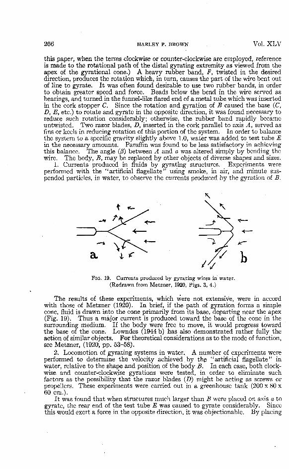
FIG. 19. Currents produced by gyrating wires in water.(Redrawn from Metzner, 1920, Figs. 3, 4.)
The results of these experiments, which were not extensive, were in accordwith those of Metzner (1920). In brief, if the path of gyration forms a simplecone, fluid is drawn into the cone primarily from its base, departing near the apex(Fig. 19). Thus a major current is produced toward the base of the cone in thesurrounding medium. If the body were free to move, it would progress towardthe base of the cone. Lowndes (1944 b) has also demonstrated rather fully theaction of similar objects. For theoretical considerations as to the mode of function,see Metzner, (1920, pp. 53-58).
2. Locomotion of gyrating systems in water. A number of experiments wereperformed to determine the velocity achieved by the "artificial flagellate" inwater, relative to the shape and position of the body B. In each case, both clock-wise and counter-clockwise gyrations were tested, in order to eliminate suchfactors as the possibility that the razor blades (D) might be acting as screws orpropellers. These experiments were carried out in a greenhouse tank (200 x 80 x60 cm.).
It was found that when structures much larger than B were placed on axis a togyrate, the rear end of the test tube E was caused to gyrate considerably. Sincethis would exert a force in the opposite direction, it was objectionable. By placing
No. 6 THE PROTOZOAN FLAGELLUM 267
around the test tube a fairly close-fitting, rigid jacket, this gyration may be min-imized. A coarse screen or hardware cloth is perhaps best, although a test tubeslightly larger than E was first used for the purpose. In the latter case, there istoo much difficulty involved in the movement of water to fill the space vacated bythe progressing system. In the set-up as shown in the diagram, the gyration ofthe test tube was sufficiently unimportant to be neglected in the gross observationsbeing made.
Velocity was measured horizontally and vertically. The latter measurementsrepresent much freer motion on the part of the "flagellate," but are more difficultto obtain under the conditions encountered. The "organism" is balanced so thatit sinks rather slowly. Then, wound up, it is held down, allowed to gyrate a fewtimes, and timed on its way up (its stable position is in the vertical axis). Forthe most part, it was timed through a distance of 20 cm. A stop watch was usedin all cases. Horizontal runs were made by placing the test tube in some suchjacket as mentioned in the preceding paragraph and holding the jacket steady inthe horizontal plane. By the nature of this set-up, such runs were confined to adistance of 5 or 6 cm. Since, in every case, B was buoyant, the gyration wasuneven in this plane. Another factor necessitating brief runs, both horizontallyand vertically, is that the force producing the gyration diminishes rapidly as therubber bands untwist. In consideration of these, and perhaps other conditions,it is obvious that the measurements are necessarily inaccurate. To assume anerror of ="=10% would be optimistic. However, the figures are at least indicative,and have some value thus.
Relative speed, as here employed, equals distance traversed by the organism inone second divided by the length of the gyrating body. Thus, if the body B were5 cm. long and the system moved at 10 cm./sec, the relative speed would be10 -r- 5, or 2. Among Protozoa which have been actually timed for rate of swimming,the relative speeds vary from 0.25 (Euglena terricola, Giinther, 1928) or less, to40.0 (Monas stigmatica, Lowndes, 1944 b, 1945 a) or more.
With a long body (B), 14 x l x l cm., at 1 to 1.1 gyrations/second, travelinghorizontally out of a vial, with angle /3 at 23°, the maximum constant velocityobserved was 1 to 1.2 cm./sec., representing a relative speed of about 0.08. Usingthe same set-up, but with angle $ greater than 90° (see Fig. 19 b), the maximumvelocity was 0.33 cm./sec. This condition hardly corresponds to any natural one.
With a short body (B) as shown in the diagram, 4.7 x 3.7 x 2.4 cm., at anestimated 6 gyrations/second, traveling vertically, with angle /3 at 23°, the max-imum constant velocity was 25 cm./sec, representing a relative speed of about 5.With angle /? at 15.2°, a maximum velocity of 33 cm./sec. was observed. Thiswas under ideal conditions and was never quite duplicated. It represents arelative speed of 7, the highest obtained in these experiments.
To test the forces produced by a flagellum undulating in a helix or spiral, thebody, B, was made in such a shape by bending a wire and coating it with 1.5 to 2.0mm. of paraffin. The form was approximately that shown in Fig. 16, but with aless complete pitch. The complete pitch would be about 20 cm. The flagellumrotated at about 18 turns/sec With the flagellum rotating counter-clockwise, thesystem moved forward at 34 to 38 cm./sec. With the flagellum rotating clockwise,the system moved backward at 20 to 22 cm./sec. If a living flagellum beats fromthe base outward, the latter is the only type of motion compatible with the system.In order for an actual flagellum to execute a movement similar to the former, thewave of contraction would have to begin at the tip of the flagellum. If thisoccurred, it would constitute a "tractellum." It probably does not occur in nature.
From our experiments with the "artificial flagellate," we learn that the mererotation and gyration of a body in water can provide sufficient force to producerapid forward locomotion of the body. This greatly strengthens the hypothesisadvanced by Lowndes (1944 a).

268 HARLEY P. BROWN Vol. XLV
UNDERWATER SWIMMING. These experiments test, in a fashion, the strength ofthe ''pull" exerted by gyrating structures. In contrast with flagellate bodies, butlike the flagella themselves, the gyrating objects do not necessarily rotate. Thearms of the swimmer serve as the gyrating structures. The rate of gyration isapproximately 1/sec. The gyrating portion is 60 cm. in length. Figures onvelocity are computed from the distance traversed in about one-half minute. Thebody weight of the swimmer is about 130 lbs. or 59 kg. In each case, enough airhas been expelled from the lungs to allow the body to sink to the bottom of thepool. All experiments were performed by the author. Timing was done by anobserver with a stop watch.
(1) The body is horizontal, with one arm extended horizontally forward andgyrating, e.g. clockwise, in a relatively narrow cone. (Fig. 20 a) Result: the bodymoves horizontally forward, rotating counter-clockwise, in this case. Velocity,10+ cm./sec. Relative speed, 0.16+. Total distance progressed, 10 ft.
FIG. 20. Underwater swimming experiments. See text for explanation.
(2) The body is vertical, with both arms extended horizontally forward, theright gyrating clockwise, the left counter-clockwise or vice versa. (Fig. 20 b) Result :the body moves horizontally forward, not rotating. Velocity, 10+ cm./sec. Rel-ative speed, 0.16+. Total distance progressed, 10 ft.
(3) The body is horizontal, with both arms extended horizontally forward, theright gyrating clockwise, the left counter-clockwise. (Fig. 20 c) Result: the bodymoves horizontally forward, not rotating. Velocity, 33 cm./sec. Relative speed,0.55. Total distance progressed, 32 ft. Reversing the gyrations, the velocity isconsiderably less, being at most about 23 cm./sec, with a relative speed of about0.39. Total distance progressed, 22 ft. This may well be due to an unintentionallyweaker stroke, as it is more difficult and tiring to the experimenter.
(4) The body is in the same position as (3), but both arms are gyrated in thesame direction. Result: the body rotates in the opposite direction at approx-imately the same rate, little forward movement being accomplished.
(5) The body is in position (3), but the arms are pendulated or swung back andforth in one plane, instead of being gyrated. Result: no detectable forwardcomponent.
Minor components are omitted in this consideration, for the sake of simplicity.In many cases they are results of the musculature of the arm.
Other experimental positions were tried, but contribute no additional significantdata and are hence omitted.
The results of these experiments provide additional evidence in support ofLowndes's hypothesis. They serve further, however, to show that rotation of thegyrating object is not necessary to the production of a forward component. Meregyration of an object (an arm or a flagellum) can produce an effective locomotor force.

NO. 6 THE PROTOZOAN FLAGELLUM 269
DISCUSSION
From the historical review given in this paper, it is apparent that a consid-erable mass of knowledge has been accumulated on the subject of flagellarstructure, but that, in large part, the data have not previously been assembled andorganized. Certain phases of the subject, to be sure, have been well summarized.For instance, Vlk (1938) treats whip- and flimmer-flagella as such rather thor-oughly, but neglects the internal structure. However, he is not to be criticizedfor this, as little new knowledge has appeared relative to the matter within severaldecades. The subject has awaited a new technique which could permit of more'minute investigation. The electron microscope provides this new angle of attackthrough its much greater resolution and magnification. This paper presents theresults of the first intensive study of the protozoan flagellum employing theelectron microscope.
By means of this instrument, we find that the flagellum, at least in the formsstudied, consists of a dense fibrillar axial region and a surrounding sheath of muchless density. This much has been previously accepted, though never before soclearly demonstrated. Just how many fibrils there are comprising the axoneme, Icannot tell. The number probably varies to some extent. The sheath seems tocontain a fiber which encircles the axoneme in the form of a helix. This sort ofcomposition of axoneme and sheath appears to be in close agreement with thestructure of mammalian sperm tails as described by Schmitt and others, but hasnever before been described in the protozoan flagellum, unless the brief remarks ofClehla (1911) be considered such. Unlike the sperm tails, certain flagella possesslateral or terminal external extensions of the sheath. These structures have beenrecorded since 1889, but are not yet universally accepted by protozoologists.Several electron micrographs of the lateral structures in question (the flimmer)are included in this paper (e. g., Plates 4, 9, 11, 12). Of course, such picturesconstitute no conclusive proof of the nature, or even natural occurrence, of theflimmer. But they certainly contribute to the evidence in favor of their accept-ance as normal structures, rather than as artifacts. If Vlk had had at his disposalthe equipment of Lowndes (1935), Harvey and Loomis (1931), or Pijper (1940),perhaps the question might have been settled for the most skeptical by the actualphotographing of flimmer on living flagella. However, the facts that they havebeen demonstrated by a number of basically different techniques, on dried ormoist flagella, following various fixatives or none at all, and that they are amazinglyconstant in appearance, position, etc., place the overwhelming weight of evidencein favor of the view that they represent normally-occurring structures in manytypes of organisms. The presence and type of flimmer or whip comprise valuabletaxonomic characters, and will doubtless be so considered when they are morereadily demonstrable.
There are a number of devices as yet untried in the study of these structures,some of which should certainly contribute to our knowledge of their nature. Imention them in the hope that someone, to whom they may be available, may beinterested in furthering this study. By means of the shadow technique ofWilliams and Wyckoff (1944, 1945 a, b), the flimmer should be made obvious. Astudy of living flagella might be made with the ultra-violet microscope (Lucas,1930, 1934; Schmitt, 1939; Lavin and Hoagland, 1943), whereby twice the resolu-tion of the ordinary microscope might be obtained. Phase difference microscopy(Richards, 1944) offers interesting possibilities in the study of living flagella,yielding the benefits of stains and fixing reagents, as does the ultraviolet micro-scope, without requiring the use of such deleterious agents. (That is, it serves todifferentiate structures and substances which we commonly bring out only bystaining.) Fluorescence microscopy (Ellinger, 1940; Metcalf and Patton, 1944)also gives promise, especially, perhaps, in the study of flagellar motion. If nat-

270 HARLEY P. BROWN Vol. X L V
urally fluorescing flagella occur, these would be ideal for such a study, but afluorescent vital stain such as Fluorescein might serve well.
It is interesting to view the results of Kunstler and Fischer in the light ofdetails brought out in electron micrographs. If the reader will again refer toFig. 1, he may notice certain similarities to some of the plates. The interpretationof the appearances, however, is rather different. The axial canal of Kunstler isprobably the axoneme. Plate 7.shows, in places, an apparent breaking up orsegmentation of the axoneme into pieces resembling the "vacuoles" of Kunstler,as seen in Fig. 1 a. Figs. 1 b and c are reminiscent of the helix of the sheath asseen in the electron micrographs, the corrugations depicted by Kunstler probablyrepresenting the surface appearance of the structures. It must be borne in mindthat Kunstler had at his disposal nothing like the resolution and magnification ofthe electron microscope; his observations were remarkably good, in considerationof this fact. Segmentation of the flagellum may somehow be produced by theaction of osmic acid, in combination with other factors. Kunstler had used thisreagent in his preparations, as have I. Gelei (1926) showed that such appearancesresulted from certain reagents, among them osmic acid in combination with otherchemicals.
Fischer apparently found a continuous axoneme in many flagella, but tookgreat pains to demonstrate or argue that it was but an artifact. He, too, did agood job with what he had, and presented a rather convincing argument. How-ever, in the light of evidence since brought forth, including much in this paper, itis highly probable that he was mistaken in his interpretation of at least the- natureof the axial fiber.
Regarding the structure of the flagellar filament, Plate 7 presents an interestingpicture, which, as Dr. Prebus points out, may well represent at the denser endthe natural appearance of the flagellum. Here the helical structure in the sheathis evident, but the inner axial fibers are obscured by the density of the surroundingmatter within the sheath. In the less dense portions of this filament we see whatis perhaps the result of a loss of material from the sheath or intermediate substance.This lighter portion corresponds to the appearance of most of the flagella shown,and brings out the internal structure, but may thus represent a state of disinte-gration of the flagellum.3 The ground substance which may have escaped isprecisely that which is considered to be the contractile portion of the flagellum bymany workers. For my part, I find it easier to attribute the major contractilityto the fibrillar structures remaining.
In this connection, I might mention a possible mechanism for the progress ofthe contractive wave which has not heretofore been suggested, so far as I know.It has no basis in experimental evidence, and is simply proposed for consideration.Since the flagellum appears to have a fibrous core which is encircled by a helicalfiber, as represented diagrammatically in Fig. 21, it is possible that the spiral fibertransmits the impulse. If along this fiber passed a wave of chemical change, com-parable to that in a firecracker fuse or a nerve fiber, perhaps releasing H ionswherever it passed over the axial fibers, it could stimulate local contraction insuch parts of the axial fibers as might be contiguous with it. In conjunction withtwisted axial fibers (Dellinger, 1909), this might account for undulatory movementsof a spiral nature. In such organisms as Peranema, the axial fibrils might bestiffened or less sensitive in the basal portion, thus accounting for the fact thatnormally only the distal part shows obvious activity, whereas, if stimulated to a
3A second possible explanation for the appearance shown in Plate VII is that a portion ofthe flagellum might have contracted, bringing the coils of the helix into closer approximationand producing the denser and thicker appearance seen in the upper portion of the flagellumshown. It is difficult to determine precisely what actually happened to produce this effect,and the interpretation of such an appearance must remain uncertain for the present.

No. 6 THE PROTOZOAN FLAGELLUM 271
greater extent, the entire flagellum is thrown into undulations. Also, if the axialfibrils along one side should be less reactive, the resultant stroke might be paddle-like. With a bit of imagination, this hypothetical mode of functioning can be fittedto most observed facts. At least, it fits the facts better than any other systemproposed, so far as I have been able to discover. However, it remains purelyspeculative.
FIG. 21. Diagram illustrating hypothetical path of impulse inflagellum. See text for explanation.
Likewise in the realm of speculation to an extent, but also of interest, in adifferent way, is the appearance of the axial core of the flagellum of Euglena (Plates2 A; 5; 9). There seem to be two equal fibers running side by side, which aredisplayed especially well where the flagellum (or merely the fibers) is twisted.These fibers are probably composed of yet finer fibrils (see Dellinger, 1909), thoughsuch are not evident in the plates. Now it happens that Euglena has a bifurcateflagellar root. Two distinct branches, arising from two separate basal granules,unite in the gullet at about the level of the stigma (eyespot) to form the singleflagellum (Wager, 1899, et al). Hartmann and Chagas (1910), in consideration ofthe bipartite root, considered Euglena as derived from a biflagellate. It seemsprobable to me that the two fibers within the flagellum definitely strengthen sucha view, and might be regarded as the two ancestral axial fibers or axonemes in closeassociation.
Another interesting source of speculation is the relationship between thebacterial flagellum and that of the protozoon. Electron studies have been madeon many bacteria. Among authors reproducing numerous electron micrographsof bacterial flagella are Piekarski and Ruska (1939) and Mudd and Anderson(1944). According to these and other authors, the flagella average from 14 to 50 m/xin diameter, and often occur in clumps or tufts, as they do in the spirochete Tre-ponema (Mudd, Polevitsky, and Anderson, 1943). Their precise nature still appearsdoubtful. The suggestion has been made (e. g., Polevitsky, 1941) that they maybe hollow tubes, but Mudd and Anderson (1942, p. 106) find no support for thisidea in cases which superficially present such an appearance. Mudd, Polevitsky,Anderson, and Chambers (1941) figure an electron micrograph of Bacillus subtiliswith the protoplasm shrunken away from the cell wall. On the other hand, Muddand Anderson (1944) figure Vibrio cholerae, showing a " single polar flagellum,which seems to traverse the cell wall to join the bacterial protoplasm." At anyrate, bacterial flagella, whatever may be their exact nature, and even though theymay not arise in a tuft, often entwine to function as a unit and may even remainadherent, forming a filament composed of many fibrils and perhaps encased in agelatinous (?) sheath. (See historical review for references.) Now recall theflagellar or sperm tail structure with its 9 to 12 fibrils, each 25 to 50 m/i in diameter,ordinarily encased in a sheath. The possibility certainly is suggested that theancestral protozoan flagellum may have been derived from such a permanentlyunited clump of fibrils as occurs in some of the bacterial forms. Should this prove

272 HARLEY P. BROWN Vol. XLV
to be the actual phylogenetic origin of the protozoan flagellum, the concept of thefibrils as contractile units, rather than as mere elastic rods, would be definitely-strengthened. However, the fibrils might well serve both as contractile and assupportive structures.
With regard to the mode of functioning of the locomotor flagellum, my observa-tions and experiments confirm the conclusions reached by Lowndes. The livingflagella observed beat in spiral undulations, the waves of contraction always pro-gressing from base to tip. My experiments with an "artificial flagellate" and withunderwater swimming demonstrated that sufficient force is produced by the simpleconical gyration of a body to account for rapid locomotion toward the base of thecone. Fig. 22 and its legend describe the propulsive mechanism as conceived byLowndes. I have never, to my knowledge, watched the swimming of this partic-ular organism, but I have observed other similar ones. From my own experimentsand the work of Mr. Lowndes, I have no doubt that the gyration of the organismcould account for a relative speed of 40 (that given by Lowndes for Monas stig-matica), if the body gyrated with sufficient rapidity. But in the forms I haveobserved, it is difficult to imagine how the body gyration alone could produce such
FIG. 22. "Monas stigmatica with its two flagella. Diagrammatic." "Waves pass alongthe long flagellum, from base to tip, in a spiral manner with an increase in velocity and ampli-tude. The force generated is transmitted to the surface of the cell at A. This causes theorganism to rotate and gyrate about the axis D. (The edge B going below the surface of thepaper and C being raised above it.) Thus the organism is converted into a rotating inclinedplane and moves forward more or less in the direction indicated by the arrow E. The veryshort flagellum F appears to act as a guiding or sensory organ during normal swimming."(Redrawn and quoted from Lowndes, 1945 a.)
speed. At a rate of body gyration of only about 1/sec. the relative speed may beat least 5. I should think that the forward component produced by the flagellumwould aid greatly in the attainment of high speed. But perhaps my imaginationis not keyed to the physics of the micro-world of the flagellum. Apparently themechanics and physical relationships (e. g., the relative viscosity of water, a majoritem in the protozoan world) to which we are accustomed are tremendouslyaltered, practically non-existent, at this level of dimensions. For an interestingdiscussion of some of the differences see Bidder (1923, pp. 304-307).
Brief mention may be made of the possibility of studying living organisms withthe electron microscope. It would obviously be desirable to study such a structureas the flagellum in the living, intact state. Several papers have appeared relative

No. 6 THE PROTOZOAN FLAGELLUM 273
to the possibility of observing living structures with the instrument, but, so far asI know, the only objects studied thus or even very practical for such study havebeen spores. Perhaps the most ambitious attempts have been made with the closedcell described by Abrams and McBain (1944). The obvious difficulties resultfrom greatly reduced contrast and Brownian movement, which prevent photo-graphing of the objects under observation. Another major factor is the killingeffect (aside from heat production) due to " . . . the electrons bounding downthrough the specimen and breaking the chemical bonds of the specimen. Breakingthe bonds is detrimental to all living organisms. Every bond in living organismsdoes not have to be intact, but the critical molecules in the living organisms musthave a specific structure in the organism for it to grow and reproduce itself. Whenyou consider that, in maximum intensity, the beam reaches a high value of 1,000electrons per square angstrom per second, you can realize that it wouldn't takethe scattering of many electrons to kill such an organism." (Anderson, 1943.)In terms more familiar to the biologist, this electron bombardment would amountto 100,000,000,000 electrons striking a surface of 1 square micron each second.The average protozoan body would present a surface of a considerable number ofsquare microns. Thus the prospects of studying a living flagellum, even if itwere perfectly stationary and met the other requirements, would not be encouraging.
SUMMARY
1. The work of previous investigators in this field is reviewed and discussed.2. The structure of the flagellum of several species of Protozoa has been
investigated by means of the electron microscope. Micrographs are includedwhich show that:
(a) The fiagella studied are of approximately uniform diameter throughouttheir entire length.
(b) Each flagellum consists of a denser axial core (axoneme) and a lessdense sheath surrounding the core.
(c) In the fiagella of Euglena and Astasia the axial core appears to consistof two closely approximated fibers of equal size.
(d) The sheath appears to contain or to consist of a coiled fiber whichencircles the axial core in a helix.
(e) The fiagella of Euglena and Astasia bear, along one side, what appearsto be a single row of delicate filaments (flimmer) extending from the sheath.These flimmer have an average length of about 5-6 times the diameter of theflagellum proper.
(f) The long flagellum of Ochromonas bears similar filaments along both(or all?) sides. ,
(g) The fiagella of Chilomonas bear no such lateral filaments.The significance of certain details present in the electron micrographs is
discussed.
3. The presence of two equal fibers in the flagellum of Euglena, together withthe fact (demonstrated by previous investigators) that the flagellum of this formhas two separate roots, indicates the ancestral formation of its flagellum by thefusion or union of two fiagella. The hypothesis of a biflagellate ancestry of Euglenais strengthened.
4. The possibility of the origin of the primitive protozojtn flagellum throughthe union within a common matrix of several simple fibrillar fiagella (as are presentin certain bacteria) is suggested. Evidence for such a theory is presented.

274 HARLEY P. BROWN Vol. X L V
5. Observations were made on the activity of living flagella. The results werein direct confirmation of Lowndes's contentions that:
(a) The flagellum beats in spiral undulations.(b) The waves of contraction progress from the base toward the tip of
the flagellum.(c) As they progress, the waves often increase in amplitude.(d) The flagellum usually serves to push, rather than to pull the organism
through the water, although it arises from the anterior end of the body.6. Experiments were performed by means of actual underwater swimming by
the author and with an "artificial flagellate" to determine the forces produced bygyrating bodies in water. In both cases, a simple conical gyratory movementwas found sufficient to produce locomotion. Employing the artificial flagellate,it was found that rotation and gyration of a small body at about 6 gyrations persecond was capable of producing a relative speed of as much as 7.0 (i. e., 7 lengthsper second), in spite of the fact that the body had to pull along after it an objectmuch larger than itself. This confirms the theory of Lowndes that the rotationand gyration of the body alone may adequately account for the locomotion of manyflagellates, without any forward component produced directly by the flagellum.
7. A hypothesis is suggested concerning the mechanism of fiagellar function:Perhaps the helical fiber of the sheath transmits an impulse which stimulates localcontraction in the underlying fibrils. Possible ways by which such a mechanismmight account for the various types of fiagellar movement are mentioned.
8. Promising techniques, recently developed in other fields, are suggested forfurther research on flagella.
LITERATURE CITED
Abrams, I. M., and J. W. McBain. 1944. A closed cell for electron microscopy. Science100 (2595): 273-274, 1 fig.
Alexeieff, A. 1924. Comparaison entre la structure des spermatozoides et celle des flagelle's.Arch. f. Protistenk, 49: 104-111, 2 figs.
Allen, E. A. 1936. A Pentatrichomonas associated with certain cases of enterohepatitis or"blackhead" of poultry. Trans. Amer. Micr. Soc. 55: 315-322, 1 pi.
Alverdes, F. 1922. Studien an Infusorien tiber Flirnmerbewegung, Lokomotion, undRejzbeantwortung. Schaxel: Arbeiten aus dem Gebiet der exp. Biol., Heft 3. 127 pp.,46 figs.
Anderson, T. F. 1943. Year Book of The Electron Microscope Society of America. (Recordof oral discussion, p. 34.)
Awerinzew, S. von. 1907. Beitrage zur Kenntnis der Flagellaten. Zool. Anz. 31 (25):834-841, 9 figs.
Ballowitz, E. 1890. Fibrillare Struktur und Contraktilitat. Pflugers Arch. ges. Physiol.46: 433-464.
1908. Uber den feineren Bau der eigenartigen, aus drei freien dimorphen Fasern bestehendenSpermien der Turbellarien. Arch. f. mikr. Anat. 71: 4-21, 3 pis.
Bancroft, F. W. 1913. Heliotropism, differential sensibility, and galvanotropism in Euglena.Jour. Exp. Zool. 15: 383-428, 5 figs.
Barker, D. 1943. Recent work on flagellar movement. New Phytol. 42: 49-53.Baylor, M. R. B., A. Nalbandov, and G. L. Clark. 1943. Electron microscope study of sperm.
Proc. Soc. Exp. Biol. and Med. 54: 229-232, 7 figs.Bidder, G. P. 1923. The relation of the form of a sponge to its currents. Quart. Jour. Micr.
Sci., N.S. 67: 293-323, 12 figs.Burton, E. F., and W. H. Kohl. 1942. The Electron Microscope. Reinhold Publishing Corp.,
New York.Biitschli, O. 1883-1887. Protozoa: Mastigophora. Bronns Klassen und Ordnungen des
Thier-reichs. Band 1, Abt. 2.1902. Bemerkungen iiber Cyanophyceen und Bacteriaceen. Arch. f. Protistenk. 1: 41-58,
1 pi.

No. 6 THE PROTOZOAN FLAGELLUM 275
Calkins, G. N. 1933. The Biology of the Protozoa, 2nd ed. rev. Lea and Febiger, Phila-delphia and New York.
Carter, G. S. 1924. On the structure and movements of the latero-frontal cilia of the gills ofMytilus. Proc. Roy. Soc. London 96 B: 115-122, 14 figs.
Claude, A., and E. F. FuUam. 1945. An electron microscope study of isolated mitochondria.Jour. Exp. Med. 81 (1): 52-62, 2 pis.
Dangeard, P. A. 1901 a. Etude sur la structure de la cellule. Le Botaniste, 8e Serie,Poitiers: 5-58, 4 figs.
1901 b. Recherches sur les eugl^niens. Ibid., 97-370, 4 pis., 53 figs.Deflandre, G. 1923. Emploi de la Nigrosin dans l'e'tude des algues infe"rieures. Bull. Soc.
bot. de France 70: 738-741.1934. Sur la structure des flagelles. Anns. Protistol. 4: 31-54, 5 pis., 14 figs.
Delage, Y., et Herouard. 1896. Traite de Zoologie Concrete, I. La Cellule et les Protozoaires.Schleicher Freres, Paris.
Dellinger, O. P. 1909. The cilium as a key to the structure of contractile protoplasm. Jour.Morph. 20: 171-210, 4 pis. (of photomicrographs), 13 figs.
Doflein, F. 1929. Lehrbuch der Protozoenkunde, 5th ed. (re-edited by E. Reichenow). Verlagvon Gustav Fischer, Jena.
Duboscq, O., et P. Grasse. 1933. L'appareil parabasal des flagelle's. Archs. Zool. Exp. etGen. 73: 381-621, 4 pis. (With an extensive bibliography.)
Ellinger, P. 1940. Fluorescence microscopy in biology. Cambridge Philosoph. Soc, Biol.Rev. 15: 323-350. (Good bibliography.)
Emmel, L., A. Jakob, und H. Golz. 1943. Elektronenoptische Untersuchungen an MalariaSporozoiten und Beobachtungen an Kulturformen von Leishmania donovani. Deutschetropenmed. Zeitschr. 46 (13): 354-358, 9 figs.
Engelmann, T. W. 1881. Uber den faserigen Bau der contraktilen Substanzen, usw. Arch.ges Physiol. 25: 538-565, 1 pi.
Entz, G. 1926. Beitrage zur Kenntnis der Peridineen. I. Zur Morphologie und Biologievon Peridinium Borgei Lemmermann. Arch. f. Protistenk. 56: 397-446, 1 pi., 33 figs.
1928. Uber den Bau und uber die Tatigkeit der Geisseln der Peridineen. Anns. Protistol.1: 75-95, 5 tables, 29 figs.
Erhard, H. 1910. Studien uber Flimmerzellen. Arch. f. Zellforsch. 4: 309-442, 2 pis., 16 figs.,250 references in bibliography.
Fischer, A. 1894. Uber die Geisseln einiger Flagellaten: Prings. Jahrb. f. wiss. Bot. 26:187-235, 2 pis.
1895. Ibid., 27: 1-163, 5 pis.Friedrich, L. 1909. Uber Bau und Naturgeschichte des Trypanoplasma helicis Leidy. Arch.
f. Protistenk. 14: 363-395, 48 figs.Gelei, J. von. 1926. Zur Kenntnis des Wimperapparates. Zeitschr. f. Anat. u. Entwick-
lungsgeschichte 81: 530-553, 24 figs.Gerloff, J. 1940. Beitrage zur Kerintnis der Variabilitat und Systematik der Gattung
Chlamydomonas. Arch. f. Protistenk. 94: 311-502, 48 figs.Gicklhorn, J. 1921. Eine einfache Methode zur Herstellung der Geissel mit Basalkorn bei
Flagellaten, besonders bei Eugleninen. Zeitschr. f. wiss. Mikr. 38: 123-129.Goldschmidt, R. 1907. Lebensgeschichte der Mastigamoben usw. Arch. f. Protistenk.,
Supplement I: 83-168, 5 pis., 20 figs.Grasse, P. P. 1926. Contribution a l'6tude des flagelles parasites. Archs. Zool. Exp. et Gen.
65: 345-602, 12 pis., 76 figs.Gray, J. 1922. The mechanism of ciliary movement. Proc. Roy. Soc. London 93 B: 104-121,
7 figs.1928. Ciliary Movement. 162 pp., 105 figs., 16 tables. Cambridge Univ. Press, London.
(Rather complete bibliography.)Giinther, F. 1928. Uber den Bau und die Lebensweise der Euglenen. Arch. f. Protistenk.
60: 511-590, 3 pis., 5 figs.*Gurwitsch, A. 1904. Morphologie und Biologie der Zelle. Jena.Hall, R. P., and T. L. Jahn. 1929. On the comparative cytology of certain euglenoid
flagellates and the systematic position of the families Euglenidae Stein and AstasiidaeButschli. Trans. Amer. Micr. Soc. 48: 388-405, 2 pis., 2 figs.
Hamburger, C. 1911. Studien uber Euglena Ehrenbergii, insbesondere iiber die Korperhiille.Sitz.-ber. d. Akad. d. Wiss., Heidelberg. Abhandlung 4.
Hartmann, M., and C. Chagas. 1910. Flagellaten-Studien. Mem. Inst. Oswaldo Cruz 2:64-125, 6 pis., figs.
Harvey, E. B., and T. F. Anderson. 1943. The spermatozoon and fertilization membrane ofArbacia punctulata as shown by the electron microscope. Biol. Bull. 85 (2): 151-156, 2 pis.
Harvey, E. N., and A. L. Loomis. 1931. High speed photomicrography of living cells subjectedto supersonic vibrations. Contrib. Biol. Labs., Princeton Univ., 10 (21). Reprintedfrom Jour. Gen. Physiol. 15 (2): 147-153, 3 figs.

276 HARLEY P. BROWN Vol. X L V
Hawley, G. G. 1945. Seeing the Invisible. The story of the Electron Microscope. A. A.Knopf, New York.
Heidenhain, M. 1911. Plasma andZelle II. Fischer, Jena.Hillier, J., and A. W. Vance. 1941. Recent developments in the electron microscope. Proc.
Inst. Radio Eng., Inc. 29: 167-176, 14 figs.Hutchinson, W. G., and M. R. McCracken. 1943. Study of flagella of a freshwater bacterium
by motion microphotography and electron micrography. (Abstract) Jour. Bact. 45: 305.Hyman, L. H. 1940. The Invertebrates: Protozoa through Ctenophora. McGraw-Hill,
New York.Kater, J. McA. 1929. Morphology and division of Chlamydomonas with reference to the
phylogeny of the flagellate neuromotor system. Univ. Calif. Publ. in Zool. 33: 125-168,6 pis., 7 figs.
Keysselitz, G. 1906. Generations- und Wirts-wechsel von Trypanoplasma borreli Laveran etMesnil. Arch. f. Protistenk. 7: 1-74, 162 figs.
Kirby, H. 1943. Observations on a trichomonad from the intestine of man. Jour. Parasitol.29 (6): 422-423.
Klebs, G. 1892. Flagellatenstudien I, II. Zeitschr. f. wiss. Zool. 55: 265-445, 6 pis., 122references.
Klein, B. M. 1929. Weitere Beitrage zur Kenntnis des Silberliniensystems der Ciliaten. Arch.f. Protistenk. 65: 183-257, 1 pi., 45 figs.
Kofoid, C. A., and O. Swezy. 1919. Studies on the parasites of termites. III. On Tricho-nympha campanula, sp. nov. Univ. Calif. Publ. in Zool. 20: 41-98, 11 pis., 4 figs.
1920. On the morphology and mitosis of Chilomastix mesnili (Wenyon), a common flagellateof the human intestine. Ibid., 117-144, 5 pis., 2 figs.
1923. On the morphology and behavior of Pentatrichomonas ardin delteili (Derrieu etRaynaud). Ibid., 373-390, 1 pi., 1 fig.
Koltzoff, N. K. 1903. Uber formbestimmende elastische Gebilde in Zellen. Biol. Centralbl.23: 680-696, 12 figs.
1909. Studien uber die Gestalt der Zelle. II. Untersuchungen tiber das Kopfskelett destierischen Spermiums. Arch. f. Zellforsch. 2: 1-65, 5 pis., 18 figs.
Korschikov, A. 1923. Uber den Bau und die Aggregation der Geisseln bei den Volvocales undden Flagellaten. Archs. Soc. Russe de Protistol. 2: 195-205, 4 figs.
Krijgsman, B. J. 1925. Beitrage zum Problem der Geisselbewegung. Arch. f. Protistenk.52: 478-488, 6 figs. '
Kudo, R. R. 1939. Protozoology, 2nd ed. C. C. Thomas, Springfield, 111.*Kunstler, J. 1882. Contribution a l'6tude des flagelle's. Bull. Soc. Zool. de France 7: 20.
1889. Recherches sur la morphologie des flagelles. Bull. Sci. de la France et de la Belgique20: 399-515, 9 pis.
Laveran, A., and F. Mesnil. 1907. Trypanosomes and Trypanosomiases. (Translation byD. Nabarro.) W. T. Keener and Co., Chicago.
Lavin, G. I., and C. L. Hoagland. 1943. Ultraviolet photomicrography of muscle. Proc.Soc. Exp. Biol. and Med. 52: 80-82, 14 figs.
Loeffler, F. 1889. Eine neue Methode zum Farben der Mikrodrganismen, im besonderenihrer Wimperhaare und Geisseln. Centralbl. f. Bakt. 6: 209-224; 2 pis.
1890. Weitere Untersuchungen uber die Beizung und Farbung der Geisseln bei denBakterien. Ibid., 7: 625.
Lowndes, A. G. 1935. The twin polygraph and strobograph. Nature 135: 1006-1007, 1 photo-graph of assembled apparatus.
1936. Flagella movement. Ibid., 138: 210-211, 4 figs.1941 a. Mechanics of a flagellum. Ibid., 148: 198, 5 figs.1941 b. On flagellar movement in unicellular organisms. Proc. Zool. Soc. London 111 A:
111-134, 20 figs.1943. The term tractellum in flagellate organisms. Nature 152: 51.1944 a. The swimming of unicellular flagellate organisms. Proc. Zool. Soc. London 113 A:
99-107, 6 figs.1944 b. The swimming of Monas stigmatica Pringsheim and Peranema trichophorum (Ehrbg.)
Stein and Volvox sp. Additional experiments on the working of a flagellum. Ibid., Il l ,114: 325-338, 10 figs.
1945 a. Swimming of Monas stigmatica. Nature 155: 579, 1 fig.1945 b. The swimming of Euglena and flagellar movement in general. The School Science
Review, No. 100, June, 1945. pp. 319-332, 2 pis., 9 figs.Lucas, F. F. 1930. The architecture of living cells.—Recent advances in methods of biological
research.—Optical sectioning with the ultra violet microscope. Tech. Publ. Bell Tele-phone System, Monograph B 514, 9 pp., 16 figs. (Also in Proc. Natl. Acad. Sci. 16:599-607.
1934. Late developments in microscopy. Ibid., Monograph B-792, 47 pp., 32 figs.
No. 6 THE PROTOZOAN FLAGELLUM 277
Mainx, F. 1928. Beitrage zur Morphologie und Physiologie der Eugleninen. Arch. f.Protistenk. 60: 305-414, 1' plate containing 18 photomicrographs, 8 figs. (Goodbibliography.)
Marsland, D. A. 1943. Quieting Paramecium for the elementary student. Science 98(2549): 414.
Mart on, L. 1941. The electron microscope. A new tool for bacteriological research. Jour.Bact. 41:397-413, 12 figs.
1943. The electron microscope in biology. Ann. Rev. Biochem. 12: 587-614. AnnualReviews Inc., Stanford University.
McClung, C. E. (editor). 1937. Handbook of microscopical technique. Paul B. Hoeber, Inc.,Medical Book Dept. of Harpers and Brothers, New York.
McDonald, J. D. 1922. On Balantidium coli (Malmsten) and Balantidium suis n. sp., with anaccount of their neuromotor apparatus. Univ. Calif. Publ. in Zool. 20: 243-300, 2 pis.,15 figs.
Metcalf, R. L., and R. L. Patton. 1944. Fluorescence microscopy applied to entomology andallied fields. Stain Technol. 19 (1): 11-27, 1 pi., 1 fig.
Metzner, P. 1920. Zur Mechanik der Geisselbewegung. Biol. Centralbl. 40 (2):* 49-87,18 figs., 48 references.
Minchin, E. A. 1922. An Introduction to the Study of the Protozoa. Edward Arnold, London.Morton, H. E., and T. F. Anderson. 1942. Some morphological features of the Nichols strain
of Treponema pallidum as revealed by the electron microscope. Amer. Jour. Syphilis,Gonorrhea, and Venereal Dis. 26 (5): 565-573, figs.
Mudd, S., and T. F. Anderson. 1942. Selective "staining" for electron micrography. Theeffects of heavy metal salts on individual bacterial cells. Jour. Exp. Med. 76 (1): 103-108,4 pis.
1944. Pathogenic bacteria, rickettsias, and viruses as shown by the electron microscope.Amer. Med. Assn., Chicago. (Reprinted, with additions, from the Jour. Amer. Med.Assn. 126: 561-571, 632-639.) 24 pp., 50 figs.
Mudd, S., K. Polevitsky, T. F. Anderson, and L. A. Chambers. 1941. Bacterial morphologyas shown by the electron microscope. II. The bacterial cell-wall in the genus Bacillus.Jour. Bact. 42: 251-264, figs.
Mudd, S., K. Polevitsky, and T. F. Anderson. 1943. Bacterial morphology as shown by theelectron microscope. V. Treponema pallidum, T. macrodentium, and T. microdentium.Jour. Bact. 46: 15-24, 8 figs.
Nigrelli, R. 1929. On the cytology and life-history of Trypanosoma diemyctyli and the poly-morphonuclear count of infected newts (Triturus viridescens). Trans. Amer. Micr. Soc.48: 366-387, 2 pis., 1 fig., 1 table.
Petersen, J. B. 1918. Om Synura uvella Stein og nogle andre Chrysomonadiner. Vidensk.Medd. fra Dansk naturhist. Foren 69: 345-357, 1 pi.
1929. Beitrage zur Kenntnis der Flagellatengeisseln. Botanisk Tidsskrift (Copenhagen)40 (5): 373-389, 1 pi.
Petrova, J. 1931. Die vermeintliche Heterokonte Botrydiopsis minor—eine Chlorophycee.Beihefte z. bot. Centralbl. 48: 221-228, 7 figs.
Piekarski, G., und H. Ruska. 1939. Ubermikroskopische Darstellung von Bakteriengeisseln.Klin. Wochenschr. 18 (1): 383-386, 11 figs.
Pijper, A. 1940. Microcinematography of the motile organs of the typhoid bacillus. Jour.Biol. Photog. Assn. 8: 158-164, figs.
1941. Microcinematography of the agglutination of typhoid bacilli. Jour. Bact. 42:395-409, figs.
Plehn, M. 1904. Trypanoplasma cyprini n. sp. Arch. f. Protistenk. 3: 175-180, 1 pi.*Plenge, H. 1898. Uber die Verbindung zwischen Geissel und Kern bei den Schwarmzellen der
Mycetozoen und bei Flagellaten. Verh. nat. Ver., Heidelberg, N.F. 6.Polevitsky, K. 1941. Pictures of bacterial forms taken with the electron microscope. (Abs.)
Jour. Bact. 41: 260.Porter, K. R., A. Claude, and E. F. Fullam. 1945. A study of tissue culture cells by electron
microscopy. Jour. Exp. Med. 81 (3): 233-246, 5 pis.Prebus, A. F. 1944. The electron microscope. Colloid Chemistry V (Edited by Jerome
Alexander). Reinhold Publ. Co., New York, pp. 152-235, figs.Prowazek, S. von. 1903. Flagellatenstudien. Arch. f. Protistenk. 2: 195-212, 2 pis.Putter, A. 1903. Die Flimmerbewegung. Ergebnisse d. Physiol. 2 (2): 1-102, 15 figs., 208
references.Reichert, K. 1909. Uber die Sichtbarmachung der Geisseln und die Geisselbewegung der
Bakterien. Centralbl. Bakt. 51: 14-94, 30 figs.Richards, O. W. 1944. Phase difference microscopy. Nature 154: 672, 1 fig.Richards, A. G., and T. F. Anderson. 1942. Electron microscope studies of insect cuticle, with
a discussion of the application of electron optics to this problem. Jour. Morph. 71:135-183, figs.

278 HARLEY P. BROWN Vol. X L V
Rosenbusch, T. 1908. Trypanosdmen-Studien. Arch. f. Protistenk. 15: 263-296, 3 pis.Roskin, G. 1922. Uber den Bau von kontraktilen Elementen und Stutzsttbstanzen einigen
Protozoen. Archs. Soc. Russe de Protistol. 1: 35-45, 1 pi., 1 fig.1923. Zur Frage uber den Bau der Geisseln. Ibid. 2; 206-209, 3 figs.1925. Uber die Axopodien der Heliozoa und die Greiftentakeln der Ephelotidae. Arch. f.
Protistenk. 52: 207-216, 9 figs.Schindera, M. 1922. Beitrage zur Biologie, Agglomeration, und Zuchtung von Trypanoplasma
helicis Leidy. Arch, f. Protistenk. 45: 200-240, 1 pi., 3 figs.Schmitt, F. O. 1939. The ultrastructure of protoplasmic constituents. Physiol. Rev. 19 (2):
270-302, 1 fig.1944. Structural proteins of cells and tissues. Advances in Protein Chem. 1: 25-68, 9 figs.
Schmitt, F. O., C. E. Hall, and M. A. Jakus. 1943. The ultrastructure of protoplasmic fibrils.Frontiers in Cytochemistry, Biological Symposia X. Jaques Cattell Press, Lancaster,Pa. pp. 261-276, 2 pis.
Schouteden, H. 1907. Notes sur quelques flagelle's. Arch. f. Protistenk. 9: 108-136, 11 figs.Schuberg A. 1905. Uber Cilien und Trichocysten einiger Infusorien. Arch. f. Protistenk.
6: 61-110, 2 pis.Smyth, J. D. 1944. A technique for mounting free-living protozoa. Science 100 (2586): 62.Ulehla, V. 1911. Ultramikroskopische Studien uber Geisselbewegung. Biol. .Centralbl. 31:
645-654, 657-676, 689-705, 721-731, 37 figs.Verworn, M. 1890. Studien zur Physiologie der Flimmerbewegung. Arch. f. ges. Physiol.
48: 149-180, 4 figs.1915. Allgemeine Physiologie, 6th ed. Fischer, Jena.
Vlk, W. 1931. Uber die Struktur der Heterokontengeisseln. Beihefte z. bot. Centralbl.48: 214-220, 15 figs.
1938. Uber den Bau der Geissel. Arch. f. Protistenk, 90: 448-488, 1 pi., 12 figs. (Goodbibliography and historical review.)
Wager, H. 1899. On the eyespot and flagellum in Euglena viridis. Jour. Linn. Soc, London,Zoology 27: 463-481, 1 pi.
Williams, L. W. 1907. The structure of cilia, especially in gastropods. The Amer. Naturalist41 (489): 545-551, 2 figs. .
Williams, R. C., and R. W. G. Wyckoff. 1944. The thickness of electron microscope objects.Jour. Appl. Phys. 15 (10): 712-715, 8 figs.
1945 a. Electron shadow-micrography of virus particles. Proc. Soc. Exp. Biol. and Med.58 (3): 265-270, 6 figs.
1945 b. Electron shadow-micrography of the tobacco mosaic virus protein.. Science 101(2632): 594-596, 3 figs.

The Protozoan Flagellum—Harley P. Brown PLATE 1
A. Portion of a diatom used for measurements and estimation of magnification. EM 1703 D.B. Astasia klebsii. Portion of flagellum showing unilateral ilimmer and indications of helixof sheath as in Plate 9. EM 1712 D. Technique #1.
279

The Protozoan Flagellum—Harley P. Brown PLATE 2
Euglena gracilis. Extremities of bodies and portions of flagella. Fig. A, EM 1704 D.Pig. B, EM 1696 D. Prepared according to technique #1.
281

The Protozoan Flagellum—Harley P. Brown PLATE 3
Euglena gracilis. Portions of flagella. In Fig. A (EM 1691 D), the helical nature of thesheath is apparent. Flimmer may be seen in Fig. B (EM 1687 D). Technique #1.
283
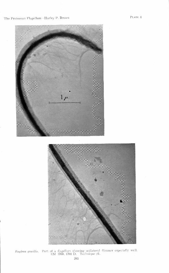
The Protozoan Flagellum—Harley P. Brown PLATE 4
Euglena gracilis Part of a flagellum showing unilateral flimmer especially well.EM 1700, 1701 D. Technique #1.
285

The Protozoan Flagellum—Harley P. Brown PLATE 5
Euglena gracilis. The flagellum extends from the body, loops, and returns to lie alongsidethe body. EM 1710, 1711 D. Technique #1.
287

The Protozoan Flagellum—Harley P. Brown PLATE 6
Euglena gracilis. In the upper section, the flagellum is seen to emerge from the body. Ithas been snapped by a rupture of the supporting film, and parts of the axoneme are exposed.The length of the flagellum is about 13 fi. EM 1697, 1698, 1699 D. Technique #1.
289

The Protozoan Flagellum—Harley P. Brown PLATE 7
Englena gracilis. Portion of the flagellum showing two very different appearances. Theupper part perhaps represents the more nearly natural condition. See discussion in text.Note the transversely striated appearance of the upper, denser portion, produced by the closelywrapped helical fiber. EM 1692, 1693, 1694 D. Technique #1.
291
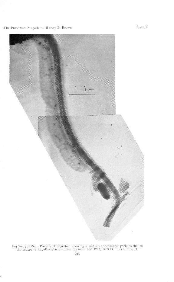
The Protozoan Flagellum—Harley P. Brown PLATE 8
Euglena gracilis. Portion of flagellum showing a swollen appearance, perhaps due tothe escape of flagellar plasm during drying. EM 1707, 1708 D. Technique #1.
293

The Protozoan Flagellum—Harley P. Brown PLATE 9
Euglena gracilis. Portion of flagellum showing unilateral flimmer. In places where theflagellum is twisted, the two equal fibers of the axoneme are made rather obvious. The helixof the sheath appears to be closely appressed to the axoneme (note regular bumps along sidesof flagellum. EM 1620, 1621 D. 'Technique #2.
295

The Protozoan Flagellum—Harley P. Brown PLATE 10
Chilomonas paramecium. Anterior end of body and the two flagella. No flimmerare to be observed. EM 1723, 1724, 1725 D. Technique #1.
297

The Protozoan Flagellum—Harley P. Brown PLATE 11
Ochromonas variabilis. Portion of body and flagellum showing flimmer on both (all ?)sides, not in a single lateral row as on the flagellum of Euglena. The dimensions of Ochromonasflagella vary with the size of the organism, but in those photographed they ranged for themost part between 4 and 6.5 in length, averaging about 0.1 in diameter. The flimmer seemto average about 0.5 in length and are less than 0.01 in diameter. Note apparent helix asin Plates 9 and 1 B. EM 1745 D. Technique #1.
29!)

The Protozoan Flagellum—Harley P. Brown PLATE 12
Ochromonas variabilis. The entire body of the organism is shown, with most of theflagellum. The small second flagellum may be seen near the base of the long flagellum. Noflimmer have been observed upon it. Apparent helix as in Plate 11. EM 1737, 1739 D.Technique #1.
301

THE EYE-BANK FOR SIGHT RESTORATION
More than half a hundred hospitals in nine states are already cooperatingwith The Eye-Bank for Sight Restoration, Inc., in a nation-wide effort to helprestore or remedy the vision of America's estimated 15,000 persons blinded becauseof comeal affections, it was announced recently by Mrs. Henry Breckenridge,executive director of the Eye Bank, 210 East 64th Street, New York.
In Greater New York alone, 32 hospitals are associated in the movement tomake available for distribution healthy corneal tissue for those whose sight maybe restored through corneal graft operations by which ocular opacity is overcome,the announcement stated.
In addition, 8 hospitals in other New York state cities, together with 6 inNew Jersey, 3 in Connecticut and one each in six other states have become activelyaffiliated with the movement.
Organized only last February, the Eye Bank was established for the collection,preservation and distribution of healthy corneas which may be obtained onlyfrom persons either living or immediately after death. Inasmuch as corneasmay be preserved and utilized for transplanting to the eyes of others for only72 hours, speedy collection and distribution is essential as soon as they are obtained.Whenever cooperating hospitals have eyes available, the Red Cross Motor Corpsrushes them to the Eye Bank for distribution to persons requiring the cornealgraft operation.
In addition to extending this activity to hospitals throughout the UnitedStates, the Eye Bank is presently engaged in a nation-wide effort to obtain supportfor its work through solicitations for memberships and donations of eyes after death.
303