Monteringsanvisning - LK Systems · 2015. 12. 9. · Monteringsanvisning | LK >BB

On the effects of structure andfunction on protein evolution
Kristoffer Illergård

c© Kristoffer Illergård, Stockholm 2010
ISBN 978-91-7155-980-7
Printed in Sweden by US-AB, Stockholm 2010
Distributor: Department of Biochemistry and Biophysics, Stockholm University

List of Papers
This thesis is based on the following papers:
I Kristoffer Illergård, David H Ardell, and Arne Elofsson (2009)Structure is three to ten times more conserved than sequence – astudy of structural response in protein cores.Proteins, 77(3):499-508
II Anni Kauko1, Kristoffer Illergård1 and Arne Elofsson (2008)Coils in the membrane core are conserved and functionallyimportant.J. Mol. Biol., 380(1):170-80
III Kristoffer Illergård, Anni Kauko, and Arne ElofssonPolar residues in the core of membrane proteins are conservedand directly involved in function.Submitted
IV Kristoffer Illergård, Simone Callegari and Arne ElofssonMPRAP: An accessibility predictor forα-helical transmembraneproteins.Submitted
Reprints were made with permission from the publishers.
Other publications:
Hedin LE1, Öjemalm K1, Bernsel A, Hennerdal A, Illergård K, Enquist K,Kauko A, Cristobal S, von Heijne G, Lerch-Bader M, Nilsson I, ElofssonA. Membrane Insertion of Marginally Hydrophobic Transmembrane HelicesDepends on Sequence Context.In press for J. Mol. Biol.
1 Authors contributed equally to the article.


Abstract
Many proteins can be described as working machines that make sure that ev-erything functions in the cell. Their specific molecular functions are largelydependent on their three-dimensional structures, which in turn are mainlypredetermined by their linear sequences of amino acid residues. Therefore,there is a relation between the sequence, structure and function of a protein, inwhich knowledge about the structure is crucial for undestanding the functions.The structure is generally difficult to determine experimentally, but should inprinciple be possible to predict from the sequence by computational methods.
The instructions of how to build the linear proteins sequences are copiedduring cell division and are passed on to successive generations. Although thecopying process is a very efficient and accurate system, it does not functioncorrectly on every occasion. Sometimes errors, or mutations can result fromthe process. These mutations gradually accumulate over time, so that the se-quences and thereby also the structures and functions of proteins evolve overtime.
This thesis is based on four papers concerning the relationship betweenfunction, structure and sequence and how it changes during the evolution ofproteins. Paper I shows that the structural change is linearly related to se-quence change and that structures are 3 to 10 times more conserved thansequences. In Paper II and Paper III we investigated non-helical structuresand polar residues, respectively, positioned in the nonpolar membrane coreenvironment ofα-helical membrane proteins. Both types were found to beevolutionary conserved and functionally important. Paper IV includes the de-velopment of a method to predict the residues inα-helical membrane proteinsthat after folding become exposed to the solvent environment.


Sammanfattning
Många proteiner kan beskrivas som arbetsmaskiner som ser till att alltfungerar i cellen. Deras specifika molekylära funktioner är till stor delberoende av deras tredimensionella strukturer, vilken i sin tur huvudsakligenär förutbestämd av deras linjära sekvenser av aminosyror. Därför finnsdet ett samband mellan sekvens, struktur och funktion hos ett protein, därkunskap om strukturen är avgörande för förståelse av funktionen. Strukturenär i allmänhet svår att bestämma experimentellt, men bör i princip varamöjlig att förutsäga från sekvensen med hjälp av beräkningsmetoder.Instruktionerna om hur man bygger de linjära proteinsekvenserna kopierasunder celldelningen och förs vidare till efterföljande generationer. Även omkopieringen är ett mycket effektivt och exakt system, fungerar det inte korrektvid varje tillfälle. Ibland kan fel eller mutationer resultera från processen.Dessa mutationer ackumuleras successivt med tiden, så att de sekvenser ochdärmed också deras strukturer och funktioner hos proteiner förändras medtiden.
Denna avhandling bygger på fyra artiklar om förhållandet mellan funktion,struktur och sekvens i proteiner och hur det förändras under evolutionensgång. Artikel I visar att den strukturella förändringen är linjär i relation tillsekvensförändring och att strukturer är 3 till 10 gånger mer bevarade jämförtmed sekvenser. I artikel II och artikel III undersöktes strukturer i icke-helixstruktur och polära aminosyror, respektive, placerade i den centrala opoläramembranmiljön iα-helix membranproteiner. Båda typerna upptäcktes varaevolutionärt bevarade och funktionellt viktiga. Artikel IV omfattar utvecklingav en metod för att förutsäga aminosyrorna iα-helikala membranproteinersom efter veckning förblir exponerade mot lösningsmedlet.


Contents
1 Introduction 1
2 The mapping between sequence and structure 52.1 Protein folding . . . . . . . . . . . . . . . . . . . . . . . . . 52.2 Structure determination. . . . . . . . . . . . . . . . . . . . . 62.3 Structure prediction. . . . . . . . . . . . . . . . . . . . . . . 7
3 Evolution of protein structures 93.1 Detecting homology . . . . . . . . . . . . . . . . . . . . . . 93.2 Evolutionary distance. . . . . . . . . . . . . . . . . . . . . . 103.3 Natural selection and genetic drift. . . . . . . . . . . . . . . 113.4 How do structure and function affect evolution?. . . . . . . . 123.5 How do structures respond to sequence changes?. . . . . . . 14
4 Membrane proteins - introduction 154.1 Biological membranes. . . . . . . . . . . . . . . . . . . . . 154.2 What do membrane proteins look like?. . . . . . . . . . . . . 164.3 Which functions do membrane proteins have?. . . . . . . . . 17
5 Membrane protein folding 195.1 Are membrane proteins inside-out of water-soluble proteins?. 195.2 Translocon-mediated folding. . . . . . . . . . . . . . . . . . 205.3 Membrane insertion of transmembrane segments. . . . . . . 215.4 Measuring free energy of insertion. . . . . . . . . . . . . . . 225.5 Assembly of transmembrane segments. . . . . . . . . . . . . 22
6 Membrane protein structure prediction 256.1 Predicting membrane protein topology. . . . . . . . . . . . . 256.2 Predicting solvent accessibility and residue contacts. . . . . . 276.3 Predicting full-atom structure. . . . . . . . . . . . . . . . . . 27
7 Membrane proteins with complex structure 297.1 Recent structures are more complex. . . . . . . . . . . . . . 297.2 Repositioning of transmembrane regions. . . . . . . . . . . . 30

CONTENTS 14
7.3 Extending topology models in structure prediction. . . . . . 32
8 Methodogy 338.1 Creation of dataset. . . . . . . . . . . . . . . . . . . . . . . 338.2 Evolutionary considerations. . . . . . . . . . . . . . . . . . 338.3 Assigning features from structure. . . . . . . . . . . . . . . 348.4 Predicting structural features. . . . . . . . . . . . . . . . . . 35
9 Summary of papers 379.1 Paper I. . . . . . . . . . . . . . . . . . . . . . . . . . . . . . 379.2 Paper II . . . . . . . . . . . . . . . . . . . . . . . . . . . . . 389.3 Paper III. . . . . . . . . . . . . . . . . . . . . . . . . . . . . 389.4 Paper IV. . . . . . . . . . . . . . . . . . . . . . . . . . . . . 399.5 Final thoughts. . . . . . . . . . . . . . . . . . . . . . . . . . 39
10 Acknowledgements 41
Bibliography 43

Chapter 1
Introduction
The basic unit of all known living organisms is the cell [1]. In each cell andat every moment a large number of biochemical reactions and processes aretaking place, which are responsible for keeping the cells alive. The main actorsto carry out these duties within the cell are proteins of different kinds.
Enzymes are proteins catalyzing chemical reactions. They are usuallyhighly specific and accelerate one or a few chemical reactions thousand-fold.Some proteins are involved in the process of cell signaling and signaltransduction. Some proteins, such as insulin, are sent to the outside of thecells to transmit a signal from the cell in which they were synthesized toother cells in distant tissues. Others are positioned in the membrane and actas receptors whose main function is to bind a signal molecule and to induce abiochemical response in the cell. Some proteins in the membrane transportions, metabolites and larger molecules such as proteins and RNA across theirmembranes. Antibodies are protein components of adaptive immune systemwhose main function is to bind foreign substances in the body, and targetthem for destruction. Transport proteins, like hemoglobin, bind usually smallbio-molecules and often transport them to other locations in the body of amulticellular organism [1]. To summarize, many proteins can be described asthe working machines that make sure that everything functions in the cell.
All instructions of how to build proteins and how to put together all partsinto livings organisms are stored in DNA. In this way genetic informationflows from DNA, via an intermediate level, RNA, to protein in a process thathas been called the central dogma of molecular biology, Figure1.1. In thisprocess the linear sequence of nucleotides in DNA is transcribed into RNA,which in turn is translated into a linear sequence of amino acid residues [1].The translated linear protein sequence folds, by a process that is not com-pletely understood, into shapes that are predetermined by their sequences andthe interaction with the solvent. In the folding, some parts become buried inthe protein interior while other remain exposed to the solvent environmentand locally contiguous segments form recurring secondary structure elementsin the form ofα-helices orβ -strands. The elements often pack into a com-

INTRODUCTION 2
Figure 1.1:DNA is replicated during cell division to successive generations. Parts ofthe linear sequence of nucleotides in DNA are transcribed into RNA, which in turn aretranslated into linear sequences of amino acid residues. The translated linear proteinsequences fold into structures that are mainly determined by their sequences. Thesestructures determine the functions of the proteins.
pact three-dimensional shape, tertiary structure, that determines its specificmolecular functions [2].
The instructions of how to build the proteins, which is stored in the DNA,are replicated (copied) during cell division so that these instructions are passedon to successive generations. Although DNA replication is a very efficient andaccuracte system, it does not function correctly on every occasion. Sometimeserrors, or mutations can occur in the process. These mutations gradually ac-cumulate over time, so that species that have diverged recently usually differby fewer mutations than those that went separate ways in the distant past, Fig-ure1.2. Moreover, the genetic changes in DNA are transferred via the flow ofinformation to protein sequences, and via protein folding to protein structuresand protein function. With knowledge about how these processes occur it istherefore possible to reconstruct the molecular history by comparing evolu-tionarily related proteins or genes [3].
Knowledge about the structure of proteins in the cell is important for char-acterizing molecular functions in e.g complex diseases. However, experimen-tal determination of protein structures is time consuming and represents a bot-tleneck in biology today. The difficulty of determining structures motivatesdevelopment of compatational methods to predict the structure (as well asfunction) from sequence. In addition, it motivates to seek an increased un-derstanding of the relationship between sequence, structure and function inproteins and also how this relation changes during evolution [5].
This thesis represent the main research results and conclusions from myPhD-studies. The main focus is the relationship between sequence, structureand function during evolution of proteins. It is based on four research articles.Paper I concerns how structures respond to evolutionary changes in proteinsequences and whether structures change slower than sequences or not. InPa-per II, Paper III andPaper IV we investigated one special type of proteins,

3
Figure 1.2:Mutations are introduced in DNA and gradually accumulate over time, sothat species that have diverged recently usually differ by fewer mutations than thosewhich went their separate ways in the distant past. The molecular history can be recon-structed by studying evolutionarily related proteins or genes. This figure show a sim-plied phylogenetic tree of how donkey, zebra, and two different kinds of horses haveevolved from a common ancestor. The phylogenetic information is extracted from datain [4].
calledα-helical membrane proteins. These proteins span the whole membraneand interact with the distinct environments both inside and outside the mem-brane. More specificallyPaper II andPaper III study non-helical structuresand polar residues, respectively, positioned in the nonpolar membrane coreenvironment, and their relation with structure and function. InPaper IV ananalysis of how the properties of protein regions that after folding becomeexposed to distinct environments differ is presented. In addition, it includes aseparate method for predicting from sequence which residues are exposed tothe solvent environment.
The first two chapters of this thesis describe some of the theoretical back-ground on how proteins fold into structures and how proteins evolve. Theseare necessary for understanding all four papers in general, butPaper I inparticular. Thereafter, four chapters are devoted to describe the relation be-tween structure, sequence and environment in the evolution of membrane pro-teins, constituting essential background knowledge forPaper II, Paper IIIandPaper IV. The following chapters contain a short overview of the generalmethodology, a brief summary of results and a discussion of my contributionsto the research field.


Chapter 2
The mapping between sequenceand structure
For some decades, much effort has been directed to predict protein structurefrom sequence by computational methods. This should in principle be possi-ble, since the three-dimensional structures of proteins are mainly determinedby their linear sequences of amino acid residues [6]. A strong motivationfor development of compational methods devoted for this purpose is that thestructure of a known sequence is functionally informative but difficult to de-termine experimentally. Although, there has been a lot of progress, the proteinstructure prediction problem is far from solved.
This chapter describes the process by which sequences fold to structures,how structures are experimentally determined and how they can be predictedfrom sequence.
2.1 Protein folding
In a series of conformational changes a fully extended and unfolded water-soluble protein often folds by lowering its free energy into a single, compact,biologically active fold. The final native state is most often believed to be theone with the lowest free energy. Its thermodynamic stability is, however, usu-ally small and depends on the differences in entropy and enthalpy betweenthe native state and the unfolded state [2]. For a protein molecule to adoptall possible conformations one at a time in a random fashion would take as-tronomically long time. However, measurements have shown that in reality aprotein typcially folds to the native structure in less than a second. This meansthat the folding must be directed in some way through a kinetic pathway ofunstable intermediates to escape sampling a large number of irrelevant con-formations [2].
Early in the folding process of water-soluble proteins hydrophobic residuestend to be buried in the interior so that hydrophobic side chains are brought out

THE MAPPING BETWEEN SEQUENCE AND STRUCTURE 6
Figure 2.1: Protein sequences fold into shapes that are predetermined by their se-quences and the interaction with the solvent. This means that there is a “mapping”between sequence and structure.
of contact with water and into contact with each other. This hydrophobic ef-fect, which minimizes unfavorable interactions between hydrophobic residuesand water, is a strong driving force of folding of this type of protein. Theburying of hydrophobic residues greatly restricts the number of possible con-formations the molecule can assume and allows proteins to fold in secondsrathers than years [2].
Some native structures are completely made up ofα-helices or ofβ -sheets,whereas others consist of a mix of both types. Many of these secondary struc-ture elements are often formed early on in the folding process of water-solubleproteins. This formation of secondary structure early in the folding processcan be regarded as a consequence of burying hydrophobic sidechains and notas the driving force for the formation of a folding intermediate [2]. Finally, thenative interactions are formed throughout the protein, including packing of theinterior as well as the fixation of surface loops. How the folding of membraneproteins differs is discussed in later chapters.
2.2 Structure determination
Determination of the structure is crucial for detailed functional mapping ofa protein, but is experimentally difficult. Two common techniques are X-raycrystallography and nuclear magnetic resonance (NMR).
X-ray crystallography is a method of determining the arrangement of atomswithin a crystal. For proteins it usually requires many different experimentsto obtain well diffracting crystals. In the method, the interaction of X-rayswith electrons in the crystal is used to obtain an electron-density map of themolecule, which can be interpreted in terms of an atomic model. The quality of

7
the derived electron-density map depends on the resolution of the diffractiondata, which in turn depends on how well-ordered the crystals are [2].
In NMR, the magnetic-spin properties of atomic nuclei within a moleculeare used to obtain a list of distance constraints between these atoms,from which a three-dimensional structure of the protein can be obtained.The method does not require protein crystals and can be used on proteinmolecules in concentrated solutions. Historically it has, however, beenrestricted in its use to small proteins [2].
Experimentally determined protein structures are deposited in Protein Data-Bank [7] as coordinate files. These files represent models that best explainexperimental data. As with all models, these structures have uncertainty asso-ciated with them owing in part to variations in experimental conditions. Anunavoidable fact is that crystallized proteins represent only a snapshot of trulyflexible structures. Two identical protein sequences crystallized under differ-ent conditions may e.g. show substantial structural differences [8]. This couldbe the case if they e.g. are caught in different functional states, such as pre-sense and absence of bound ligands, or open or closed conformation of an ionchannel.
2.3 Structure prediction
The three-dimensional structure of a protein is guided by two distinct sets ofprinciples operating at vastly different time scales: the laws of physics and thetheory of evolution [9]. From a physical point of view the protein molecule isa consequence of a variety of electrostatic interactions that under native con-ditions usually fold a protein into a stable well-defined structure in less thana second by minimizing its free energy. From an evolutionary point of viewthe protein molecule can be seen as a result of gradual changes in sequenceand structure over millions of years. These two view points give rise to twodifferent approaches to the protein structure prediction problem [10]: de novoand comparative modeling.
De novomethods predict the structure from the sequence alone, withoutrelying on similarity between the modeled sequence and any known struc-ture. These methods assume that the native structure corresponds to the globalfree energy minimum of the protein and attempts to find this minimum byan exploration of many possible protein conformations. Two key steps indenovomethods are the procedure for efficiently carrying out the conformationalsearch and the free energy function used for evaluating possible conforma-tions. Success in water-soluble protein structure prediction has so far beenrestricted to rather small proteins.
The aim of all comparative modeling methods is to build a model for a targetprotein of unknown structure using similarity to a template protein of knownstructure. In principle it consists of four steps: i) finding a template of known

THE MAPPING BETWEEN SEQUENCE AND STRUCTURE 8
structure to the target sequence to be modelled, ii) aligning the sequence tothe template, iii) building a model and iv) assessing the model. The qualityof the model is largely dependent on the similarity in sequence between thetarget and the template, and therefore also on how much information can betransferred from template to target in the model building process [9].
Despite recent progress in thede novoprediction field, the comparativemodeling approach is by far more accurate thande novopredictions. Since thequality of the structures determines the information that can be extracted fromthem, comparative modeling is therefore always the recommended choicewhen a close homolog of known structure is known [9].

Chapter 3
Evolution of protein structures
The genetic changes in DNA are transferred via the flow of information toprotein sequences, and via protein folding to protein structures and proteinfunction. With knowledge about the process, by which evolutionary changesoccur, it is possible to reconstruct the molecular history by comparing evolu-tionarily related proteins or genes [3].
This chapter describes i) the processes of detection of homology betweenproteins, ii) measurement of evolutionary distances between them, iii) howstructure and function affect evolution and iv) finally estimation of how struc-tures respond ot evolutionary changes in sequence.
3.1 Detecting homology
Since proteins gradually change, two proteins can be evolutionarily relatedand have a common ancestor, Figure3.1. Per definition, two proteins are saidto be homologous if they share a common ancestor. The concept of homol-ogy is central in molecular biology and is often inferred between proteins ifthe similarity in sequence or structure is high. Often, the publically availablerepositories of known protein sequences and structures are scanned in auto-matic ways to detect homologous proteins [5].
In the protein homology detection process it is important to create align-ments, which are sets of protein sequences that specifies the correspondenceamong the residues of those sequences. An evolutionary alignment is needede.g. to be able to reconstruct ancient evolutionary changes. In this kind ofalignment every pair of characters at a site (meaning within a column of thealignment) are to be interpreted as having evolved from an ancestral residue inan ancestral sequence. Regions of either sequence left unpaired in this align-ment are interpresented as having been inserted or deleted since the divergencefrom their common ancestor [3].

EVOLUTION OF PROTEIN STRUCTURES 10
Figure 3.1:Divergence of two proteins from a common ancestor. Changes in proteinsequences are transferred to protein structures and protein function. Some changes insequence result in major changes in structure and function (a), whereas other changesonly result in minor changes in structure and function (b).
For proteins a distinction can be made between sequence alignments, whichare derived from only protein sequences, and structural alignments, which arederived from protein structures.
A pairwise sequence alignment method often optimize the evolutionaryalignment by optimizing over a molde of possible evolutionary histories oftwo sequences assumed to have diverged from a common ancestor. The align-ment optimization is calculated with the assumption that different sites evolveindependently of one another, and that the sequences have a certain commonand specific composition and pattern of substitution [5].
Protein structural alignment methods assign a correspondence among thestructurally similar parts in the structural models of two or more protein struc-tures. In general, they optimize a measure of structural similarity of parts,rather than any evolutionary quantity. One structural similarity measure com-monly included in the optimization score is root mean square deviation ofthe atomic coordinates of the superposable regions. The structural alignmentmight be represented in the same way as a sequence alignment, i.e. with anumber of residue correspondences [11].
3.2 Evolutionary distance
When an alignment between two homologous proteins has been constructedit is often of interest to determine how much change that has occurred be-tween these two sequences since divergence. The simplest measure of thedistance between two sequences is to count the number of sites in the align-ment at which the two sequences differ. Often, the fraction of identical or

11
Figure 3.2:The observed number or fraction of non-identical sites in a pairwise align-ment is easy to calculate. However, since multiple substitutions can occur at thesame site this measure underestimates the actual number of substitutions that havetaken place since divergence from the common ancestor. The figure is partly adaptedfrom [3].
non-identical sites is used. Although, this kind of measure is easy to calculateit is a poor measure of the actual number of evolutionary changes. The rea-son is that multiple substitutions can occur at the same site, which results inunderestimation of the actual number of substitutions that have taken place,Figure3.2. The sequence identity measure saturates as more substitutions areaccumulated and severely undestimates the sequence divergence at larger evo-lutionary distances [3].
There has been a considerable amount of research on developing methodsto estimate the actual number of evolutionary changes in DNA sequences.Most methods are based on four assumptions, which in many cases mightbe unrealistic: (i) all sites change independently, (ii) the substitution rate isconstant over time and in different lineages, (iii) the sequence compositions isat equlibrium, (iv) the conditional probabilities of substitutions are the samefor all sites and do not change over time. In addition, some methods modela variation in substitution rate among sites, most often by discretizationing agamma distribution [3].
There are relatively few methods for computing distances for protein se-quences, which is probably largely due to the greater complexity of the prob-lem; instead of four states required for DNA data, protein sequences comprisesome 20 different amino acids.
3.3 Natural selection and genetic drift
A central task of molecular evolution is to determine the processes by whichgene sequences change over time. Today we know that the specific molecularfunctions associated with proteins are the result of randomized processes suchas mutation and genetic drift and organizing processes like natural selection.

EVOLUTION OF PROTEIN STRUCTURES 12
Natural selection is the process by which heritable traits that make it morelikely for an organism to survive and successfully reproduce become morecommon in a population over successive generations. Mutations that are notsubject to natural selection are called neutral and are lost (usually) or fixed(very occasionally) by the process of genetic drift. Non-neutral mutations canbe of two types; advantageous for the organism, which are subject of positiveselection, and disadvantageous, which are subject of negative selection [3].
Reconstructions of ancient mutations that have survived evolution and be-come fixed in populations have shown that most common types of changeswithin proteins are short (non-frame shifting) insertions or deletions and re-placements of single amino acid residues that conserve the hydrophobicity [5].Among these mutations, the majority can be believed to have been neutral ornearly neutral, whereas most of the deleterious ones have been selectively re-moved [3].
Although most evolutionary changes in proteins are believed to occur bysmall changes that are neutral for structure and function there is evidence thatother types of changes also occur. It has e.g. been shown that a few non-neutralchanges in protein sequences can cause dramatic changes to protein structuresand functions with large evolutionary consequences [19, 20]. However, it isnot known how common and important such events are.
3.4 How do structure and function affect evolution?
In proteins, some sites evolve faster than others. From multiple sequencealignments the types and rates of substitutions can be reconstructed. Earlierstudies have claimed that there is an inverse correlation between the rate ofsubstitution and the functional constraint acting on a site [3]. Indeed, it iswell-estalished that the slowest evolving sites are most often directly function-ally important and might be responsible for e.g. binding of ligands or catalysis.
If the structure of one of the proteins in an alignment is known it is possi-ble to “map” the features from the structure to the alignment (or vice versa).By this can kind of mapping it is e.g. possible to investigate whether localstructures of distinct secondary structure or surface accessibility classes ex-hibit different amino acid substitution patterns and rates of substitution, Fig-ure 3.3. The most evident such trend for water-soluble proteins seems to bethat residues at exposed sites are often hydrophilic and replaced at a fast raterthan the often hydrophobic and buried sites are. Similarly, residues at posi-tions of non-regular secondary structures (loops and coils) are replaced at afaster rate than regular secondary structure (α-helices andβ -sheets) [12, 13].
Similarly, if the function of one of the proteins in an alignment is knownit is possible to “map” the functional features with “evolutionary features”.Functional annotations of specific residues in well-characterized proteins are,however, most often restricted to just a few sites that are responsible for e.g

13
Figure 3.3:If the structure and function of one of the proteins in an alignment is knownit is possible to “map” the features from the structure and function to the alignment(or vice versa). Structural and functional features could be e.g. secondary structureand ligand binding sites. Features derived from a multiple alignment could be e.g.type and rate of substitution. Both type of features can be done at a residue level.
binding of ligands, while the majority of sites lack information. One observa-tion of the functionally important sites is, as previously mentioned, that theyoften are evolutionary conserved. Another observation, is that they often arefound in rare, unfavorable or unusual energetic environments, such as left-handed helices [14], ionizable groups with perturbed titration curves [15], andpolar residues engaged in unfavorable electrostatic interactions in enzyme ac-tive sites [16]. Therefore, it has been suggested that residues involved in func-tion often contribute unfavorably to the stability of the native state and thatevolution often optimizes functional properties at the expense of thermody-namic stability [17, 18]. However, the generality of the rule is not known.
Another application of mapping the structure or function of one protein toan alignment than correlating the structural and functional features with sub-stitution patterns is to infer them to residues of unknown or less-characterizedstructure and function. This is done by assuming that the structural or func-tional features are similar in regions that correspond to each other in the align-ment (i.e. being in the same column in the alignment). This type of inferenceby homology is commonly used in structure and function prediction and is ingeneral more accurate the more similar the local protein sequence regions are.However, the local structure and function might occasionally differ drasticallyeven at conserved sites.
EVOLUTION OF PROTEIN STRUCTURES 14
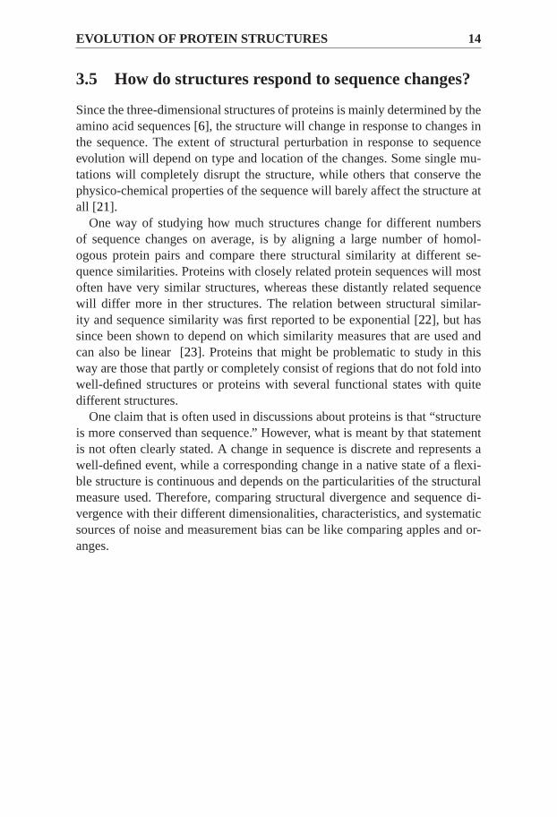
3.5 How do structures respond to sequence changes?
Since the three-dimensional structures of proteins is mainly determined by theamino acid sequences [6], the structure will change in response to changes inthe sequence. The extent of structural perturbation in response to sequenceevolution will depend on type and location of the changes. Some single mu-tations will completely disrupt the structure, while others that conserve thephysico-chemical properties of the sequence will barely affect the structure atall [21].
One way of studying how much structures change for different numbersof sequence changes on average, is by aligning a large number of homol-ogous protein pairs and compare there structural similarity at different se-quence similarities. Proteins with closely related protein sequences will mostoften have very similar structures, whereas these distantly related sequencewill differ more in ther structures. The relation between structural similar-ity and sequence similarity was first reported to be exponential [22], but hassince been shown to depend on which similarity measures that are used andcan also be linear [23]. Proteins that might be problematic to study in thisway are those that partly or completely consist of regions that do not fold intowell-defined structures or proteins with several functional states with quitedifferent structures.
One claim that is often used in discussions about proteins is that “structureis more conserved than sequence.” However, what is meant by that statementis not often clearly stated. A change in sequence is discrete and represents awell-defined event, while a corresponding change in a native state of a flexi-ble structure is continuous and depends on the particularities of the structuralmeasure used. Therefore, comparing structural divergence and sequence di-vergence with their different dimensionalities, characteristics, and systematicsources of noise and measurement bias can be like comparing apples and or-anges.

Chapter 4
Membrane proteins -introduction
There are two main types of solvent environments in which proteins performtheir functions. The first environment is hydrophilic, with water as the mostimportant component and the second is hydrophobic, with lipids as the maincomponent. The proteins in these environments are in the thesis called water-soluble proteins and membrane proteins, respectively.
This chapter describes what a membrane is and what the main types ofstructures and functions of membrane proteins are. It serves as an introductionto membrane proteins.
4.1 Biological membranes
A biological membrane functions as a barrier between living cells and theirenvironment, and to compartmentalize intracellular organelles within eukary-otes. Therefore, it needs to be both mechanistically strong and flexible. It alsoneeds to be impermeable to compounds that are unwanted in the cell and tohave mechanisms for passage of desired compounds into the cell [24].
The fundamental structural component of biological membranes is lipid.This lipid molecule often consists of a hydrophilic headgroup, and one ormore hydrophobic fatty acid hydrocarbon tails. As a consequence of theiramphiphilic nature, lipids aggregate and spontaneously self-organize inwater, generally into either micelles or bilayers. The biological membraneis a bilayer, where the hydrophobic fatty acid chains point away from thehydrophilic water molecules, arranged into two oppositely oriented fluidicleaflets [24], Figure4.1.
The lipid composition varies between membranes and determines thethickness, fluidity and curvature of the bilayer. The actual distribution ofthe functional groups across a 1,2-Dioleoyl-sn-Glycero-3-phosphocholine(DOPC) lipid bilayer have been measured experimentally [25], and simulated

MEMBRANE PROTEINS - INTRODUCTION 16
Figure 4.1: Left) Structure of 1,2-Dioleoyl-sn-Glycero-3-phosphocholine (DOPC)lipid bilayer after 1.5 ns molecular dynamics simulation. The figure is from [27] andadapted from [26]. Right) The distribution of the functional groups across aDOPClipid bilayer, as determined by X-ray and neutron diffraction measurements. The colorcoding in left and right are similar. The figure is from [27] and adapted from [25].
by molecular dynamics [26], Figure 4.1. The emerging picture is a highlyheterogeneous environment, where the chemical environment across themembrane varies markedly over short distances.
4.2 What do membrane proteins look like?
In biological membranes, proteins are interspersed among the lipids and asmuch as half of the mass fraction might consist of proteins embedded withinthe lipids [24]. Broadly, membrane proteins can be classified as peripheralor integral, Figure4.2 a. Peripheral membrane proteins are only loosely at-tached to the membrane through electrostatic or van der Waals interactionswith the lipid headgroups or other membrane proteins, or through a cova-lently attached anchor [24]. Integral membrane proteins most often span thewhole membrane, and can be divided into two type of structures; i) closed bar-rels of amphipathicβ -strands and ii) as bundles of tightly packedα-helices,Figure4.2b.
β -sheets are used in theβ -barrel type of transmembrane proteins. Residuesin the β -sheets alternatively point outwards, facing the lipids, and inwards,facing the inside of the barrel, resulting in a sequence pattern in which theresidues are typically alternatively polar and hydrophobic. The outcome is apolar channel through which water-soluble molecules can cross. It has beenestimated that around 2–3 % of the genes in bacteria encodesβ -barrel mem-brane proteins [28] and that they can be found in the outer membrane of mitho-chondria and cloroplasts.

17
Figure 4.2:Different types of membrane proteins. a) Membrane proteinscan be di-vided into peripherial and integral. b) Integral membrane proteins can be divided intoα-helical bundle andβ -barrel types.
Theα-helical bundle type of membrane proteins is the most abundant andwell-studied. Theα-helices are in general longer than theβ -strands ofβ -barrel proteins and are easier to predict from sequence. A recent study estimatethat, in humans, this type of membrane protein constitute around 27 % of thegenes that codes for membrane proteins [29].
Hereafter, the term “membrane protein” will refer toα- helical membraneproteins.
4.3 Which functions do membrane proteins have?
One of the most effective way to increase the knowledge of specific functionsof proteins is to determine their structure. In the case of membrane proteins,it is experimentally particularly difficult and have led to a 100-fold underrep-resentation in structural databanks compared to non-membrane proteins [30].Although, the number of experimentally determined structures grows expo-nentially, the number of available sequences grows faster. Current estimatespredicts that it will take at least three decades before 80 % of the membranesuperfamilies will have at least one determined structure, which can be usedas template for comparative modeling [30]. This means that there is a hugeneed of structural information about membrane proteins at the moment andprobably for a long time in the future.
Membranes contain a substantial amount of proteins with diverse functions,Figure 4.3. They transport ions, metabolities and larger molecules such asproteins and RNA across their membranes. Membrane proteins are also re-sponsible for sending and receiving chemical signals, to propagate electricalimpulses, attach to neighbouring cells, anchor other proteins to specific lo-cations in the cell. Other functions include regulating intracellular vesiculartransport, controlling membrane lipid composition, organizing and maintain-ing the shape of organelles and the cell itself [24].

MEMBRANE PROTEINS - INTRODUCTION 18
Figure 4.3:The functions of membrane proteins are diverse. The proteins are e.g. in-volved in energy transport during photosynthesis or respiration, transport of moleculesacoss their membranes, transmission of chemical signals orcatalyzation of chemi-cal reactions. Their important functions make them highly interesting from a medicalpoint of view.
Due to their important functions, membrane proteins are highly interestingfrom a medical point of view. Cystic fibrosis is caused by misfolding muta-tions in a chloride transporter protein in the lung. Neurological and cardiacdiseases are caused by nonfunctional photoreceptors (rhodopsins) in the eye.Water-channel proteins (aquaporins) are critical for kidney function but alsoinvolved in stroke. Finally, mutations in membrane proteins involved in pro-tein import into organelles such as peroxisomes and mitochondria underliemany serious (but fortunately rare) heritable disorders [31]. Due to this medi-cal relevance, many membrane proteins are prime drug targets, and it has beenestimated that more than half of all drugs currently on the market are directedagainst membrane proteins.

Chapter 5
Membrane protein folding
The solvent environments and the folding process of membrane proteins isdifferent from water-soluble proteins.
The following chapter describes how linear amino acid sequences ofmembrane protein fold into three-dimensional structures. The process canbe described according to a two-stage model with initial formation oftransmembrane-helices followed by assembly of transmembrane regions intotertiary structure.
5.1 Are membrane proteins inside-out of water-solubleproteins?
In the folding of a protein some residues will remain exposed to the environ-ment while others will become buried in the protein interior. For water-solubleproteins it is energetically favorable to bury hydrophobic residues and exposepolar and charged residues to the surrounding water. However, transmembraneproteins face three distinct environments; a hydrophobic lipid environment in-side the membrane, a hydrophilic water environment outside the membraneand an interface region rich in phospholipid headgroups. Because of the en-ergetic conditions resulting from these distinct environments the accessiblesurfaces of transmembrane proteins need to expose different types of residuesat different locations.
As previously explained, the dominant driving force behind folding ofwater-soluble proteins is the hydrophobic effect, that minimizes unfavorableinteractions between hydrophobic residues and water [32]. Therefore, watersoluble proteins generally have a hydrophobic interior and hydrophilicexterior. In contrast, the drive to bury polar residues from the solventenvironment within the membrane is much weaker. Early studies of thebacteriorhodopsin structure suggest that membrane proteins are “inside-out”, i.e. that they consist of a hydrophilic interior and a hydrophobic exterior,Figure5.1. However, later studies indicated that the “inside-out” rule is not

MEMBRANE PROTEIN FOLDING 20
Figure 5.1:Water-soluble proteins (a) generally have a hydrophobic (orange) interiorand hydrophilic (green) exterior. It has been suggested that membrane proteins (b)are inside-out of water-soluble proteins, with a hydrophilic interior and hydrophobicexterior. However, such a (inside-out) rule is generally not applicable to all membraneproteins.
generally applicable to allα-helical membrane proteins [33, 34, 35]. Sincemembrane proteins are exposed to distinctly different chemical surroundingacross the membrane that varies markedly over short distances, they willhave to adapt their surface to the different environments. Therefore, the maindriving forces of folding and stabilization are different and less understoodfor membrane proteins. However, it is likely that stably folded proteins inmembrane regions also reside in a free energy minimum [35].
5.2 Translocon-mediated folding
Most α-helical transmembrane proteins are targeted by an N-terminal signalsequence to the endoplasmatic reticula membrane. For such proteins, the ri-bosome, which is the unit that synthesizes the polypeptide sequence, bindsto a channel complex known as the Sec translocon during translation. There-after, translocation is resumed and the nascent molecule is threaded directlythrough the channel as it is being synthesized [36]. In bacteria, the mecha-nism for insertion of proteins into the plasma membrane is similar, whereasinsertion of proteins into the bacterial periplasm or the outer membranes ofmitochondria differs. Since protein translation and translocation of membraneinsertion occur simulataneously the process is termed co-translational [24].
The translocon apparatus can be seen as a switching station [36] that re-ceives elongating peptide sequences from the ribosome, and directs them ei-ther across the membrane or into the membrane bilayer, depending upon thesegment’s properties, Figure5.2. Whether the molecule is folded or unfolded

21
Figure 5.2:The translocon apparatus can be seen as a switching station that receiveselongating peptide sequences from the ribosome, and directs them either across themembrane or into the membrane bilayer, depending upon the segment’s properties.The figure is from [27] with ideas influenced by [37].
upon exit from the ribosome tunnel and the molecule is entering the transloconchannel is not known [36].
5.3 Membrane insertion of transmembrane segments
Somehow the ribosome-translocon complex manages to recognize the incipi-ent transmembrane helices in the nascent polypeptide and allows them to in-sert into the lipid bilayer rather than being fully translocated across the mem-brane into the lumen of the endoplasmatic reticulum. Most probably the trans-membrane segments leave the channel through a lateral gate in the channelwall that opens sideways toward the bilayer. In a high-resolution structure ofa translocon one can see a possible opening from the interior into the lipid bi-layer, which is hypothesized to control the passage of nascent transmembranehelices into the bilayer from the interior of translocon [36].
It has been assumed that transmembrane helices move into the lipid mem-brane from the protein-conducting channel by a partitioning process, wheresufficiently hydrophobic helices prefer the bilayer, whereas more polar he-lices favor the translocon. Therefore, the process would appear as an equlib-rium process in which favourable interactions between lipids and amino acidside chains promote membrane integration rather than translocation [36]. Theefficiency with which a transmembrane segment is integrated by the translo-con should then be sensitive to the relative positions of amino acids withinthe segment. This trend is evident when looking at amino acid distributions inknown membrane protein structures.

MEMBRANE PROTEIN FOLDING 22
Figure 5.3:An experimental model to determine insertion efficiency of designed pu-tative transmembrane segments in the membrane [38]. With the model, the require-ments in terms of amino acid composition for a sequence segment to be recognizedas a transmembrane segment and inserted into the membrane, as opposed to beingtranslocated across can be investigated. The figure is adapted from [38].
5.4 Measuring free energy of insertion
Recently, von Heijne and co-workers have made progress in describing the re-quirements in terms of amino acid composition for a sequence segment to berecognized as a transmembrane segment and inserted into the membrane, asopposed to being translocated across [38]. The experimental data comes frominsertion efficiency measurements of designed putative transmembrane seg-ments in a model membrane protein, Figure5.3. A large number of designedand natural test segments were systematically introduced in the experimentalsetup. From the measured relative amounts of inserted and translocated formsof the proteins, an appearant free energy of membrane insertion of the seg-ment,∆ Gapp, was calculated. A model for predicting the membrane insertionefficiency of transmembrane segments was derived, which takes amino acidposition, length of the segment and hydrophobic moment into account [38].
The experiments showed that strongly polar residues give highly position-specific contributions to membrane insertion, and are much better toleratedtowards the water-lipid interface regions than inside the hydrophobic core.Hydrophobic residues give favorable contributions and are less position spe-cific [38].
5.5 Assembly of transmembrane segments
The selection of transmembrane segments is only the first step in the complexprocess of gathering the transmembrane segments together to form the nativeprotein structure. After translocon-guided insertion, the transmembrane seg-ments of multispanning membrane proteins must condense to form the finalfolded three-dimensional structure.

23
For single-spanning proteins it seems that a “threshold hydrophobicity”can be defined with the experimentally derived∆ Gapp-scale that determineswhether or not a given polypeptide segment will form a transmembrane seg-ment. However, this kind of approach does not appear to work for multi-spanning membrane proteins. Actually, a surprisingly large fraction (25 %)of the transmembrane helices in the multi-spanning proteins of known three-dimensional structure have∆ Gapp > 0 kcal/mol, which suggest that they arenot sufficiently hydrophobic to insert efficiently as single transmembrane seg-ments [39].
The relatively large fraction of transmembrane segments that cannot insertefficiently into the membrane in the absence of other parts of the protein sug-gests that some transmembrane segments in multi-spanning proteins may de-pend on other parts of the same protein for efficient insertion. Possibly pairsor higher assemblages of transmembrane helices interact among themselvesbefore partioning into the bilayer [36].


Chapter 6
Membrane protein structureprediction
The need of computational methods for predicting structures from sequence iseven more pronounced for membrane proteins than for water-soluble proteins[24, 31, 40]. As was decribed in the previous chapter, membrane proteinscan conceptually be decomposed into two consequtive steps: folding of theindividual hydrophobic segments into transmembrane segments, often corre-sponding to helices, followed by association of the different transmembranesegments. In this chapter it will be described that structure prediction ofα-helical membrane proteins can, similarly, be broken down into two steps:(i) determining the topology of the proteins, which includes delineating theboundaries of the transmembrane segments and (ii) predicting the tertiary con-formation of the protein, which includes determining how the transmembranesegments pack together [41].
6.1 Predicting membrane protein topology
The topology of membrane proteins refers to a description of the way in whicha polypeptide chain weaves back and forth across the membrane [40]. Thesimplest description is a list of the different segments of the polypeptide chainthat form the transmembrane helices and their orientation relative to the mem-brane. The concept of topology is widely used in membrane protein structureprediction. Its determination can be a crucial preliminary step to modeling thestructure as it constrains the way individual transmembrane segments couldassociate within the membrane [40].
The first predictors forα-helical transmembrane proteins calculated seg-mental hydrophobicity averaged over 10 to 20 residues along the sequence.They used the fact that transmembrane helices are on average more hydropho-bic than the connecting loop regions. First, only the so called topography waspredicted, meaning that no attempt was made to predict the orientation of the

MEMBRANE PROTEIN STRUCTURE PREDICTION 26
Figure 6.1: The membrane protein Bacteriorhodposin represented as a topology (left)and as a full atom structure (right). Structure prediction of α-helical membrane pro-teins can be broken down into two steps: (i) determining the topology of the proteins,which includes delineating the boundaries of the transmembrane segments and (ii)predicting the tertiary conformation of the protein, whichincludes determining howthe transmembrane segments pack together.
protein. The topographic signal based on the segmental hydrophobicity seemsto be a rather symmetric property and depends mainly on the distances fromthe membrane center [40, 41].
With the observation that the loops connecting the transmembrane helicesdiffer in amino acid composition, depending on whether they face the insideor the outside of the cell, it became possible to extend predictions to includethe orientation of the proteins [42]. This, so called positive inside rule [43], isstill widely used although it is still unclear whether it is the result of propertiesof the translocon or the cytoplasmic membrane [41].
Later significant improvements of topology predictions is to include infor-mation from evolutionarily related sequences and the use of machine learningtechniques, such as Hidden Markov Models, to extract the relevant sequencefeatures [40, 41]. Today the best topology predictors can assign the correctnumber of transmembrane regions, their approximate locations and the cor-rect side of the termini with an accuracy of 70–90 % [40]. Some problemsthat remain difficult is how to predict the exact location of transmembrane re-gions, to distinguish between signal peptides and transmembrane regions, toidentify marginally hydrophobic transmembrane segments and to predict thetopology of more complex structures.

27
6.2 Predicting solvent accessibility and residuecontacts
Most efforts in structure prediction ofα-helical membrane proteins have beenfocused at improving the predictions of the topology [40]. Although, a pre-dicted topology might be a useful initial step, it does not give any informationof how different segments in the membrane are packed together into bundlesin the tertiary structure. Two common steps in structure prediction of water-soluble proteins to aid prediction of the packing of different segments are pre-diction of solvent accessibility and residue contacts. These structural featuresare in the case of membrane proteins complementary to the topology.
A solvent accessibility predictor assigns which residues are exposed to thesolvent environment in the native structure, and which are buried in the proteininterior. Residues responsible for catalysis or substrate binding, positionedwithin the membrane region are often partly buried in the protein interior,while temporary protein-protein-interactions naturally occur on solvent ex-posed sites. Therefore, a predictor of solvent accessibility is interesting forpredicting functional relevance of individual residues. The attempts, hitherto,made to predict accessibility of membrane proteins [44] have been developedto predict the exposure within the membrane environment and, thus, requirean initial prediction step for determining if a residue is located within themembrane or not. Such an initial prediction step is coupled with errors at theresidue level, since the exact start and stop points of the transmembrane seg-ment is hard to assign correctly.
A contact predictor assigns which residue pairs are in close contact in thestructure. One common way to identify contacts is to look for sites that un-dergo similar or compensating substitutions. Such sites have been shown tobe close in structure more than randomly picked pairs of sites. Since net-works of correlated mutations have been found to appear near binding re-gions and active sites [45], identification of these could potentially be used toidentify functionally important regions. Predicting contacts in water-solubleproteins has successfully been utilized to constrain structure prediction andto identify incorrect models generated by structure prediction methods. How-ever, for membrane proteins this type of preditions have received little atten-tion [46]. Recently, there have been a few attempts with varying success topredict contacts in membrane proteins with the aim of identifying interactingα-helices [46].
6.3 Predicting full-atom structure
Predicted structural features like topology, surface accessibility and residuecontacts might be useful, but, the goal of structure prediction is to get a full-atom model. Structure prediction can, as previously described, be divided into

MEMBRANE PROTEIN STRUCTURE PREDICTION 28
de novoprediction and comparative modeling. In the case of membrane pro-teins, both methods are quite difficult, which might explain the effort to de-velop methods for predicting structural information of low resulution, liketopology.
In principle de novoprediction of membrane proteins could be done withthe same methods and protocols as for water-soluble proteins. However, thereare two main differences in which the predictions of membrane proteins dif-fer from water-soluble proteins; i) the solvent environment is different andmore complex and ii) many membrane proteins are large [40, 41]. As an ex-ample of the latter, G-protein coupled receptors contains 7-transmembraneregions and several hundred residues per chain. Another example is the pho-tosynthetic reaction center which consist of many chains that constitute a verylarge complex. Historically, succesfulde novopredictions has been limitedto rather small proteins. Together, these difficulties with predictions of mem-brane proteins have effectively restricted the development so far. However,recently Rosetta, which has been successful for water-soluble proteins by us-ing a fragment-based assembly algorithm, was adopted for transmembraneprotein structures [47]. Another, interesting attempt is FILM [48], whichsimilarly might be seen as a adaption for membrane proteins of a previoussuccesful method.
The most obvious problem for comparative modeling of membrane proteinsis the lack of templates with known structures. Many methods have hithertoused quite similar protocols as for prediction of water-soluble proteins [49].An interesting attempt to model the structures of most of the 900 human G-protein couple receptors was recently performed by Skolnick and coworkersusing the TASSER algorithm [50], resulting in quite good accuracy of thepredicted rhodopsin structure. However, as for other methods the correctnessof the structures can not be verified until more structures are available [40, 41].
Topology, surface accessibility, residue contacts and other structural fea-tures can be interpreted as low-resolution data. Predictions of such featurescan be used as constraints for “complete” structure prediction of all atoms.Alternatively, low resolution data can be derived experimentally from e.g. site-specific mutagenesis, chemical crosslinking, NMR, masspectrometry, glyco-sylation or binding affinities, protein-protein interactions. Low-resolution datacan also be used to evaluate agreement between full atom models and pre-dicted features [40, 41]. Combining different types of low-resolution data instructure prediction might be a field that will grow in the future.

Chapter 7
Membrane proteins withcomplex structure
The folding ofα-helical membrane proteins is often described as a two-stepprocess, where the insertion of individual transmembrane helices into the lipidbilayer in their energetically most favored position is followed by a fold-ing process where the formed helices find their optimal packing interactions.While the two-stage model is still a useful first-order approximation of thefolding process, both structural and biochemical studies have begun to un-ravel a more complex reality [51]. Many recently determined structures ofα-helical membrane proteins have a complex type of structure, possibly alsomore complex folding and reaction cycle than the first structures that weredetermined.
This chapter describes the discovery that has highlighted that many proteinsdo not fit in the old models for folding and structure prediction ofα-helicalmembrane proteins.
7.1 Recent structures are more complex
For a long time the general view was that membrane proteins form simple he-lix bundles, with their hydrophobic transmembrane helices crisscrossing themembrane in more or less perpendicular orientations. Indeed, many mem-brane proteins, abide by this principle. Bacteriorhodopsin is one of them, Fig-ure7.1. It is a seven-helix bundle and functions as a light-driven proton pump:small, light-induced movements in its transmembrane helices entice protonsto translocate across the membrane against an electrochemical gradient. Thehelices lie almost straight in the membrane and pack with typical knobs-into-holes packing angles [36].
However, some more recently solved membrane protein structures showthat reality is not always this simple. Membrane-embedded helices can beshort, long, kinked or interrupted in the middle of the membrane, they can

MEMBRANE PROTEINS WITH COMPLEX STRUCTURE 30
Figure 7.1:Illustration of the concept of membrane topology, which is adescription ofthe way in which a polypeptide chain weaves back and forth across the membrane [40].Bacteriorhodpsin (right) represents a protein with simplemembrane topology. Gluta-mate transporter homolog (left) represents a protein with acomplex membrane topol-ogy. It contains e.g. kinked (yellow) and breaked (green) transmembrane helices, reen-trant regions (red) and coils in the deep membrane core (blue).
be almost perpendicular to the membrane plane, be strongly tilted, lie flat onthe surface of the membrane, or even span only a part of the membrane andthen turn back. In addition, some do not pack to each other according to sim-ple “knobs-into-holes” geometries [36]. The glutamate transporter homologis one protein with many such deviations and with a complex topology, Fig-ure7.1. This protein contains long steeply inclined helices and short, closelyspaced pairs of helices that penetrate only halfway through the membrane(forming so-called reentrant loops). There are also stretches of non-helicalstructure deep within the membrane that are largely buried between the trans-membrane helices [40].
7.2 Repositioning of transmembrane regions
The underlying assumption in many topology predictions is that the most hy-drophobic sequence segments are centered in the membrane core. However,the positioning in the membrane of transmembrane regions in the folded struc-ture does not always correspond to the thermodynamically favored positionin the membrane of the isolated region. This is e.g. the case for some ofthe transmembrane segments in glutamate homolog transporter, Figure7.2.Long-range tertiary interactions might make it more energetically favorablefor transmembrane regions to have an altered (or “shifted”) position in themembrane. Occurrence of such shifts suggest that transmembrane regionsmay undergo rather dramatic repositioning in the membrane during the fold-ing and oligomerization process. From the point of view of structure predic-tion, repositioning of transmembrane regions may explain why topology pre-

31
Figure 7.2:The topology of Glutamate transporter homolog. The underlying assump-tion in many topology predictions is that the most hydrophobic sequence (green) seg-ments of length 19-23 residues are centered in the membrane core. However, the po-sitioning in the membrane of transmembrane regions in the folded structure does notalways correspond to the thermodynamically favored position in the membrane of theisolated region.
dictions are at best mediocre at predicting the exact length and location oftransmembrane regions as they are found in high-resolution structures [40,36].
Another expectation is that the transmembrane region is hydrophobic. How-ever, as previously mentioned a large fraction of the transmembrane segmentsin known multispanning structures are marginally hydrophobic and containmany polar residues in the membrane core. How, and at which stage duringfolding, these regions are inserted into the membrane is not completely under-stood. Neither is it known at which stage during folding reentrant regions andresidues in coil regions in membrane core are formed. A better understandingregarding these issues of the folding processes will probably increase structureprediction performance [40, 36].
Observations of transmembrane and reentrant regions that are in thermo-dynamically disfavored positions in the membrane in the folded structuresuggest that membrane proteins are rather dynamic entities in the early fold-ing stages and that repositioning of transmembrane regions might be com-mon [36]. Larger repositioning might also occur after folding during the reac-tion cycle of the proteins. Small-molecule transporters must flip between dras-tically different conformations so that the protein are open either towards theexternal or the internal side of the cell. Large-scale conformational changes,including repositioning of transmembrane helices, have e.g. been documentedby X-ray crystallography during the catalytic cycle of the Ca2+ATPase [36].Most often the structure of only one of the states is available.
Other membrane proteins form stable structures with little flexibility. Pro-teins involved in proton and electron transfer typically coordinate a range ofcofactors that need to be positioned relative to each other with high precisionand hence must be quite rigidly packed [36].
MEMBRANE PROTEINS WITH COMPLEX STRUCTURE 32
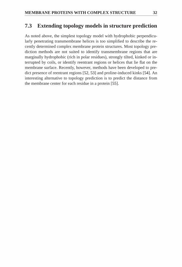
7.3 Extending topology models in structure prediction
As noted above, the simplest topology model with hydrophobic perpendicu-larly penetrating transmembrane helices is too simplified to describe the re-cently determined complex membrane protein structures. Most topology pre-diction methods are not suited to identify transmembrane regions that aremarginally hydrophobic (rich in polar residues), strongly tilted, kinked or in-terrupted by coils, or identify reentrant regions or helices that lie flat on themembrane surface. Recently, however, methods have been developed to pre-dict presence of reentrant regions [52, 53] and proline-induced kinks [54]. Aninteresting alternative to topology prediction is to predict the distance fromthe membrane center for each residue in a protein [55].

Chapter 8
Methodogy
This chapter describes which databases and evolutionary considerations thatwere used to create the datasets in the investigations. Further, it explainshowstructural annotations were derived from known structures and predicted an-notations from sequence-derived information.
8.1 Creation of dataset
The included analysis datasets of determined structures were SCOP 1.74 [56](Paper I) and Orientation of Proteins in Membrane (OPM) [57] (Paper II,Paper III andPaper IV).
In SCOP, protein domains of known structures are divided into classeslike “All- α”, “All- β ”, “ α-β (a/b)” and “α-β (a+b)” and fold descriptions like“layer”,“sandwich”, “barrel” and “bundle”. Based on evolutionary related-ness, the structures are also hierarchably divided into superfamilies and fami-lies .
OPM [57] is specialized on membrane protein structures. The database hasclassified the structures into superfamilies and families as well as includedfunctional information. The database have, by energetic calculations, orientedthe structures so that the predicted membrane center are located at the X-Yplane. This means that the absolute value of the Z-coordinate corresponds tothe distance from the membrane center.
8.2 Evolutionary considerations
In Paper I, the homology among protein pairs was inferred from SCOP an-notations if they belong to they same superfamily or family. Pairs of proteinswere structurally aligned with STRUCTAL [58]. From the resulting align-ments the evolutionary distance (average number of substitutions since diver-gence of common ancestor) were estimated using the maximum likelihood

METHODOGY 34
estimates with the JTT+Γ substitution model [59], allowing site-rate hetero-geneity, computed using Tree-Puzzle v.5.2 [60].
In Paper II, Paper III andPaper IV, the local sequence alignment programPSIBLAST was used to identify homologous proteins to the ones of knownstructure in OPM. In this process, proteins within the database Uniprot90,that contain many proteins of unknown structure, with high sequence similar-ity to the known structures were identified as evolutionary related. From themultiple alignments, amino acid substitution (replacement) rates within eachprotein were estimated using the program rate4site [61]. The program com-putes a phylogenetic tree from the alignment and uses the stochastic processunderlying sequence evolution within protein families to calculate a conserva-tion score using an empirical Bayesian method for each residue. In Rate4site,the rate (conservation score) of each site (residue) i in protein j is normalizedby subtracting the average conservation score and dividing by the averagestandard deviation of the reference protein j.
To avoid overrepresentation of proteins from a few evolutionary families thedata must often be homology reduced. This was done after some intitial filter-ing based on structural quality. The main dataset inPaper I was reduced bytaking one protein pair per family or superfamily, respectively. The homologyreduction of the membrane proteins in OPM inPaper II, Paper III andPa-per IV used the program CD-hit [62] to select representative structures withmaximum 40 % sequence identity to each other. In addition,Paper III andPaper IV also took OPM superfamily annotations into account to make sureto only pick maximum one representative structure from each superfamily.
8.3 Assigning features from structure
The assignment of protein secondary structure and relative surface accessi-bility from coordinates can be done by fast and consistent automatic compu-tational methods. Secondary structure assignment were, Figure8.1, assignedwith STRIDE (Paper I) and DSSP [63] (Paper II, Paper III andPaper IV).The assignment of the relative surface accessibility of residues in a proteincan be calculated from the coordinate file by rolling a probe over the wholestructure. The absolute calculated area per residue is normalized in order toget the relative surface accessibility, for example by an extended Ala-X-Alatri-peptide conformation or the maximum surface accessibility. InPaper I,Paper II, Paper III andPaper IV the surface accessibility was calculated byNaccess 2.1.1 [64] with the probe size 1.4 Å, corresponding to the size of wa-ter. The output from all three programs gives structural states for each residuein the polypeptide sequence. The many secondary structural states given bySTRIDE and DSSP were merged into three classes; helix, sheet and coil, andthe accessibility values were classified into two groups, buried and exposed.

35
Figure 8.1: If the structure is known, features such as secondary structure can be as-signed from coordinates by fast and consistent automatic computational methods. Thisor other types of features can also be predicted from sequence-derived information.
In Paper II, Paper III andPaper IV the Z-coordinates from OPM wereused to classify all residues into different groups: membrane core region,water-lipid interface region and water-soluble region (outside the membrane).The topology was automatically classified from the structures according to aprotocoll as described in [65]. Here each residue is classified to belong to oneout of four topological states: transmembrane, reentrant, inside or outside.
In all these methods one annotation per residue is obtained in an automaticand objective way. This made it possible to e.g. analyze the distribution ofamino acids, surface accessibility, secondary structure as a function of Z-coordinate.
8.4 Predicting structural features
A few features were predicted by sequence-derived information, Figure8.1.These features were Z-coordinate (by Zpred [55]), topology (by Octopus [52])and free energy of membrane insertion (by∆G-predictor [38]). The ability topredict the relative solvent accessibility and secondary structure was investi-gated using the machine learning technique called support vector machines,as implemented in the svmlight package [66]. During evaluation and opti-mization of the training, the homology reduced dataset of known structureswas divided into five groups. All combinations of four groups were used totrain while the fifth was used to evaluate the performance (cross-validated).By comparing the predicted with the known structural features the matthewcorrelation coefficient or the mean average error was calculated. A numberof sequence derived parameters, such as amino acid scores from PSI-BLAST,

METHODOGY 36
substitution rate, and predicted distance from membrane center, as well asSVM model parameters, were evaluated as input to the SVM.

Chapter 9
Summary of papers
Paper I concerns how structures respond to evolutionary changes in proteinsequences and whether structures change slower than sequences or not.Pa-per II andPaper III concerns non-helical structures and polar residues, re-spectively, positioned in the non-polar membrane core environment, and theirrelation with structure and function withinα-helical membrane proteins. InPaper IV an analysis is presented of how the properties of membrane proteinregions that after folding become exposed to distinct environments differ. Inaddition, it includes a separate method for predicting which residues are ex-posed to the solvent environment from sequence. Below you will find a shortsummary of each of the papers that are included in the thesis. At last, there aresome final thoughts.
9.1 Paper I
We investigated how protein structures respond to sequence changes (struc-tural response) by comparing the structural similarity between a large num-ber of structurally aligned and evolutionarily related proteins at different dis-tances of divergence. Initially, we used the commonly used root mean squaredeviation as a measure of structural similarity. The first finding was that bychanging the sequence similarity measure from fraction identity to number ofamino acid changes since divergence, a measure called evolutionary distance,the relationship between sequence and structure transforms from exponentialto linear. Moreover, we developed three other measures of structure simil-iarity at the residue level. The analysis of these measures showed that thenumber of changes that have occurred in secondary structure, relative surfaceaccessibility and residue contacts are linearly related to the average number ofchanges that have occurred in amino acid sequence. According to these mea-sures, structure is 15 to 30 times more conserved than sequence. Thereafter,we grouped the amino acid residues according to physico-chemical similarity.It was found that by grouping the amino acids into the same number of states

SUMMARY OF PAPERS 38
as the structural state sequences, structure is 3 to 10 times more conservedthan sequence.
Although the structural response is linear on average, the variation is large.We tried to identify structural or functional features connected to proteins withhigh or low structural response. The only strong trend we identified was thatprotein domains with a high fraction ofβ -sheets change states in secondarystructure sequence “faster” than proteins with high fraction ofα-helices dofor same amount of sequence change.
9.2 Paper II
In this paper, we systematically analyzed the 7 % of residues in non-helical(coil) conformation in the membrane core in knownα-helical membrane pro-teins. The coils can be found in transmembrane-helical kinks, as major breaksin transmembrane-helices and as parts of reentrant regions. On average, thesites in coil conformation are more evolutionary conserved than those of othersites, particularly within reentrants and breaks. The backbone around coilresidues has a polar character and the residues are often either buried or lo-cated near aqueous channels. The residues are frequently found within chan-nel and transporter proteins where they introduce the flexibility and polarityrequired for transport across the membrane. A literature study confirmed thatcoils are essential for the function of the proteins.
9.3 Paper III
In this paper, we characterized the 9 % of strongly polar residues (D, E, K, R,H, N, P and Q) that after folding withinα-helical membrane proteins end upin the middle of the membrane. These residues are more conserved than otherresidues. This is connected to their tendency to be buried in the protein inte-rior and to their high functional importance. The environment that surroundsstrongly polar residues in the membrane core resembles the environment thatsurrounds strongly polar residues in water-soluble proteins. Frequently, theirpolar groups form hydrogen bonds with water and line functionally importantcavities. In addition, strongly polar residues in the membrane core are often di-rectly involved in binding of small compounds in channels and transporters orlong-term interactions to prosthetic groups within electron transporters. Fur-ther, it was predicted that in human membrane proteins polar residues areover-represented among transport proteins and G-Protein Coupled Receptors.Finally, we saw a trend towards more polar residues in proteins with severaltransmembrane regions. In particular, the first transmembrane region containsfewer polar residues than the others.

39
9.4 Paper IV
Here, we analyzed the properties of different classes of sites within a set ofmembrane proteins of known structures. We found that sites that face the sol-vent environments, are very different inside and outside the membrane. Incontrast, residues buried within the interior of the protein are rather similar.As in water-soluble proteins, the non-membrane regions ofα- helical mem-brane proteins have a hydrophilic exterior and a hydrophobic interior. Withinthe membrane, both the exposed surface and the buried surface of membraneproteins are hydrophobic, but the difference between the buried sites is muchsmaller.
We found that state-of-the-art predictors for surface area are optimized forone of the environments and therefore performs poorly in the other envi-ronment. To circumvent this problem we included complete transmembraneproteins in a training set and developed a new predictor, membrane proteinresidue surface accessibility predictor (MPRAP). The new predictor performswell both inside and outside the membrane, and it is the best predictor in-side the membrane and in the water-lipid interface region. Further, it is bet-ter than the combination of previous specialized predictors. Potential uses ofthe predictor were demonstrated by identification of error-containing proteinstructures.
9.5 Final thoughts
In Paper I we investigated how structures respond to sequence changes bycomparing four measures of structural similarity to two measures of sequencesimilarity, for a large number of protein pairs. We found a linear relationbetween evolutionary distance and the number of changes in discrete struc-tural states in structural core. The finding made it possible to quantify howmuch more conserved structure is than sequence. By comparing the numberof changes in discrete structural states with changes in amino acid sequencewe got a simple way to test these features that normally occur in differentdimensions. Although, structure previously have been stated to be more con-served than sequences and the sequence-structure similarity relationship havebeen investigated before, we believe this was the first attempt to quantify howmuch more conserved structure is compared to sequence.
Although, it is well known that evolutionary distance is a better measure ofsequence divergence, it is mostly used for closely related proteins that can bealigned by sequence alignment methods. In this study, structural alignmentswere used, which made it possible to include both closely and distantly relatedproteins. This paper, therefore, in addition illustrate a new use of the measure.
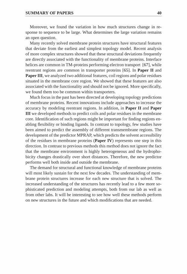
SUMMARY OF PAPERS 40
Moreover, we found the variation in how much structures change in re-sponse to sequence to be large. What determines the large variation remainsan open question.
Many recently solved membrane protein structures have structural featuresthat deviate from the earliest and simplest topology model. Recent analysisof more complex structures showed that these structural deviations frequentlyare directly associated with the functionality of membrane proteins. Interfacehelices are common in TM-proteins performing electron transport [67], whilereentrant regions are common in transporter proteins [65]. In Paper II andPaper III, we analyzed two additional features, coil regions and polar residuessituated in the membrane core region. We showed that these features are alsoassociated with the functionality and should not be ignored. More specifically,we found them too be common within transporters.
Much focus in the past has been directed at developing topology predictionsof membrane proteins. Recent innovations include approaches to increase theaccuracy by modeling reentrant regions. In addition, inPaper II andPaperIII we developed methods to predict coils and polar residues in the membranecore. Identification of such regions might be important for finding regions en-abling flexibility or binding ligands. In contrast to topology, few studies havebeen aimed to predict the assembly of different transmembrane regions. Thedevelopment of the predictor MPRAP, which predicts the solvent accessibilityof the residues in membrane proteins (Paper IV) represents one step in thisdirection. In contrast to previous methods this method does not ignore the factthat the membrane environment is highly heterogeneous and the hydropho-bicity changes drastically over short distances. Therefore, the new predictorperforms well both inside and outside the membrane.
The demand for structural and functional knowledge of membrane proteinswill most likely sustain for the next few decades. The understanding of mem-brane protein structures increase for each new structure that is solved. Theincreased understanding of the structures has recently lead to a few more so-phisticated prediction and modeling attempts, both from our lab as well asfrom other labs. It will be interesting to see how well these methods performon new structures in the future and which modifications that are needed.

Chapter 10
Acknowledgements
During my work I have recieved assistance and encouragements from manypeople. I would like to thank all of you so much! Here are some persons thatdeserve some special thanks.
Arne, my supervisor, thank you for scientific contributions, inspiration, re-laxed attitude and stimulating time of research. In short, it has been a greattime!
Anni, my main collaborator, thank you for all loudly enthusiasm about“anomalies”, for being “open” in funny and unexpected moments and forletting me discover what “circular writing” means. Most of all, thank you forall nice time in the lab.
Simone and Yogesh, my best students, thank you for always being so inter-ested and curious about all sorts of things. Simone, thank you for trying toconvince me that imperial stouts taste better than belgium-style ales, no mat-ter of how stupid it sounds. Yogesh, thank you for all stories about Nepal. Iwhish that you some day can show me your home country.
Linnea and Aron, thank you for scientific collaboration. Who would believe 9years ago that we would publish a scientific paper together (not only us, butanyway...)? Thank you for helping me out with all sorts of work- and non-work related problems. You are the best friends!
Öjvind, Håkan, Andreas, Sara, Erik and Björn, my past and present roommates, thank you for all work-related help. Thank you for daily non-workrelated discussions about things such as “kvintcirkeln” and the philosophy of“att snitta ned”. Sara, thank you for exxccellent proofreading of an earlierversion of this thesis.
Uno Carlsson, Lars-Göran Mårtensson, Per Hammarström, Bengt Persson andDavid Ardell, my former supervisors, thank you for your relaxed atmoshperein your labs. Your enthusiasm for doing protein science research convincedme to stay in the “business” after graduation from Linköping University.
ACKNOWLEDGEMENTS 42
Pauwel Kwak, thank you for creating the best beer. Ever!
All not previously mentioned co-workers, administrators, and friends, thankyou for making my time so pleasant at work. You are so many that I cannotacknowledge you all by name. I would like to thank you for help at work,nice company at the lunch tables, cake clubs, journal clubs, seminars andbadminton-games. Thank you also for nice “after works”, poker evenings andsocial activities with the Pärk team Svedmyra 1.
Mamma, Pappa and Johanna, my family, thank you for all encouragement,support and for aways being so kind. You are the best family (I have everhad)!
Josefin, my wife and personal illustrator, thank you for creating the beatifulfigures in this thesis. Thank you for all understanding, especially during somelong working days during the writing of this thesis. Thank you for letting mefinding out that everything in life is not about work. Thank you for makingmy life so enjoyable, partly because of your “kyckling med jordnötssås” andpartly for making future so interesting and unpredictable. I love you so much!

Bibliography
[1] H. Lodish; A. Berk; S.L. Zipursky; P. Matsudaira; D. Baltimore; J.Darnell.Molecular Cell Biology. W.H. Freeman and Company, New York, 1999.
[2] C. Branden; J. Tooze.Introduction to Protein Structure. Garland PublishingInc, 1999.
[3] R.D.M. Page; E.C. Holmes.Molecular Evolution - A Phylogentic Approach.Blackwell Science Ltd, 1998.
[4] E.H. Edwards.Bonniers stora bok om hästar. Bokförlaget Bonnier Alba AB,Stockholm, 1995.
[5] M. Zvelebil; J.O. Baum.Understanding Bioinformatics. Garland Science,Taylor and Francis Group, LLC, an informa buisiness, 2008.
[6] C.B. Anfinsen. Principles that govern the folding of protein chains.Science,181(96):223–230, Jul 1973.
[7] Protein data bank; a resource for studying biological macromolecules, Dec 2009.http://www.rcsb.org/pdb/home/home.do.
[8] M. Kosloff and R. Kolodny. Sequence-similar, structure-dissimilar protein pairsin the PDB.Proteins, 71(2):891–902, May 2008.
[9] J.M. Bujnicki. Practical Bioinformatics. Springer-Verlag Berlin Heidelberg,2004.
[10] D. Baker and A. Sali. Protein structure prediction and structural genomics.Sci-ence, 294(5540):93–96, Oct 2001.
[11] M.L. Sierk and G.J. Kleywegt. Deja vu all over again: finding and analyzingprotein structure similarities.Structure, 12(12):2103–2111, Dec 2004.
[12] A.M. Dean, C. Neuhauser, E. Grenier, and G.B. Golding. The pattern of aminoacid replacements in alpha/beta-barrels.Mol Biol Evol, 19(11):1846–1864,Nov 2002.
[13] L. Marsh and C.S. Griffiths. Protein structural influences in rhodopsin evolution.Mol Biol Evol, 22(4):894–904, Apr 2005.

BIBLIOGRAPHY 44
[14] M. Novotny and G.J. Kleywegt. A survey of left-handed helices in protein struc-tures.J Mol Biol, 347(2):231–241, Mar 2005.
[15] M.J. Ondrechen. Identifying functional sites based on prediction of chargedgroup behavior.Curr Protoc Bioinformatics, Chapter 8:Unit 8.6, Sep 2004.
[16] R. Greaves and J. Warwicker. Active site identification through geometry-basedand sequence profile-based calculations: burial of catalytic clefts.J Mol Biol,349(3):547–557, Jun 2005.
[17] B.H. Dessailly, M.F. Lensink, and S.J. Wodak. Relating destabilizingregions toknown functional sites in proteins.BMC Bioinformatics, 8:141, 2007.
[18] G. Cheng, B. Qian, R. Samudrala, and D. Baker. Improvementin proteinfunctional site prediction by distinguishing structural and functional constraintson protein family evolution using computational design.Nucleic Acids Res,33(18):5861–5867, 2005.
[19] G.B. Golding and A.M. Dean. The structural basis of molecular adaptation.MolBiol Evol, 15(4):355–369, Apr 1998.
[20] N.V. Grishin. Fold change in evolution of protein structures.J Struct Biol,134(2-3):167–185, May 2001.
[21] B.W. Matthews. Studies on protein stability with t4 lysozyme.Adv ProteinChem, 46:249–278, 1995.
[22] C. Chothia and A.M. Lesk. The relation between the divergence of sequenceand structure in proteins.EMBO J, 5(4):823–826, Apr 1986.
[23] A. R. Panchenko and T. Madej. Structural similarity of loops in proteinfamilies:toward the understanding of protein evolution.BMC evolutionary biology,5(1):10, Feb 3 2005.
[24] M. Luckey. Membrane Structural Biology - With Biochemical and Bio-physical Foundations. Cambridge University Press, 2008.
[25] M.C. Wiener and S.H. White. Structure of a fluid dioleoylphosphatidylcholinebilayer determined by joint refinement of x-ray and neutron diffractiondata. III.complete structure.Biophys J, 61(2):434–447, Feb 1992.
[26] S.E. Feller, D. Yin, R.W. Pastor, and A.D. MacKerell, Jr. Moleculardynamicssimulation of unsaturated lipid bilayers at low hydration: parameterization andcomparison with diffraction studies.Biophys J, 73(5):2269–2279, Nov 1997.
[27] A. Bernsel. Sequenced-based predictions of membrane-protein topology.US-AB, Printed in Sweden 2008, 2008.

45
[28] A.G. Garrow, A. Agnew, and D.R. Westhead. TMB-hunt: an aminoacid com-position based method to screen proteomes for beta-barrel transmembrane pro-teins.BMC Bioinformatics, 6:56, 2005.
[29] M.S. Almen, K.J. Nordstrom, R. Fredriksson, and H.B. Schioth.Mapping thehuman membrane proteome: a majority of the human membrane proteins can beclassified according to function and evolutionary origin.BMC Biol, 7:50, 2009.
[30] A. Oberai, Y. Ihm, S. Kim, and J.U. Bowie. A limited universe of membraneprotein families and folds.Protein Sci, 15(7):1723–1734, Jul 2006.
[31] G. von Heijne. The membrane protein universe: what’s out thereand whybother?J Intern Med, 261(6):543–557, Jun 2007.
[32] L. Lins and R. Brasseur. The hydrophobic effect in protein folding. FASEB J,9(7):535–540, Apr 1995.
[33] T.J. Stevens and I.T. Arkin. Turning an opinion inside-out: Reesand eisenberg’scommentary (proteins 2000;38:121-122) on "are membrane proteins ’inside-out’ proteins?" (proteins 1999;36:135-143).Proteins, 40(3):463–464, Aug2000.
[34] L. Adamian and J. Liang. Prediction of transmembrane helix orientation inpolytopic membrane proteins.BMC Struct Biol, 6:13, 2006.
[35] S.H. White and W.C. Wimley. Membrane protein folding and stability: physicalprinciples.Annu Rev Biophys Biomol Struct, 28:319–365, 1999.
[36] G. von Heijne. Membrane-protein topology.Nat Rev Mol Cell Biol,7(12):909–918, Dec 2006.
[37] S.H. White and G. von Heijne. The machinery of membrane protein assembly.Curr Opin Struct Biol, 14(4):397–404, Aug 2004.
[38] T. Hessa, N.M. Meindl-Beinker, A. Bernsel, H. Kim, Y. Sato, M. Lerch-Bader,I. Nilsson, S.H. White, and G. von Heijne. Molecular code for transmembrane-helix recognition by the sec61 translocon.Nature, 450(7172):1026–1030, Dec2007.
[39] L.E. Hedin, K. Ojemalm, A. Bernsel, A. Hennerdal, K. Illergard,K. Enquist,A. Kauko, S. Cristobal, G. von Heijne, M. Lerch-Bader, I. Nilsson, andA. Elof-sson. Membrane insertion of marginally hydrophobic transmembrane helicesdepends on sequence context.J Mol Biol, Nov 2009.
[40] A. Elofsson and G. von Heijne. Membrane protein structure: prediction versusreality. Annu Rev Biochem, 76:125–140, 2007.
[41] S.J. Fleishman and N. Ben-Tal. Progress in structure prediction of alpha-helicalmembrane proteins.Curr Opin Struct Biol, 16(4):496–504, Aug 2006.

BIBLIOGRAPHY 46
[42] M.G. Claros and G. von Heijne. Toppred II: an improved softwarefor membraneprotein structure predictions.Comput Appl Biosci, 10(6):685–686, Dec 1994.
[43] G. von Heijne. Control of topology and mode of assembly of a polytopic mem-brane protein by positively charged residues.Nature, 341(6241):456–458, Oct1989.
[44] Y. Park, S. Hayat, and V. Helms. Prediction of the burial status of transmembraneresidues of helical membrane proteins.BMC Bioinformatics, 8:302, 2007.
[45] G.B. Gloor, L.C. Martin, L.M. Wahl, and S.D. Dunn. Mutual information in pro-tein multiple sequence alignments reveals two classes of coevolving positions.Biochemistry, 44(19):7156–7165, May 2005.
[46] A. Fuchs, A. Kirschner, and D. Frishman. Prediction of helix-helix contactsand interacting helices in polytopic membrane proteins using neural networks.Proteins, 74(4):857–871, Mar 2009.
[47] P. Barth, B. Wallner, and D. Baker. Prediction of membrane protein structureswith complex topologies using limited constraints.Proc Natl Acad Sci U S A,106(5):1409–1414, Feb 2009.
[48] M. Pellegrini-Calace, A. Carotti, and D.T. Jones. Folding in lipid membranes(FILM): a novel method for the prediction of small membrane protein 3d struc-tures.Proteins, 50(4):537–545, Mar 2003.
[49] L.R. Forrest, C.L. Tang, and B. Honig. On the accuracy of homology modelingand sequence alignment methods applied to membrane proteins.Biophys J,91(2):508–517, Jul 2006.
[50] Y. Zhang, M.E. Devries, and J. Skolnick. Structure modeling of allidentified gprotein-coupled receptors in the human genome.PLoS Comput Biol, 2(2):e13,Feb 2006.
[51] S.H. White and G. von Heijne. How translocons select transmembrane helices.Annu Rev Biophys, 37:23–42, 2008.
[52] H. Viklund and A. Elofsson. OCTOPUS: improving topology prediction bytwo-track ANN-based preference scores and an extended topological grammar.Bioinformatics, 24(15):1662–1668, Aug 2008.
[53] D.T. Jones. Improving the accuracy of transmembrane proteintopology pre-diction using evolutionary information.Bioinformatics, 23(5):538–544, Mar2007.
[54] S. Yohannan, S. Faham, D. Yang, J.P. Whitelegge, and J.U. Bowie. The evolutionof transmembrane helix kinks and the structural diversity of g protein-coupledreceptors.Proc Natl Acad Sci U S A, 101(4):959–963, Jan 2004.

47
[55] E. Granseth, H. Viklund, and A. Elofsson. ZPRED: predicting the distance tothe membrane center for residues in alpha-helical membrane proteins.Bioin-formatics, 22(14):e191–6, Jul 2006.
[56] A.G. Murzin, S.E. Brenner, T. Hubbard, and C. Chothia. SCOP:a structuralclassification of proteins database for the investigation of sequences andstruc-tures.J Mol Biol, 247(4):536–540, Apr 1995.
[57] M.A. Lomize, A.L. Lomize, I.D. Pogozheva, and H.I. Mosberg.OPM: orienta-tions of proteins in membranes database.Bioinformatics, 22(5):623–625, Mar2006.
[58] S. Subbiah, D.V. Laurents, and M. Levitt. Structural similarity of DNA-bindingdomains of bacteriophage repressors and the globin core.Curr Biol, 3(3):141–148, Mar 1993.
[59] D.T. Jones, W.R. Taylor, and J.M. Thornton. The rapid generation of mutationdata matrices from protein sequences.Comput Appl Biosci, 8(3):275–282, Jun1992.
[60] H.A. Schmidt, K. Strimmer, M. Vingron, and A. von Haeseler. TREE-PUZZLE:maximum likelihood phylogenetic analysis using quartets and parallel comput-ing. Bioinformatics, 18(3):502–504, Mar 2002.
[61] I. Mayrose, D. Graur, N. Ben-Tal, and T. Pupko. Comparison of site-specificrate-inference methods for protein sequences: empirical bayesian methods aresuperior.Mol Biol Evol, 21(9):1781–1791, Sep 2004.
[62] W. Li and A. Godzik. Cd-hit: a fast program for clustering and comparing largesets of protein or nucleotide sequences.Bioinformatics, 22(13):1658–1659, Jul2006.
[63] W. Kabsch and C. Sander. Dictionary of protein secondary structure: pat-tern recognition of hydrogen-bonded and geometrical features.Biopolymers,22(12):2577–2637, Dec 1983.
[64] Thornton JM. Hubbard SJ. Naccess, computer program.Department of Biochemistry and Molecular Biologyhttp://wolf.bi.umist.ac.uk/unix/naccess.html, 1(1):1–2, Jan 1993.
[65] H. Viklund, E. Granseth, and A. Elofsson. Structural classification and predic-tion of reentrant regions in alpha-helical transmembrane proteins: application tocomplete genomes.J Mol Biol, 361(3):591–603, Aug 2006.
[66] T. Joachims. Making large-scale svm learning practical. in: B schölkopf and cburges and a smola, (eds), advances in kernel methods - support vector learning.MIT Press, Cambridge Massachusetts, London England, 1999.
BIBLIOGRAPHY 48
[67] E. Granseth, D.O. Daley, M. Rapp, K. Melen, and G. von Heijne. Experimen-tally constrained topology models for 51,208 bacterial inner membrane proteins.J Mol Biol, 352(3):489–494, Sep 2005.