Ommaya Reservoir Integrity Computed Tomography Evaluation ... · Computed Tomography Evaluation of...
7
Received 02/26/2017 Review began 03/08/2017 Review ended 03/11/2017 Published 03/15/2017 © Copyright 2017 Moraff et al. This is an open access article distributed under the terms of the Creative Commons Attribution License CC-BY 3.0., which permits unrestricted use, distribution, and reproduction in any medium, provided the original author and source are credited. Real-Time Fluoroscopic and C-Arm Computed Tomography Evaluation of Ommaya Reservoir Integrity Adrienne M. Moraff , Melanie Hayden Gephart , Larry M. Shuer , Jeremy J. Heit 1. Department of Neurosurgery, Stanford University School of Medicine 2. Stanford University School of Medicine 3. Department of Radiology, Stanford University School of Medicine Corresponding author: Jeremy J. Heit, [email protected] Disclosures can be found in Additional Information at the end of the article Abstract We describe a case of a 24-year-old patient with relapsed acute myelogenous leukemia involving the central nervous system. After placement of an Ommaya reservoir for intrathecal chemotherapy administration, the patient developed progressive headache, nausea, and drowsiness and was found to have an enlarging subdural collection underlying the Ommaya. To exclude leakage of the Ommaya system into the subdural space, real-time fluoroscopic and C- arm computed tomographic evaluation of the Ommaya reservoir was performed after iodinated contrast injection into the reservoir. This novel technique demonstrated complete integrity of the Ommaya reservoir without evidence of blockage or leakage of the system. The patient underwent uncomplicated evacuation of the subdural collection without replacement of the Ommaya reservoir and made an excellent recovery. This technique for real-time interrogation of the Ommaya reservoir may have additional utility in the evaluation for Ommaya reservoir dysfunction. Categories: Neurology, Neurosurgery, Radiology Keywords: ommaya reservoir, c-arm ct, leakage, tubing, subdural hematoma, flat panel ct Introduction Ommaya reservoirs are surgically implanted closed catheter systems with a needle-accessible subcutaneous reservoir [1]. This reservoir is in continuity with the cerebrospinal fluid (CSF) through tubing that extends from the reservoir to the ventricular system, which allows for repeated CSF sampling and intrathecal medication delivery [2]. Ommaya implantation is generally well tolerated, but complications related to the device implantation occur in 15% of patients [3-4]. Infection, intraparenchymal or intraventricular hemorrhage, catheter malposition, CSF leakage, tubing blockage, and development of a subdural hematoma or hygroma are among the most commonly encountered complications, and these complications may require surgical revision [4-5]. Neuroimaging is performed to evaluate Ommaya reservoir placement and to assess for complications related to placement. Most commonly computed tomography (CT) or magnetic resonance imaging (MRI) is performed to determine the location of the Ommaya catheter tip, to assess for intracranial hemorrhage, and to identify extra cerebral collections following placement. Although CT and MRI offer excellent spatial resolution and information regarding the adjacent brain parenchyma and ventricular system, these techniques do not evaluate the dynamic flow of fluid through the Ommaya reservoir, which limits its ability to detect tubing blockage or the site of leakage. 1 1 2 3 Open Access Case Report DOI: 10.7759/cureus.1097 How to cite this article Moraff A M, Hayden gephart M, Shuer L M, et al. (March 15, 2017) Real-Time Fluoroscopic and C-Arm Computed Tomography Evaluation of Ommaya Reservoir Integrity. Cureus 9(3): e1097. DOI 10.7759/cureus.1097
Transcript of Ommaya Reservoir Integrity Computed Tomography Evaluation ... · Computed Tomography Evaluation of...

Received 02/26/2017 Review began 03/08/2017 Review ended 03/11/2017 Published 03/15/2017
© Copyright 2017Moraff et al. This is an open accessarticle distributed under the terms ofthe Creative Commons AttributionLicense CC-BY 3.0., which permitsunrestricted use, distribution, andreproduction in any medium,provided the original author andsource are credited.
Real-Time Fluoroscopic and C-ArmComputed Tomography Evaluation ofOmmaya Reservoir IntegrityAdrienne M. Moraff , Melanie Hayden Gephart , Larry M. Shuer , Jeremy J. Heit
1. Department of Neurosurgery, Stanford University School of Medicine 2. Stanford University School ofMedicine 3. Department of Radiology, Stanford University School of Medicine
Corresponding author: Jeremy J. Heit, [email protected] Disclosures can be found in Additional Information at the end of the article
AbstractWe describe a case of a 24-year-old patient with relapsed acute myelogenous leukemiainvolving the central nervous system. After placement of an Ommaya reservoir for intrathecalchemotherapy administration, the patient developed progressive headache, nausea, anddrowsiness and was found to have an enlarging subdural collection underlying the Ommaya. Toexclude leakage of the Ommaya system into the subdural space, real-time fluoroscopic and C-arm computed tomographic evaluation of the Ommaya reservoir was performed after iodinatedcontrast injection into the reservoir. This novel technique demonstrated complete integrity ofthe Ommaya reservoir without evidence of blockage or leakage of the system. The patientunderwent uncomplicated evacuation of the subdural collection without replacement of theOmmaya reservoir and made an excellent recovery. This technique for real-time interrogationof the Ommaya reservoir may have additional utility in the evaluation for Ommaya reservoirdysfunction.
Categories: Neurology, Neurosurgery, RadiologyKeywords: ommaya reservoir, c-arm ct, leakage, tubing, subdural hematoma, flat panel ct
IntroductionOmmaya reservoirs are surgically implanted closed catheter systems with a needle-accessiblesubcutaneous reservoir [1]. This reservoir is in continuity with the cerebrospinal fluid (CSF)through tubing that extends from the reservoir to the ventricular system, which allows forrepeated CSF sampling and intrathecal medication delivery [2]. Ommaya implantation isgenerally well tolerated, but complications related to the device implantation occur in 15% ofpatients [3-4]. Infection, intraparenchymal or intraventricular hemorrhage, cathetermalposition, CSF leakage, tubing blockage, and development of a subdural hematoma orhygroma are among the most commonly encountered complications, and these complicationsmay require surgical revision [4-5].
Neuroimaging is performed to evaluate Ommaya reservoir placement and to assess forcomplications related to placement. Most commonly computed tomography (CT) or magneticresonance imaging (MRI) is performed to determine the location of the Ommaya catheter tip, toassess for intracranial hemorrhage, and to identify extra cerebral collections followingplacement. Although CT and MRI offer excellent spatial resolution and information regardingthe adjacent brain parenchyma and ventricular system, these techniques do not evaluate thedynamic flow of fluid through the Ommaya reservoir, which limits its ability to detect tubingblockage or the site of leakage.
1 1 2 3
Open Access CaseReport DOI: 10.7759/cureus.1097
How to cite this articleMoraff A M, Hayden gephart M, Shuer L M, et al. (March 15, 2017) Real-Time Fluoroscopic and C-ArmComputed Tomography Evaluation of Ommaya Reservoir Integrity. Cureus 9(3): e1097. DOI10.7759/cureus.1097

Shunt interrogation studies (termed “shuntograms”) have also been used to assess the integrityof other ventriculoperitoneal shunts. In these studies, iodinated contrast is injected into theshunt tubing, and serial radiographs are taken every 15-20 minutes to evaluate for contrastextravasation along the course of the tubing rather than in the peritoneum, where the tubingterminates. This technique may also be adapted to Ommaya reservoirs, although such anadaptation has not been reported.
Radioisotopes, including 111In-DPTA or 99mTc-DPTA, may be introduced through an Ommayato assess the CSF flow blockage, Ommaya tubing blockage, or Ommaya leakage by radionuclideimaging techniques [6-8]. However, radionuclide imaging has poor spatial and temporalresolution, which may prevent clear identification of the site of leakage or blockage [9]. Toprevent unnecessary Ommaya replacement when compromise of the reservoir or tubing isincorrectly suspected, better minimally-invasive dynamic diagnostic testing of Ommaya systemintegrity is needed.
Here we describe a novel real-time imaging technique for evaluation of Ommaya reservoirintegrity in a patient with a symptomatic post-operative subdural collection. The Ommayareservoir was injected with iodinated contrast, and transit of this contrast through the Ommayasystem into the ventricular system was well visualized with real-time fluoroscopy to excludeleakage of the Ommaya system into the subdural collection. A flat panel CT was obtained in theneuroendovascular suite for further evaluation of the subdural collection after contrastinjection, and reformatted images from this study demonstrated no contrast extension into thesubdural collection. Based on these imaging findings, the patient underwent uneventfulevacuation of the subdural collection without replacement of the Ommaya shunt, and he madean excellent recovery.
Case PresentationA 24-year-old man with a nine-month history of acute myelogenous leukemia (AML) presentedto our hospital with progressive headache, nausea, and drowsiness. The patient initiallypresented 11 months earlier with fatigue, and he was admitted to an outside hospital. He wasfound to be anemic, with a complete cell count demonstrating evidence of AML with a
hemoglobin level of 5.1 g/dl, a white blood cell count of 14.1 x 10³/µL with 89% blasts, and
platelets of 29 x 103/µL. A complete evaluation at the time of diagnosis demonstrated bonemarrow infiltration, but no evidence of central nervous system involvement. The patientunderwent induction chemotherapy treatment with high dose pravastatin, idarubicin, andcytarabine, and he required re-induction for a persistent blast level of 78% on a bone marrowaspiration that was performed 14 days after induction. Following re-induction therapy, arepeat bone marrow biopsy showed no blasts, and he underwent consolidation chemotherapywith high-dose cytarabine followed by a matched myeloablative stem cell transplantation.
The patient initially did well after his bone marrow transplant, but nine months later hedeveloped neck spasms and right upper extremity neuropathy. He underwent an MRI scan ofthe right brachial plexus (Figure 1), which demonstrated nerve root thickening that was foundto represent leukemic infiltration. A lumbar puncture demonstrated evidence of blasts in theCSF. There was no evidence of peripheral disease recurrence upon repeat bone marrow biopsyand a complete imaging evaluation. The patient underwent treatment with high-dosecytarabine and dexamethasone. A right frontal Ommaya reservoir was placed using stereotacticguidance for weekly intrathecal chemotherapy.
2017 Moraff et al. Cureus 9(3): e1097. DOI 10.7759/cureus.1097 2 of 7

FIGURE 1: Brachial plexus magnetic resonance imagingdemonstrating leukemic infiltration of the bilateral nerve roots.(A, B) Short tau inversion recovery (STIR) images demonstrate nerve root thickening andincreased T2-weighted signal abnormality (arrows) in the sagittal (A) and coronal plane (B). (C,D) Post contrast images show diffuse enhancement within thickened nerve roots of the brachialplexus in the sagittal (C) and coronal plane (D).
The patient developed a headache seven days after placement of the Ommaya reservoir, andthere was difficulty in accessing the Ommaya. A head CT demonstrated appropriate positioningof the Ommaya reservoir tip in the frontal horn of the right lateral ventricle, but a newhypodense 5-mm extra-axial collection overlying the right frontal lobe was present (Figure 2).The patient’s headache worsened, and he developed nausea and vomiting with intrathecalchemotherapy administration. A follow-up head CT demonstrated an interval increase inthe size of the right frontal extra-axial collection with an associated increase in local masseffect on the adjacent cerebral sulci and modest midline shift.
2017 Moraff et al. Cureus 9(3): e1097. DOI 10.7759/cureus.1097 3 of 7

FIGURE 2: Head computed tomography images before andafter Ommaya reservoir placement demonstrate increasing sizeof a right subdural collection.Axial (A-C) and coronal reformations (D-F) from a non-contrast head computed tomogramdemonstrate a normal appearance of the brain prior to Ommaya reservoir placement (A, D).After Ommaya reservoir placement, the catheter (B, C, E, F, arrowheads) traverses the rightfrontal lobe (B, C) and terminates in the right lateral ventricle near the foramen of Monro (E, F).There is a hypodense subdural collection (B, C, E, R, arrows) overlying the right frontal lobe thatwas initially small (B, E) but increased on a follow-up study (C, F).
The patient’s symptoms persisted for an additional two weeks, and he developed increasingconfusion and somnolence. The right frontal extra-axial collection remained stable in size, butthe concern was raised that the Ommaya reservoir system had been damaged during needleaccess for intrathecal chemotherapy administration. It was further postulated that leakage ofthe Ommaya system into the subdural space overlying the right frontal lobe might account forthe right frontal lobe extra-axial collection. Other considerations for the patient’s symptomsand imaging findings included brain shift and draining vein tension with fluid removal andadministration of chemotherapy, medication leakage into the subdural space duringchemotherapy administration, or intracranial hypotension given the patient’s history of priorlumbar punctures. The patient was planned for surgical drainage of the extra-cerebralcollection and possible replacement of the Ommaya reservoir. Given his neutropenic status, itwas preferred that the Ommaya only be removed and replaced if it was damaged.
To assess the integrity of the Ommaya system and leakage into the subdural space, the patientwas brought to the neuroendovascular suite for real time fluoroscopic evaluation of theOmmaya following contrast injection of the reservoir. The neuroendovascular suite is equippedwith a Siemens Artis Zee biplane fluoroscopy unit with flat panel CT capability. The patient wasplaced supine on the angiography table. The skin overlying the Ommaya reservoir was sterilelyprepared and draped in a standard fashion, and the reservoir was accessed with a 22-guagebutterfly needle. Return of clear CSF was verified after this access, and 5 ml of Omnipaque 300contrast dye was slowly injected into the Ommaya reservoir. The contrast injection was
2017 Moraff et al. Cureus 9(3): e1097. DOI 10.7759/cureus.1097 4 of 7
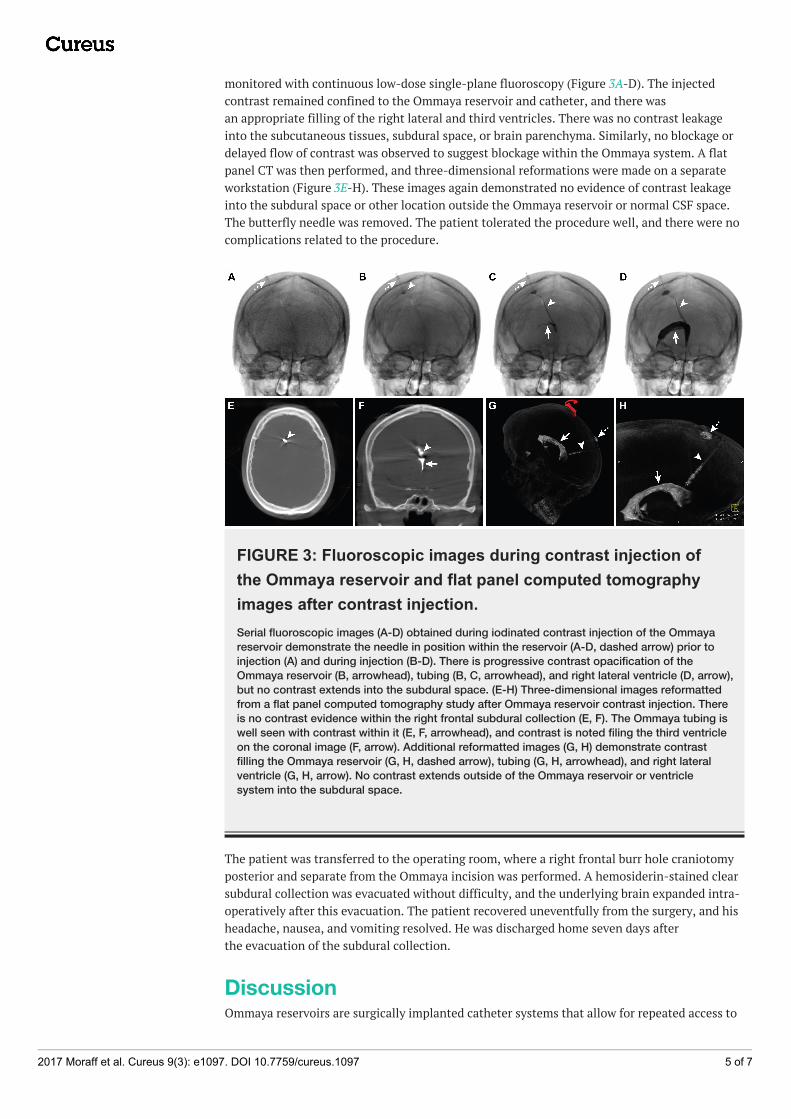
monitored with continuous low-dose single-plane fluoroscopy (Figure 3A-D). The injectedcontrast remained confined to the Ommaya reservoir and catheter, and there wasan appropriate filling of the right lateral and third ventricles. There was no contrast leakageinto the subcutaneous tissues, subdural space, or brain parenchyma. Similarly, no blockage ordelayed flow of contrast was observed to suggest blockage within the Ommaya system. A flatpanel CT was then performed, and three-dimensional reformations were made on a separateworkstation (Figure 3E-H). These images again demonstrated no evidence of contrast leakageinto the subdural space or other location outside the Ommaya reservoir or normal CSF space.The butterfly needle was removed. The patient tolerated the procedure well, and there were nocomplications related to the procedure.
FIGURE 3: Fluoroscopic images during contrast injection ofthe Ommaya reservoir and flat panel computed tomographyimages after contrast injection.Serial fluoroscopic images (A-D) obtained during iodinated contrast injection of the Ommayareservoir demonstrate the needle in position within the reservoir (A-D, dashed arrow) prior toinjection (A) and during injection (B-D). There is progressive contrast opacification of theOmmaya reservoir (B, arrowhead), tubing (B, C, arrowhead), and right lateral ventricle (D, arrow),but no contrast extends into the subdural space. (E-H) Three-dimensional images reformattedfrom a flat panel computed tomography study after Ommaya reservoir contrast injection. Thereis no contrast evidence within the right frontal subdural collection (E, F). The Ommaya tubing iswell seen with contrast within it (E, F, arrowhead), and contrast is noted filing the third ventricleon the coronal image (F, arrow). Additional reformatted images (G, H) demonstrate contrastfilling the Ommaya reservoir (G, H, dashed arrow), tubing (G, H, arrowhead), and right lateralventricle (G, H, arrow). No contrast extends outside of the Ommaya reservoir or ventriclesystem into the subdural space.
The patient was transferred to the operating room, where a right frontal burr hole craniotomyposterior and separate from the Ommaya incision was performed. A hemosiderin-stained clearsubdural collection was evacuated without difficulty, and the underlying brain expanded intra-operatively after this evacuation. The patient recovered uneventfully from the surgery, and hisheadache, nausea, and vomiting resolved. He was discharged home seven days afterthe evacuation of the subdural collection.
DiscussionOmmaya reservoirs are surgically implanted catheter systems that allow for repeated access to
2017 Moraff et al. Cureus 9(3): e1097. DOI 10.7759/cureus.1097 5 of 7

the CSF and intrathecal chemotherapy administration, which decreases patient morbidityrelated to repeated lumbar puncture [1-2]. However, Ommaya reservoirs may develop blockagewithin the catheter or leakage due to damage of the reservoir, damage to the catheter, ordisconnection of the reservoir and catheter components [4-5]. Compromise of the Ommayasystem may result in non-target delivery of chemotherapy or a CSF leak that may lead to thedevelopment of an extra-axial fluid collection. Surgical replacement of the Ommaya may berequired in the setting of Ommaya reservoir compromise, and there is an increased risk ofinfection and bleeding complications in this patient population that is frequently neutropenicand thrombocytopenic.
Non-invasive evaluation of Ommaya reservoir integrity is challenging given the need for hightemporal and spatial resolution when interrogating the system. Here we describe, to ourknowledge, the first description of real-time fluoroscopic interrogation of Ommaya reservoirintegrity. Fluoroscopy provides excellent temporal and spatial resolution, and iodinatedcontrast injection of the Ommaya reservoir allowed for real-time evaluation of the system’sintegrity. Appropriate flow of contrast from the reservoir to the tubing and into the ventricleswas easily visualized fluoroscopically. Digital storage of the real-time fluoroscopy imagesallowed for repeated review of the contrast injection to ensure that subtle areas of contrastextravasation outside the reservoir, tubing, or ventricles were not overlooked. The sensitivity ofthe examination was increased further by performing a flat panel CT scan after contrast wasclearly identified extending throughout the ventricles. The data from this three-dimensionalscan was reformatted to clearly demonstrate contrast confined to the Ommaya system andventricles. Furthermore, the extra-axial collection overlying the right frontal lobe was shown tobe free of contrast with excellent anatomic correlation to the prior non-invasive imaging. Thisstudy nicely demonstrated the integrity of the Ommaya system and affected the clinicaldecision to avoid Ommaya reservoir replacement at the time of subdural collection evacuation.
Although not performed in this case report, real time fluoroscopic evaluation of Ommayareservoir integrity may be best performed in hybrid neurointerventional-surgical suites. Thesemodern suites offer state of the art fluoroscopic equipment in an operative suite that wouldallow for evaluation of the Ommaya reservoir integrity as described in this report followed bythe neurosurgical intervention that is tailored to the imaging findings. This streamlinedapproach would save time and resources by reducing patient transfer times, the need for repeatanesthesia set up, and repeat procedural room turn around. It will be of interest to see howsimilar diagnostic questions may be answered in an innovative manner using these hybridsuites.
ConclusionsReal-time fluoroscopic and flat panel CT evaluation of Ommaya reservoir integrity by iodinatedcontrast injection may be safely performed to evaluate the integrity of the catheter system.Demonstration of Ommaya system integrity without evidence of blockage or leakage in thiscase report allowed for tailored neurosurgical treatment.
Additional InformationDisclosuresHuman subjects: Consent was obtained by all participants in this study. Informed consentobtained. Conflicts of interest: In compliance with the ICMJE uniform disclosure form, allauthors declare the following: Payment/services info: All authors have declared that nofinancial support was received from any organization for the submitted work. Financialrelationships: All authors have declared that they have no financial relationships at present orwithin the previous three years with any organizations that might have an interest in the
2017 Moraff et al. Cureus 9(3): e1097. DOI 10.7759/cureus.1097 6 of 7
submitted work. Other relationships: All authors have declared that there are no otherrelationships or activities that could appear to have influenced the submitted work.
References1. Ommaya AK: Subcutaneous reservoir and pump for sterile access to ventricular cerebrospinal
fluid. Lancet. 1963, 282:983–984. 10.1016/S0140-6736(63)90681-02. Ommaya AK: Implantable devices for chronic access and drug delivery to the central nervous
system. Cancer drug deliv. 1984, 1:169–179. 10.1089/cdd.1984.1.1693. Machado M, Salcman M, Kaplan RS, et al.: Expanded role of the cerebrospinal fluid reservoir
in neurooncology: indications, causes of revision, and complications. Neurosurgery. 1985,17:600–603.
4. Lishner M, Perrin RG, Feld R, et al.: Complications associated with Ommaya reservoirs inpatients with cancer. The Princess Margaret Hospital experience and a review of theliterature. Arch Intern Med. 1990, 150:173–176. 10.1001/archinte.1990.00390130145023
5. de Waal R, Algra PR, Heimans JJ, et al.: Methotrexate induced brain necrosis and severeleukoencephalopathy due to disconnection of an Ommaya device. J. Neurooncol. 1993,15:269–273. 10.1007/BF01050074
6. Larson SM, Johnston GS, Ommaya AK, et al.: The radionuclide ventriculogram. Jama. 1973,224:853–857. 10.1001/jama.1973.03220200023007
7. Korkmaz M, Kim EE, Wong FC, et al.: In-111 DTPA Ommayagrams in leptomeningealcarcinomatosis. Clin Nucl Med. 1995, 20:610–612.
8. Glantz MJ, Hall WA, Cole BF, et al.: Diagnosis, management, and survival of patients withleptomeningeal cancer based on cerebrospinal fluid-flow status. Cancer. 1995, 75:2919–2931.10.1002/1097-0142(19950615)75:12<2919::AID-CNCR2820751220>3.0.CO;2-9
9. Stone JA, Castillo M, Neelon B, et al.: Evaluation of CSF leaks: high-resolution CT comparedwith contrast-enhanced CT and radionuclide cisternography. AJNR Am J Neuroradiol. 1999,20:706–712.
2017 Moraff et al. Cureus 9(3): e1097. DOI 10.7759/cureus.1097 7 of 7