Innovative radiotherapy techniques: a multicentre time-driven - KCE

STUDY PROTOCOL Open Access
OLIGOPELVIS – GETUG P07: a multicentrephase II trial of combined salvageradiotherapy and hormone therapy inoligometastatic pelvic node relapses ofprostate cancerStephane Supiot1,2*, Emmanuel Rio1, Valérie Pacteau3, Marie-Hélène Mauboussin3, Loïc Campion2,4
and François Pein3
Abstract
Background: Metastatic prostate cancer remains a common cause of death in Europe, and improvements inmanagement of the disease are urgently needed. The advent of positron-emission tomography (PET) imagingenhanced with fluorocholine has led to the identification of a new sub-group of metastatic prostate cancerpatients: those with so-called oligometastatic disease. Presenting with a low burden of metastatic disease (≤5lesions), this new sub-group lies between true metastatic prostate cancer patients for whom androgen- deprivationtherapy (ADT) is the mainstay of treatment, and patients with a rising PSA, but no visible lesion on conventionalimaging, in whom intermittent ADT has been shown to be no less effective than continuous ADT. One mightconclude that intermittent ADT would also be the standard of care for oligometastatic prostate cancer patients, butradical strategies such as extensive lymphadenectomy or high-dose radiotherapy have been suggested as anothermeans to delay the need for ADT, and increase its effectiveness once initiated. This study will explore the role ofsalvage pelvic image-guided intensity-modulated radiation therapy (IMRT) combined with ADT administered for 6months in pelvic oligometastatic patients in prolonging the failure-free interval between two consecutive ADTcourses, or even to cure selected patients with limited metastatic burden.
Methods/Design: We plan to assess the two year outcome in oligometastatic prostate cancer patients (1–5 pelvicoligometastases) treated concomitantly with high-dose IMRT (54 Gy, 30 fractions to the pelvis and 66 Gy, 30fractions to the lymph nodes) and ADT for six months.
Discussion: This multicenter prospective phase II study will yield new data regarding the safety and efficacy ofhigh-dose radiotherapy combined with ADT and will provide a basis for a larger phase III study to examine the roleof radiotherapy in this population currently treated only with hormone therapy.
Trial registration: NCT02274779, date of registration: 23/10/14
* Correspondence: [email protected] of Radiation Oncology, Institut de Cancérologie de l’Ouest,Boulevard J. Monod, F-44800 Nantes, St-Herblain, France2Centre de Recherche en Cancérologie Nantes-Angers (CRCNA), UMR 892Inserm - 6299 CNRS, Institut de Recherche en Santé de l’Université deNantes, 8 quai Moncousu, BP 7072144007 Nantes cedex 1, FranceFull list of author information is available at the end of the article
© 2015 Supiot et al. Open Access This article is distributed under the terms of the Creative Commons Attribution 4.0International License (http://creativecommons.org/licenses/by/4.0/), which permits unrestricted use, distribution, andreproduction in any medium, provided you give appropriate credit to the original author(s) and the source, provide alink to the Creative Commons license, and indicate if changes were made. The Creative Commons Public DomainDedication waiver (http://creativecommons.org/publicdomain/zero/1.0/) applies to the data made available in thisarticle, unless otherwise stated.
Supiot et al. BMC Cancer (2015) 15:646 DOI 10.1186/s12885-015-1579-0

BackgroundImprovements in the management of metastatic prostatecancer are urgently needed given the high number ofprostate cancer deaths in Europe. The advent of FCH-PET imaging has recently led to identification of moreprostate cancer patients now termed oligometastatic(five or fewer lesions) who would otherwise be consid-ered clinically negative using whole body bone scanningand CT [1]. Oligometastatic disease has also been recog-nized in other tumor types such as colon, lung, or breastcancers, and has influenced their management, withmore radical treatments such as surgical resection orradical radiotherapy being employed [2].Continuous Androgen deprivation therapy (ADT) is
recommended in metastatic prostate cancer patients [3].At the opposite, the European Association of Urologystates that no ADT is recommended in patients with arising PSA but no visible lesion on conventional imaging(http://uroweb.org/individual-guidelines/oncology-guide-lines). The group of oligometastatic prostate cancer pa-tients clearly falls between these two categories andintermittent ADT could represent a practical option foroligometastatic patients [4, 5].”Radical strategies such as extensive lymphadenectomy
or high-dose radiotherapy have been explored in thequest to improve the efficacy of ADT or delay its initi-ation [6]. Extended lymph node dissection is a good op-tion for selected patients: one single institution seriesshowed an encouraging rate of long-term biochemicalcontrol of 29 % at 5 years among patients demonstratingan initial response [7]. Similar results have been ob-tained using radiotherapy, with a limited number ofmostly single-institution retrospective studies reportinggood biochemical control in a majority of patients, with30-month progression-free survival rates of more than40 %, with some, low, toxicity [8, 9]. Overall, 51 % of pa-tients are progression free one to three years after sal-vage surgery or radiotherapy in oligometastatic prostatecancer patients with disease confined to the pelvis [10].Different radiotherapy strategies are possible. High-
dose stereotactic body radiotherapy (SBRT) offers thepotential of irradiating each individual metastatic lymphnode using with a limited number of fractions and iswell-tolerated overall [2]. It is also possible to irradiateall pelvic lymph nodes with high doses [11] combinedwith a boost to PET-positive lymph nodes using image-guided, intensity-modulated radiation therapy (IG-IMRT). The choice between the two strategies is influ-enced by surgical series that suggest that FCH-PETmay miss micrometastatic disease in neighboring lymphnodes [12]. We have therefore favoured IG-IMRT,treating all of the pelvic lymph nodes, with a boost toPET-positive lymph nodes. Prostate cancer patients mayrelapse in pelvic lymph nodes as well as in retroperitoneal
lymph nodes up to the level of L2-L3 [13]. However, toavoid potentially increased bowel toxicity, the Grouped’Etude des Tumeurs Uro-Génitales (GETUG) committeerecommends setting the upper limit of the radiation fieldto the level of the bifurcation of the aorta.There is a limited knowledge about the importance of
aggressive salvage treatments of oligometastatic prostatecancer [14]. Most studies are single-institution retro-spective studies. This study will be the first prospectivemulticenter study of a homogenous patient populationtreated with high-dose IG-IMRT. Other clinical trials in-volving oligometastatic prostate cancer patients are cur-rently exploring different options:
– NCT01859221: A single-institution phase II stereo-tactic body radiotherapy and stereotactic hypofrac-tionated radiotherapy for oligometastatic hormonesensitive and hormone-resistant prostate cancer
– NCT0219278: SBRT as a treatment ofoligometastases of prostate cancer
– NCT01558427: a randomized Phase II Trialcomparing salvage treatment (surgery or SBRT) oractive clinical surveillance for oligometastaticprostate cancer [15]
In summary, the literature suggests that oligometa-static prostate cancer patients may be successfullytreated with intermittent ADT and high-dose irradi-ation to the metastatic lesions. The GETUG strategy isnow to treat these metastatic patients with curative in-tent rather than with palliative hormone therapy ascurrently. This strategy aims to prolong the failure-freeinterval between two consecutive ADT courses, oreven cure selected patients with limited metastaticburden. In this phase II study we plan to assess thetwo-year outcomes of oligometastatic prostate cancerpatient (1–5 pelvic oligometastases) treated concomi-tantly with high-dose IMRT and 6-month LH-RHagonists.
Methods/DesignHypothesisWe hypothesize that salvage pelvic lymph node IG-IMRTcombined with 6-month ADT will achieve a 2-yearrelapse-free survival of 70 % in hormone-sensitive prostatecancer patients. Study design is described in Fig. 1.
Study objectivesThe main objective is to assess biochemical-clinical failure:defined by a cluster of events including PSA progression(defined below) or clinical evidence of local (definedbelow) or metastatic progression (defined below), or post-treatment initiation of hormonal therapy by the treatingphysician, or prostate cancer-related death.
Supiot et al. BMC Cancer (2015) 15:646 Page 2 of 9

Fig. 1 Study design
Supiot et al. BMC Cancer (2015) 15:646 Page 3 of 9
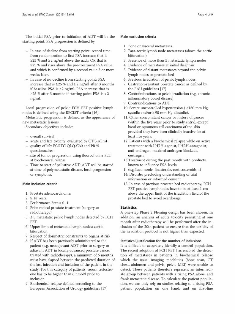
The initial PSA prior to initiation of ADT will be thestarting point. PSA progression is defined by
– In case of decline from starting point: record timefrom randomization to first PSA increase that is≥25 % and ≥ 2 ng/ml above the nadir OR that is≥25 % and rises above the pre-treatment PSA valueand which is confirmed by a second value 3 or moreweeks later.
– In case of no decline from starting point: PSAincrease that is ≥25 % and ≥ 2 ng/ml after 3 monthsif baseline PSA is ≥2 ng/ml. PSA increase that is≥25 % after 3 months if starting point PSA is < 2ng/ml.
Local progression of pelvic FCH PET-positive lymph-nodes is defined using the RECIST criteria [16].Metastatic progression is defined as the appearance of
new metastatic lesions.Secondary objectives include:
– overall survival– acute and late toxicity: evaluated by CTC-AE v4– quality of life: EORTC QLQ-C30 and PR25
questionnaires– site of tumor progression: using fluorocholine PET
at biochemical relapse– Time to start of palliative ADT: ADT will be started
at time of polymetastatic disease, local progressionor symptoms.
Main inclusion criteria
1. Prostate adenocarcinoma;2. ≥ 18 years3. Performance Status 0–14. Prior radical prostate treatment (surgery or
radiotherapy)5. ≤ 5 metastatic pelvic lymph nodes detected by FCH
PET.6. Upper limit of metastatic lymph nodes: aortic
bifurcation7. Respect of dosimetric constraints to organs at risk8. If ADT has been previously administered to the
patient (e.g. neoadjuvant ADT prior to surgery oradjuvant ADT in locally-advanced prostate cancertreated with radiotherapy), a minimum of 6 monthsmust have elapsed between the predicted duration ofthe last injection and inclusion of the patient in thestudy. For this category of patients, serum testoster-one has to be higher than 6 nmol/l prior toinclusion.
9. Biochemical relapse defined according to theEuropean Association of Urology guidelines [17]
Main exclusion criteria
1. Bone or visceral metastases2. Para-aortic lymph node metastases (above the aortic
bifurcation)3. Presence of more than 5 metastatic lymph nodes4. Evidence of metastases at initial diagnosis5. Evidence of distant metastases beyond the pelvic
lymph nodes or prostate bed6. Previous irradiation of pelvic lymph nodes7. Castration-resistant prostate cancer as defined by
the EAU guidelines [17]8. Contraindications to pelvic irradiation (e.g. chronic
inflammatory bowel disease)9. Contraindications to ADT10. Severe uncontrolled hypertension ( ≥160 mm Hg
systolic and/or ≥ 90 mm Hg diastolic).11. Other concomitant cancer or history of cancer
(within the five years prior to study entry), exceptbasal or squamous cell carcinoma of the skinprovided they have been clinically inactive for atleast five years.
12. Patients with a biochemical relapse while on activetreatment with LHRH-agonist, LHRH-antagonist,anti-androgen, maximal androgen blockade,oestrogen.
13.Treatment during the past month with productsknown to influence PSA levels
1. (e.g.fluconazole, finasteride, corticosteroids…)14. Disorder precluding understanding of trial
information or informed consent15. In case of previous prostate bed radiotherapy, FCH
PET-positive lymphnodes have to be at least 1 cmabove the upper limit of the irradiation field of theprostate bed to avoid overdosage.
StatisticsA one-step Phase 2 Fleming design has been chosen. Inaddition, an analysis of acute toxicity persisting at onemonth after radiotherapy will be performed after the in-clusion of the 20th patient to ensure that the toxicity ofthe irradiation protocol is not higher than expected.
Statistical justification for the number of inclusionsIt is difficult to accurately identify a control population.The recent adoption of FCH PET has enabled the detec-tion of metastases in patients in biochemical relapsewhich the usual imaging modalities (bone scan, CTchest, abdomen and pelvis, pelvic MRI) were unable todetect. These patients therefore represent an intermedi-ate group between patients with a rising PSA alone, andfrank metastatic disease. To calculate the patient popula-tion, we can only rely on studies relating to a rising PSApatient population on one hand, and on first-line
Supiot et al. BMC Cancer (2015) 15:646 Page 4 of 9

metastatic patients on the other hand. Prospective ran-domized data gathered from patients with a rising PSAshowed that the median duration of biochemical controlwas 20 months with intermittent ADT [1]. Prospectiverandomized data concerning intermittent ADT in meta-static patients showed that 75 % of patients had resumedADT after a mean duration of 15 months off [3]. Basedon retrospective data, Ost et al. estimated that 51 % ofpatients are progression-free one to three years after sal-vage surgery or radiotherapy for pelvic oligometastaticdisease [10]. Moreover, Rischke et al. showed a 50 %clinical recurrence at 2 year for patients undergoingboth salvage lymphnode dissection and pelvic radiother-apy [18]. We therefore estimate that a biochemicalrelapse-free survival at 2 years of 70 % would be a sig-nificant result.Based on a one-step Fleming design [19], we wish to
be 95 % (α < 5 %) certain that any difference observedbetween the standard and experiment treatment groupsis not due to the play of chance. We also want to be ableto detect such a difference with 95 % power (b = 5 %).With these α and b, rejecting the null hypothesis H0:Π ≤ 50 %, and accepting the alternative hypothesis H1:Π ≥ 70 %, will require 63 evaluable patients.Around 10 % of patients may be lost to follow-up be-
fore the evaluation of biochemical relapse at 2 years, soto ensure that we retain at least 63 patients until thisevaluation it will be necessary to recruit 70 patients tothis study.If 39 or more of these 63 evaluable patients are still
free of biochemical relapse at 24 months, we may rejectthe null hypothesis H0: Π ≤ 50 % with a risk of error of3.85 % and a power of 93.5 % to detect the alternativehypothesis H1: Π ≥ 70 %.
Level of statistical significance setThe power (1 - β) is 93.5 % and the α risk <5 %.
Statistical criteria for stopping the studyIf, among the first 20 patients, 10 or more patientspresent with grade 2 toxicity, we can state that the rateof grade 2 complications is greater than the referencenorm of 30 % (p = 0.048). In this case the Data SafetyMonitoring Board (DSMB) will meet to analyze the tox-icity and decide whether to continue the study.If, among the first 20 patients, 2 or more patients
present with grade 3 toxicity, we can state that the rate ofgrade 3 complications is above the reference norm of 2 %(p = 0.059). In this case the DSMB will meet to analyze thetoxicity and decide whether to continue the study.
Detailed description of techniques to be used
1. Androgen Deprivation Therapy (ADT)
The recommended ADT is Eligard ® 45 mg actingfor 6 months. The use of other ADTs (LH-RHagonists or antagonists) is possible. The overalltreatment duration is 6 months. It will be ideallyadministered on the first day of radiotherapy, orwithin the 3 months prior to the first day ofradiotherapy.
2. Radiation Therapy
� General recommendations– The use of Intensity-Modulated Radiation Ther-
apy (IMRT) is mandatory.– Irradiation will be delivered with a non-empty
bladder and an empty rectum. It is recommendedthat the patient empties his bladder and rectumone hour before the planning CT-scan and beforeeach radiotherapy treatment, and then drink atleast 1/3 L of water after voiding. The one hourdelay will be adapted to the patient’s continence.
– Daily verification of patient’s organ positioningusing IGRT is mandatory
– Verification of in vivo dose delivery is mandatory– The use of a record and verify processing
software is mandatory.� Preparation of treatment
Planning CT of the pelvic cavity that includes allof the pelvic lymph nodes will be performed in thesupine position, with iv injection of contrast agentto help visualize the pelvic vessels. Oral contrastagent will be administered to assist in the thedetermination of the small bowel volume. Theminimum CT resolution will be at least 3 mmthick slices every 3 mm, but thinner slices may benecessary for very small volume lymphadenopathy.Merging the CT with pelvic MRI may helpvisualize the vascular axes and pelvic lymph nodes.Fusion with FCH PET:The FCH PET images will be segmented using athreshold adapted to the size of thelymphadenopathy. These images will be used tolocate the pathological lymph nodes, but thetumor volume (GTV) will be contoured on theplanning CT.Target volumesMetastatic lymph nodes
GTV1 to GTV5 will be delineated on the planningCT-scan with the aid of FCH PET.A margin of 5 mm around the GTVs will define the
clinical target volumes (CTVs).An additional margin of 5 mm around the CTVs will de-
fine the Planned Target Volumes (PTVs). The intersection
Supiot et al. BMC Cancer (2015) 15:646 Page 5 of 9
between PTVs and small intestine will be removed fromthe PTVs.
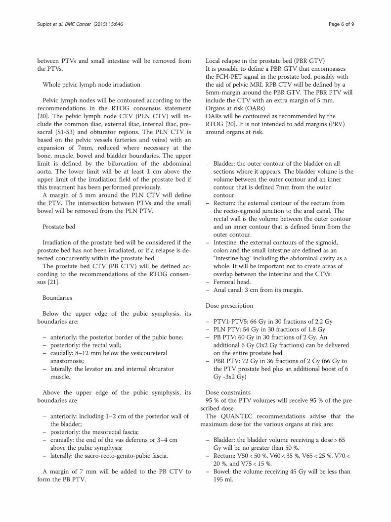
Whole pelvic lymph node irradiation
Pelvic lymph nodes will be contoured according to therecommendations in the RTOG consensus statement[20]. The pelvic lymph node CTV (PLN CTV) will in-clude the common iliac, external iliac, internal iliac, pre-sacral (S1-S3) and obturator regions. The PLN CTV isbased on the pelvic vessels (arteries and veins) with anexpansion of 7mm, reduced where necessary at thebone, muscle, bowel and bladder boundaries. The upperlimit is defined by the bifurcation of the abdominalaorta. The lower limit will be at least 1 cm above theupper limit of the irradiation field of the prostate bed ifthis treatment has been performed previously.A margin of 5 mm around the PLN CTV will define
the PTV. The intersection between PTVs and the smallbowel will be removed from the PLN PTV.
Prostate bed
Irradiation of the prostate bed will be considered if theprostate bed has not been irradiated, or if a relapse is de-tected concurrently within the prostate bed.The prostate bed CTV (PB CTV) will be defined ac-
cording to the recommendations of the RTOG consen-sus [21].
Boundaries
Below the upper edge of the pubic symphysis, itsboundaries are:
– anteriorly: the posterior border of the pubic bone;– posteriorly: the rectal wall;– caudally: 8–12 mm below the vesicoureteral
anastomosis;– laterally: the levator ani and internal obturator
muscle.
Above the upper edge of the pubic symphysis, itsboundaries are:
– anteriorly: including 1–2 cm of the posterior wall ofthe bladder;
– posteriorly: the mesorectal fascia;– cranially: the end of the vas deferens or 3–4 cm
above the pubic symphysis;– laterally: the sacro-recto-genito-pubic fascia.
A margin of 7 mm will be added to the PB CTV toform the PB PTV.
Local relapse in the prostate bed (PBR GTV)It is possible to define a PBR GTV that encompassesthe FCH-PET signal in the prostate bed, possibly withthe aid of pelvic MRI. RPB CTV will be defined by a5mm-margin around the PBR GTV. The PBR PTV willinclude the CTV with an extra margin of 5 mm.Organs at risk (OARs)OARs will be contoured as recommended by theRTOG [20]. It is not intended to add margins (PRV)around organs at risk.
– Bladder: the outer contour of the bladder on allsections where it appears. The bladder volume is thevolume between the outer contour and an innercontour that is defined 7mm from the outercontour.
– Rectum: the external contour of the rectum fromthe recto-sigmoid junction to the anal canal. Therectal wall is the volume between the outer contourand an inner contour that is defined 5mm from theouter contour.
– Intestine: the external contours of the sigmoid,colon and the small intestine are defined as an“intestine bag” including the abdominal cavity as awhole. It will be important not to create areas ofoverlap between the intestine and the CTVs.
– Femoral head.– Anal canal: 3 cm from its margin.
Dose prescription
– PTV1-PTV5: 66 Gy in 30 fractions of 2.2 Gy– PLN PTV: 54 Gy in 30 fractions of 1.8 Gy– PB PTV: 60 Gy in 30 fractions of 2 Gy. An
additional 6 Gy (3x2 Gy fractions) can be deliveredon the entire prostate bed.
– PBR PTV: 72 Gy in 36 fractions of 2 Gy (66 Gy tothe PTV prostate bed plus an additional boost of 6Gy -3x2 Gy)
Dose constraints95 % of the PTV volumes will receive 95 % of the pre-
scribed dose.The QUANTEC recommendations advise that the
maximum dose for the various organs at risk are:
– Bladder: the bladder volume receiving a dose > 65Gy will be no greater than 50 %.
– Rectum: V50 < 50 %, V60 < 35 %, V65 < 25 %, V70 <20 %, and V75 < 15 %.
– Bowel: the volume receiving 45 Gy will be less than195 ml.
Supiot et al. BMC Cancer (2015) 15:646 Page 6 of 9

– Femoral head: <5 % of each femoral head shouldreceive a dose > 50 Gy.
– Anal canal: D55 < 100 %.
Where the PTV coverage conflicts with the OAR con-straints, the OAR protection will be privileged. For thisreason, the PTV may be reduced to the CTV.
Image guidance
Image-guided radiotherapy (IGRT) will be performeddaily. The daily registration will be based at least onbone structures. It will be possible to readjust to thenodal structures in case of displacements > 1 cm.
Quality control.
� Quality control of target volumes and critical organs
A GETUG RT quality assurance committee has beenestablished consisting of a radiation oncologist and a ra-diation physicist. This committee is available for consult-ation should questions arise concerning the treatmentprotocol. The committee will initially accredit eachcentre on the basis of an electronic copy of a single caseplan that demonstrates:
� CTV and PTV contoured according to protocol;� organs at risk (bladder and rectal walls and femoral
heads) contoured according to protocol;� DVH for intestine, bladder and rectal wall, PTV and
CTV that meet dose constraints;� a statement that patients will be treated with an
approved daily image guidance technique.
All treatment plans will be sent via a web-based plat-form for central reviewing and approval prior to the be-ginning of the radiotherapy treatment.
� Quality control of IMRTAll IMRT plans will undergo quality assuranceevaluation with ion chamber measurements or anequivalent method of dose verification to verify theabsolute dose for each IMRT field and film dosimetryto measure the relative dose for each IMRT field, as instandard clinical practice. Independent Monitor Unitscalculation may be substituted for ion chamberdosimetry when available.
Follow-upFollow-up is described in Table 1. Quality of life will beevaluated prior to treatment and one month after
completion of radiotherapy, and then every 6 months for2 years using EORTC QLQ C30 and PR25. PSA andtestosterone will be determined prior to radiotherapy,then 1 month after completion and then every 6months. Repeat FCH-PET will be performed at bio-chemical recurrence.
DiscussionThis trial addresses the use of radiotherapy and androgendeprivation therapy (ADT) in that emerging category ofpatients whose oligometastatic disease has been identifiedon fluorocholine PET imaging. This new category falls be-tween true metastatic disease where ADT is a mainstay,and those patients with a rising PSA without visible le-sions on conventional imaging, in whom intermittentADT has been shown to be no less effective than continu-ous ADT. In this patient population, the role of radiother-apy is undetermined and two different approaches arecurrently in progess. The first approach is to consider thatradiotherapy may delay the need for ADT by treating eachvisible oligometastasis using SBRT. Many single-centerstudies have been published (for review: [6]) and a ran-domized phase II study is comparing surveillance andsalvage stereotactic radiotherapy or surgery to PETCholine-positive lymph nodes [15]. The second approachis that salvage radiotherapy may increase relapse-free sur-vival or even cure some patients as based on recent retro-spective series [9, 22]. Choline PET is currently the mostvalidated prostate cancer staging modality, as compared topelvic MRI, total bone scan or CT-scan. Other imagingmodalities might perform better in the near future, suchas whole body MRI or PSMA targeting radiolabeledagents. These imaging modalities are likely to becomestandard of care very soon and may further better defineoligometastatic burden in future clinical trials. We feelthat it is important to explore different approaches totreatment to inform the oncologists and urologists whoare now routinely faced with the decision as to the bestcourse of action in this new category of patients.
� Project planning:– The role of each team
It is expected that each team will recruit a mean num-ber of 4 patients during the inclusion period. Each teamwill consist of a local radiation oncologist and his or hernuclear oncology and urology colleagues.
– Project calendarThe study has been submitted and approved byregulatory authorities (ANSM; date of approval: 13/06/14) and ethics committee (Centre de Protectiondes Personnes Ouest IV - Nantes; date of approval:3/05/14). The study opened in september 2014. A
Supiot et al. BMC Cancer (2015) 15:646 Page 7 of 9

written informed consent will be obtained from thestudy participants. Recruitment is planned until theend of 2016. After a two-year minimum follow-up,final results will be presented during the last trimes-ter 2018.
– Project co-ordinationThe project has been extensively discussed andapproved by the GETUG. Patient recruitment willbe monitored at each quarterly GETUG meeting.
AbbreviationsADT: Androgen deprivation therapy; PET: Positron emission tomography; FCH:18F-Choline, fluorocholine; SBRT: Stereotactic body radiotherapy; IG-IMRT: Imageguided-intensity modulated radiation therapy; GETUG: Groupe d’Etude desTumeurs Uro-Génitales; PLN CTV: Pelvic lymph node clinical target volume; PBCTV: Prostate bed CTV; PBR GTV: Prostate bed relapse gross tumor volume.
Competing interestsThis study is funded by an unrestricted research grant from Astellas France.
Authors’ contributionsSS, ER, VP, MHM, FP participated in the design of the study and LC designedthe statistical analysis. SS conceived the study, and participated in its designand coordination and helped to draft the manuscript. All authors read andapproved the final manuscript.
AcknowledgmentsThis study is sponsored by a research grant from Astellas pharmaceuticals,France.
List of investigatorsDr Stéphane SUPIOT, ICO site René Gauducheau, NANTES-ST HERBLAIN
Dr Patrice CELLIER, ICO site Paul Papin, ANGERSDr Paul SARGOS, Institut Bergonié, BORDEAUXDr Marlon SILVA, Centre François Baclesse, CAENDr Geneviève LOOS, Centre Jean Perrin, CLERMONT-FERRANDDr Yazid BELKACEMI, CHU Henri Mondor, APHP CRETEILDr Gilles CREHANGE , Centre GF Leclerc, DIJONDr Fabrice DENIS, Clinique Victor Hugo, LE MANSDr David PASQUIER, Centre Oscar Lambret, LILLEDr Pascal POMMIER, Centre Léon Bérard, LYONDr Didier PEIFFERT, IC Lorraine, NANCYDr Xavier BUTHAUD, Centre Catherine de Sienne, NANTESDr Renaud DE CREVOISIER, Centre Eugène Marquis, RENNESDr Guy DE BRISSON DE LA ROCHE, IC de la Loire Lucien Neuwirth, STETIENNEDr Igor LATORZEFF, ONCORAD Garonne, Clinique Pasteur, TOULOUSEDr Ali HASBINI, Clinique Pasteur, BREST.
Author details1Department of Radiation Oncology, Institut de Cancérologie de l’Ouest,Boulevard J. Monod, F-44800 Nantes, St-Herblain, France. 2Centre deRecherche en Cancérologie Nantes-Angers (CRCNA), UMR 892 Inserm - 6299CNRS, Institut de Recherche en Santé de l’Université de Nantes, 8 quaiMoncousu, BP 7072144007 Nantes cedex 1, France. 3Department of Research,Institut de Cancérologie de l’Ouest-René Gauducheau, boulevard JacquesMonod, Saint Herblain 44800, France. 4Department of Statistics, Institut deCancérologie de l’Ouest-René Gauducheau, boulevard Jacques Monod, SaintHerblain 44800, France.
Received: 2 January 2015 Accepted: 27 July 2015
References1. Umbehr MH, Muntener M, Hany T, Sulser T, Bachmann LM. The role of 11C-
choline and 18F-fluorocholine positron emission tomography (PET) and
Table 1 Study calendar
Visits Inclusion Treatmentperiod
follow-up
Visits Max 2monthsprior toRT day 1
M1 1monthafterend of RT
Before progression After progression
Every 6 months during2 years
Every 6 monthsuntil progression
Every 6 monthsuntil death
Inclusion/exclusion criteria X
Signed consent form X
Inclusion (max 2 months prior to RT) X
FCH PET X (a) X (b)
Physical examination with PS X X (d) X X X X
Prior history, tumor characteristics X
Blood Pressure X X (d) X X
Acute toxicity during ADT and RT (f) X (d) X
Late toxicity (f) X
QLQ-C30 et QLQ-PR25 X X X
PSA X X (f) X (f) X (f)
Testosterone X (g) X X X
ADT (c) + RT X
(a) maximum 3 months prior to initiation of ADT(b) at progression(c) 6 months treatment staring on RT day 1 or maximum 3 months prior to RT(d) once per week during RT(e) biochemical progression = PSA higher than inclusion PSA with 2 successive rises in the same laboratory(f) NCI-CTCAE v4.0(g) 28 days prior to ADT
Supiot et al. BMC Cancer (2015) 15:646 Page 8 of 9

PET/CT in prostate cancer: a systematic review and meta-analysis. Eur Urol.2013;64(1):106–17.
2. Almaghrabi MY, Supiot S, Paris F, Mahe MA, Rio E. Stereotactic bodyradiation therapy for abdominal oligometastases: a biological and clinicalreview. Radiat Oncol. 2012;7:126.
3. Hussain M, Tangen CM, Berry DL, Higano CS, Crawford ED, Liu G, et al.Intermittent versus continuous androgen deprivation in prostate cancer.N Engl J Med. 2013;368(14):1314–25.
4. Crook JM, O'Callaghan CJ, Duncan G, Dearnaley DP, Higano CS, Horwitz EM,et al. Intermittent androgen suppression for rising PSA level afterradiotherapy. N Engl J Med. 2012;367(10):895–903.
5. Niraula S, Le LW, Tannock IF. Treatment of prostate cancer with intermittentversus continuous androgen deprivation: a systematic review ofrandomized trials. J Clin Oncol. 2013;31(16):2029–36.
6. De Bari B, Alongi F, Buglione M, Campostrini F, Briganti A, Berardi G, et al.Salvage therapy of small volume prostate cancer nodal failures: a review ofthe literature. Crit Rev Oncol Hematol. 2014;90(1):24–35.
7. Suardi N, Gandaglia G, Gallina A, Di Trapani E, Scattoni V, Vizziello D, et al.Long-term outcomes of salvage lymph node dissection for clinicallyrecurrent prostate cancer: results of a single-institution series with aminimum follow-up of 5 years. Eur Urol. 2014;67:299–309.
8. Jereczek-Fossa BA, Beltramo G, Fariselli L, Fodor C, Santoro L, Vavassori A,et al. Robotic image-guided stereotactic radiotherapy, for isolated recurrentprimary, lymph node or metastatic prostate cancer. Int J Radiat Oncol BiolPhys. 2012;82(2):889–97.
9. Picchio M, Berardi G, Fodor A, Busnardo E, Crivellaro C, Giovacchini G, et al.(11)C-Choline PET/CT as a guide to radiation treatment planning of lymph-node relapses in prostate cancer patients. Eur J Nucl Med Mol Imaging.2014;41(7):1270–9.
10. Ost P, Bossi A, Decaestecker K, De Meerleer G, Giannarini G, Karnes RJ, et al.Metastasis-directed therapy of regional and distant recurrences aftercurative treatment of prostate cancer: a systematic review of the literature.Eur Urol. 2014;67:852–63.
11. Adkison JB, McHaffie DR, Bentzen SM, Patel RR, Khuntia D, Petereit DG, et al.Phase I trial of pelvic nodal dose escalation with hypofractionated IMRT forhigh-risk prostate cancer. Int J Radiat Oncol Biol Phys. 2012;82(1):184–90.
12. Passoni NM, Suardi N, Abdollah F, Picchio M, Giovacchini G, Messa C, et al.Utility of [11C]choline PET/CT in guiding lesion-targeted salvage therapies inpatients with prostate cancer recurrence localized to a single lymph nodeat imaging: results from a pathologically validated series. Urol Oncol.2014;32(1):38. e9–16.
13. Lepinoy A, Cochet A, Cueff A, Cormier L, Martin E, Maingon P, et al. Patternof occult nodal relapse diagnosed with (18)F-fluoro-choline PET/CT inprostate cancer patients with biochemical failure after prostate-onlyradiotherapy. Radiother Oncol. 2014;111(1):120–5.
14. Crehange G, Roach 3rd M, Martin E, Cormier L, Peiffert D, Cochet A, et al.Salvage reirradiation for locoregional failure after radiation therapy forprostate cancer: who, when, where and how? Cancer Radiother.2014;18(5–6):524–34.
15. Decaestecker K, De Meerleer G, Ameye F, Fonteyne V, Lambert B, Joniau S,et al. Surveillance or metastasis-directed Therapy for OligoMetastaticProstate cancer recurrence (STOMP): study protocol for a randomized phaseII trial. BMC Cancer. 2014;14:671.
16. Eisenhauer EA, Therasse P, Bogaerts J, Schwartz LH, Sargent D, Ford R, et al.New response evaluation criteria in solid tumours: revised RECIST guideline(version 1.1). Eur J Cancer. 2009;45(2):228–47.
17. Heidenreich A, Bastian PJ, Bellmunt J, Bolla M, Joniau S, van der Kwast T,Mason M, Matveev V, Wiegel T, Zattoni F, Mottet N: EAU guidelines onprostate cancer. part II: treatment of advanced, relapsing, and castration-resistant prostate cancer. Eur Urol 2014;65(2):467–79.
18. Rischke HC, Schultze-Seemann W, Wieser G, Kronig M, Drendel V, StegmaierP, et al. Adjuvant radiotherapy after salvage lymph node dissection becauseof nodal relapse of prostate cancer versus salvage lymph node dissectiononly. Strahlenther Onkol. 2015;191(4):310-20.
19. Fleming TR. One-sample multiple testing procedure for phase II clinicaltrials. Biometrics. 1982;38(1):143-51.
20. Lawton C, Michalski J, El-Naqa I, Buyyounouski MK, Lee WR, Menard C, et al.RTOG GU Radiation oncology specialists reach consensus on pelvic lymphnode volumes for high-risk prostate cancer. Int J Radiat Oncol Biol Phys.2009;74(2):383-7
21. Michalski JM, Lawton C, el Naqa I, Ritter M, O'Meara E, Seider MJ, et al.Development of RTOG consensus guidelines for the definition of the clinicaltarget volume for postoperative conformal radiation therapy for prostatecancer Int J Radiat Oncol Biol Phys. 2010;76(2):361-8
22. Miralbell et Buchegger. PET/CT imaging and the oligometastatic prostatecancer patient: an opportunity for a curative approach with high-doseradiotherapy? Eur J Nucl Med Mol Imaging. 2014;41(7):1267-9
Submit your next manuscript to BioMed Centraland take full advantage of:
• Convenient online submission
• Thorough peer review
• No space constraints or color figure charges
• Immediate publication on acceptance
• Inclusion in PubMed, CAS, Scopus and Google Scholar
• Research which is freely available for redistribution
Submit your manuscript at www.biomedcentral.com/submit
Supiot et al. BMC Cancer (2015) 15:646 Page 9 of 9