Oim 2005
39
Materials Characterization Lab www.mri.psu.edu/mcl OIM/EBSD Maria Klimkiewicz [email protected] 865-3624 July 20, 2005
-
Upload
langtudaikieu -
Category
Business
-
view
706 -
download
8
Transcript of Oim 2005
Materials Characterization Labwww.mri.psu.edu/mcl
OIM/EBSD
Maria Klimkiewicz
865-3624
July 20, 2005
Materials Characterization Labwww.mri.psu.edu/mcl
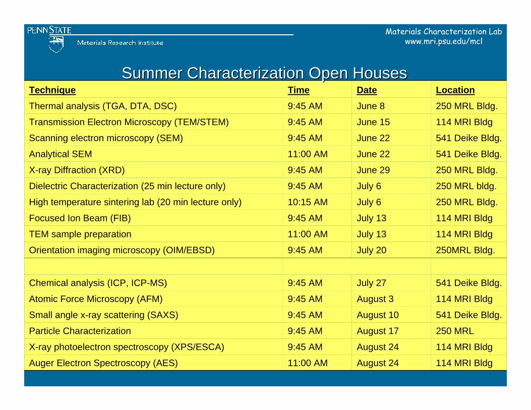
250 MRLAugust 179:45 AMParticle Characterization
114 MRI BldgAugust 249:45 AMX-ray photoelectron spectroscopy (XPS/ESCA)
114 MRI BldgAugust 2411:00 AMAuger Electron Spectroscopy (AES)
541 Deike Bldg.July 279:45 AMChemical analysis (ICP, ICP-MS)
541 Deike Bldg.August 109:45 AMSmall angle x-ray scattering (SAXS)
114 MRI Bldg August 39:45 AMAtomic Force Microscopy (AFM)
250MRL Bldg.July 209:45 AMOrientation imaging microscopy (OIM/EBSD)
114 MRI BldgJuly 1311:00 AMTEM sample preparation
114 MRI BldgJuly 139:45 AMFocused Ion Beam (FIB)
250 MRL Bldg.July 610:15 AMHigh temperature sintering lab (20 min lecture only)
250 MRL bldg.July 69:45 AMDielectric Characterization (25 min lecture only)
250 MRL Bldg.June 299:45 AMX-ray Diffraction (XRD)
541 Deike Bldg.June 2211:00 AMAnalytical SEM
541 Deike Bldg.June 229:45 AMScanning electron microscopy (SEM)
114 MRI BldgJune 159:45 AMTransmission Electron Microscopy (TEM/STEM)
250 MRL Bldg.June 89:45 AMThermal analysis (TGA, DTA, DSC)
LocationDateTimeTechniqueSummer Characterization Open HousesSummer Characterization Open Houses
Materials Characterization Labwww.mri.psu.edu/mcl
BeaverStadium
Park Ave.
Park Ave.
Porter RoadPollock Road
University Drive
College Ave.
ShortlidgeR
oad North
Bur ro w
esR
oa d
00
00
00
00
00
00
00
00
00
Centre Community
Hospital
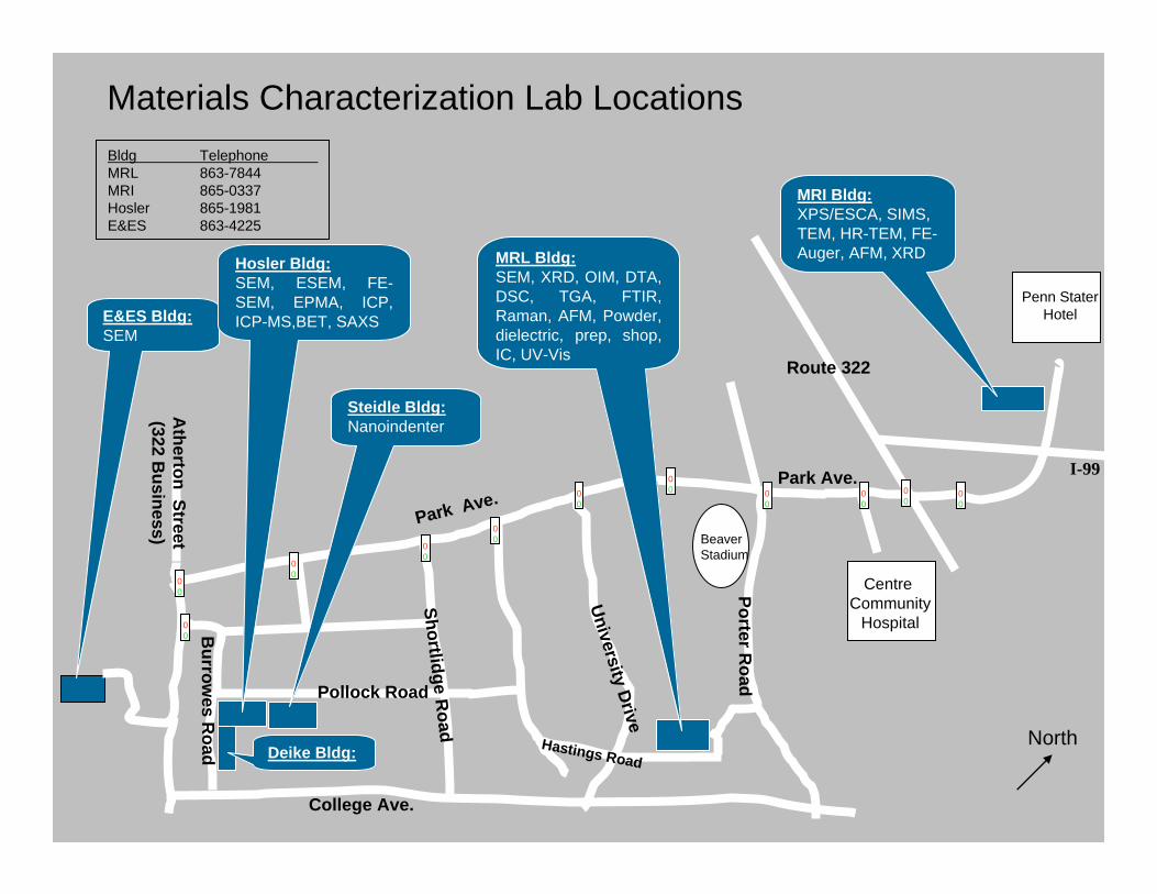
E&ES Bldg:SEM
Hosler Bldg:SEM, ESEM, FE-SEM, EPMA, ICP, ICP-MS,BET, SAXS
MRI Bldg:XPS/ESCA, SIMS, TEM, HR-TEM, FE-Auger, AFM, XRD
Atherton Street
(322 Business)
MRL Bldg:SEM, XRD, OIM, DTA, DSC, TGA, FTIR, Raman, AFM, Powder, dielectric, prep, shop, IC, UV-Vis
Hastings Road
Penn StaterHotel
00
Materials Characterization Lab LocationsBldg TelephoneMRL 863-7844MRI 865-0337Hosler 865-1981E&ES 863-4225
Route 322
I-99 00
Steidle Bldg:Nanoindenter
Deike Bldg:
Materials Characterization Labwww.mri.psu.edu/mcl
Outline
― Introduction to OIM/EBSD― OIM examples― how to get started ―OIM resources― a brief lab tour
Materials Characterization Labwww.mri.psu.edu/mcl
Introduction to OIM/EBSD
Materials Characterization Labwww.mri.psu.edu/mcl
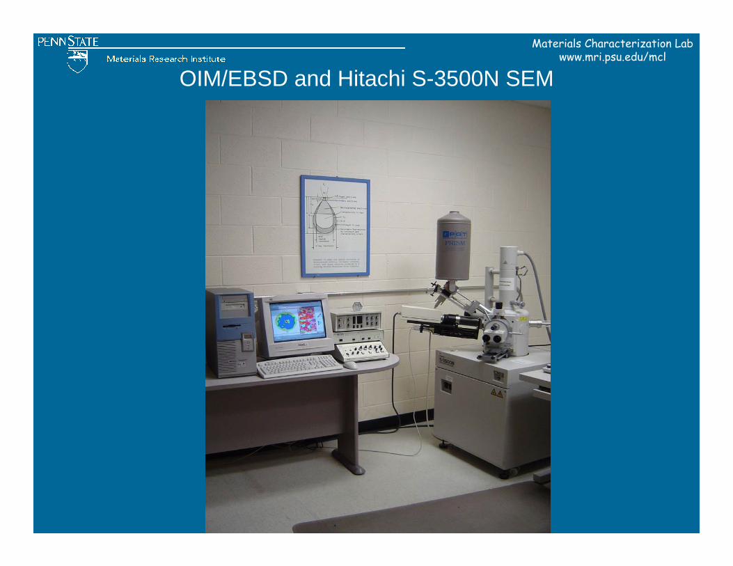
OIM/EBSD and Hitachi S-3500N SEM
Materials Characterization Labwww.mri.psu.edu/mcl
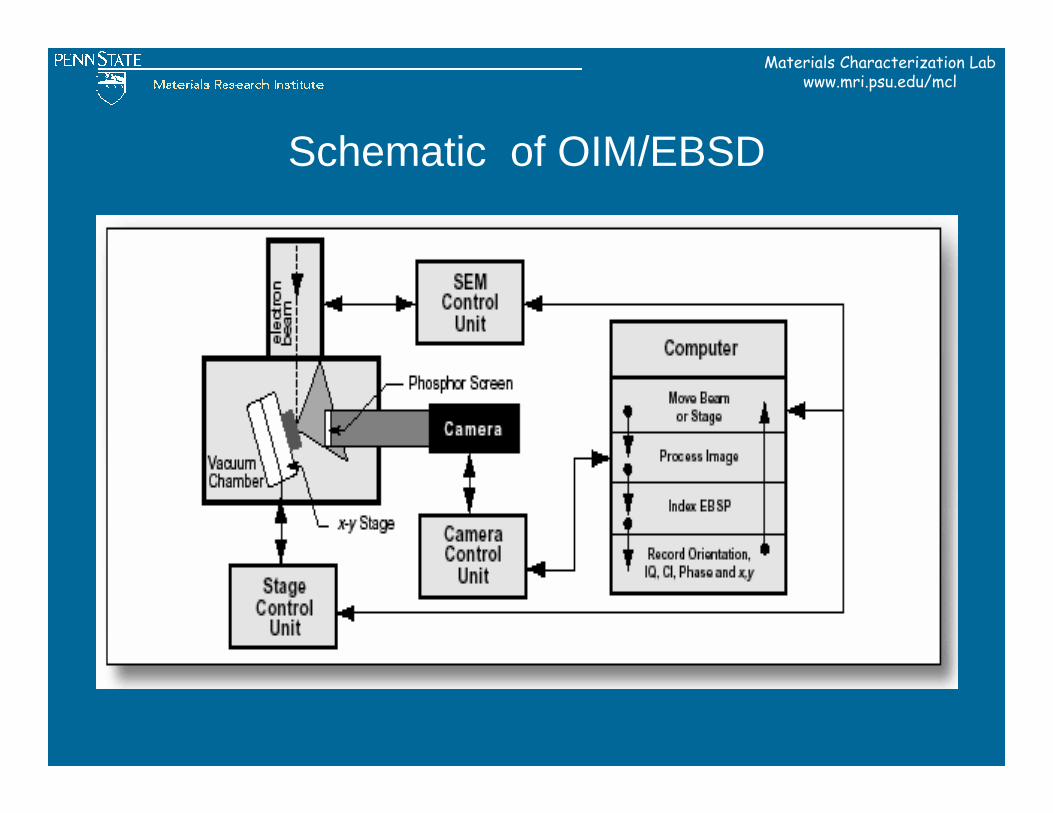
Schematic of OIM/EBSD
Materials Characterization Labwww.mri.psu.edu/mcl
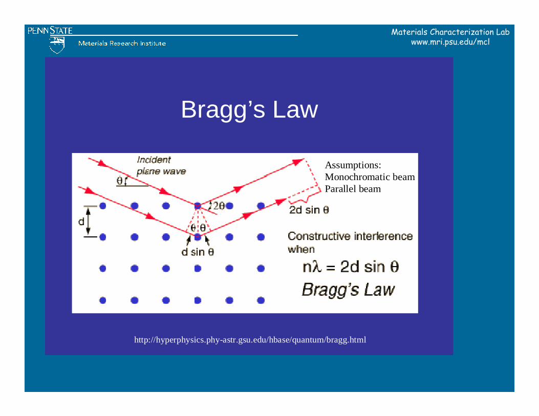
Bragg’s Law
Assumptions:Monochromatic beamParallel beam
http://hyperphysics.phy-astr.gsu.edu/hbase/quantum/bragg.html
Materials Characterization Labwww.mri.psu.edu/mcl
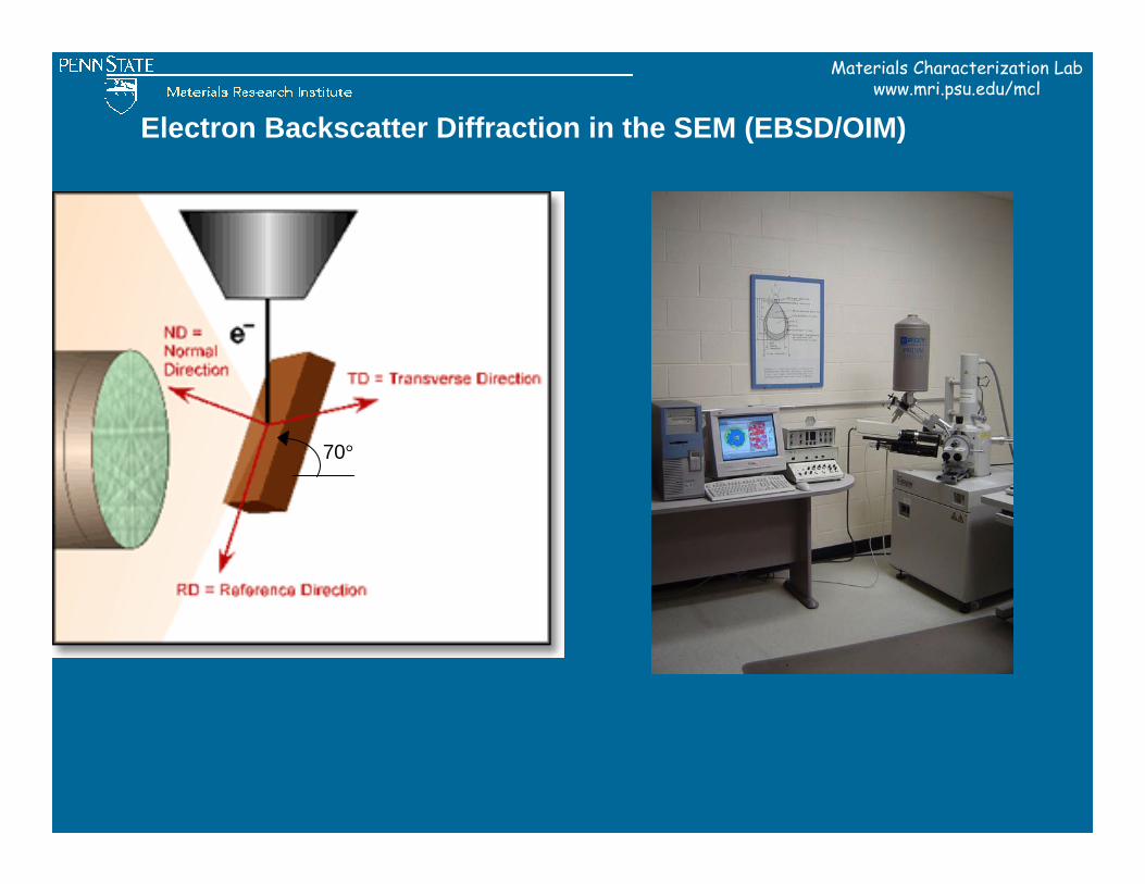
70°
Electron Backscatter Diffraction in the SEM (EBSD/OIM)
Materials Characterization Labwww.mri.psu.edu/mcl
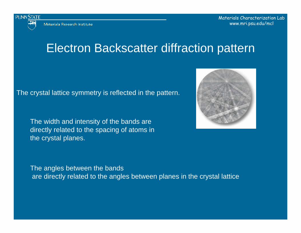
Electron Backscatter diffraction pattern
The crystal lattice symmetry is reflected in the pattern.
The width and intensity of the bands are directly related to the spacing of atoms in the crystal planes.
The angles between the bandsare directly related to the angles between planes in the crystal lattice
Materials Characterization Labwww.mri.psu.edu/mcl
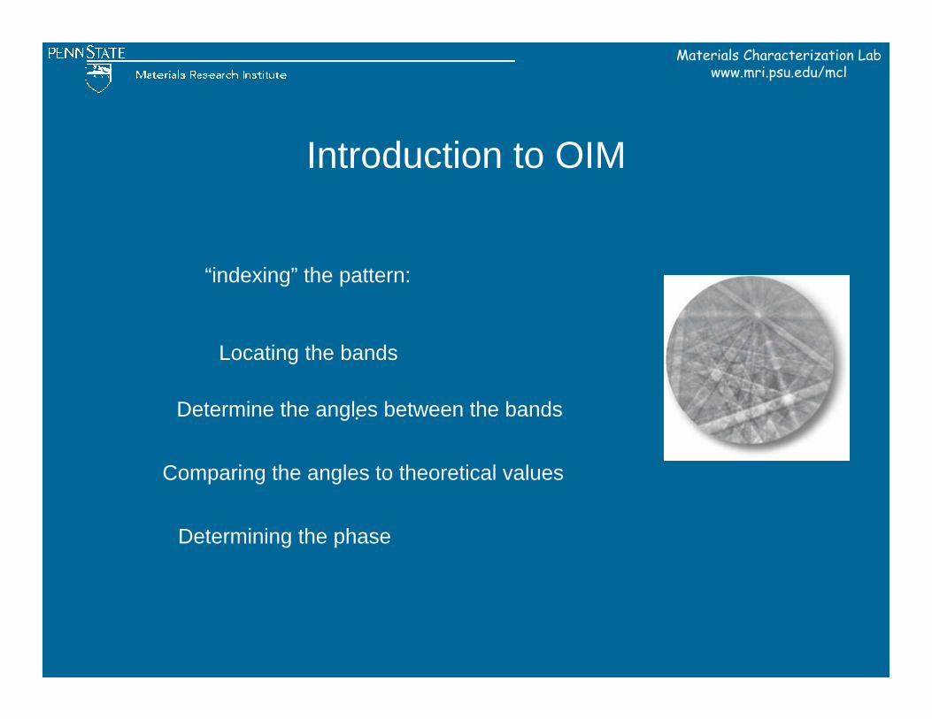
Introduction to OIM
.
“indexing” the pattern:
Locating the bands
Determine the angles between the bands
Comparing the angles to theoretical values
Determining the phase
Materials Characterization Labwww.mri.psu.edu/mcl
112323
211
111
125 013
233323
121
251 141
152
011
031 152
114
125
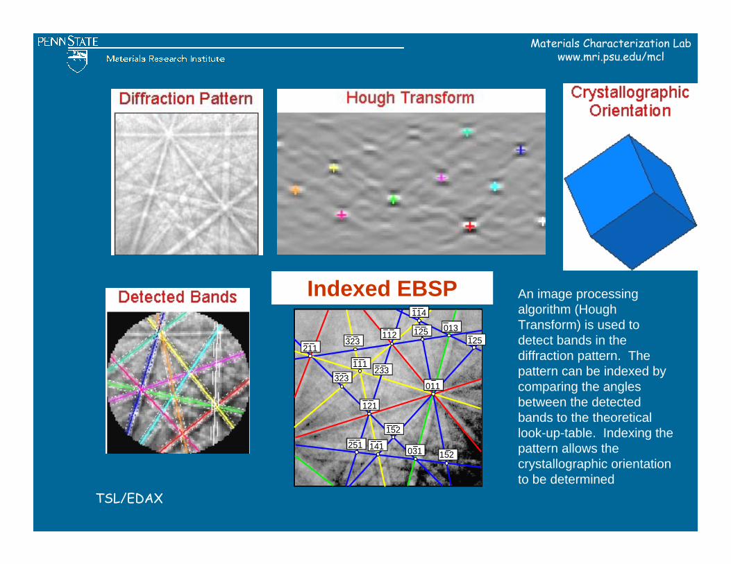
Indexed EBSPIndexed EBSP
TSL/EDAX
An image processing algorithm (Hough Transform) is used to detect bands in the diffraction pattern. The pattern can be indexed by comparing the angles between the detected bands to the theoretical look-up-table. Indexing the pattern allows the crystallographic orientation to be determined
Materials Characterization Labwww.mri.psu.edu/mcl
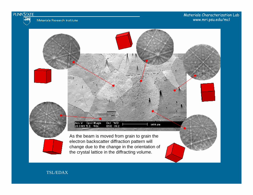
TSL/EDAX
As the beam is moved from grain to grain the electron backscatter diffraction pattern will change due to the change in the orientation of the crystal lattice in the diffracting volume.
Materials Characterization Labwww.mri.psu.edu/mcl
OIM consists of two parts:
• Data Collection- “On Line”
• and Analysis-” Off Line”
Materials Characterization Labwww.mri.psu.edu/mcl
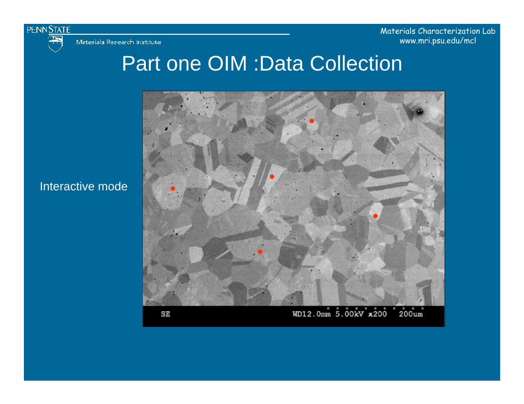
Part one OIM :Data Collection
Interactive mode
Materials Characterization Labwww.mri.psu.edu/mcl
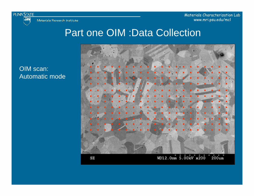
Part one OIM :Data Collection
OIM scan:Automatic mode
. . . . . . . . . . . . . . . . . . . . . . . . . . . . . . . . . . . . . . . . . . . . . . . . . . . . . . . . . . . . . . . . . . . . . . . . . . . . . . . . . . . . . . . . . . . . . . . . . . . . . . . . . . . . . . . . . . . . . . . . . . . . . . . . . . . . . . . . . . . . . . . . . . . . . . . . . . . . . . . . . . . . . . . . . . . . . . . . . . . . . . . . . . . . . . . . . . . . . . . . . . . . . . . . . . . . . . . . . . . . . . . . . . . . . . . . . . . . . . . .
Materials Characterization Labwww.mri.psu.edu/mcl
Part two OIM- Analysis Application
The stored data (location, orientation, image quality, confidence index, and phase) can be processed to create Orientation Imaging Micrographs
Materials Characterization Labwww.mri.psu.edu/mcl
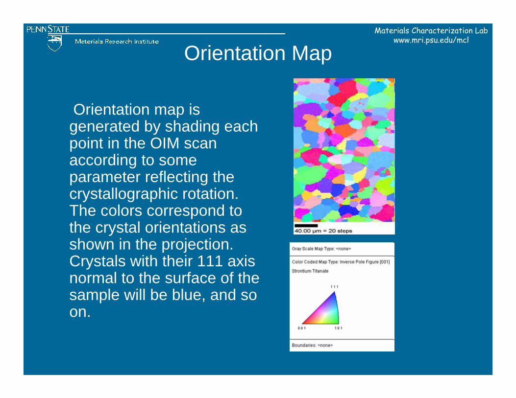
Orientation Map
Orientation map is generated by shading each point in the OIM scan according to some parameter reflecting the crystallographic rotation. The colors correspond to the crystal orientations as shown in the projection. Crystals with their 111 axis normal to the surface of the sample will be blue, and so on.
Materials Characterization Labwww.mri.psu.edu/mcl
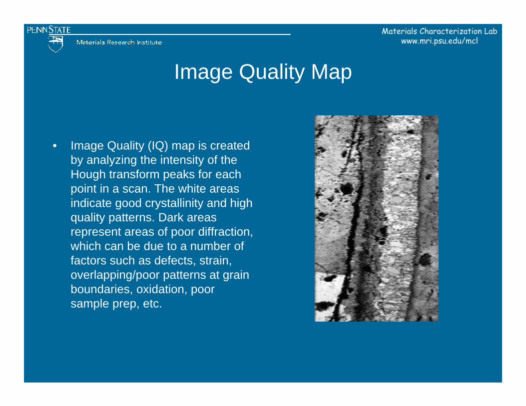
Image Quality Map
• Image Quality (IQ) map is created by analyzing the intensity of the Hough transform peaks for each point in a scan. The white areas indicate good crystallinity and high quality patterns. Dark areas represent areas of poor diffraction, which can be due to a number of factors such as defects, strain, overlapping/poor patterns at grain boundaries, oxidation, poor sample prep, etc.
Materials Characterization Labwww.mri.psu.edu/mcl
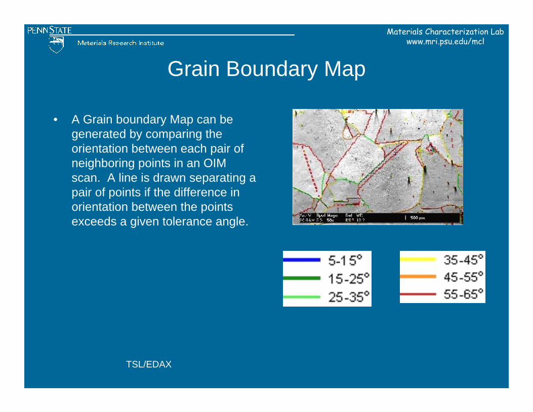
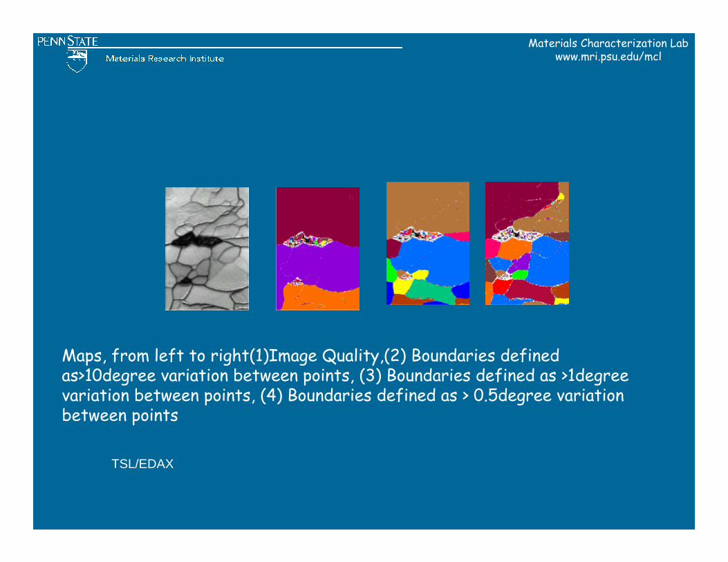
Grain Boundary Map
• A Grain boundary Map can be generated by comparing the orientation between each pair of neighboring points in an OIM scan. A line is drawn separating a pair of points if the difference in orientation between the points exceeds a given tolerance angle.
TSL/EDAX
Materials Characterization Labwww.mri.psu.edu/mcl
Maps, from left to right(1)Image Quality,(2) Boundaries defined as>10degree variation between points, (3) Boundaries defined as >1degree variation between points, (4) Boundaries defined as > 0.5degree variation between points
TSL/EDAX
Materials Characterization Labwww.mri.psu.edu/mcl
OIM examples
Materials Characterization Labwww.mri.psu.edu/mcl
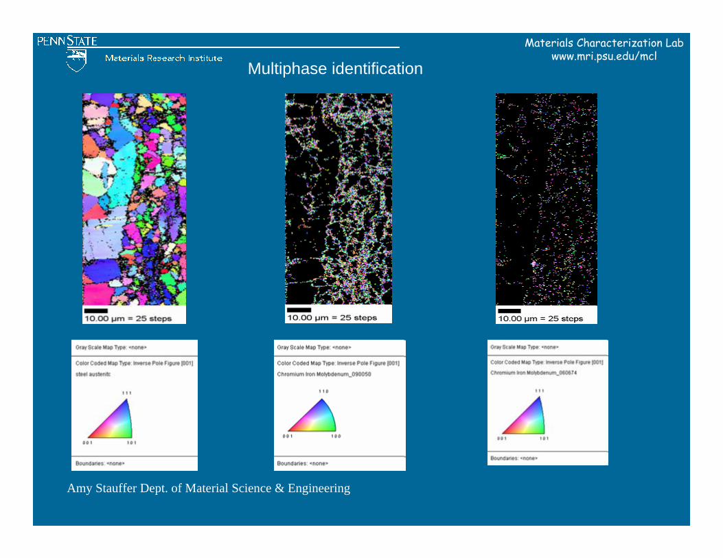
Amy Stauffer Dept. of Material Science & Engineering
Multiphase identification
Materials Characterization Labwww.mri.psu.edu/mcl
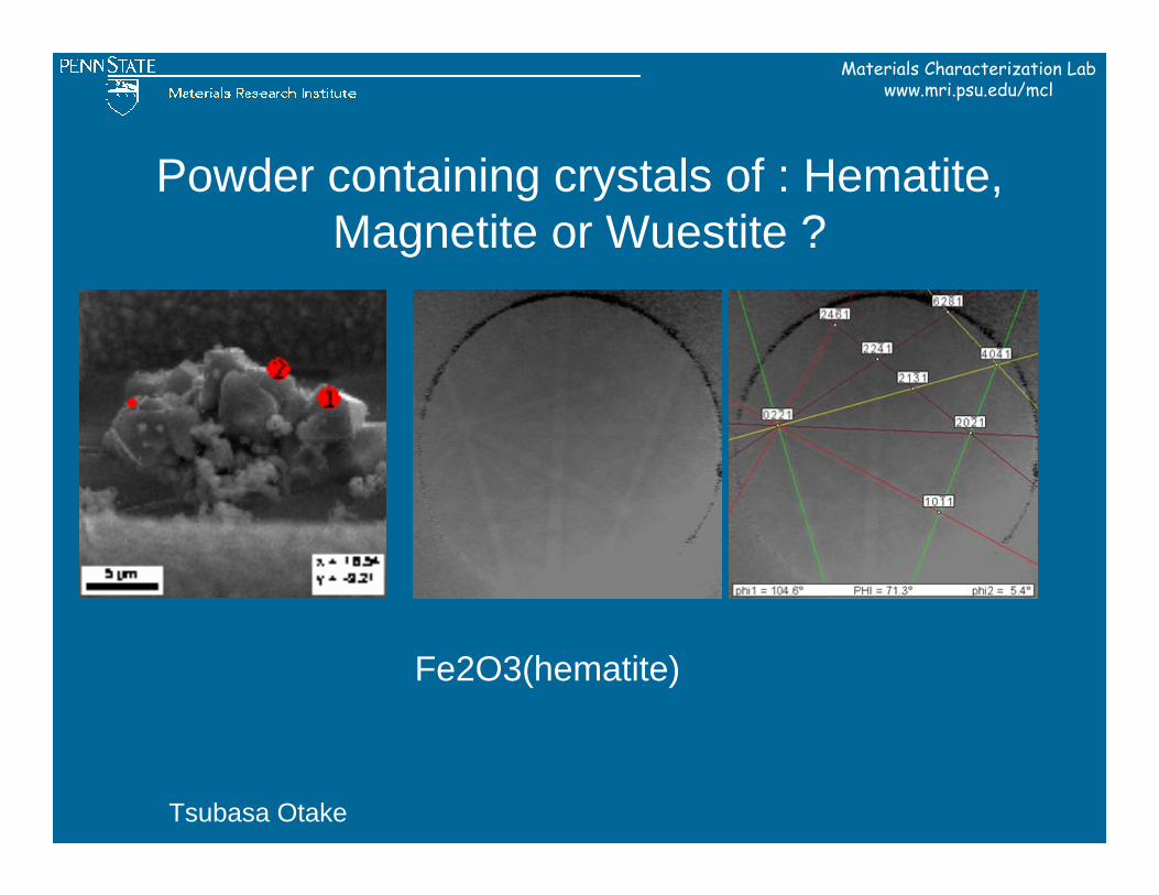
Powder containing crystals of : Hematite, Magnetite or Wuestite ?
Fe2O3(hematite)
Tsubasa Otake
Materials Characterization Labwww.mri.psu.edu/mcl
Tsubasa Otake
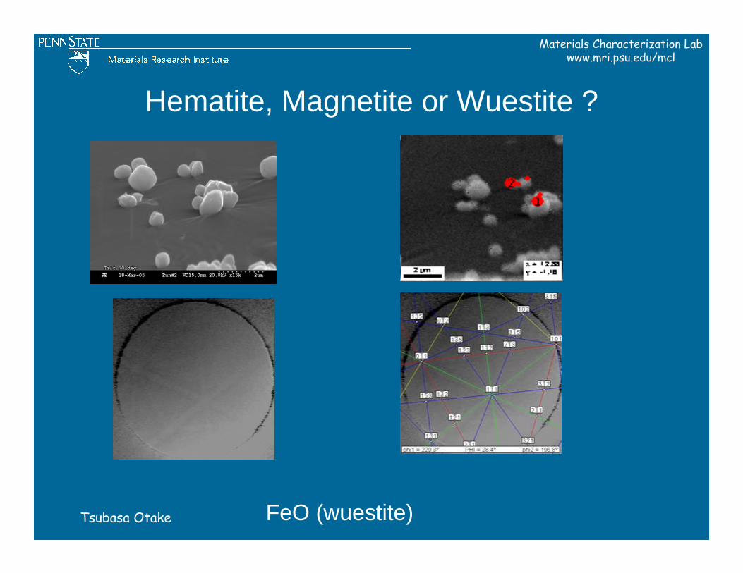
Hematite, Magnetite or Wuestite ?
FeO (wuestite)
Materials Characterization Labwww.mri.psu.edu/mcl
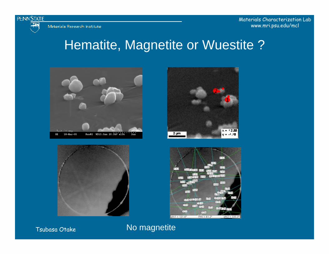
Tsubasa Otake
Hematite, Magnetite or Wuestite ?
No magnetite
Materials Characterization Labwww.mri.psu.edu/mcl
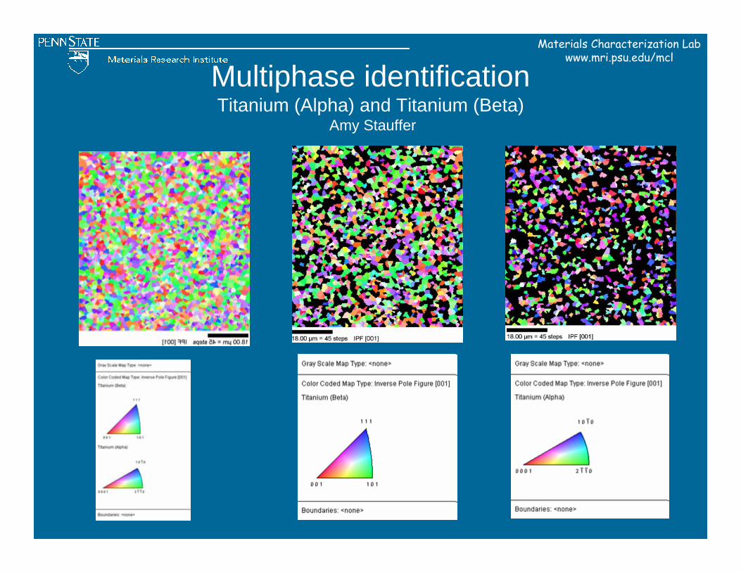
Multiphase identificationTitanium (Alpha) and Titanium (Beta)
Amy Stauffer
Materials Characterization Labwww.mri.psu.edu/mcl
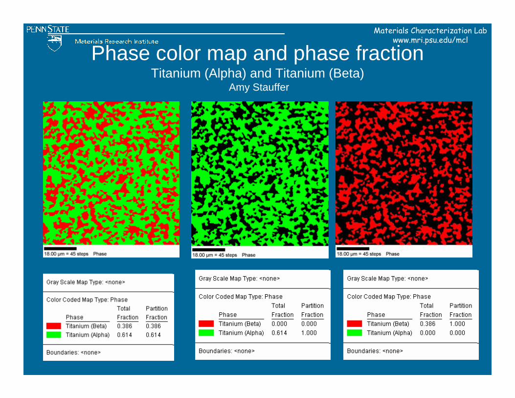
Phase color map and phase fractionTitanium (Alpha) and Titanium (Beta)
Amy Stauffer
Materials Characterization Labwww.mri.psu.edu/mcl
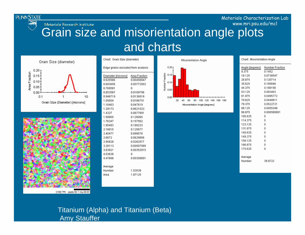
Grain size and misorientation angle plots and charts
Titanium (Alpha) and Titanium (Beta)Amy Stauffer
Materials Characterization Labwww.mri.psu.edu/mcl
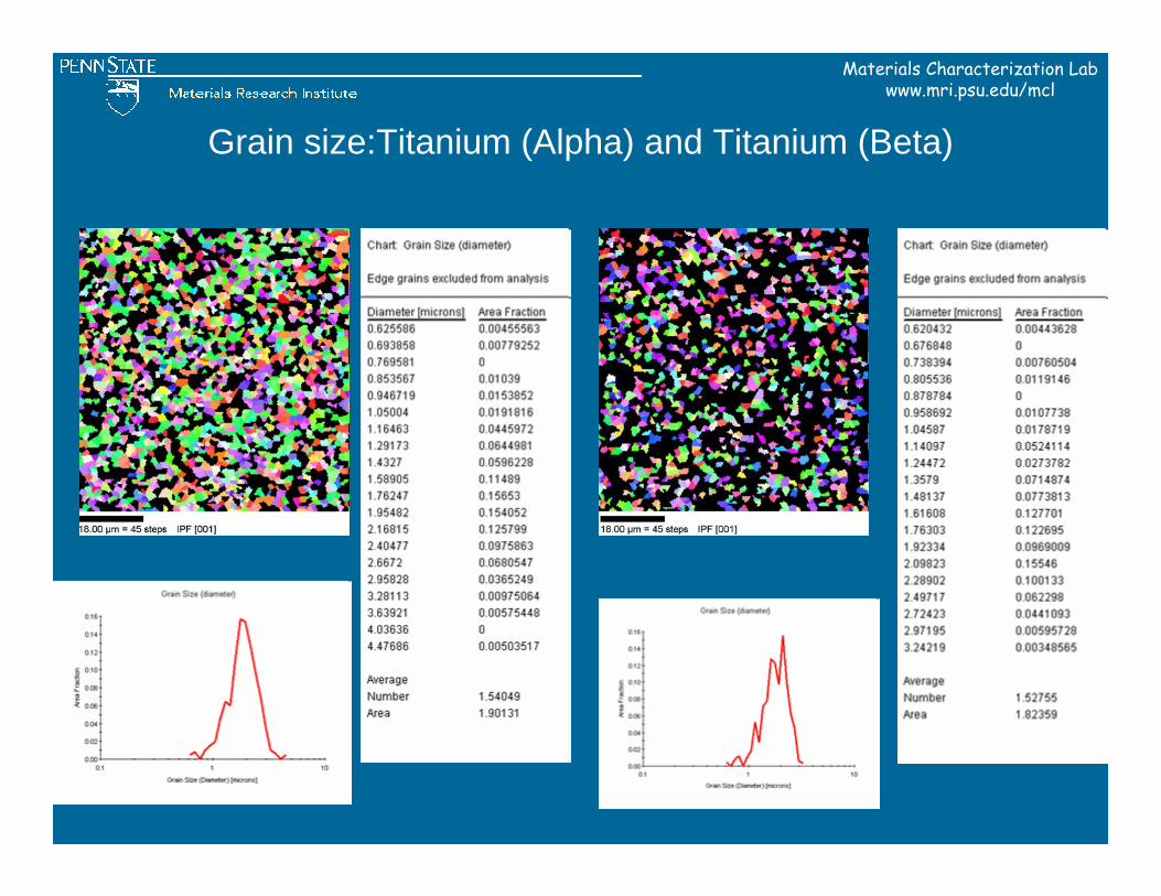
Grain size:Titanium (Alpha) and Titanium (Beta)
Materials Characterization Labwww.mri.psu.edu/mcl
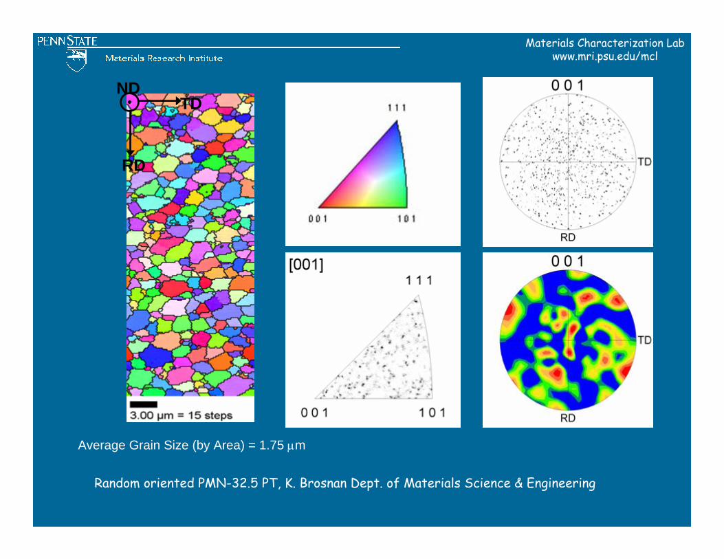
Average Grain Size (by Area) = 1.75 µm
RD
TDND
Random oriented PMN-32.5 PT, K. Brosnan Dept. of Materials Science & Engineering
Materials Characterization Labwww.mri.psu.edu/mcl
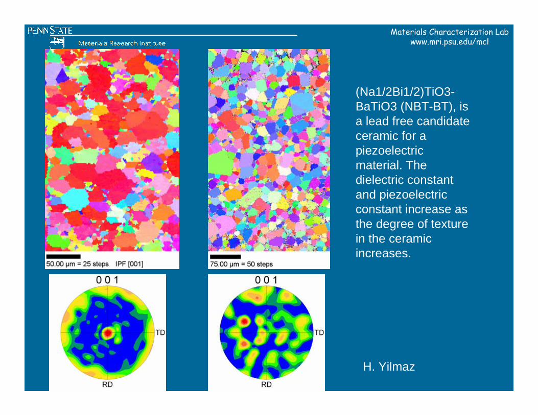
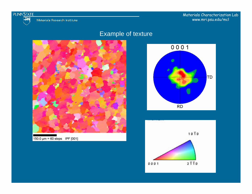
(Na1/2Bi1/2)TiO3-BaTiO3 (NBT-BT), is a lead free candidate ceramic for a piezoelectric material. The dielectric constant and piezoelectric constant increase as the degree of texture in the ceramic increases.
H. Yilmaz
Materials Characterization Labwww.mri.psu.edu/mcl
Example of texture
Materials Characterization Labwww.mri.psu.edu/mcl
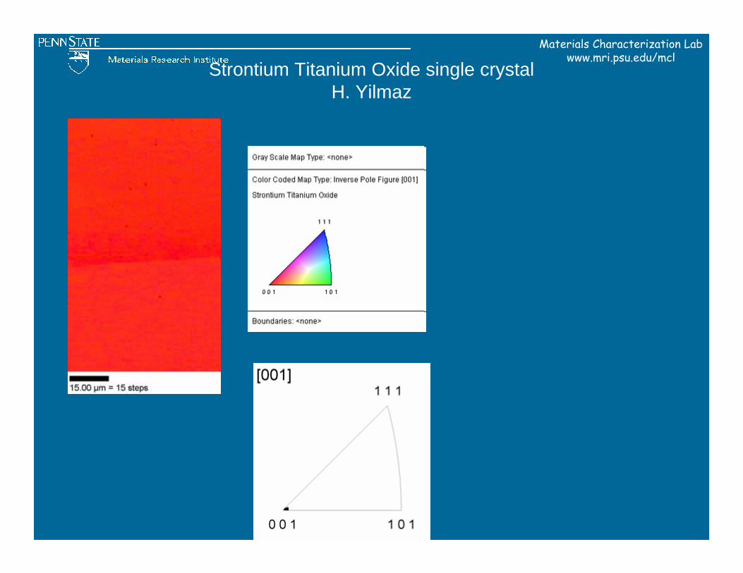
Strontium Titanium Oxide single crystalH. Yilmaz
Materials Characterization Labwww.mri.psu.edu/mcl
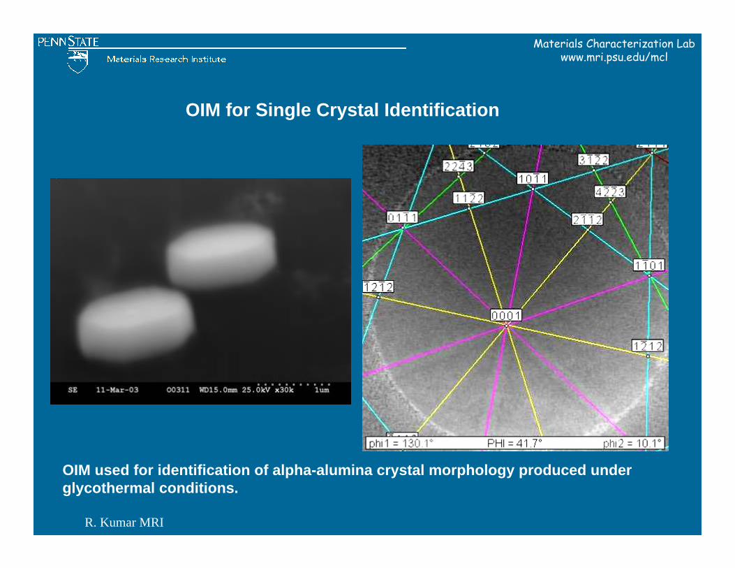
OIM used for identification of alpha-alumina crystal morphology produced under glycothermal conditions.
OIM for Single Crystal Identification
R. Kumar MRI
Materials Characterization Labwww.mri.psu.edu/mcl
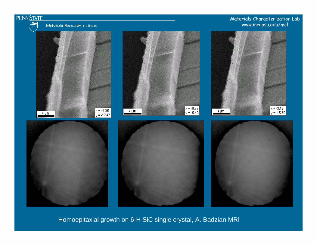
Homoepitaxial growth on 6-H SiC single crystal, A. Badzian MRI
Materials Characterization Labwww.mri.psu.edu/mcl
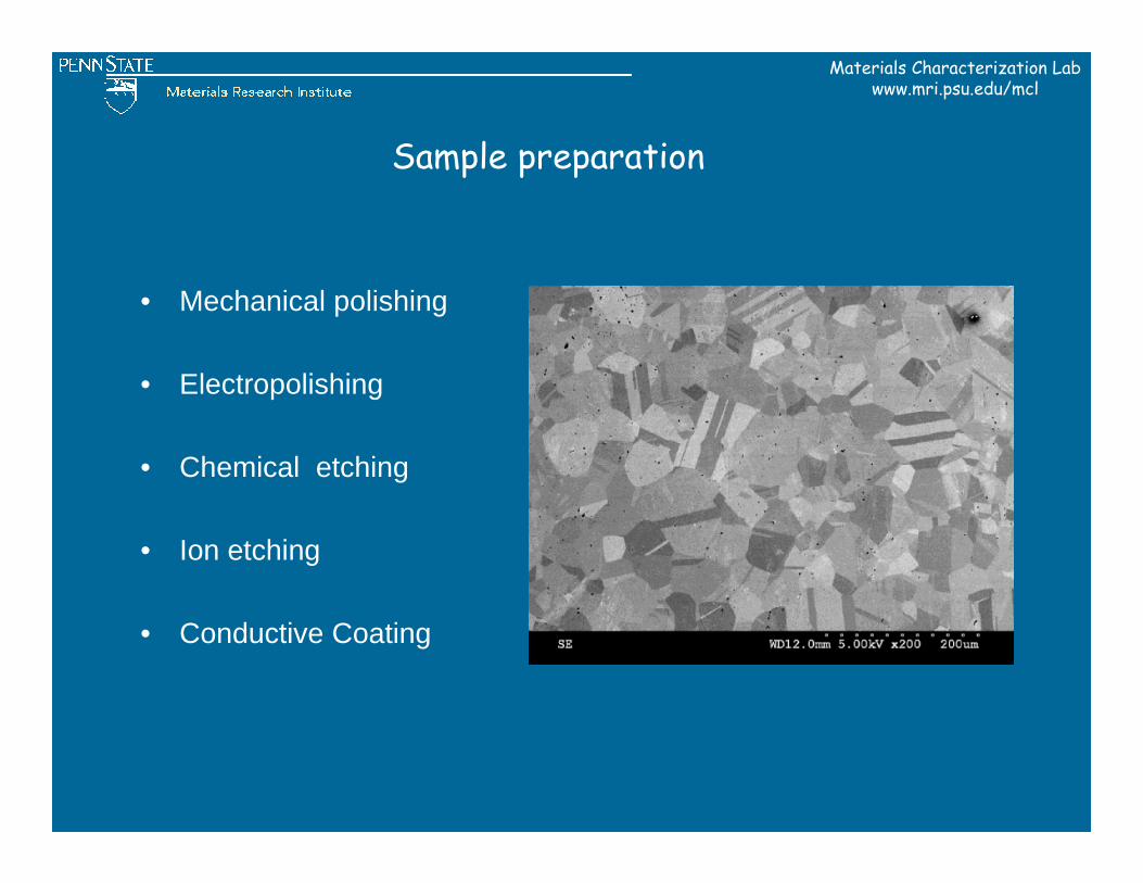
• Mechanical polishing
• Electropolishing
• Chemical etching
• Ion etching
• Conductive Coating
Sample preparation
Materials Characterization Labwww.mri.psu.edu/mcl
Get Started
Contact: Maria Klimkiewicz
1. SEM training2. Sample preparation3. OIM training
Materials Characterization Labwww.mri.psu.edu/mcl
OIM resources
• OIM users manual• Electron Backscatter Diffraction in Materials Science-
book edited by A. J. Schwartz M. Kumar and B. L. Adams
• http://www.edax.com/