Of Model Pets and Cancer Models: An Introduction to Mouse ...
16
Topic Introduction Of Model Pets and Cancer Models: An Introduction to Mouse Models of Cancer Andrea Lunardi, Caterina Nardella, John G. Clohessy, and Pier Paolo Pandolfi 1 Cancer Genetics Program, Beth Israel Deaconess Cancer Center, Department of Medicine and Pathology, Beth Israel Deaconess Medical Center, Harvard Medical School, Boston, Massachusetts The extraordinary endeavor to faithfully model human disorders in mice began in the early 1900s, and in the century since has delivered fundamental advances in our understanding and treatment of human disease. Although it could not be appreciated at the time, 99% of mouse protein-coding genes have an equivalent homolog in the human genome, despite the striking differences in appearance between mouse and man. This remarkable genetic similarity, together with our ability to finely engineer the murine genome, has made the mouse the ideal animal in which to model and analyze human biology and disease. Here we describe this remarkable shared journey between human and mouse, and envisage the next generation of mouse models, which will no doubt prove increasingly sophisticated and even more faithful to human disease. We also address the strategic use of mice in the fight against cancer, and the role they will play in the development of therapies to eradicate this disease. INTRODUCTION Of the many legacies bequeathed to the world by Asia, one of the most surprising is the ancient Chinese passion for “fancy mice,” or mice bred as unique and invaluable pets. Written accounts from as long ago as 1100 B.C.E. describe a tradition that matured into an incredible system of breeding strategies focused on somatic traits such as the color of the coat, of the eyes, and the body size. Although China may have been the cradle of the selective breeding of these little mammals, the popular practice soon spread to the rest of the ancient world, and the interest in mouse breeding first arrived in Europe in the early part of the 17th century. The use of mice for scientific purposes, however, would not commence for almost 200 years. Indeed, it was not until 1900 that Harvard researcher William Castle and a contemporaneous team led by Leo Loeb and mouse breeder Abbie Lathrop at the University of Pennsylvania began to study the genetic determinants responsible for the inheritance of specific somatic traits in mice and the potential correlation between the incidence of spontaneous tumors and specific mouse strains (Lathrop and Loeb 1915a,b, 1918). The idea that mice might represent useful models to disentangle the genetic forces driving human cancer susceptibility and resistance also sparked the interest of a promising undergraduate working with Castle, Clarence Cook Little. But while studying the role of the immune system in cancer transplantation, Little first encountered one of the fundamental problems in mouse modeling: the genetic heterogeneity caused by crossing different mouse strains. The extreme variability of Little’s initial results, and the observation that the inheritance of somatic traits was not solely Mendelian, dictated his decision to generate pure “inbred” genetic strains by systematically crossing 1 Correspondence: [email protected] © 2014 Cold Spring Harbor Laboratory Press Cite this introduction as Cold Spring Harb Protoc; doi:10.1101/pdb.top069757 17 Cold Spring Harbor Laboratory Press on December 28, 2021 - Published by http://cshprotocols.cshlp.org/ Downloaded from
Transcript of Of Model Pets and Cancer Models: An Introduction to Mouse ...

Topic Introduction
Of Model Pets and Cancer Models: An Introductionto Mouse Models of Cancer
Andrea Lunardi, Caterina Nardella, John G. Clohessy, and Pier Paolo Pandolfi1
Cancer Genetics Program, Beth Israel Deaconess Cancer Center, Department of Medicine and Pathology,Beth Israel Deaconess Medical Center, Harvard Medical School, Boston, Massachusetts
The extraordinary endeavor to faithfully model human disorders in mice began in the early 1900s, andin the century since has delivered fundamental advances in our understanding and treatment of humandisease. Although it could not be appreciated at the time, 99% of mouse protein-coding genes have anequivalent homolog in the human genome, despite the striking differences in appearance betweenmouse and man. This remarkable genetic similarity, together with our ability to finely engineer themurine genome, has made the mouse the ideal animal in which to model and analyze human biologyand disease. Here we describe this remarkable shared journey between human andmouse, and envisagethe next generation of mouse models, which will no doubt prove increasingly sophisticated and evenmore faithful to human disease. We also address the strategic use of mice in the fight against cancer,and the role they will play in the development of therapies to eradicate this disease.
INTRODUCTION
Of the many legacies bequeathed to the world by Asia, one of the most surprising is the ancientChinese passion for “fancy mice,” or mice bred as unique and invaluable pets. Written accounts fromas long ago as 1100 B.C.E. describe a tradition that matured into an incredible system of breedingstrategies focused on somatic traits such as the color of the coat, of the eyes, and the body size.Although China may have been the cradle of the selective breeding of these little mammals, thepopular practice soon spread to the rest of the ancient world, and the interest in mouse breedingfirst arrived in Europe in the early part of the 17th century.
The use of mice for scientific purposes, however, would not commence for almost 200 years.Indeed, it was not until 1900 that Harvard researcherWilliam Castle and a contemporaneous team ledby Leo Loeb and mouse breeder Abbie Lathrop at the University of Pennsylvania began to study thegenetic determinants responsible for the inheritance of specific somatic traits in mice and the potentialcorrelation between the incidence of spontaneous tumors and specific mouse strains (Lathrop andLoeb 1915a,b, 1918). The idea that mice might represent useful models to disentangle the geneticforces driving human cancer susceptibility and resistance also sparked the interest of a promisingundergraduate working with Castle, Clarence Cook Little. But while studying the role of the immunesystem in cancer transplantation, Little first encountered one of the fundamental problems in mousemodeling: the genetic heterogeneity caused by crossing differentmouse strains. The extreme variabilityof Little’s initial results, and the observation that the inheritance of somatic traits was not solelyMendelian, dictated his decision to generate pure “inbred” genetic strains by systematically crossing
1Correspondence: [email protected]
© 2014 Cold Spring Harbor Laboratory PressCite this introduction as Cold Spring Harb Protoc; doi:10.1101/pdb.top069757
17
Cold Spring Harbor Laboratory Press on December 28, 2021 - Published by http://cshprotocols.cshlp.org/Downloaded from

siblings. This approach not only reduced the variability of Little’s results, but also represented aturning point in the use of mice as animal models in medical research. Little went on to author arange of pioneering studies, was the first to propose mice as faithful models of human cancer and itsgenetics (Castle and Little 1909, 1910; Little 1911, 1913, 1914), and eventually founded The JacksonLaboratory, a world-renowned institution for the application of genetic animal modeling to the studyof human pathologies (http://jaxmice.jax.org) (Fig. 1).
WHEN GENIUS MEETS TECHNOLOGY
The transition from passive observation of spontaneous tumors in specific inbred strains to activegenetic manipulation of the mouse genome began during the second half of the 20th century. In April1974, Rudolf Jaenisch and Beatrice Mintz (Fig. 2A,B) published an account, in the Proceedings of theNational Academy of Science (PNAS), of the first genetically modified mouse to carry viral DNA stablyinserted into the genome of its somatic cells. Unexpectedly, however, no signs of tumor developmentwere found in the resulting 1-yr-old mice and, because there was no integration of the SV40 DNA intothe germline, the founders did not pass the transgene on to their offspring (Jaenisch andMintz 1974).The first stable and heritable insertion of exogenous DNA into the mouse genome was obtained twoyears later, in 1976, when Rudolf Jaenisch infected mouse embryos at the preimplantation stage offour to eight cells with the Moloney leukemia virus (M-MuLV), creating the first transgenic mousemodel of cancer (specifically leukemia) that transmitted both transgene and phenotype to the off-spring following Mendelian rules (Jaenisch 1976).
Although Jaenisch and Mintz’s work was essential to pave the way for murine engineering, afurther technical breakthrough for the generation of stable “transgenic” mouse lines came with thedevelopment of a method to inject exogenous recombinant DNA into the pronuclei of fertilizedmouse oocytes. The first publication describing the stable insertion of an exogenous DNA fragmentinto the mouse genome via this method appeared in Nature on November 5, 1981. Franklin Cos-tantini and Elizabeth Lacy of Oxford University were able to integrate into the genome of (C57BL/6xCBA/H) fertilized eggs. Thirty percent of the resulting second generation (F2) was characterized bythe presence in their genome of the λRβG2 transgene, definitively proving the generation of the firststable engineered transgenic mouse line (Costantini and Lacy 1981). One month later, on December11, 1981, Jon W. Gordon and Frank H. Ruddle (Yale University) published, in Science, the generationof another engineered mouse model, characterized this time by the stable genomic insertion ofhuman interferon cDNA cloned in the pBR322 vector. pIf-4, the name of the founder female, wasable to transmit with a Mendelian ratio the exogenous DNA to her offspring (F1) when crossed with a
FIGURE 1. Clarence Cook Little, founding father of The Jackson Laboratory and an early pioneer in mouse genetics.(Reprinted from Nadeau 2002, by permission of Macmillan Publishers Ltd, © 2002.)
18 Cite this introduction as Cold Spring Harb Protoc; doi:10.1101/pdb.top069757
A. Lunardi et al.
Cold Spring Harbor Laboratory Press on December 28, 2021 - Published by http://cshprotocols.cshlp.org/Downloaded from

wild-type male, which served as a proof of principle for the beginning of a new era of geneticengineering and mouse modeling (Gordon and Ruddle 1981; Ruddle 1981).
THE ONCOMOUSE
Even as awareness grew that the generation of transgenic mice was now a tractable and reproduciblemethodology, excitement was also building in the cancer research community over the successfulmolecular cloning of viral and cellular oncogenes (Bishop 1985a,b,c). Inevitably, the two areas ofresearch came together with the generation of the first series of “oncomice,” transgenic mouse modelsoverexpressing different oncogenes and characterized by the heritable predisposition to developspecific types of cancer.
The first “oncomouse” was published in Cell in June 1985 thanks to a fruitful collaborationbetween Richard Palmiter and Ralph Brinster (Palmiter and Brinster 1985). They were initiallyinterested in studying the effect of rat growth hormone (GH) overexpression in transgenic mice(Mt1-GH). To counteract the low levels of the hormone and obtain a more reliable expression ofthe integrated transgene, Palmiter fused the entire SV40 early region, including enhancer, promoter,and large/small T-antigens coding sequence, upstream of the Mt1-GH synthetic gene (MGH) but inthe opposite direction (SV-MGH), with the intent being to increase the activity of theMt1 promoterthrough the proximity of the SV40 enhancer (Palmiter and Brinster 1985).
The result of this intervention was totally unexpected. Although Rudolf Jaenisch and BeatriceMintz had previously published that the SV40 large/small T-antigens were absolutely unable to drivecell transformation in vivo in engineered mice (Jaenisch and Mintz 1974), the SV-MGH transgenicmice of Palmiter and Brinster were clearly predisposed to develop and transmit to their offspringbrain tumors from the epithelial cell layer of the choroid plexus, as well as additional sporadic tumorsin other organs, thus becoming the first transgenic mouse model of cancer (Palmiter and Brinster1985).
This breakthrough discovery, mainly due to serendipity, was immediately followed by the gener-ation of further transgenic mice overexpressing potential oncogenes such as v-Src or Myc. Althoughthe transgenic mouse for v-Src (Mt1-v-Src) showed no compelling evidence of tumor development,the hypothesis-driven generation of the Eμ-Mycmouse model, a collaboration between Palmiter andBrinster with Jerry Adams and Suzanne Cory, was an incredible success. The overexpression of Myc inthe lymphoid cell lineage, thanks to the immunoglobulin enhancer (Eμ), was sufficient to drive B-celllymphomas in the Eμ-Myc transgenic mice (both founder and offspring) (Adams et al. 1985), defi-nitely proving the oncogenic essence of the Ig-Myc translocation in human malignancies. In Decem-
FIGURE 2. Rudolf Jaenisch (A) and Beatrice Mintz (B) generating the first genetically modified mouse to carry viralDNA stably inserted into the genome of its somatic cells. (A, Image courtesy of R. Jaenisch; B, Reprinted, withpermission, from Acc. 90–105—Science Service, Records, 1920s–1970s, Smithsonian Institution Archives.)
Cite this introduction as Cold Spring Harb Protoc; doi:10.1101/pdb.top069757 19
Of Model Pets and Cancer Models
Cold Spring Harbor Laboratory Press on December 28, 2021 - Published by http://cshprotocols.cshlp.org/Downloaded from

ber 1985, Nature published the Eμ-Myc transgenic mice (Adams et al. 1985). At the same time, theteam of Philip Leder and Timothy Stewart was working on Myc. Their attention was focused on thegeneration of a transgenic mouse overexpressing Myc in the breast. Thanks to Leder’s knowledge ontheMyc gene and its biology, Stewart’s ability to generate transgenic mice by pronuclear injection, andthe characterization of the regulatory region of a mouse retrovirus called MMTV (mouse mammarytumor virus), Stewart and Leder were able to successfully generate MMTV-Myc transgenic mice in1983. As the two researchers hoped, mammary tumors began to appear in the transgenic females aftera few rounds of pregnancy, further demonstrating the oncogenic potential of Myc in this specifictissue. Published in the October 1984 (Stewart et al. 1984), the MMTV-Myc was the first transgenicmousemodel of cancer to be identified by the term “oncomouse.” Thereafter, the generation ofmousemodels for cancer research exploded in the work of many pioneering researchers, further strengthen-ing the results obtained by Palmiter/Brinster and Leder/Stewart teams, and validating the “onco-mouse” as one of the most transformational developments in the history of cancer research, with atremendous impact on the future of this and many other scientific fields (Fig. 3).
EMBRYONIC STEM CELLS AND HOMOLOGOUS RECOMBINATION:THE OTHER SIDE OF THE MOON
However, the advent of the technology to generate transgenic mice was not the only breakthrough inmouse genetics of the 20th century. In 1981, two independent groups published a methodology forisolating and culturing in vitromouse embryonic stem (ES) cells collected from the inner cell mass of a3.5-d-old postcoitummouse blastocyst (Evans and Kaufman 1981; Martin 1981). Importantly, the EScells maintained their pluripotency in vitro and were able to generate chimericmice when reimplantedin a new blastocyst, contributing to different cell lineages comprising the germline (Bradley et al. 1984).Within a few years, the discovery and characterization of the homologous recombination (HR) mech-anisms in mammalian cells (Capecchi 1989) led to the development of techniques for the insertion ofspecific exogenous DNA molecules into planned genomic loci, or “gene targeting” (Capecchi 1989).
FIGURE 3. Flyer advertising Philip Leder’s 1986 “NIH Lecture”on early gene targeting efforts in the mouse. The NIH Lectureseries was established to facilitate interchange of informationand to give appropriate recognition for outstanding scientificaccomplishment. (Image reprinted from National Institutes ofHealth [U.S.], Medical Arts and Photography Branch, Bethesda,MD, 1986.)
20 Cite this introduction as Cold Spring Harb Protoc; doi:10.1101/pdb.top069757
A. Lunardi et al.
Cold Spring Harbor Laboratory Press on December 28, 2021 - Published by http://cshprotocols.cshlp.org/Downloaded from

It was in 1987 that Oliver Smithies successfully applied a gene-targeting strategy to mouse ES cellsto replace a mutant form of the Hprt gene with its wild-type exogenous counterpart (Doetschmanet al. 1987). The same year, Mario R. Capecchi published in Cell the generation of the first knockout(KO)mouse (Hrpt−) obtained by the illuminated functional combination of ES cell technology and anHR-based gene-targeting approach (Thomas and Capecchi 1987). The publication of the Hrpt−
mouse model soon after inspired the generation of a number KO mice models, which promisedincredible discoveries in the fields of developmental biology and human health.
Over time, the ability to ablate a specific gene from the mouse genome, or insert a particularhuman disease–associated genetic mutation into a wild-type allele, became the cornerstone of asystematic effort to validate and characterize in vivo the function of genes considered oncosuppres-sors. Perhaps, the initial climax in these efforts was reached in 1992, when Allen Bradley’s group at theBaylor College ofMedicine in Houston and RobertWeinberg’s group at theWhitehead Institute of theMassachusetts Institute of Technology in Boston published their p53 and retinoblastoma (Rb) knock-out mouse models in Nature (Donehower et al. 1992; Jacks et al. 1992). Both models definitivelyproved the fundamental oncosuppressive nature of these two genes.
The power to delete or mutate any gene of interest in themouse genome has forever revolutionizedthe study of human genetic disease, and the development of these methods earnedMario R. Capecchi,Martin J. Evans, and Oliver Smithies the 2007 Nobel Prize in Physiology or Medicine.
THE ONCOMOUSE PATENT
The final decades of the 20th century would become known as an era of revolutionary changes in thelandscape of biomedical research and drug development. At the same time, however, society began toconsider biological discoveries as intellectual property, and hence patentable—a practice that wouldhave a significant impact on the course of cancer research in mouse models.
In 1980 the U.S. Congress approved the Bayh–Dole Act specifically to encourage academic insti-tutions to patent and transfer scientific innovations (developed with government funds) to industry.Many scientists, such as Palmiter and Brinster, never opted to patent their research, but three differentU.S. patents were issued (in 1988, 1992, and 1999) to Harvard University based on Philip Leder andTimothy Stewart’s work on the generation of Oncomice (“a transgenic non-human animal, whosegermline and somatic cells contain an activated oncogene”), the derivation and use of cell lines fromtumor-bearing Oncomice, and the use of Oncomice for testing carcinogenic compounds as well asanticancer drugs.
Problems quickly arose for researchers when Harvard University decided to exclusively license allthree patents to the DuPont Corporation. Although the rationale for patenting scientific discoverieshad been to motivate both holders and licensees to invest in further research and transformativeproducts, the onerous fees and restrictions that DuPont imposed on sublicensees hindered the use ofthe Oncomice for academic research oriented toward cancer treatment. To overcome this stumblingblock to research, in 2000 the National Institutes of Health (NIH) signed a series of agreements withDuPont allowing U.S. government employees and funding recipients to use Oncomice in basicbiological science conducted in academic laboratories. Although of fundamental importance, thisnegotiation did not entirely release the research community from the broad purview of the patentsand fees associated with these licenses, particularly in regard to the use of transgenic cancer-pronemouse models in the private sector and in joint projects with academia.
With the expiration of all three U.S. patents by 2007, further efforts were quickly focused onameliorating the restrictions imposed on research use (both commercial and nonprofit) of Onco-mice in drug testing, which is probably the most important commercial application of the technology.A 2005 decision by the U.S. Supreme Court strengthened the concept of “Food and Drug Adminis-tration (FDA) safe harbor,” which was embedded in the Hatch–Waxman exemption (1984), and gavefurther protection to research in academic laboratories. The concurrent decisions of different phar-
Cite this introduction as Cold Spring Harb Protoc; doi:10.1101/pdb.top069757 21
Of Model Pets and Cancer Models
Cold Spring Harbor Laboratory Press on December 28, 2021 - Published by http://cshprotocols.cshlp.org/Downloaded from

maceutical companies to avoid licensing agreements with DuPont was further evidence of thecommon willingness to resolve this long-standing problem and move forward with the developmentof mouse models for human disease treatment optimization.
ARE MICE RELEVANT MODELS FOR HUMAN DISEASE? THE ACUTEPROMYELOCYTIC LEUKEMIA (APL) PARADIGM
Although they look very different from humans, the results of mouse (2002) and human (2003)genome sequencing have revealed that we share >99% of our protein-coding genes with these littlerodents. If this discovery still surprises the layman, it nevertheless strikingly supports the rationale forusing mice as faithful model organisms in the study of genetic human diseases and possible therapies.
By now it is simply a matter of fact that mouse models have led to incredible advances in thetreatment of serious and sometimes lethal pathological conditions. To select only one among a plethoraof examples, the cure for a previously lethal form of hematopoietic malignancy, acute promyelocyticleukemia (APL), was optimized through experimentation on oncomice.
APL is characterized by a block in the promyelocytic stage of myeloid hemopoietic differentiationand is invariably associated with reciprocal and balanced chromosomal translocations involvingchromosome 17. In 1991, several groups simultaneously identified the two genes at the breakpointof the t(15;17) translocation: PML on chromosome 15q22 and the RARα gene on chromosome 17q2(Longo et al. 1990; de Thé et al. 1991; Goddard et al. 1991; Kakizuka et al. 1991; Pandolfi et al. 1991).Subsequent investigations, however, revealed that APL is genetically heterogeneous and identified butsix different hematologic malignancies characterized by translocations where RARα is invariablyinvolved, but its partners vary (Pandolfi 1996). This genetic diversity proved to have an impact onthe way in which APL responds to treatment (Lo Coco et al. 1991).
The modeling efforts for APL proved complex, especially at that time, because of a lack ofappropriate expression vectors, promoters, and regulatory elements to accurately direct in vivo,transgenic expression in the hemopoietic cellular compartments. In 1997, however, three groupslead by Mike Bishop, Timothy Ley, and Pier Paolo Pandolfi, respectively, reported the first faithfulmouse models of PML-RARα-driven APL (Brown et al. 1997; Grisolano et al. 1997; He et al. 1997).The Pandolfi laboratory went on to model the other subtypes of APL. Critically, the generation ofthese models showed that clinically, molecularly, and in their response to therapy, these leukemiasbehaved in a similar manner to human APL. For example, PML-RARαmice were found to transientlyrespond to retinoic acid (RA) exactly as human t(15;17) APL. As in human APL, RAwas able to inducecomplete remission in the mouse, but leukemia would eventually relapse if treated with RA alone(Brown et al. 1997; Grisolano et al. 1997; He et al. 1997). In addition, leukemias in APL PML-RARαmouse models would often become RA resistant, as does human t(15;17) APL treated solely with RA(He et al. 1998). Furthermore, as in human t(11;17) APL, leukemia in PLZF-RARαmice was found tobe resistant to RA at presentation (He et al. 1998) (Fig. 4).
Subsequently, the mouse models of APL began to inform the design of clinical trials. Resultsshowed that leukemia in PML-RARαmice was extremely responsive to As2O3 (which later proved tobe another powerful weapon for the treatment of APL), whereas once again leukemia in PLZF-RARαmice was found to be resistant (see also below) (Lallemand-Breitenbach et al. 1999; Rego et al. 2000).By using information accrued in preclinical testing of different APLmousemodels, clinicians began tostratify patients for clinical trials on the basis of genetic criteria; thus, t(15;17) APL patients weretreated successfully with As2O3, whereas t(11;17) APL patients were not (Nasr et al. 2008).
Critically, different drug combinations were tested in preclinical trials using, in a randomizedfashion, the various subtypes of the APL mouse model, an effort that in a clinical setting would belengthy and almost impossible because of the rarity of these subtypes and the prohibitive number ofpatients to be recruited for such screening. This preclinical approach incorporating APL mousemodels proved decisive in the eradication of human APL (Lallemand-Breitenbach et al. 1999; Rego
22 Cite this introduction as Cold Spring Harb Protoc; doi:10.1101/pdb.top069757
A. Lunardi et al.
Cold Spring Harbor Laboratory Press on December 28, 2021 - Published by http://cshprotocols.cshlp.org/Downloaded from
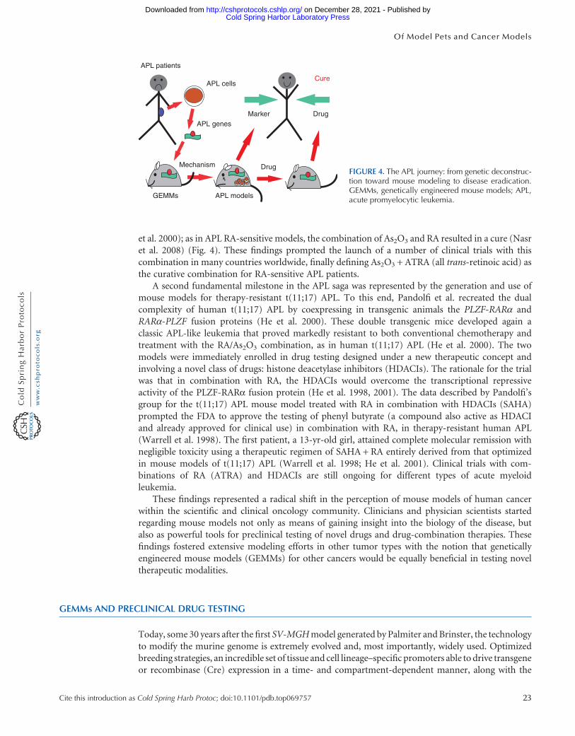
et al. 2000); as in APL RA-sensitive models, the combination of As2O3 and RA resulted in a cure (Nasret al. 2008) (Fig. 4). These findings prompted the launch of a number of clinical trials with thiscombination in many countries worldwide, finally defining As2O3 + ATRA (all trans-retinoic acid) asthe curative combination for RA-sensitive APL patients.
A second fundamental milestone in the APL saga was represented by the generation and use ofmouse models for therapy-resistant t(11;17) APL. To this end, Pandolfi et al. recreated the dualcomplexity of human t(11;17) APL by coexpressing in transgenic animals the PLZF-RARα andRARα-PLZF fusion proteins (He et al. 2000). These double transgenic mice developed again aclassic APL-like leukemia that proved markedly resistant to both conventional chemotherapy andtreatment with the RA/As2O3 combination, as in human t(11;17) APL (He et al. 2000). The twomodels were immediately enrolled in drug testing designed under a new therapeutic concept andinvolving a novel class of drugs: histone deacetylase inhibitors (HDACIs). The rationale for the trialwas that in combination with RA, the HDACIs would overcome the transcriptional repressiveactivity of the PLZF-RARα fusion protein (He et al. 1998, 2001). The data described by Pandolfi’sgroup for the t(11;17) APL mouse model treated with RA in combination with HDACIs (SAHA)prompted the FDA to approve the testing of phenyl butyrate (a compound also active as HDACIand already approved for clinical use) in combination with RA, in therapy-resistant human APL(Warrell et al. 1998). The first patient, a 13-yr-old girl, attained complete molecular remission withnegligible toxicity using a therapeutic regimen of SAHA + RA entirely derived from that optimizedin mouse models of t(11;17) APL (Warrell et al. 1998; He et al. 2001). Clinical trials with com-binations of RA (ATRA) and HDACIs are still ongoing for different types of acute myeloidleukemia.
These findings represented a radical shift in the perception of mouse models of human cancerwithin the scientific and clinical oncology community. Clinicians and physician scientists startedregarding mouse models not only as means of gaining insight into the biology of the disease, butalso as powerful tools for preclinical testing of novel drugs and drug-combination therapies. Thesefindings fostered extensive modeling efforts in other tumor types with the notion that geneticallyengineered mouse models (GEMMs) for other cancers would be equally beneficial in testing noveltherapeutic modalities.
GEMMs AND PRECLINICAL DRUG TESTING
Today, some 30 years after the first SV-MGHmodel generated by Palmiter andBrinster, the technologyto modify the murine genome is extremely evolved and, most importantly, widely used. Optimizedbreeding strategies, an incredible set of tissue and cell lineage–specific promoters able to drive transgeneor recombinase (Cre) expression in a time- and compartment-dependent manner, along with the
APL patients
APL cells
MarkerAPL genes
Mechanism
GEMMs
Drug
Drug
Cure
APL models
FIGURE 4. The APL journey: from genetic deconstruc-tion toward mouse modeling to disease eradication.GEMMs, genetically engineered mouse models; APL,acute promyelocytic leukemia.
Cite this introduction as Cold Spring Harb Protoc; doi:10.1101/pdb.top069757 23
Of Model Pets and Cancer Models
Cold Spring Harbor Laboratory Press on December 28, 2021 - Published by http://cshprotocols.cshlp.org/Downloaded from

power to finely tune Cre activity or transgene expression through Tet- (Tet-On; Tet-Off) or estrogenreceptor–inducible systems, have given rise to a wide array of GEMMs thatmore faithfully recapitulatemost of the different types of human cancer.
Although of fundamental importance to the study of the biology of cancer, mouse models alsorepresent by now one of the most useful in vivo platforms for drug development and the investigationof drug resistance. A key advantage of mouse models is that they allow tumors to grow in anenvironment characterized by a normal immune system, in the organ of origin, and surrounded bythe correct stroma, all conditions that strongly affect tumor progression and response to treatment.Furthermore, tumor response to specific therapeutic regimens can be monitored by using exactly thesame procedures as would be administered to human patients, such as palpation, serological mea-surement of tumor markers, magnetic resonance imaging (MRI), positron emission tomography(PET), computed tomography (CT)-PET, or bioluminescence, and the response classified by usingthe same criteria (e.g., RECIST). Finally, mice are relatively inexpensive, treatments can be easily runon a large number of homogeneous animals (genotype, age, background, sex), responses are fasterthan in human patients, and genetic and molecular analyses to define mechanisms of de novo oracquired drug resistance are always applicable.
Accordingly, in the last decade, GEMMs have been enrolled in a wide variety of preclinical studieswith such differing goals as the following.
• Target validation. GEMMs are often used to determine whether the potential target of a newcompound is indeed a key factor for tumor cell survival, proliferation, or migration (Martinet al. 2005; Toogood et al. 2005).
• Testing new and more sophisticated drugs for target therapy.GEMMs are routinely enrolled to assesstumor response to new experimental therapeutics directed against specific targets known to bederegulated in specific types of tumors. These therapeutics have included (among others) retinoicacid, tyrosine kinase receptor inhibitors, rapamycin analogs, PARP inhibitors, cyclin-dependentkinase inhibitors, PI3K/mTOR/AKT inhibitors, neoangiogenesis inhibitors, farnesyl transferaseinhibitors, prostaglandin inhibitors, androgen receptor inhibitors, and DNA methyltransferaseinhibitors. These, to name a few, and multiple other compounds have been tested in preventionor treatment settings in different types of mouse models (Politi and Pao 2011; Sharpless andDepinho 2006).
• Mechanisms of de novo or acquired tumor resistance.Unfortunately, to date no drug has the power tocure a cancer as a single agent. Furthermore, the most effective treatments normally show differentmagnitudes of response in different patients, and invariably the tumor in questionwill acquire someresistance to treatment. GEMMs represent an invaluable tool to quickly anticipate (years in advanceof comparable studies in human patients) the duration of tumor response and the mechanisms ofacquired resistance when it kicks in. Importantly, efforts to characterize themolecularmechanismsand pathways responsible for acquired resistance to specific treatment have proven GEMMs to befaithful and essential to unraveling these mechanisms. Numerous examples of such contributionsbyGEMMs exist, and include, amongmany other examples, the fact that RA treatment is ineffectivein t(11;17) APL (He et al. 2000, 2001), that chemotherapy cytotoxicity is tightly linked to Trp53status (Lowe et al. 1993a,b), that sensitivity to EGFR inhibition can be bypassed by RAS or PI3Khyperactivity (Mellinghoff et al. 2005), that KRAS/TP53 and KRAS/LKB1 double-mutant lungcancers are resistant to docetaxel (Chen et al. 2012), and that androgen deprivation therapy isineffective in a context where XAF1/XIAP and SRD5A1 genes are deregulated (Lunardi et al. 2013).
• Combinational therapy. Finally, GEMMs are a perfect investigational platform to test drug com-binations focused to target-specific pathways identified as candidate mechanisms for tumor re-sistance to particular treatments. As previously described, a paramount example of theprominence of using GEMMs in this preclinical arm is represented by the discovery, initially inmouse models and then successfully translated to human patients, of the curative effect of the RAand As2O3 combination for the t(15;17) APL (Rego et al. 2000; Nasr et al. 2008).
24 Cite this introduction as Cold Spring Harb Protoc; doi:10.1101/pdb.top069757
A. Lunardi et al.
Cold Spring Harbor Laboratory Press on December 28, 2021 - Published by http://cshprotocols.cshlp.org/Downloaded from

Accordingly, in recent years different groups worldwide have poured an exponentially in-creasing amountof effort in thisdirection,with important resultsnowcoveringmanydifferent typesof tumors. Combination treatment with VEGFR plus bFGF inhibitors, for instance, has beeneffective in a mouse model of pancreatic islet carcinogenesis resistant to VEGFR inhibition alone(Casanovas et al. 2005).Other research has shown that PARP inhibition sensitizesBRCA1-null p53-hetmouse model of breast cancer to PI3K inhibition (Juvekar et al. 2012); AKT/mTOR and ERK/MAPK double targeting inhibits tumor growth in the castration-resistant Pten/Nkx3.1 mutantmouse model of prostate cancer (Kinkade et al. 2008; Floc’h et al. 2012); XIAP and SRD5A1double inhibition through Embelin plus Dutasteride treatment rescues ADT efficacy in castra-tion-resistant Pten/Lrf and Pten/p53 double null prostate cancers mouse models (Lunardi et al.2013); and MEK inhibitor selumetinib provides substantial benefit to Kras/Trp53 but not to Kras/Lkb1doublenull lung cancermousemodels resistant todocetaxel asmonotherapy (Chen et al. 2012).
Although these studies represent only a few of the many published examples, they are, we hope,plentiful to convey the clinical relevance of GEMMs to optimize the treatment of human geneticdiseases such as cancer.
XENOGRAFTS, ALLOGRAFTS, AND CARCINOGEN-INDUCED TUMORS
Transplantation models and chemical inducers have been used routinely for decades in cancerresearch. These technologies are in continuous development and, despite their well-known limita-tions, represent additional important tools for cancer biology and drug screening studies.
The term xenograft, from the Greek xenos plus graphion, indicates a graft of tissue obtained from amember of one species and transplanted into a member of another species. The most commonxenograft approach used in cancer research laboratories is the injection of human tumor cell linesmixed with Matrigel under the skin of immunocompromised mice (i.e., nude, SCID, NOG, NSG, orNOD/SCID/γ). Injected cells normally engraft, and in few weeks a solid tumoral mass is palpable.Although this method is extremely fast and reproducible, they use tumor cell lines that have beengrowing in vitro for decades (HeLa cell line was generated in 1951, HEK293 in early 1970, PC3 in1979), and so carry an unknown number of mutations, genomic aberrations, and metabolic dysfunc-tions, which has sometimes led to complications in the correct interpretation of positive as well asnegative results.
A more complex, in general less efficient, but absolutely valid alternative is the transplantation ofpart of a primary human tumor into mice. This kind of procedure yields the best results when thetumor is transplanted into the same organ of the host as where it arose in the patient, thus assuring acomparable growth environment. The mice commonly enrolled in xenograft transplantation studies,however, are often moderately to severely immunocompromised; although this condition is necessaryto permit the transplant, the engraftment can be considered extremely artifactual and observationsderived from it therefore can potentially be misleading.
Similar in concept but different in execution, transplanted tumor cells or primary tumors derivedfrom anothermouse (instead of from a human being) are known as allografts (alias syngeneicmodels).Accordingly, transplants can be done in immune-competent wild-type host mice without any risk ofrejection, because they do not suffer from distinct immune responses, a condition that more closelymimics the classical tumor’s microenvironment.
Last, but hardly of minor relevance, is the use of carcinogens to induce spontaneous tumors inmice. Starting in the 1940s with the use of mice to study the tumoral effects of polycyclic aromatichydrocarbons on the digestive organ, today the list of tumor types induced in murine models includesbreast cancer through the MMTV virus, oral and lung cancers by tobacco-derived compounds, skincancer through the irradiation with ultraviolet light or the administration of TPA/DMBA, specifictypes of leukemia by ionizing radiation, and multiple types of malignancies through cadmium andasbestos. Although these models may be considered faithful platforms for selected cancer biology
Cite this introduction as Cold Spring Harb Protoc; doi:10.1101/pdb.top069757 25
Of Model Pets and Cancer Models
Cold Spring Harbor Laboratory Press on December 28, 2021 - Published by http://cshprotocols.cshlp.org/Downloaded from

studies and drug testing, generally they suffer from the same limitations as primary tumors in humans,which they may or may not mirror.
THE NEW AGE: THE CO-CLINICAL TRIAL PROJECT
In the last decade, tremendous advances in technology have allowed scientists to gain powerful insightinto the molecular and genetic determinants that drive cancer. The access to technologies such aswhole exome sequencing and their diminishing cost are providing new challenges to cancer careproviders. With molecular pathology increasingly impacting patient care protocols, the ability toidentify and evaluate novel drugs and drug combinations in molecularly categorized cancers usinga randomized trial approach becomes increasingly challenging. This shift in how we believe aboutcancer, not only from a pathological subtype but now also by molecular subtype, heralds a new era forthe mouse in cancer biology, presenting the opportunity to play a key role in redefining the paradigmby which we develop treatments and therapies for cancer patients.
Furthermore, the advancement of new engineering technologies now makes possible the gener-ation of increasingly sophisticated and faithful mouse models that recapitulate the complex geneticevents characterizing human cancer. However, although a “GEMM to human” or “preclinical toclinical” approach has already proven to be hugely effective toward the eradication of a subset ofdiseases (e.g., acute promyelocytic leukemia [APL]) where the genetic defects are well defined and“druggable” targets have been identified, the rate at which all this advanced knowledge is translatedinto effective therapeutics is still pitifully slow.
To fill this void, in 2007 Pier Paolo Pandolfi designed with Lewis C. Cantley the “Co-Clinical TrialProject” and implemented it at the Beth Israel Deaconess Cancer Center of Harvard Medical Schoolunder the auspices of the National Cancer Institute (Nardella et al. 2011).
The Co-Clinical Trial Project stems from a paradigmatic realization regarding the power of testingnew experimental therapeutics and traditional standard-of-care drugs, as single agents or in combi-nation, in faithful GEMMs, according to the simple yet transformative principle of conducting thetreatments in parallel, and with exactly the same protocols as current clinical trials in human patients(Nardella et al. 2011) (Fig. 5).
The keys to this concept are “synchronicity” and “integration.” More specifically, by treatingGEMMs and human patients in exactly the same way, it will be possible to accrue relevant clinical,biological, and pharmacologic data (i.e., sensitivity vs. resistance to specific regimens, imaging anal-ysis, microarray and proteomic profiling, somatic mutation, nucleotide polymorphisms, etc.) fromboth sources, analyze the data in parallel, integrate the information obtained, and, finally, identifybiomarkers that can help to genetically or molecularly stratify patients by predicting positive ornegative responses to the treatment (Nardella et al. 2011) (Fig. 6). Importantly, these efforts willuncover new mechanisms from which to built new experimental therapeutic modalities.
Drug
Drug
Drug
GEMMs
NOGs
Real-timeintegratedinformation
FIGURE 5. A new working paradigm: the “Co-ClinicalTrial Project.” Real-time integrated analysis of treat-ments in human patients with corresponding treat-ments in mouse models that faithfully model humandisease can enable more efficient and effective stratifi-cation for patient treatment and result in successful clin-ical outcomes.
26 Cite this introduction as Cold Spring Harb Protoc; doi:10.1101/pdb.top069757
A. Lunardi et al.
Cold Spring Harbor Laboratory Press on December 28, 2021 - Published by http://cshprotocols.cshlp.org/Downloaded from

Two outstanding examples regarding the feasibility and the power of the “Co-Clinical” approachhave come from the laboratories of Pandolfi and Wong on prostate (Lunardi et al. 2013) and lungcancer (Chen et al. 2012), respectively.
The mainstay of systemic therapy for advanced prostate cancer (CaP) for the last 70 years has beenandrogen deprivation therapy (ADT). This form of therapy is generally effective, with an initial successrate of almost 90%. However, almost invariably the disease becomes resistant to the treatment.Interestingly, a wide variability in the endurance of ADT is well documented: Response to ADTcan last anywhere from a few months to more than a decade, but little is known about the geneticand molecular determinants of the inevitable resistance that develops.
Given the variability of genetic patterns of CaP, it has been hypothesized that the genetic forcesdriving progression of the disease would also affect the response to therapy.
By studying in parallel a wide cohort of human prostate cancer patients with a panel of differentcastration-resistant GEMMs characterized by common genetic lesions identified in human CaP, Pan-dolfi’s group showed that this is the case (Lunardi et al. 2013). Indeed, they observed that differentgenetic alterations dictate a differential response to ADT in both GEMMs and human patients. Addi-tionally, Pandolfi et al. were able to identify XAF1/XIAP- and SRD5A1-specific pathways as mainlyderegulated in genetically and molecularly stratified mouse and human prostate tumors. Importantly,both pathways were found to be “druggable,” and in vitro studies of human CaP cell lines as well as invivo treatment of castration-resistant prostate cancer (CRPC) GEMMs showed a strong efficacy ofEmbelin (natural XIAP inhibitor) and Dutasteride (SRD5A1 inhibitor) when combined with ADT,clearly suggesting that such a drug regimen could benefit CRPC patients (Lunardi et al. 2013).
Thus, this “Co-Clinical” strategy quickly identified predictive genetic biomarkers and novelmechanisms of resistance to therapy that, if further validated in perspective clinical trial, willserve as an additional proof of principle regarding the power of this approach to facilitate theirreal-time identification and potentially redirect therapy-resistant patients to more effective types oftreatment.
The second and very recent example of a successful application of a “Co-Clinical” strategy was theresult of a collaboration focused on lung cancer between the laboratory of Kwok KinWong and manyother investigators, including Jeffrey Engelman, Pier Paolo Pandolfi, and Lewis Cantley (Chen et al.2012).
Lung cancer is the leading cause of cancer death in the United States and lung cancer patients aregenerally stratified according to a single “oncogenic states,” such as mutant KRAS, LKB1, or TP53; yetcancers harboring an identical genetic aberration may be characterized by sizable differences in theirresponse to a specific targeted therapy. As in the case of CaP, the genetic and molecular mechanismsunderlying this heterogeneous response are not fully understood.
In an attempt to gain new insight into this vexing question, Dr. Wong et al. enrolled Kras, Kras/Trp53, and Kras/Lkb1 GEMMs in a “Co-Clinical” trial mirroring an ongoing clinical trial of acombination of the MEK inhibitor selumetinib (AZD6244) with docetaxel in patients with
Drug X
Using the very sameclinical protocol....
Acquiredresistance
Remission
D D
C
B
A
D
Real-timeintegratedinformation
FIGURE 6. “The Co-Clinical Trial Project” beyond thepreclinical (mouse) to clinical (man) dogma. A coclin-ical approach to the development of personalizedpatient care will take advantage of multiple GEM—Sof human disease to identify in real time an appropriatecourse of treatment for successful eradication ofdisease.
Cite this introduction as Cold Spring Harb Protoc; doi:10.1101/pdb.top069757 27
Of Model Pets and Cancer Models
Cold Spring Harbor Laboratory Press on December 28, 2021 - Published by http://cshprotocols.cshlp.org/Downloaded from

KRAS-mutant lung cancer. Their studies showed that concomitant loss of either p53 or Lkb1 wassufficient to transform docetaxel-sensitive Kras-mutant cancers into resistant tumors. Importantly,this study showed that Kras and Kras/p53 lung cancer GEMMs were sensitive to the combination ofselumetinib plus docetaxel, whereas the Kras/Lkb1 mutant mice showed de novo resistance to thetherapy. Critically, these data were predictive of what was observed in human lung cancer (Chenet al. 2012).
Thus, these initial coclinical efforts already show significant promise for the coclinical platform tobe a powerful approach toward a real-time development and optimization of patient stratification inthe successful treatment of cancer.
BUILDING THE MOUSE HOSPITAL
Although GEMMs are playing a fundamental role in unraveling the cornerstone genetic mechanismsof many different human diseases, their use is still imperfect. To improve the applicability of GEMMsto help us better defeat scourges such as cancer we need to (i) define in greater detail the moleculargenetic events that underlie cancer (and additionally, in those cases where the resistance to a particulartreatmentmay be a secondary event that is completely unrelated to the initial genetic lesions), which isto say that we have to enroll more and better patients in our mouse hospital; (ii) develop standardoperating procedures (SOPs) to standardize how experiments are performed and analyzed acrossmultiple research centers; and (iii) generate an infrastructure to allow for preclinical trials in GEMMsto be performed and integrated in real time with human clinical trial data, for rapid translation ofresults to the clinic in a coclinical fashion.
In line with these critical needs, Pier Paolo Pandolfiwith Lewis Cantley designed and launched thefirst “Mouse Hospital” at the BIDMCCancer Center and HarvardMedical School, with the support ofNCI in 2009 (Nardella et al. 2011). Other “Mouse Hospitals” are now operational at the NIH,Columbia University, Cold Spring Harbor Laboratory, Jackson Laboratory, and in Europe.
The Mouse Hospital represents an ideal realization of the multidisciplinary environment requiredfor the centralized execution of drug testing in mice and its subsequent integration with data fromhuman patients (i.e., the “Co-Clinical” approach described above).
Some of the pivotal components of the Mouse Hospital are (i) a state-of-the-art vivarium with astaff specialized in mouse husbandry, treatment, and veterinary care; (ii) a mouse pharmacy wherecompounds used in the trials in mice are synthesized, thereby bypassing the complexity of thematerialtransfer agreement (MTA) process with pharmaceutical companies; (iii) in vivo imaging platformswith protocols that align with those performed in human patients both in terms of data acquisition andin terms of analysis; (iv) a center for biomarkers and comparative pathology, where human/mousemarkers are evaluated for the purposes of diagnosis and prognosis, and that includes the developmentof tissue-based assays aimed at identifying, validating, and assessing these biomarkers in cancers and inresponse to treatments; (v) a bioinformatics data analysis center that assures open access to bioinfor-matics tools for integrated analysis of data as well as toxicity, along with IC50 and protocols for drugadministration, with the ultimate goal of generating databases for genetically stratified human andmouse “patient” samples and their response to treatments; and (vi) a center for the development ofnew and improved mouse models that faithfully recapitulate the genetics of disease.
Thus, the “Mouse Hospital” represent an extension of the clinical cancer center, whose successfulintegration requires strong interaction between clinical experts and scientific staff to ensure thattrials in mice are designed in a manner that faithfully mirrors human trials for the benefit of thepatients, which can subsequently inform clinicians regarding patient stratification and allow forimplementation of personalized treatment protocols. It is this collaboration among geneticists,biologists, bioinformatics, chemists, pharmacologists, radiologists, pathologists, and clinicians thatlies at the core of the mouse hospital and represents a necessary prerequisite for the valid applicationof the coclinical and postclinical platforms (Nardella et al. 2011; Chen et al. 2012; Lunardi et al.2013) (Fig. 7).
28 Cite this introduction as Cold Spring Harb Protoc; doi:10.1101/pdb.top069757
A. Lunardi et al.
Cold Spring Harbor Laboratory Press on December 28, 2021 - Published by http://cshprotocols.cshlp.org/Downloaded from

It is what we owe to patients, who still daily succumb to this horrible disease. We simply cannotfail them.
CONCLUSIONS
In the past two decades the rising improvement of sequencing technologies has enabled the attain-ment of an incredible amount of molecular and genetic information regarding the mechanismsdriving human diseases, primarily cancer, and in doing so is changing the face of how we think ofcancer and manage patient care. Concurrent advances in genetic engineering have likewise providedus the tools to generate GEMMs that finely mirror many of the genetic aberrations characterizinghuman diseases. The sophisticated mouse models generated over the past few years have contributedtremendously to disentangling the basic mechanisms responsible for various tumor types, neural andimmune disorders, diabetes, and other diseases and have allowed us to screen the efficacy of newcompounds in thousands of preclinical studies along the way. However, positive results obtained inpreclinical studies using mouse models have not always translated to successful clinical trials inpatients. Accordingly, the generation of ever more faithful, robust, and cost-effective mousemodels, together with the realization of centralized “Mouse Hospitals” able to conduct large-scalemouse trials by using preclinical, coclinical, or postclinical platforms, are necessary to further improvethe predictive potential of GEMMs in clinical studies (Nardella et al. 2011; Chen et al. 2012; Lunardiet al. 2013) (Fig. 7).
Although the use of GEMMs and high-tech in vivo analysis may be considered expensive, andobtaining useful quantities of investigational drugs from pharmaceutical companies remains chal-lenging, the costs are minimal when compared with the millions of dollars wasted on failed humanclinical trials. Importantly, the coclinical approach may represent a real breakthrough in the effort toquickly stratify responsive patients from resistant patients enrolled in phase I/II clinical trials, on thebasis of precise biomarkers. Simultaneously, this approach may allow us to identify the molecularmechanisms dictating drug resistance. Such analysis will accelerate the process for approval of newdrugs, diminish clinical trial failure, and serve as a rationale for quickly enrolling resistant patients innew combination treatments based on the molecular diagnosis of their disease.
ACKNOWLEDGMENTS
We dedicate this to all the people who along the way gave their contributions, famous or unknown, tothe development of mouse genetic engineering, one of the most important and effective instrumentsfor the study of human diseases.
Mousemodeling of
human diseaseImagingcenter
Comparativepathology
center
Education/legaloffice
The mousepharmacy
The ‘‘MouseHospital’’
Pharmas Synthesislab
Bioinformaticand
genomic center
FIGURE 7. Classification of the various strategic ap-proaches that fall under the “Co-Clinical” paradigm.
Cite this introduction as Cold Spring Harb Protoc; doi:10.1101/pdb.top069757 29
Of Model Pets and Cancer Models
Cold Spring Harbor Laboratory Press on December 28, 2021 - Published by http://cshprotocols.cshlp.org/Downloaded from

We thank Cheryl Marks, Dinah Singer, and Lewis C. Cantley for the fundamental critical discus-sions that paved the way for the realization of the Co-Clinical approach. A special thanks to ThomasGarvey for insightful editing. The Co-Clinical paradigm whose principles and preliminary importantresults are described here has been supported by the Mission Hope Cancer Center/National CancerInstitute (MHCC/NCI) through theRC2 (CA147940-01) and theLeadership (CA141457-04)Grants toP.P.P. A.L. has been supported in part by a fellowship from the Istituto Toscano Tumori (ITT, Italy).
REFERENCES
Adams JM, Harris AW, Pinkert CA, Corcoran LM, Alexander WS, Cory S,Palmiter RD, Brinster RL. 1985. The c-myc oncogene driven by immu-noglobulin enhancers induces lymphoid malignancy in transgenicmice. Nature 318: 533–538.
Bishop JM. 1985a. Exploring carcinogenesis with retroviral and cellular on-cogenes. Progress Med Virol 32: 5–14.
Bishop JM. 1985b. Viral oncogenes. Cell 42: 23–38.Bishop JM. 1985c. Viruses, genes, and cancer. II. Retroviruses and cancer
genes. Cancer 55: 2329–2333.Bradley A, Evans M, Kaufman MH, Robertson E. 1984. Formation of germ-
line chimaeras from embryo-derived teratocarcinoma cell lines. Nature309: 255–256.
Brown D, Kogan S, Lagasse E, Weissman I, Alcalay M, Pelicci PG, Atwater S,Bishop JM. 1997. A PMLRARα transgene initiates murine acute pro-myelocytic leukemia. Proc Natl Acad Sci 94: 2551–2556.
Capecchi MR. 1989. The new mouse genetics: Altering the genome by genetargeting. Trends Genetics 5: 70–76.
Casanovas O, Hicklin DJ, Bergers G, Hanahan D. 2005. Drug resistance byevasion of antiangiogenic targeting of VEGF signaling in late-stage pan-creatic islet tumors. Cancer Cell 8: 299–309.
Castle WE, Little CC. 1909. The peculiar inheritance of pink eyes amongcolored mice. Science 30: 313–314.
Castle WE, Little CC. 1910. On a modified Mendelian ratio among yellowmice. Science 32: 868–870.
Chen Z, Cheng K,Walton Z,Wang Y, Ebi H, Shimamura T, Liu Y, Tupper T,Ouyang J, Li J, et al. 2012. A murine lung cancer co-clinical trial iden-tifies genetic modifiers of therapeutic response. Nature 483: 613–617.
Costantini F, Lacy E. 1981. Introduction of a rabbit β-globin gene into themouse germ line. Nature 294: 92–94.
de Thé H, Lavau C, Marchio A, Chomienne C, Degos L, Dejean A. 1991. ThePML-RARα fusion mRNA generated by the t(15;17) translocation inacute promyelocytic leukemia encodes a functionally altered RAR. Cell66: 675–684.
Doetschman T, Gregg RG, Maeda N, Hooper ML, Melton DW, ThompsonS, Smithies O. 1987. Targetted correction of a mutant HPRT gene inmouse embryonic stem cells. Nature 330: 576–578.
Donehower LA, Harvey M, Slagle BL, McArthur MJ, Montgomery CA Jr,Butel JS, Bradley A. 1992. Mice deficient for p53 are developmentallynormal but susceptible to spontaneous tumours. Nature 356: 215–221.
Evans MJ, Kaufman MH. 1981. Establishment in culture of pluripotentialcells from mouse embryos. Nature 292: 154–156.
Floc’h N, Kinkade CW, Kobayashi T, Aytes A, Lefebvre C, Mitrofanova A,Cardiff RD, Califano A, Shen MM, Abate-Shen C. 2012. Dual targetingof the Akt/mTOR signaling pathway inhibits castration-resistant pros-tate cancer in a genetically engineered mouse model. Cancer Res 72:4483–4493.
Goddard AD, Borrow J, Freemont PS, Solomon E. 1991. Characterization ofa zinc finger gene disrupted by the t(15;17) in acute promyelocyticleukemia. Science 254: 1371–1374.
Gordon JW, Ruddle FH. 1981. Integration and stable germ line transmissionof genes injected into mouse pronuclei. Science 214: 1244–1246.
Grisolano JL, Wesselschmidt RL, Pelicci PG, Ley TJ. 1997. Altered myeloiddevelopment and acute leukemia in transgenic mice expressing PML-RARα under control of cathepsin G regulatory sequences. Blood 89:376–387.
He LZ, Tribioli C, Rivi R, Peruzzi D, Pelicci PG, Soares V, Cattoretti G,Pandolfi PP. 1997. Acute leukemia with promyelocytic features in PML/RARα transgenic mice. Proc Natl Acad Sci 94: 5302–5307.
He LZ, Guidez F, Tribioli C, Peruzzi D, Ruthardt M, Zelent A, Pandolfi PP.1998. Distinct interactions of PML-RARα and PLZF-RARα with co-repressors determine differential responses to RA in APL. Nat Genetics18: 126–135.
He LZ, Bhaumik M, Tribioli C, Rego EM, Ivins S, Zelent A, Pandolfi PP.2000. Two critical hits for promyelocytic leukemia. Mol Cell 6: 1131–1141.
He LZ, Tolentino T, Grayson P, Zhong S, Warrell RP Jr, Rifkind RA, MarksPA, Richon VM, Pandolfi PP. 2001. Histone deacetylase inhibitorsinduce remission in transgenic models of therapy-resistant acute pro-myelocytic leukemia. J Clin Invest 108: 1321–1330.
Jacks T, Fazeli A, Schmitt EM, Bronson RT, Goodell MA, Weinberg RA.1992. Effects of an Rb mutation in the mouse. Nature 359: 295–300.
Jaenisch R. 1976. Germ line integration and Mendelian transmission of theexogenous Moloney leukemia virus. Proc Natl Acad Sci 73: 1260–1264.
Jaenisch R, Mintz B. 1974. Simian virus 40 DNA sequences in DNA ofhealthy adult mice derived from preimplantation blastocysts injectedwith viral DNA. Proc Natl Acad Sci 71: 1250–1254.
Juvekar A, Burga LN, Hu H, Lunsford EP, Ibrahim YH, Balmana J, Rajen-dran A, Papa A, Spencer K, Lyssiotis CA, et al. 2012. Combining a PI3Kinhibitor with a PARP inhibitor provides an effective therapy forBRCA1-related breast cancer. Cancer Discovery 2: 1048–1063.
Kakizuka A, Miller WH Jr, Umesono K, Warrell RP Jr, Frankel SR, MurtyVV, Dmitrovsky E, Evans RM. 1991. Chromosomal translocation t-(15;17) in human acute promyelocytic leukemia fuses RARα with anovel putative transcription factor, PML. Cell 66: 663–674.
Kinkade CW, Castillo-Martin M, Puzio-Kuter A, Yan J, Foster TH, Gao H,Sun Y, Ouyang X, Gerald WL, Cordon-Cardo C, et al. 2008. TargetingAKT/mTOR and ERK MAPK signaling inhibits hormone-refractoryprostate cancer in a preclinical mouse model. J Clin Invest 118: 3051–3064.
Lallemand-Breitenbach V, Guillemin MC, Janin A, Daniel MT, Degos L,Kogan SC, Bishop JM, de Thé H. 1999. Retinoic acid and arsenic syn-ergize to eradicate leukemic cells in a mouse model of acute promye-locytic leukemia. J Exp Med 189: 1043–1052.
Lathrop AE, Loeb L. 1915a. Further investigations on the origin of tumorsin mice: I. Tumor incidence and tumor age in various strains of mice. JExp Med 22: 646–673.
Lathrop AE, Loeb L. 1915b. Further investigations on the origin of tumors inmice: II. Tumor incidence and tumor age in hybrids. J Exp Med 22:713–731.
Lathrop AE, Loeb L. 1918. Further investigations on the origin of tumors inmice: V. The tumor rate in hybrid strains. J Exp Med 28: 475–500.
Little CC. 1911. The “Dilute” forms of yellow mice. Science 33: 896–897.Little CC. 1913. “Yellow” and “Agouti” factors in mice. Science 38: 205.Little CC. 1914. A possible Mendelian explanation for a type of inheritance
apparently non-Mendelian in nature. Science 40: 904–906.Lo Coco F, Avvisati G, Diverio D, Biondi A, Pandolfi PP, AlcalayM, De Rossi
G, Petti MC, Cantu-Rajnoldi A, Pasqualetti D, et al. 1991. Rearrange-ments of the RAR-α gene in acute promyelocytic leukaemia: Correla-tions with morphology and immunophenotype. Br J Haematol 78:494–499.
Longo L, Pandolfi PP, Biondi A, Rambaldi A, Mencarelli A, Lo Coco F,Diverio D, Pegoraro L, Avanzi G, Tabilio A, et al. 1990. Rearrangementsand aberrant expression of the retinoic acid receptor α gene in acutepromyelocytic leukemias. J Exp Med 172: 1571–1575.
Lowe SW, Ruley HE, Jacks T, Housman DE. 1993a. p53-dependent apopto-sis modulates the cytotoxicity of anticancer agents. Cell 74: 957–967.
30 Cite this introduction as Cold Spring Harb Protoc; doi:10.1101/pdb.top069757
A. Lunardi et al.
Cold Spring Harbor Laboratory Press on December 28, 2021 - Published by http://cshprotocols.cshlp.org/Downloaded from
Lowe SW, Schmitt EM, Smith SW, Osborne BA, Jacks T. 1993b. p53 is re-quired for radiation-induced apoptosis in mouse thymocytes. Nature362: 847–849.
Lunardi A, Ala U, Epping MT, Salmena L, Webster KA, Wang G, Mazzuc-chelli R, Stack E, Lis R, Patnaik A, et al. 2013. A co-clinical approachidentifies mechanisms and potential therapies for androgen deprivationresistance in prostate cancer. Nat Genet 45: 747–755.
Martin GR. 1981. Isolation of a pluripotent cell line from early mouseembryos cultured in medium conditioned by teratocarcinoma stemcells. Proc Natl Acad Sci 78: 7634–7638.
Martin A, Odajima J, Hunt SL, Dubus P, Ortega S, Malumbres M, BarbacidM. 2005. Cdk2 is dispensable for cell cycle inhibition and tumor sup-pression mediated by p27Kip1 and p21Cip1. Cancer Cell 7: 591–598.
Mellinghoff IK, Wang MY, Vivanco I, Haas-Kogan DA, Zhu S, Dia EQ, LuKV, Yoshimoto K, Huang JH, Chute DJ, et al. 2005. Molecular deter-minants of the response of glioblastomas to EGFR kinase inhibitors. NEngl J Med 353: 2012–2024.
Nadeau JH. 2002. Single-nucleotide polymorphisms: Tackling complexity.Nature 420: 517–518.
Nardella C, Lunardi A, Patnaik A, Cantley LC, Pandolfi PP. 2011. The APLparadigmand the “co-clinical trial”project.CancerDiscovery1: 108–116.
Nasr R, Guillemin MC, Ferhi O, Soilihi H, Peres L, Berthier C, Rousselot P,Robledo-Sarmiento M, Lallemand-Breitenbach V, Gourmel B, et al.2008. Eradication of acute promyelocytic leukemia-initiating cellsthrough PML-RARA degradation. Nat Med 14: 1333–1342.
Palmiter RD, Brinster RL. 1985. Transgenic mice. Cell 41: 343–345.Pandolfi PP. 1996. PML, PLZF and NPM genes in the molecular pathogen-
esis of acute promyelocytic leukemia. Haematologica 81: 472–482.
Pandolfi PP, Grignani F, Alcalay M, Mencarelli A, Biondi A, LoCoco F,Grignani F, Pelicci PG. 1991. Structure and origin of the acutepromyelocytic leukemia myl/RARα cDNA and characterization of itsretinoid-binding and transactivationproperties.Oncogene6:1285–1292.
Politi K, Pao W. 2011. How genetically engineered mouse tumor modelsprovide insights into human cancers. J Clin Oncol 29: 2273–2281.
Rego EM, He LZ, Warrell RP Jr, Wang ZG, Pandolfi PP. 2000. Retinoic acid(RA) and As2O3 treatment in transgenic models of acute promyelocyticleukemia (APL) unravel the distinct nature of the leukemogenic processinduced by the PML-RARα and PLZF-RARα oncoproteins. Proc NatlAcad Sci 97: 10173–10178.
Ruddle FH. 1981. A new era in mammalian gene mapping: Somatic cellgenetics and recombinant DNA methodologies. Nature 294: 115–120.
Sharpless NE, Depinho RA. 2006. The mighty mouse: Genetically engi-neered mouse models in cancer drug development. Nat Rev DrugDiscov 5: 741–754.
Stewart TA, Pattengale PK, Leder P. 1984. Spontaneous mammary adeno-carcinomas in transgenic mice that carry and express MTV/myc fusiongenes. Cell 38: 627–637.
Thomas KR, Capecchi MR. 1987. Site-directed mutagenesis by gene target-ing in mouse embryo-derived stem cells. Cell 51: 503–512.
Toogood PL, Harvey PJ, Repine JT, Sheehan DJ, VanderWel SN, Zhou H,Keller PR, McNamara DJ, Sherry D, Zhu T, et al. 2005. Discovery of apotent and selective inhibitor of cyclin-dependent kinase 4/6. J MedChem 48: 2388–2406.
Warrell RP Jr, He LZ, Richon V, Calleja E, Pandolfi PP. 1998. Therapeutictargeting of transcription in acute promyelocytic leukemia by use of aninhibitor of histone deacetylase. J Natl Cancer Inst 90: 1621– 1625.
Cite this introduction as Cold Spring Harb Protoc; doi:10.1101/pdb.top069757 31
Of Model Pets and Cancer Models
Cold Spring Harbor Laboratory Press on December 28, 2021 - Published by http://cshprotocols.cshlp.org/Downloaded from

doi: 10.1101/pdb.top069757Cold Spring Harb Protoc; Andrea Lunardi, Caterina Nardella, John G. Clohessy and Pier Paolo Pandolfi Of Model Pets and Cancer Models: An Introduction to Mouse Models of Cancer
ServiceEmail Alerting click here.Receive free email alerts when new articles cite this article -
CategoriesSubject Cold Spring Harbor Protocols.Browse articles on similar topics from
(436 articles)Mouse (923 articles)Laboratory Organisms, general
http://cshprotocols.cshlp.org/subscriptions go to: Cold Spring Harbor Protocols To subscribe to
© 2014 Cold Spring Harbor Laboratory Press
Cold Spring Harbor Laboratory Press on December 28, 2021 - Published by http://cshprotocols.cshlp.org/Downloaded from