
Ocular Trauma and Emergencies - UK HealthCare · PDF fileOcular Trauma and Emergencies Jacob...
143
Ocular Trauma and Emergencies Jacob J. Yunker, M.D. Retina & Vitreous Surgery Macular Diseases and Degeneration Assistant Professor, Department of Ophthalmology University of Kentucky College of Medicine
Transcript of Ocular Trauma and Emergencies - UK HealthCare · PDF fileOcular Trauma and Emergencies Jacob...
Ocular Trauma and
Emergencies
Jacob J. Yunker, M.D.
Retina & Vitreous Surgery
Macular Diseases and Degeneration
Assistant Professor, Department of Ophthalmology
University of Kentucky College of Medicine
Epidemiology
• Accidental eye injury is one of the leading
causes of visual impairment
• >2.4 million eye injuries in the US per year
• 90% are preventable
• Most common cause of visual loss in
persons under age 25
3
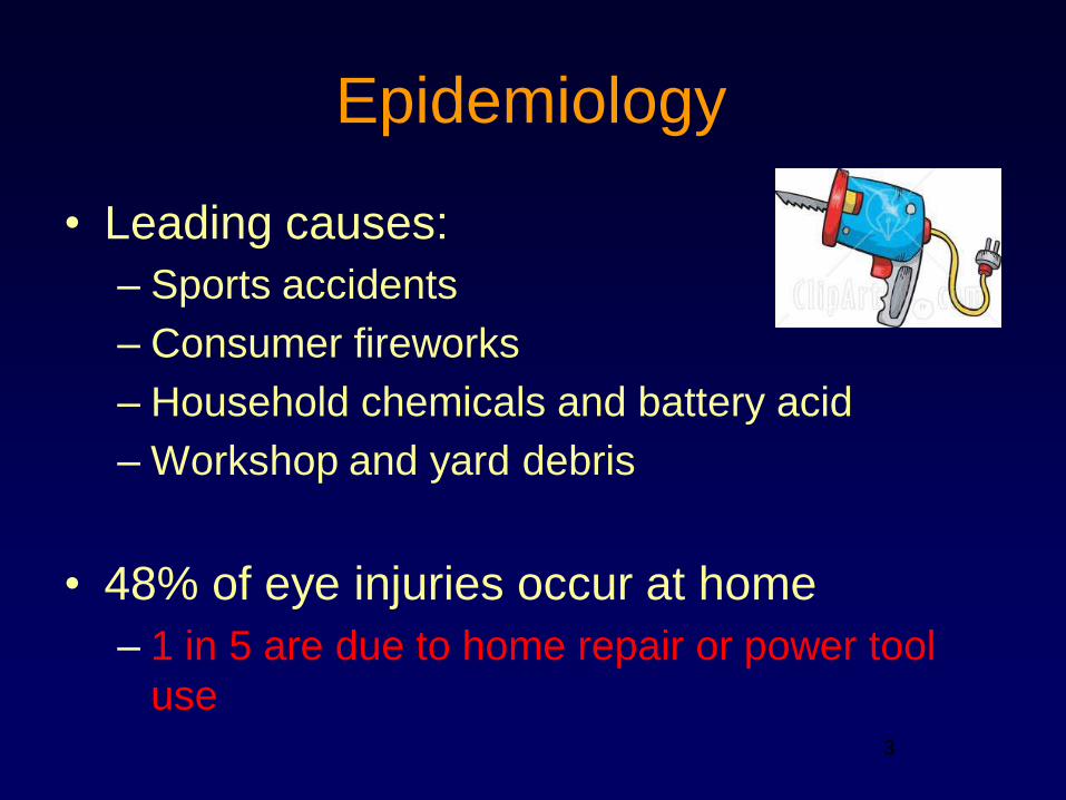
Epidemiology
• Leading causes:
– Sports accidents
– Consumer fireworks
– Household chemicals and battery acid
– Workshop and yard debris
• 48% of eye injuries occur at home
– 1 in 5 are due to home repair or power tool
use3
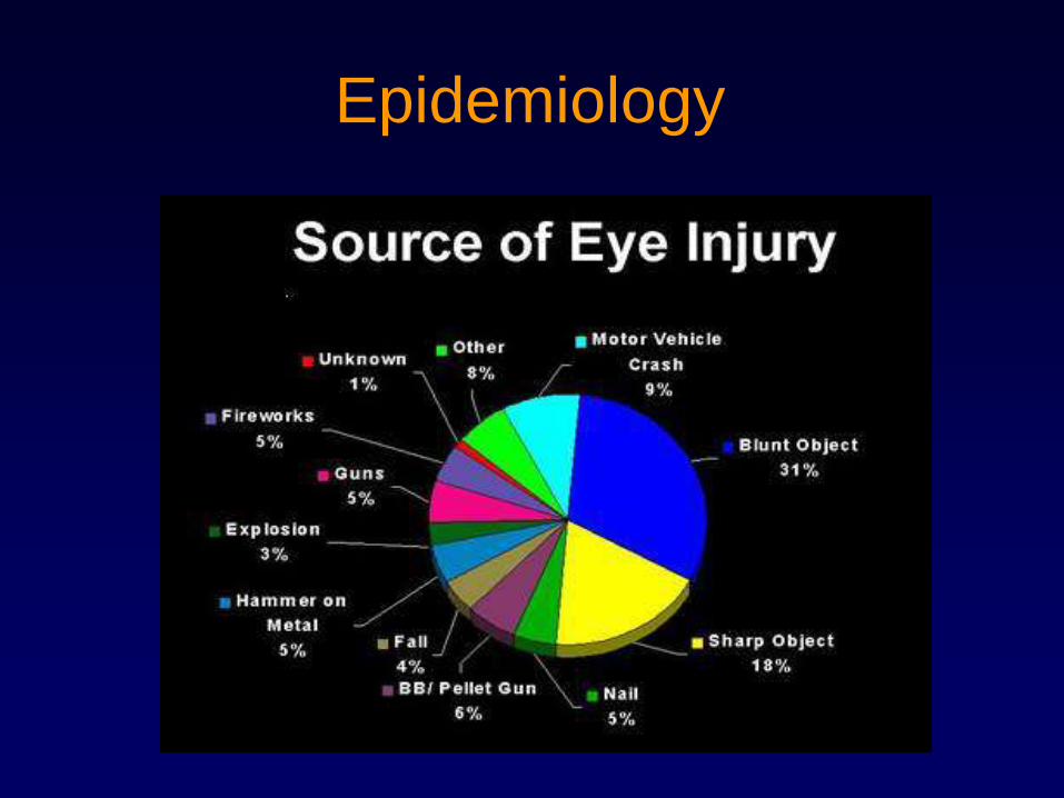
Epidemiology
History
• Age
• Occupation
• Brief history of accident
• Specific symptoms
• Prior condition of eyes
• General health
• Allergies
• Tetanus prophylaxis
Examination - Inspection
• Gross appearance
• Hand held light or penlight
• Slit lamp
• Fluorescein and Wood’s lamp
• Direct ophthalmoscope
Examination
• Visual Acuity
• Motility
• Pupils
• Visual Field
• Inspection
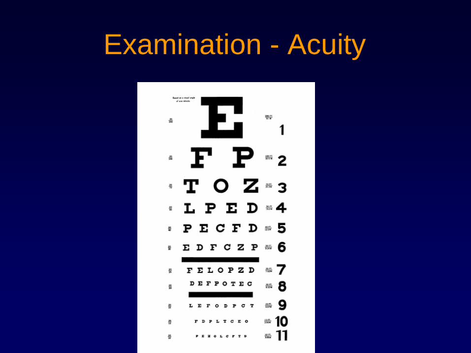
Examination - Acuity
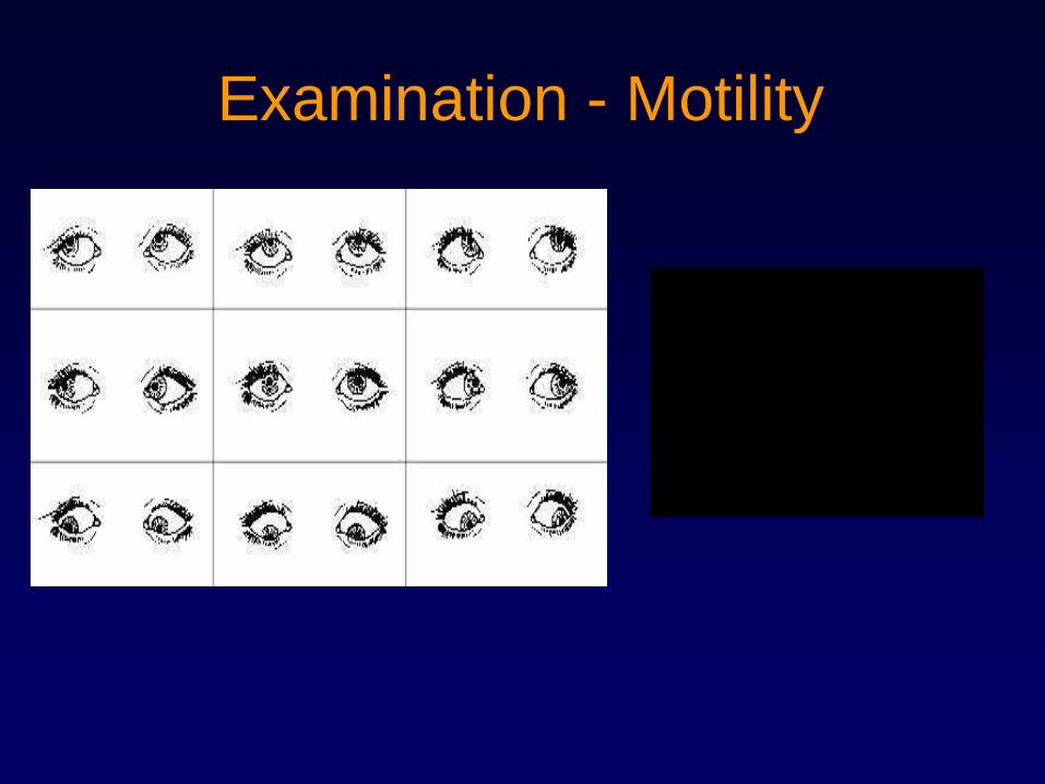
Examination - Motility
10
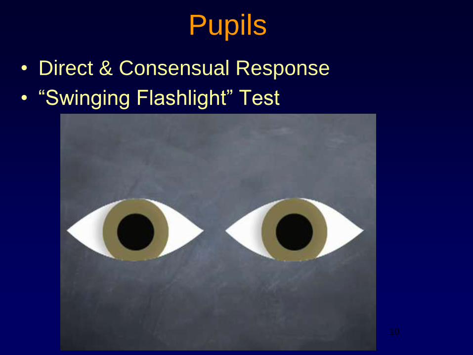
Pupils
• Direct & Consensual Response
• ―Swinging Flashlight‖ Test
11
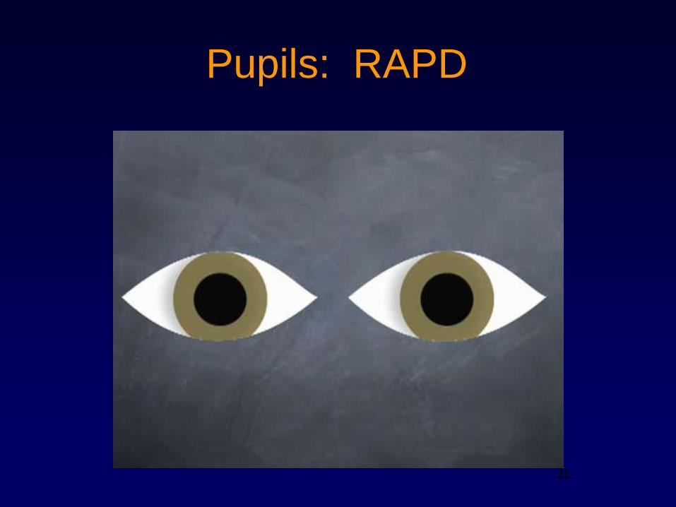
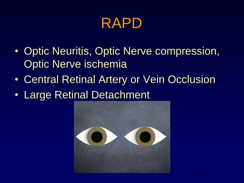
Pupils: RAPD
12
RAPD
• Optic Neuritis, Optic Nerve compression,
Optic Nerve ischemia
• Central Retinal Artery or Vein Occlusion
• Large Retinal Detachment
12
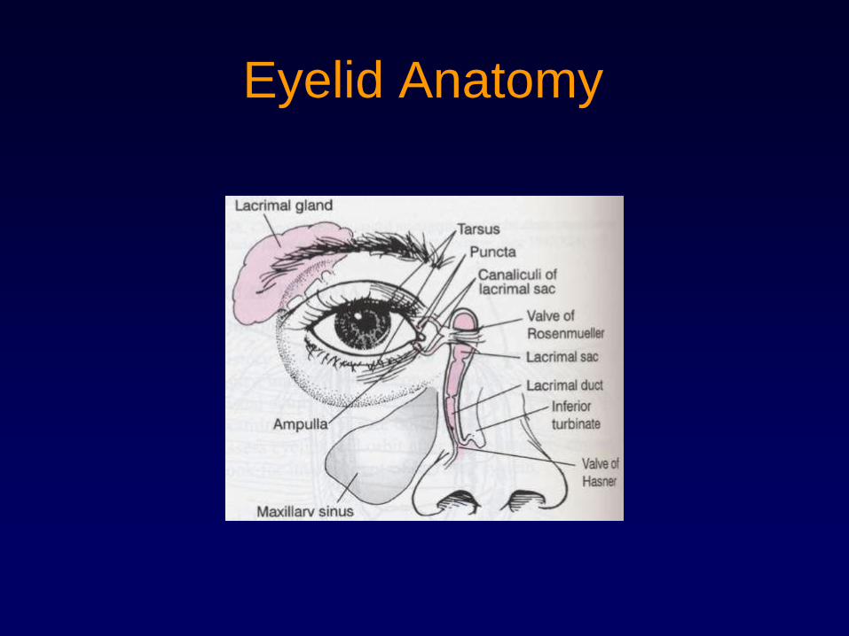
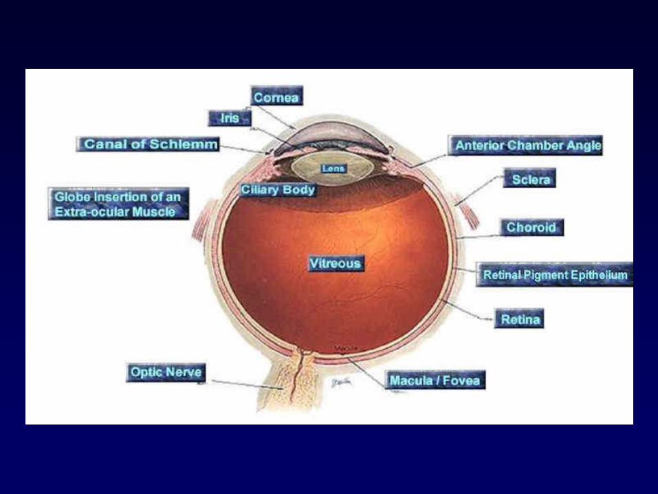
Eyelid Anatomy
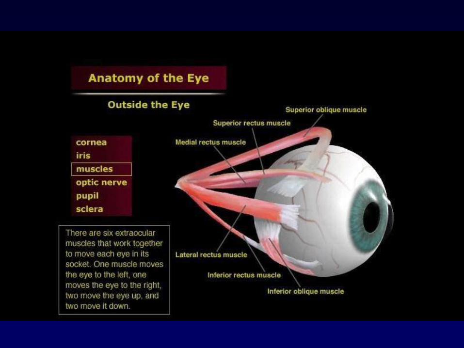
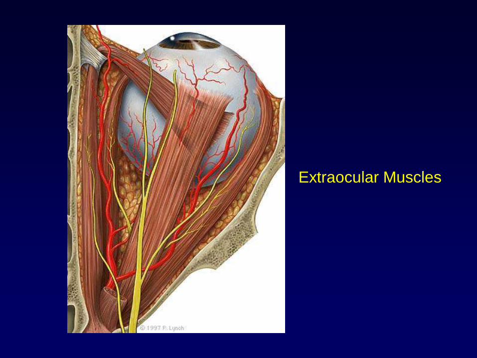
Extraocular Muscles

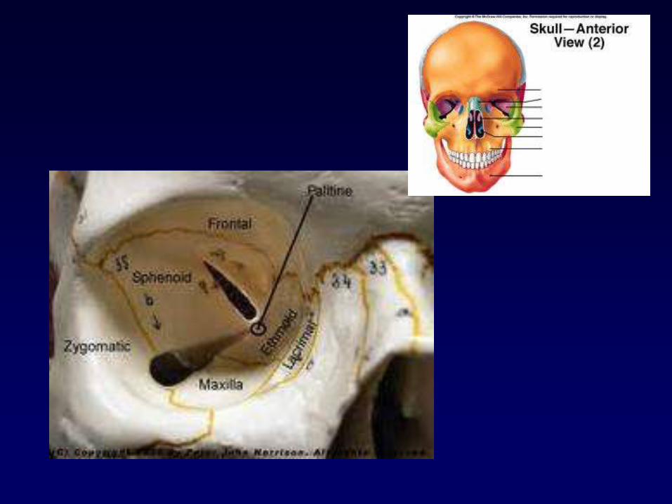
Review of Anatomy
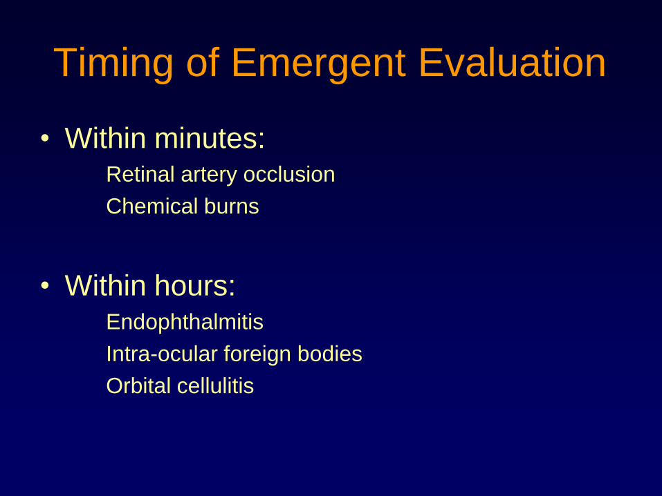
Timing of Emergent Evaluation
• Within minutes:Retinal artery occlusion
Chemical burns
• Within hours:Endophthalmitis
Intra-ocular foreign bodies
Orbital cellulitis
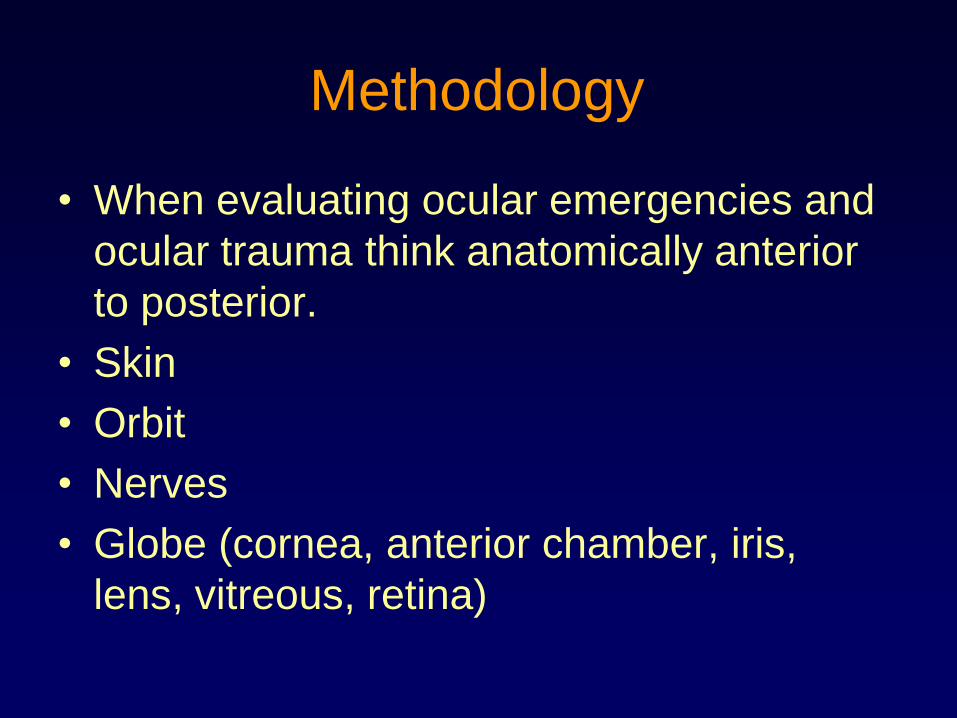
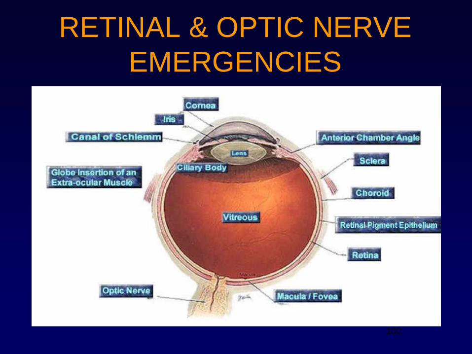
Methodology
• When evaluating ocular emergencies and
ocular trauma think anatomically anterior
to posterior.
• Skin
• Orbit
• Nerves
• Globe (cornea, anterior chamber, iris,
lens, vitreous, retina)
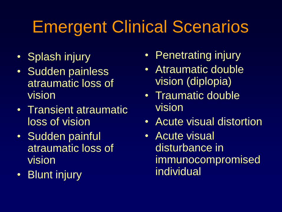
Emergent Clinical Scenarios
• Splash injury
• Sudden painless atraumatic loss of vision
• Transient atraumatic loss of vision
• Sudden painful atraumatic loss of vision
• Blunt injury
• Penetrating injury
• Atraumatic double vision (diplopia)
• Traumatic double vision
• Acute visual distortion
• Acute visual disturbance in immunocompromised individual
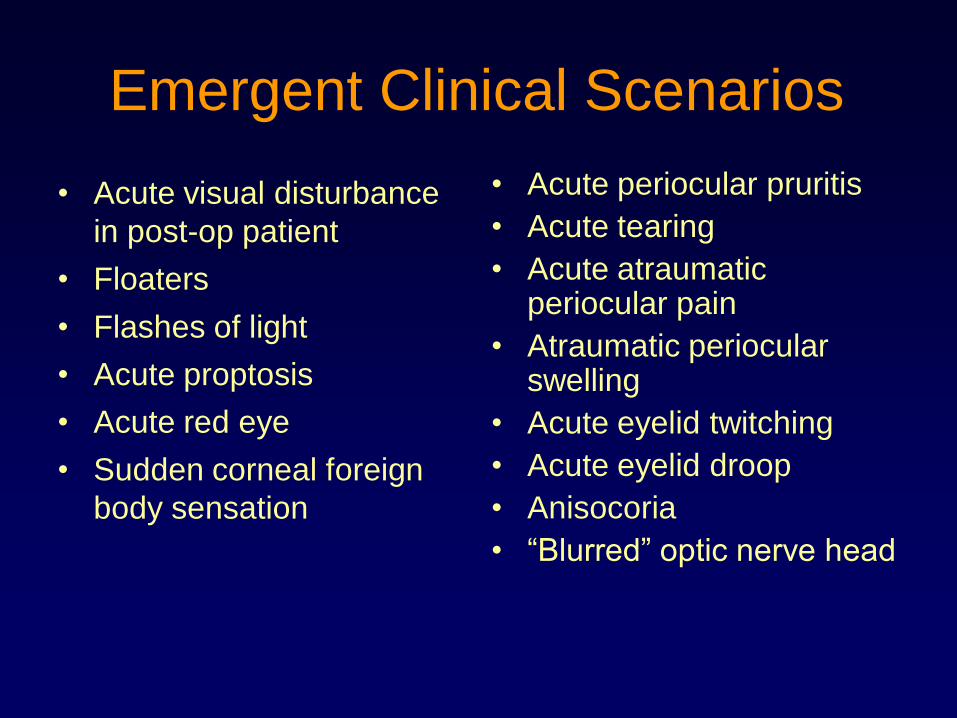
Emergent Clinical Scenarios
• Acute visual disturbance
in post-op patient
• Floaters
• Flashes of light
• Acute proptosis
• Acute red eye
• Sudden corneal foreign
body sensation
• Acute periocular pruritis
• Acute tearing
• Acute atraumatic periocular pain
• Atraumatic periocular swelling
• Acute eyelid twitching
• Acute eyelid droop
• Anisocoria
• ―Blurred‖ optic nerve head
23
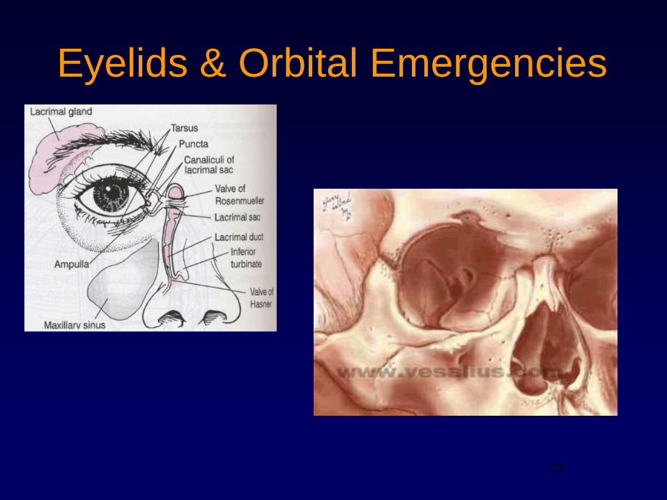
Eyelids & Orbital Emergencies
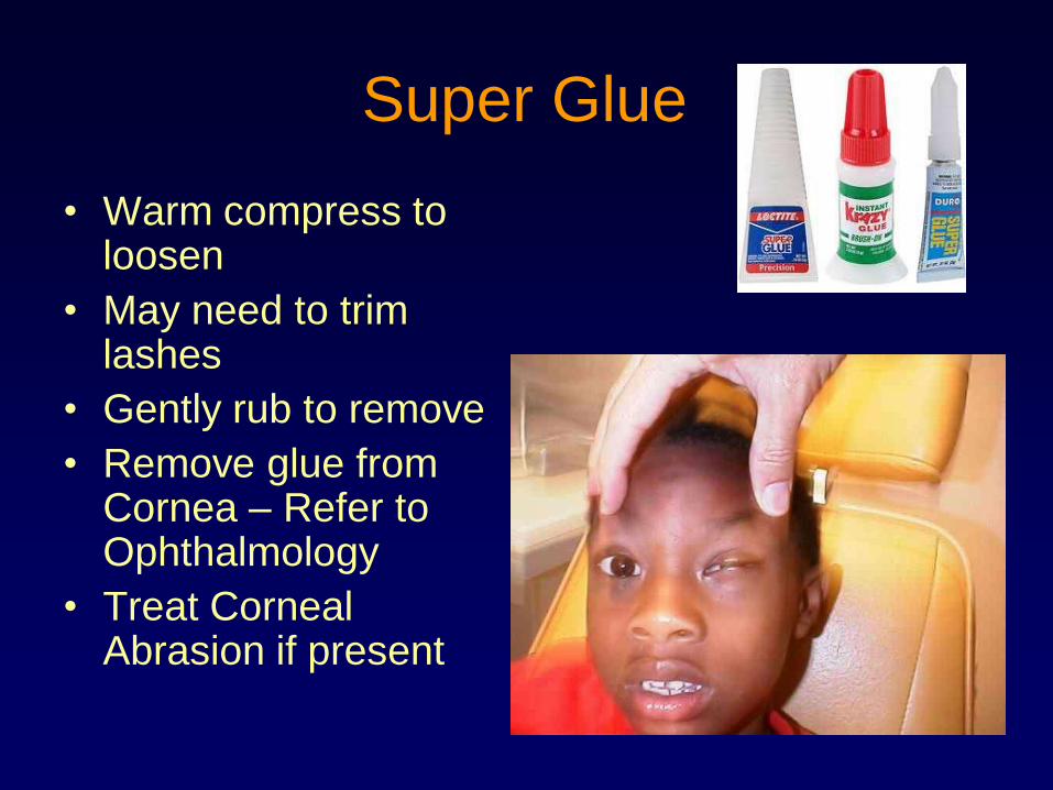
Super Glue
• Warm compress to loosen
• May need to trim lashes
• Gently rub to remove
• Remove glue from Cornea – Refer to Ophthalmology
• Treat Corneal Abrasion if present
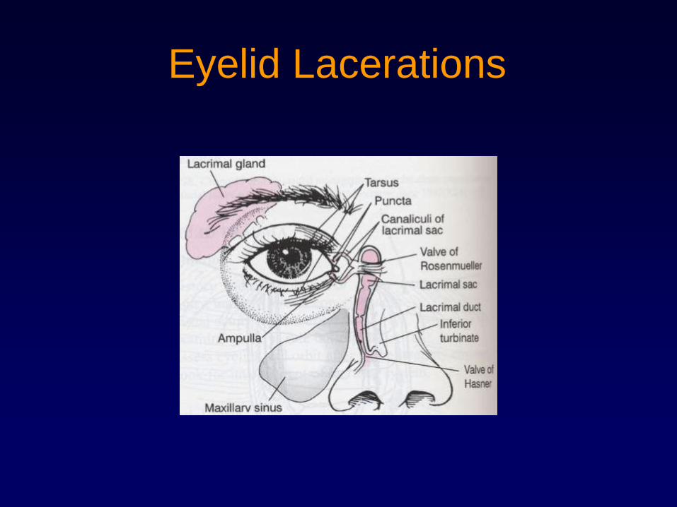
Eyelid Lacerations
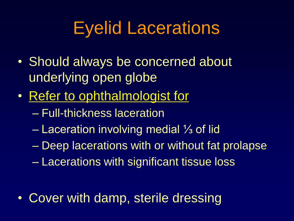
Eyelid Lacerations
• Should always be concerned about
underlying open globe
• Refer to ophthalmologist for
– Full-thickness laceration
– Laceration involving medial ⅓ of lid
– Deep lacerations with or without fat prolapse
– Lacerations with significant tissue loss
• Cover with damp, sterile dressing
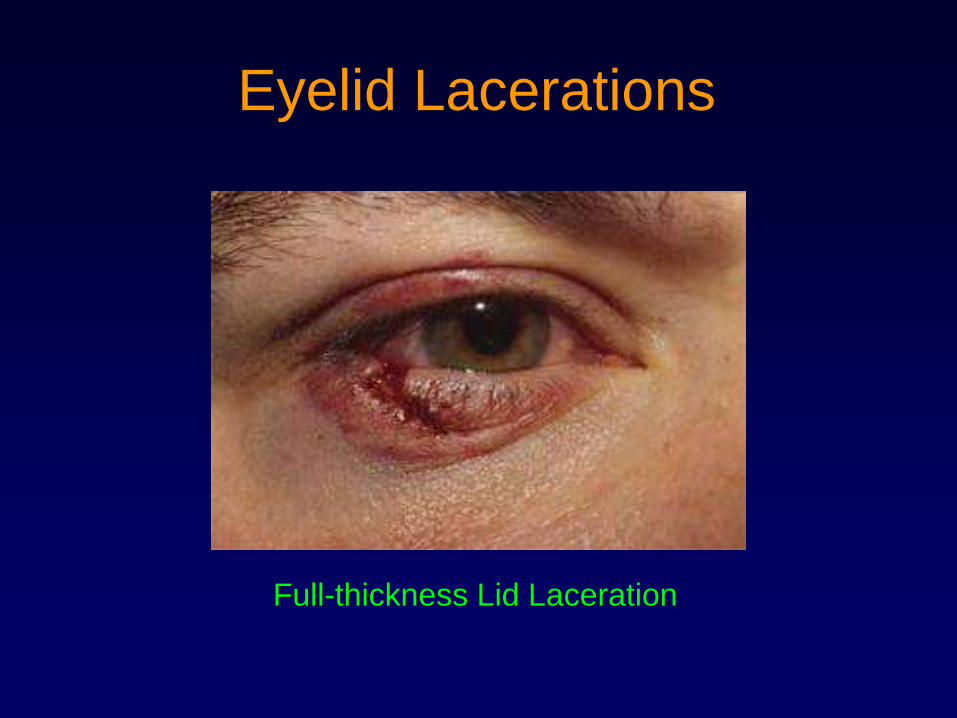
Eyelid Lacerations
Full-thickness Lid Laceration
28
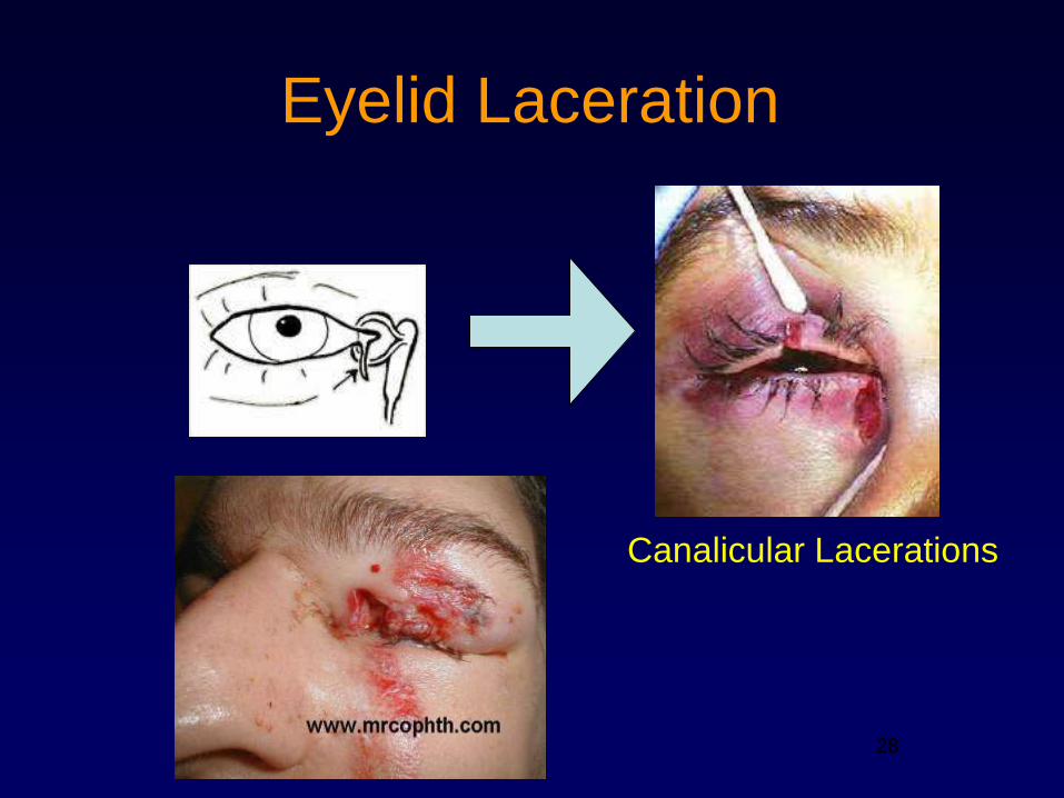
Eyelid Laceration
28
Canalicular Lacerations
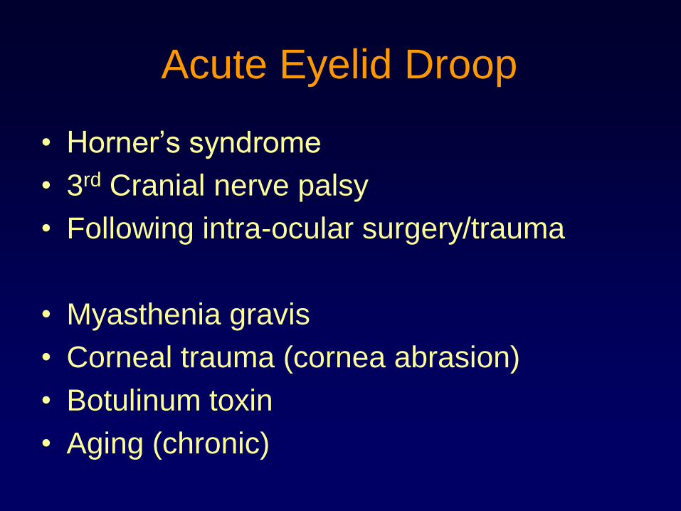
Acute Eyelid Droop
• Horner’s syndrome
• 3rd Cranial nerve palsy
• Following intra-ocular surgery/trauma
• Myasthenia gravis
• Corneal trauma (cornea abrasion)
• Botulinum toxin
• Aging (chronic)
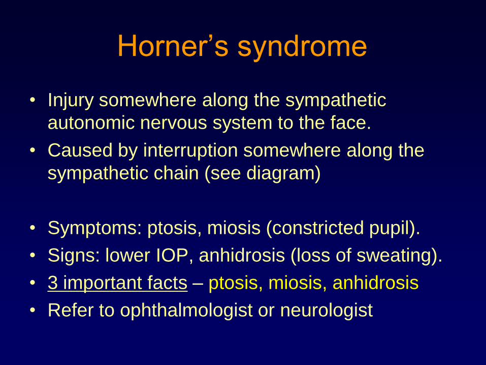
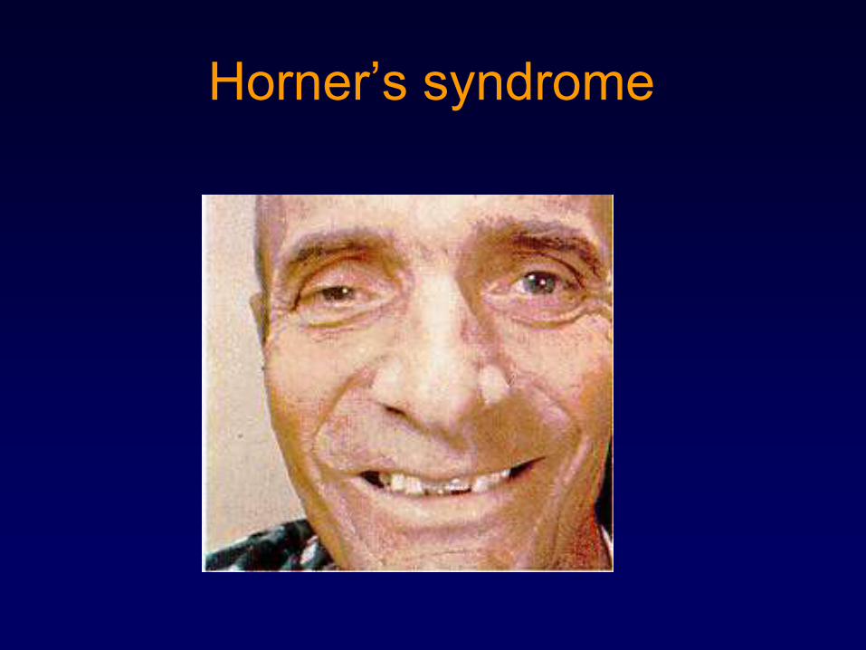
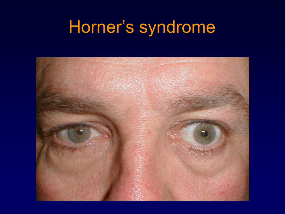
Horner’s syndrome
• Injury somewhere along the sympathetic
autonomic nervous system to the face.
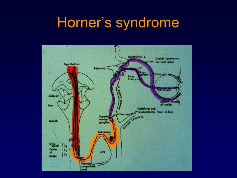
• Caused by interruption somewhere along the
sympathetic chain (see diagram)
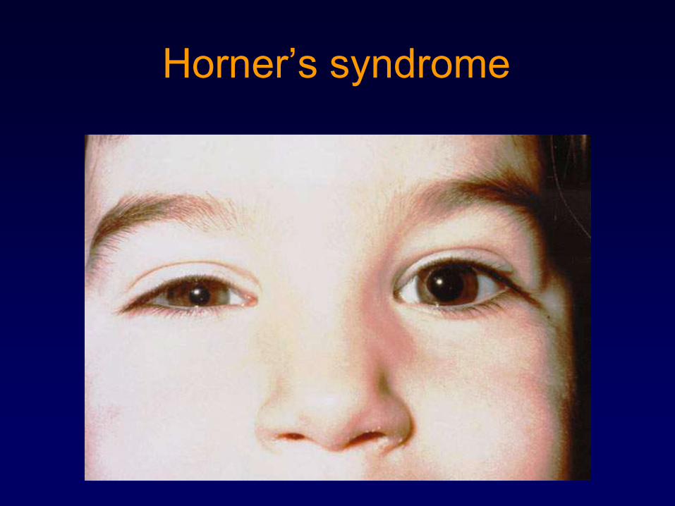
• Symptoms: ptosis, miosis (constricted pupil).
• Signs: lower IOP, anhidrosis (loss of sweating).
• 3 important facts – ptosis, miosis, anhidrosis
• Refer to ophthalmologist or neurologist
Horner’s syndrome
Horner’s syndrome
Horner’s syndrome
Horner’s syndrome
3rd Cranial nerve palsy
• Acute onset of double vision, may be horizontal or vertical, disappears when one eye is closed.
• Ptosis, eye is ―down and out‖ (CN IV and VI nl) with limited mobility.
• If pupil involved (dilated relative to other eye) then immediate imaging is required (to rule out mass lesion compressing brain stem).
• Can be painful if diabetes is the etiology
• Consult with an ophthalmologist or neurologist immediately
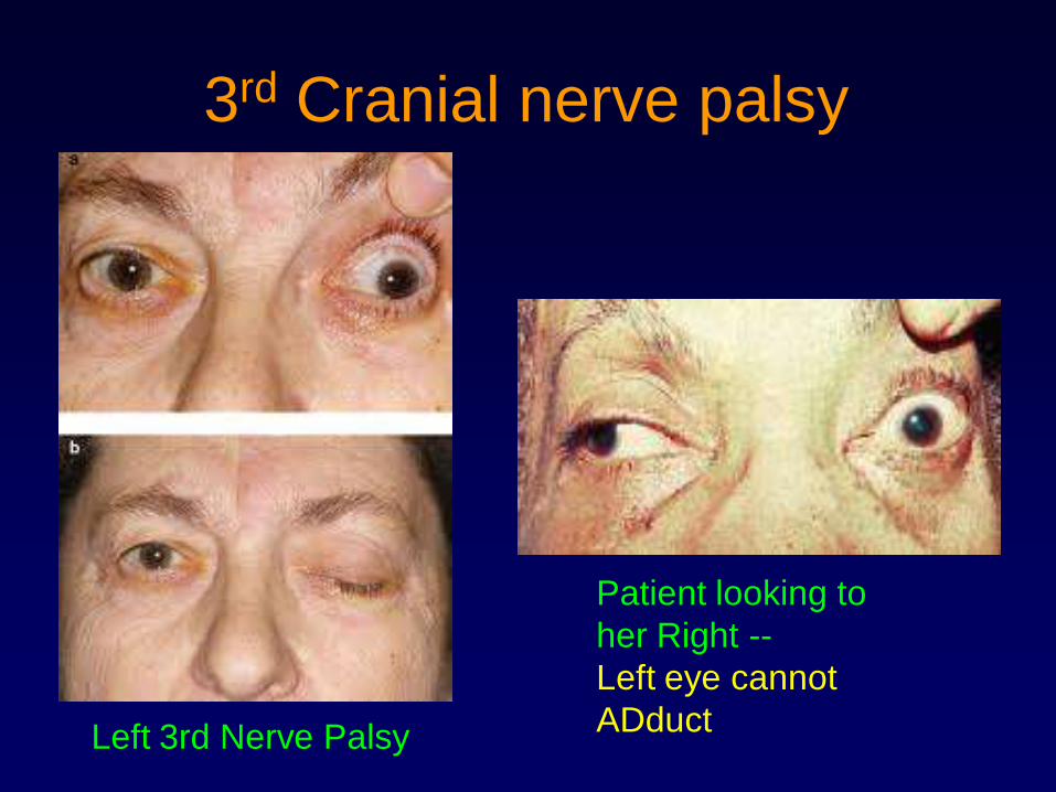
3rd Cranial nerve palsy
Left 3rd Nerve Palsy
Patient looking to
her Right --
Left eye cannot
ADduct
37
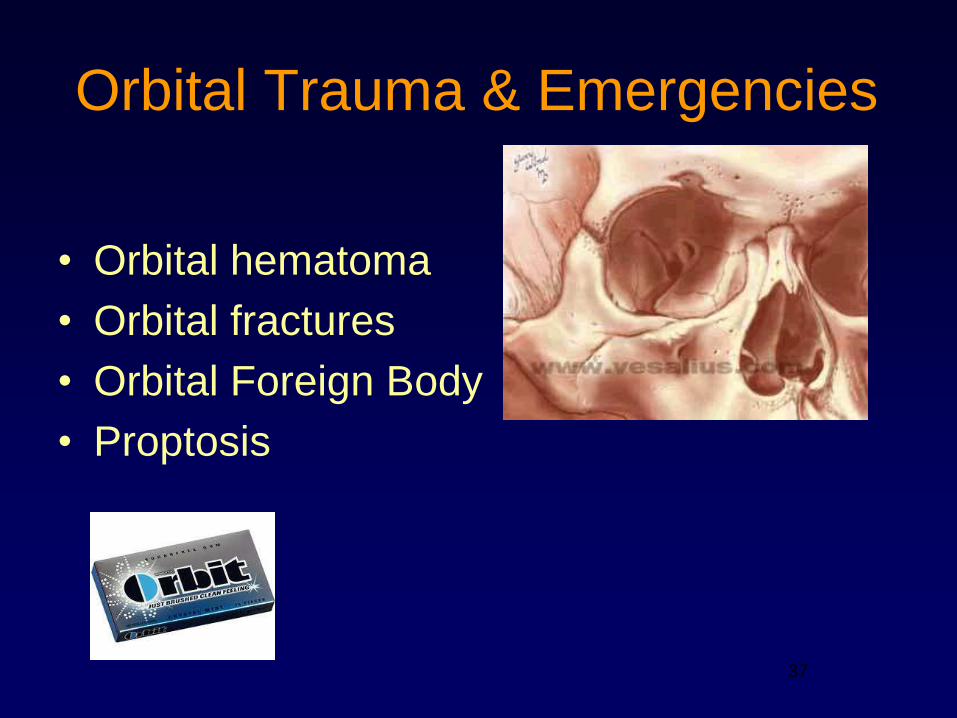
Orbital Trauma & Emergencies
• Orbital hematoma
• Orbital fractures
• Orbital Foreign Body
• Proptosis
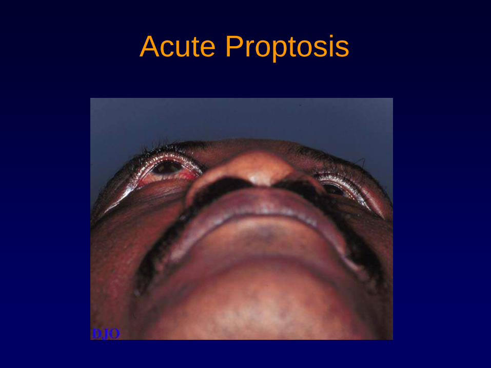
Acute Proptosis

Acute Proptosis
• Orbital cellulitis
• Orbital pseudotumor
• Vascular abnormalities: carotid-cavernous sinus fistula, varix
• Retrobulbar hemorrhage
• Graves' orbitopathy
• Orbital vasculitis: polyarteritis nodosa, Wegener's granulomatosis, temporal arteritis
• Granulomatous disease: sarcoidosis
• Orbital tumors: primary, secondary, metastatic
Acute Periocular Pain
• Sinusitis
• Dry eyes
• Orbital pseudotumor
• Optic neuritis
• Diabetic cranial nerve palsy
• Orbital cellulitis
• Preseptal cellulitis
• (many others)
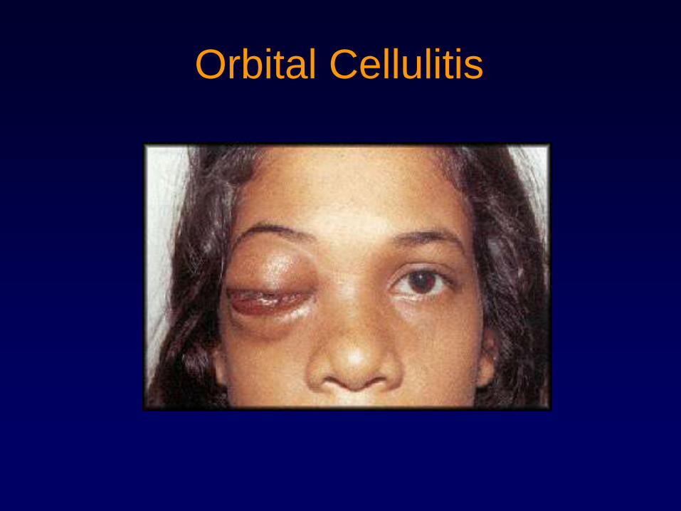
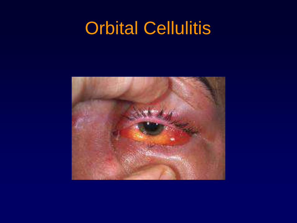
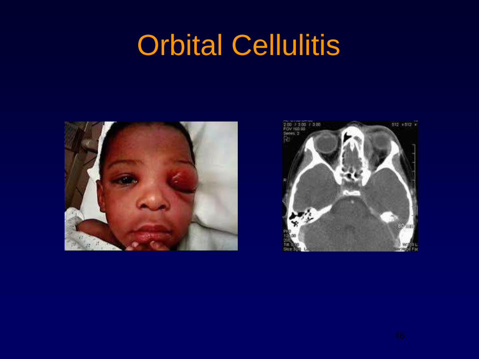
Orbital Cellulitis
• Cellulitis posterior to the orbital septum
• Symptoms - Red eye, pain, blurred vision, headache,
double vision
• Signs - Eyelid edema, erythema, warmth, tenderness.
Proptosis, restricted ocular motility with pain on
attempted movement.
• Tx – consult ophthalmologist and obtain orbital CT. Will
require oral/IV antibiotics.
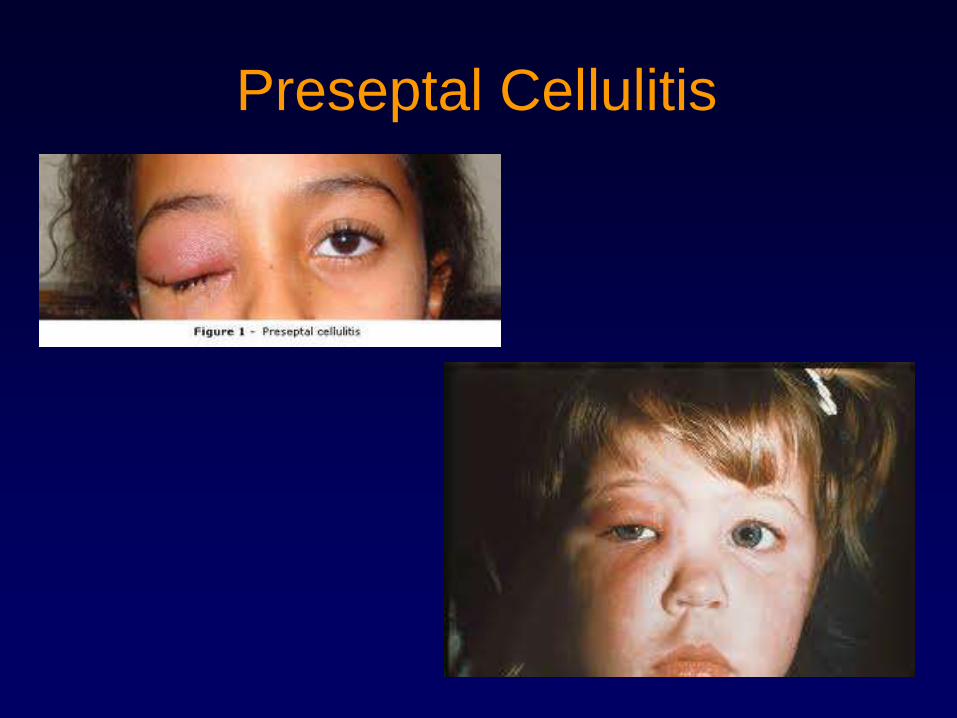
• Needs to be differentiated from preseptal cellulitis which
has salient features that differentiate the two including
no vision changes, no restriction of eye movements
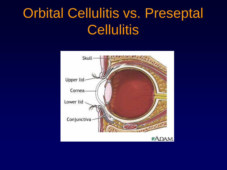
Orbital Cellulitis vs. Preseptal
Cellulitis
43
Preseptal Cellulitis
Orbital Cellulitis
Orbital Cellulitis
46
Orbital Cellulitis
Orbital Hematoma
• If mild, treat with cool compresses
• If large amount of hemorrhage, especially
behind the globe (Retrobulbar
hemorrhage)
– may require emergency surgery to reduce
intraocular pressure and protect corneal
surface
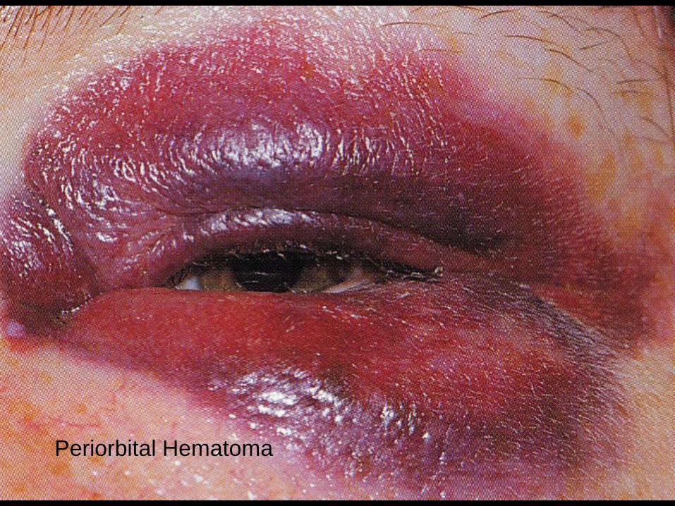
Periorbital Hematoma
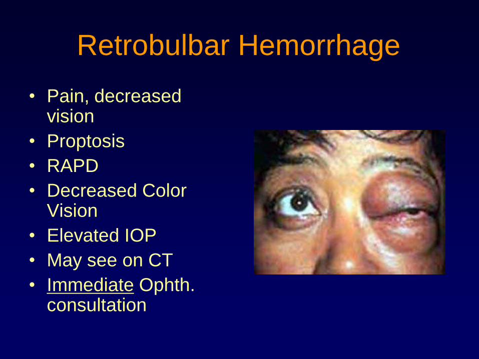
Retrobulbar Hemorrhage
• Pain, decreased vision
• Proptosis
• RAPD
• Decreased Color Vision
• Elevated IOP
• May see on CT
• Immediate Ophth. consultation
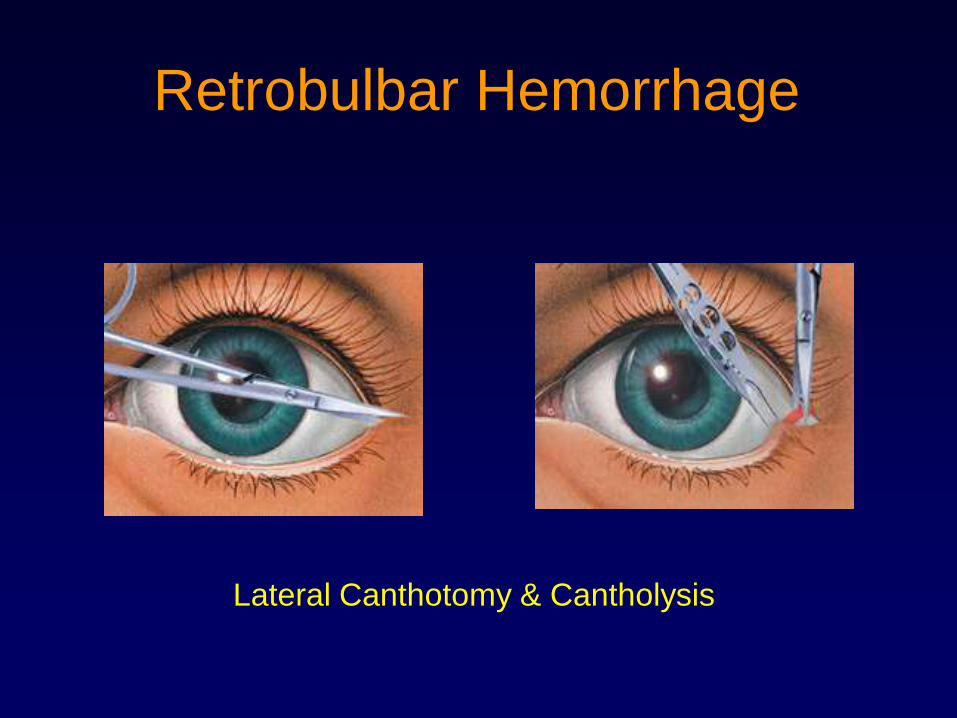
Retrobulbar Hemorrhage
Lateral Canthotomy & Cantholysis
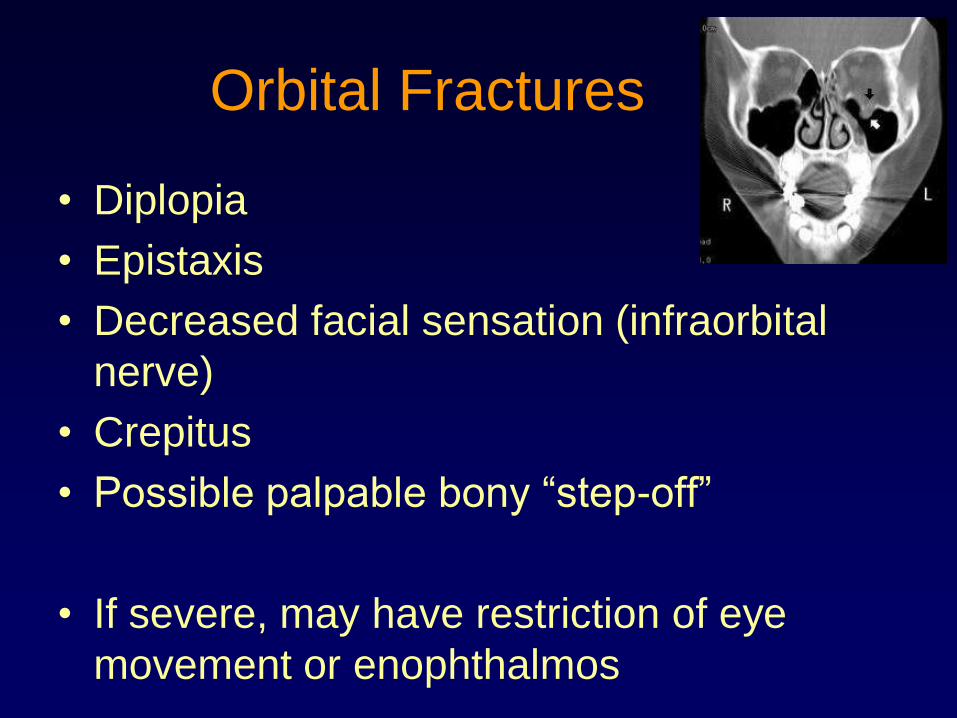
Orbital Fractures
• Diplopia
• Epistaxis
• Decreased facial sensation (infraorbital
nerve)
• Crepitus
• Possible palpable bony ―step-off‖
• If severe, may have restriction of eye
movement or enophthalmos
52
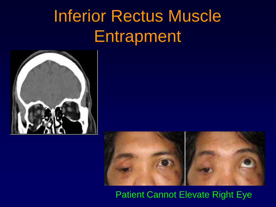
Inferior Rectus Muscle
Entrapment
Patient Cannot Elevate Right Eye
53
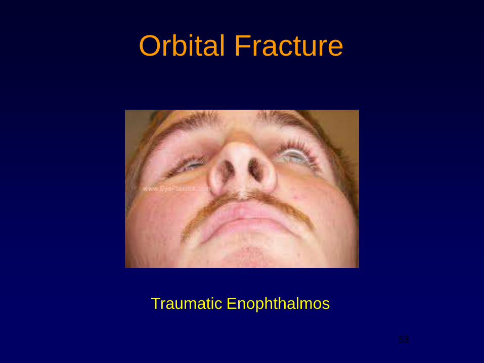
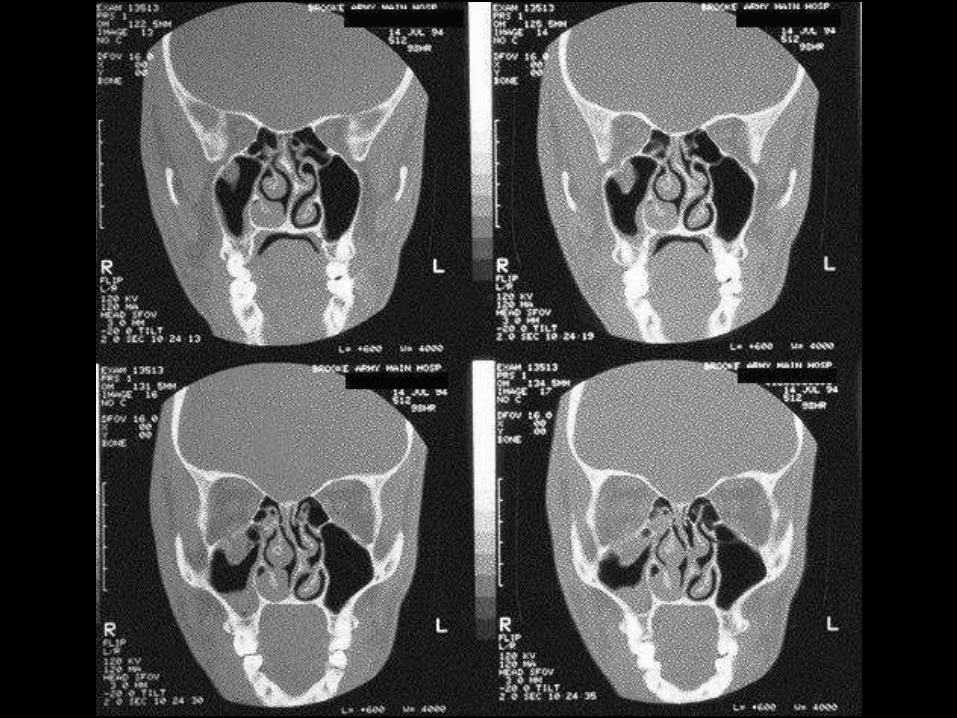
Orbital Fracture
Traumatic Enophthalmos
Orbital Fractures
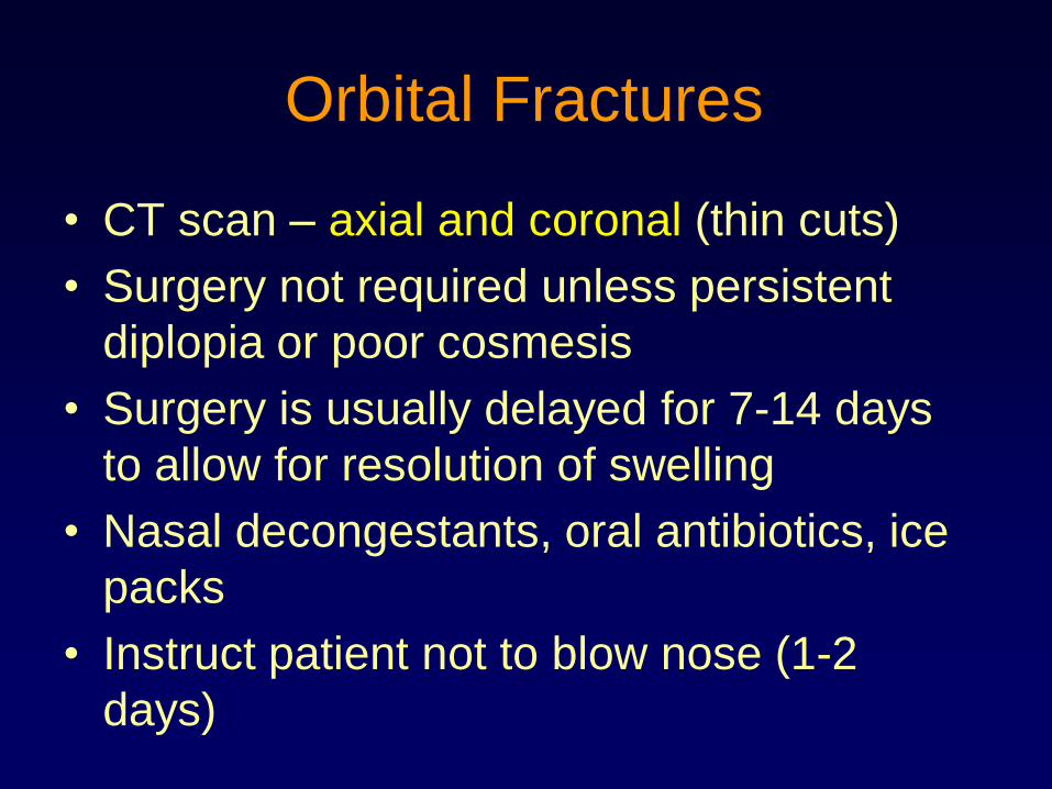
• CT scan – axial and coronal (thin cuts)
• Surgery not required unless persistent
diplopia or poor cosmesis
• Surgery is usually delayed for 7-14 days
to allow for resolution of swelling
• Nasal decongestants, oral antibiotics, ice
packs
• Instruct patient not to blow nose (1-2
days)
56
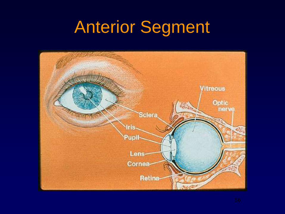
Anterior Segment
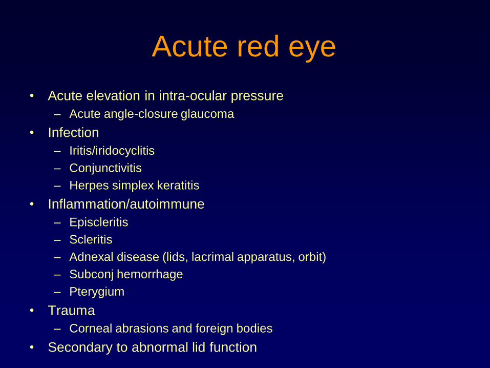
Acute red eye
• Acute elevation in intra-ocular pressure
– Acute angle-closure glaucoma
• Infection
– Iritis/iridocyclitis
– Conjunctivitis
– Herpes simplex keratitis
• Inflammation/autoimmune
– Episcleritis
– Scleritis
– Adnexal disease (lids, lacrimal apparatus, orbit)
– Subconj hemorrhage
– Pterygium
• Trauma
– Corneal abrasions and foreign bodies
• Secondary to abnormal lid function
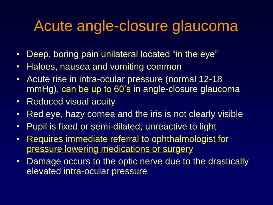
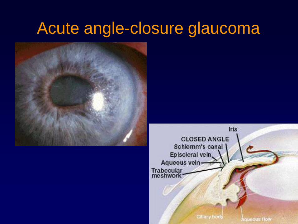
Acute angle-closure glaucoma
• Deep, boring pain unilateral located ―in the eye‖
• Haloes, nausea and vomiting common
• Acute rise in intra-ocular pressure (normal 12-18 mmHg), can be up to 60’s in angle-closure glaucoma
• Reduced visual acuity
• Red eye, hazy cornea and the iris is not clearly visible
• Pupil is fixed or semi-dilated, unreactive to light
• Requires immediate referral to ophthalmologist for pressure lowering medications or surgery
• Damage occurs to the optic nerve due to the drastically elevated intra-ocular pressure
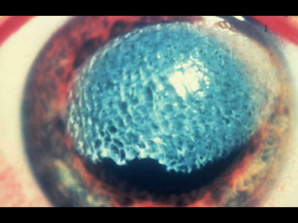
Acute angle-closure glaucoma
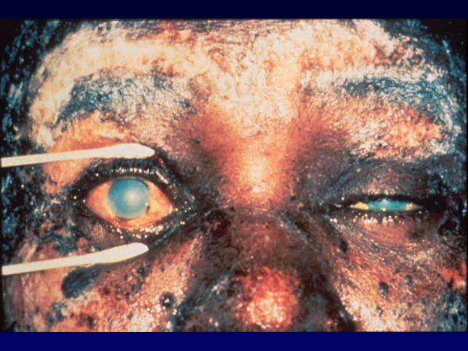
Chemical Burns
Irrigate immediately
before
anything else
Alkaline (bases)
• Fertilizers
• Cleaning products (ammonia)
• Drain cleaners (lye)
• Oven cleaners
• Bleach (sodium hydroxide)
• Fireworks (magnesium hydroxide)
• Cement (lime)
Alkaline (bases)
• High pH
• Especially damaging – will denature
proteins and lyse cell membranes which
enhances penetration
Acids
• Battery acid (sulfuric acid)
• Glass polish/etching (hydrofluoric acid)
• Vinegar, nail polish remover (acetic acid)
Acids
• Low pH
• Depth of penetration usually less due to
precipitation of proteins
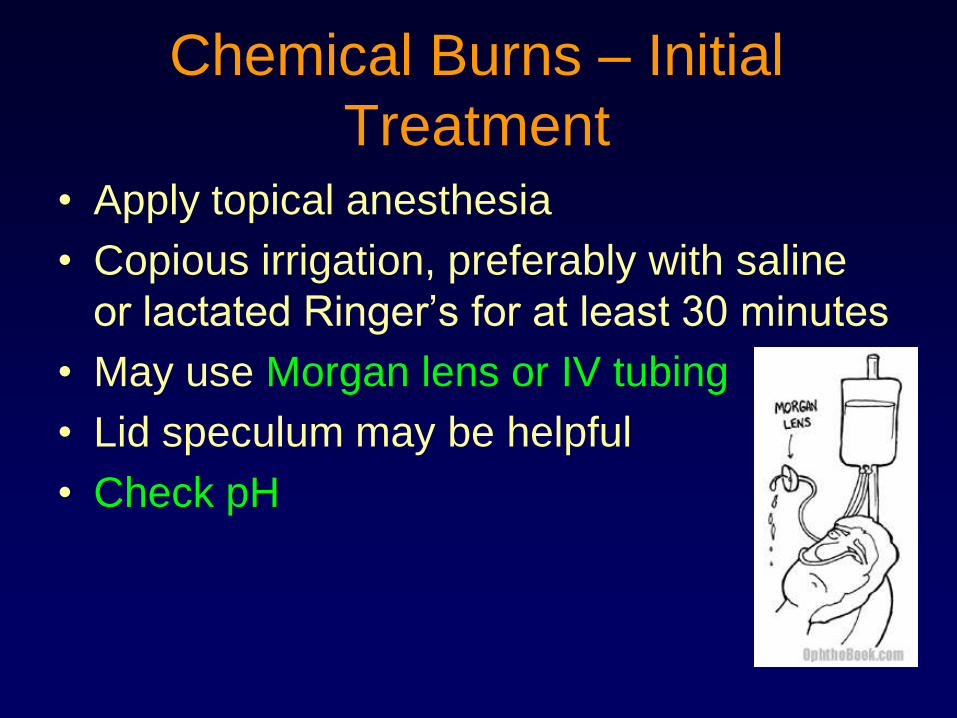
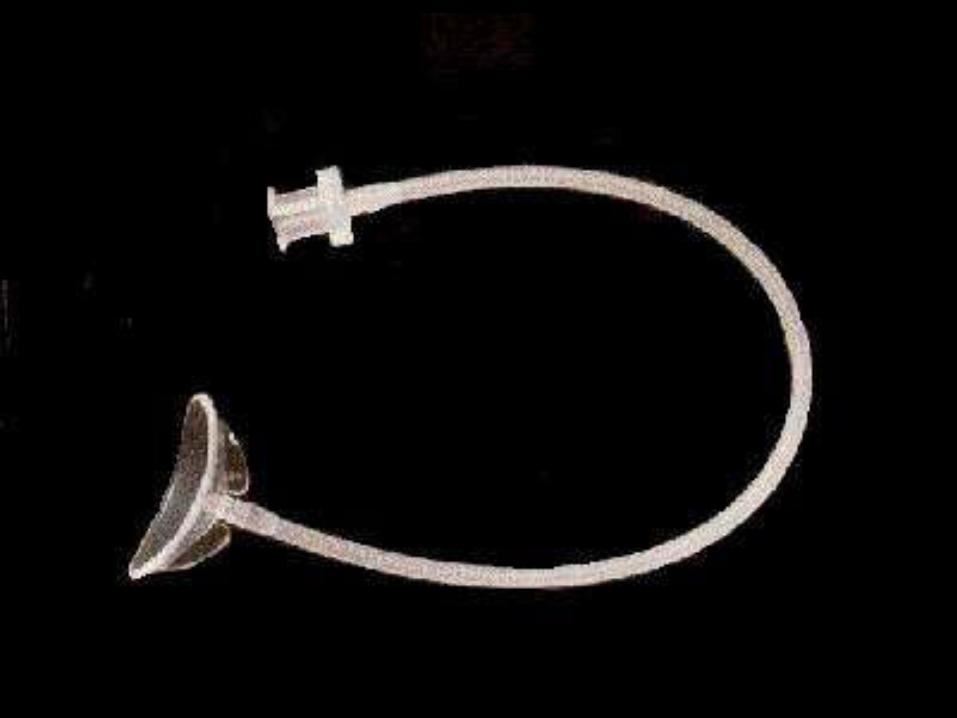
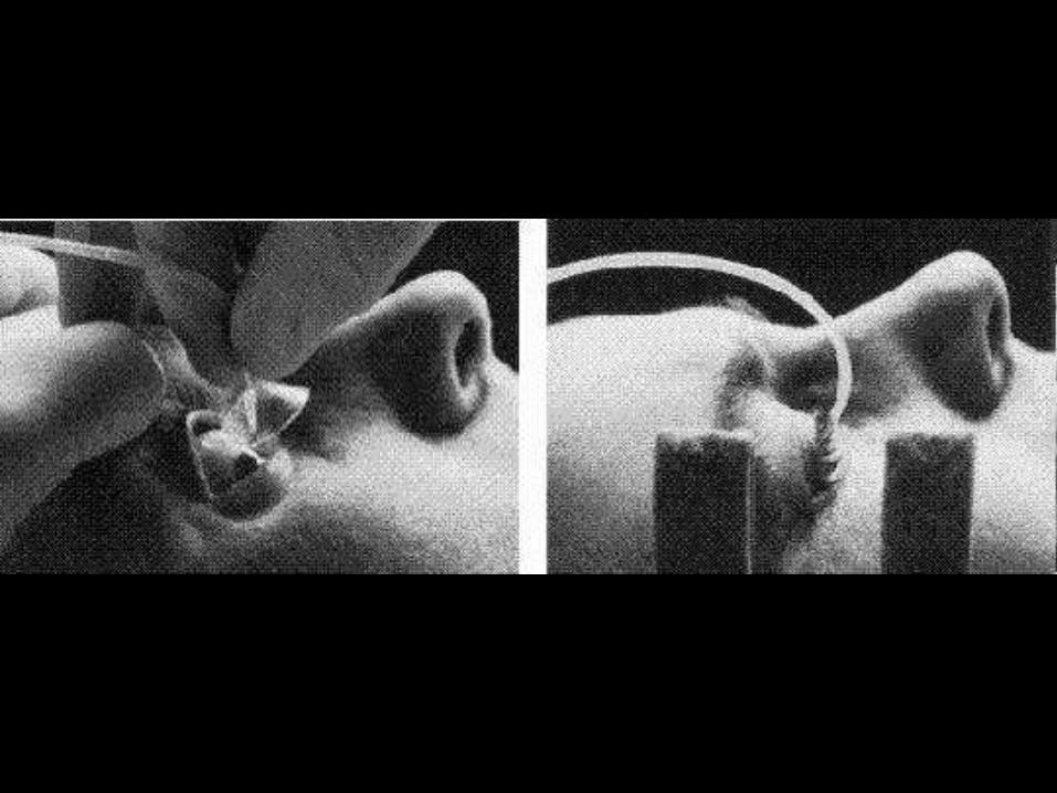
Chemical Burns – Initial
Treatment
• Apply topical anesthesia
• Copious irrigation, preferably with saline
or lactated Ringer’s for at least 30 minutes
• May use Morgan lens or IV tubing
• Lid speculum may be helpful
• Check pH

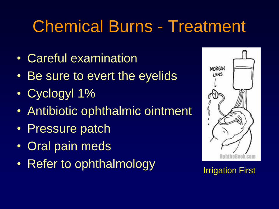
Chemical Burns - Treatment
• Careful examination
• Be sure to evert the eyelids
• Cyclogyl 1%
• Antibiotic ophthalmic ointment
• Pressure patch
• Oral pain meds
• Refer to ophthalmologyIrrigation First
Thermal Burn
• Most common – cigarettes and curling iron
• Usually superficial burns
• Treat like chemical burn except no
irrigation needed
• May need debridement of burned tissue
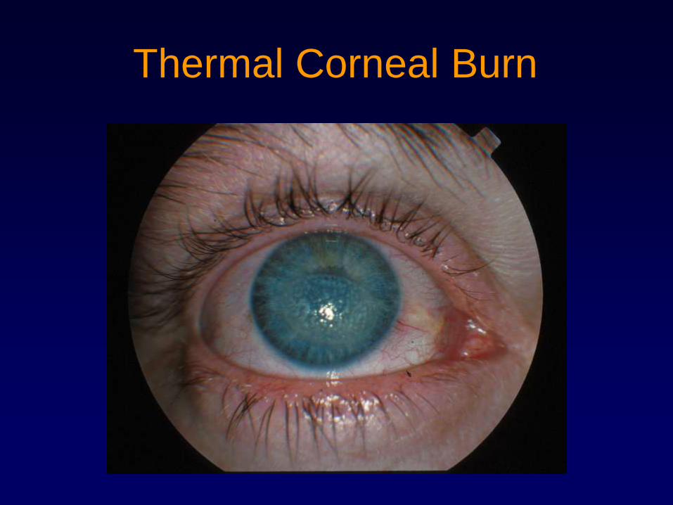
Thermal Corneal Burn
Ultraviolet Burn
• Welding or sun lamps without eye
protection
• Produces small, diffuse epithelial defects
which stain with fluorescein
• Becomes severely painful several hours
after exposure
• Treat with Cyclogyl, antibiotic ointment,
and pressure patching
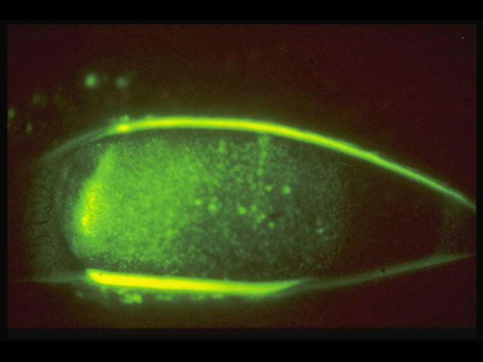
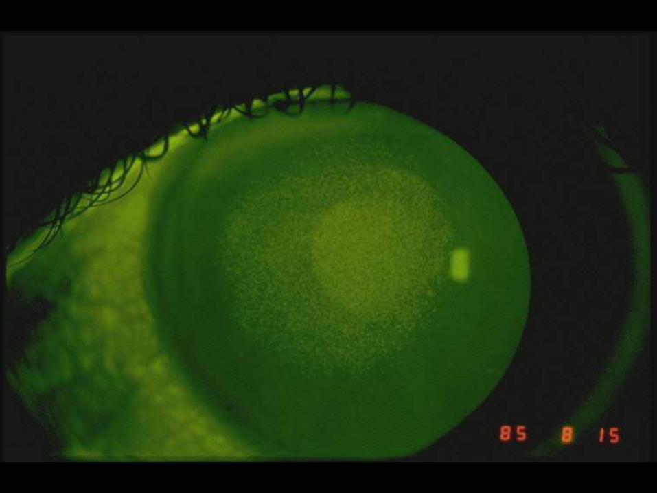
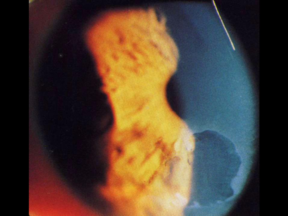
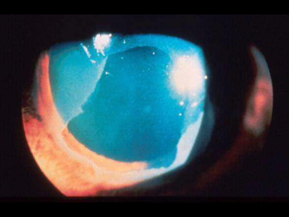
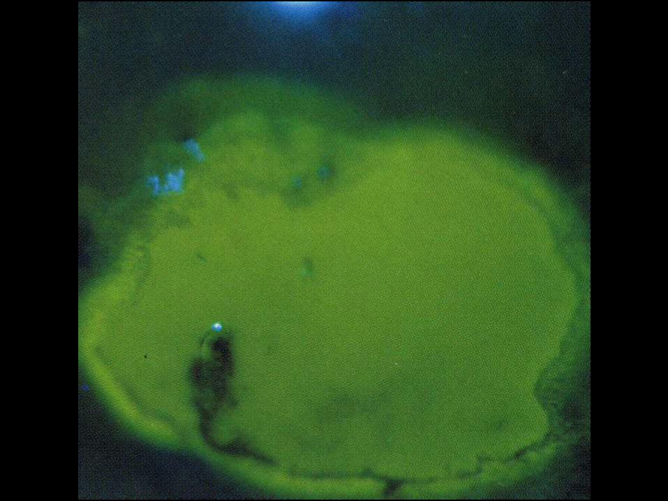
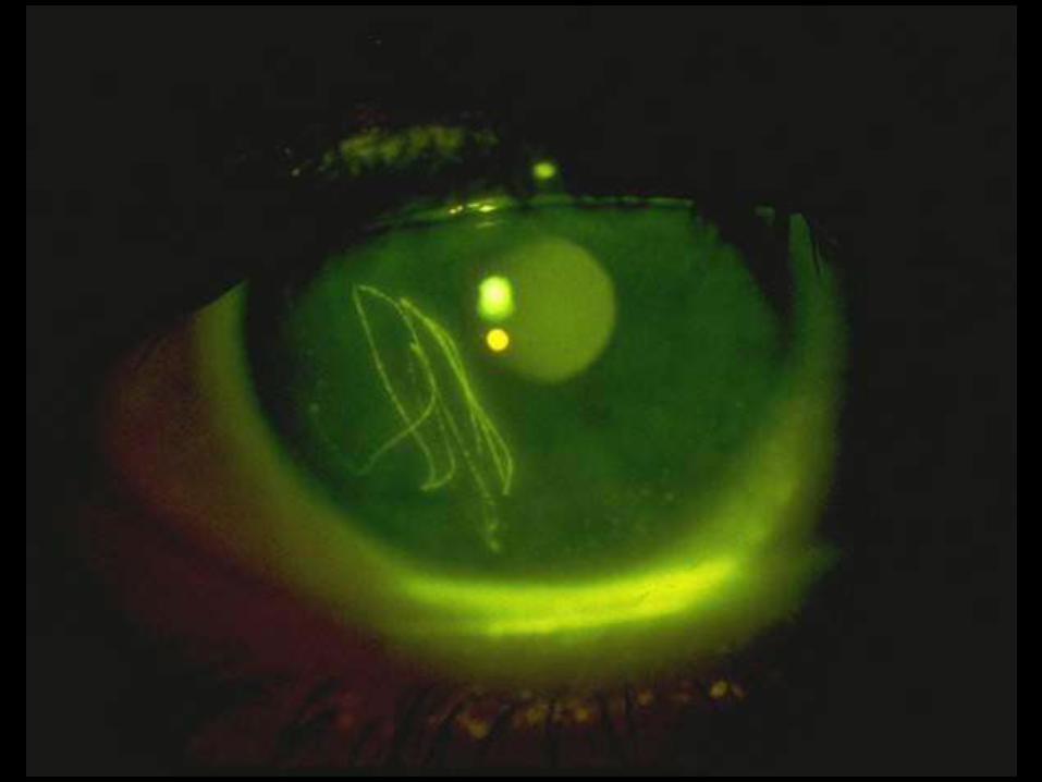
Corneal Abrasions
• Usually a defect in superficial layer of cornea
• Can usually be seen without fluorescein
• Glows yellow/green with fluorescein and blue light
• Treat with Cyclogyl (dilating drop), antibiotic ointment or drops, and possibly pressure patch. Needs follow up exam
• NEVER prescribe topical anesthetic
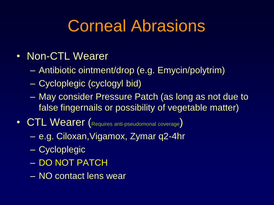
Corneal Abrasions
• Non-CTL Wearer
– Antibiotic ointment/drop (e.g. Emycin/polytrim)
– Cycloplegic (cyclogyl bid)
– May consider Pressure Patch (as long as not due to
false fingernails or possibility of vegetable matter)
• CTL Wearer (Requires anti-pseudomonal coverage)
– e.g. Ciloxan,Vigamox, Zymar q2-4hr
– Cycloplegic
– DO NOT PATCH
– NO contact lens wear
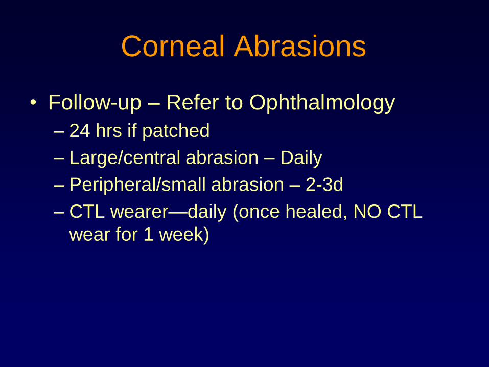
Corneal Abrasions
• Follow-up – Refer to Ophthalmology
– 24 hrs if patched
– Large/central abrasion – Daily
– Peripheral/small abrasion – 2-3d
– CTL wearer—daily (once healed, NO CTL
wear for 1 week)
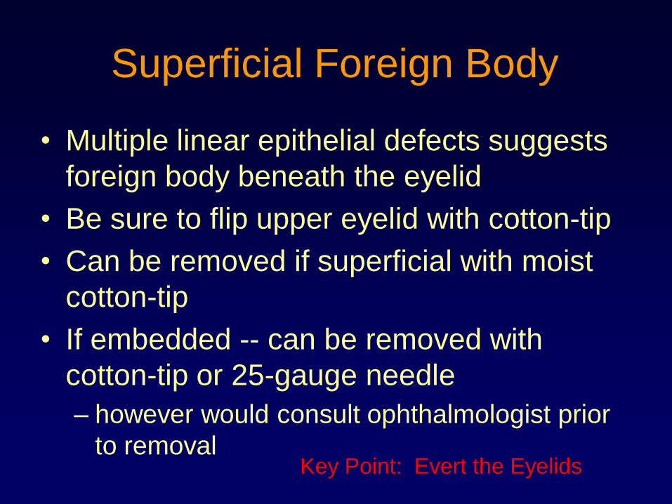
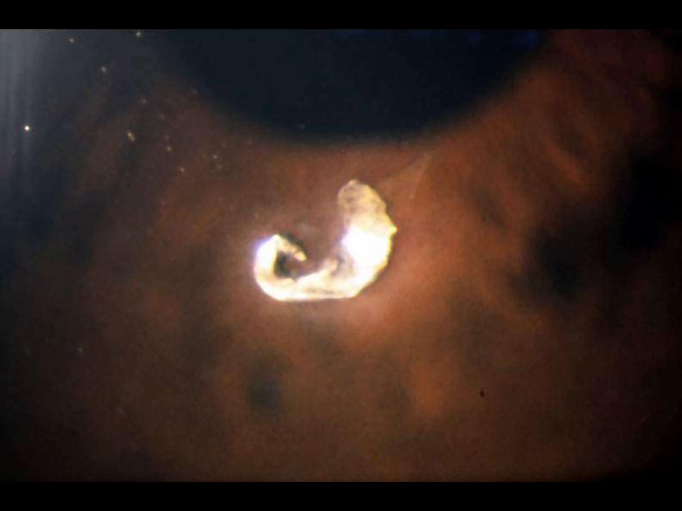
Superficial Foreign Body
• Multiple linear epithelial defects suggests
foreign body beneath the eyelid
• Be sure to flip upper eyelid with cotton-tip
• Can be removed if superficial with moist
cotton-tip
• If embedded -- can be removed with
cotton-tip or 25-gauge needle
– however would consult ophthalmologist prior
to removalKey Point: Evert the Eyelids

85
Evert the Eyelids
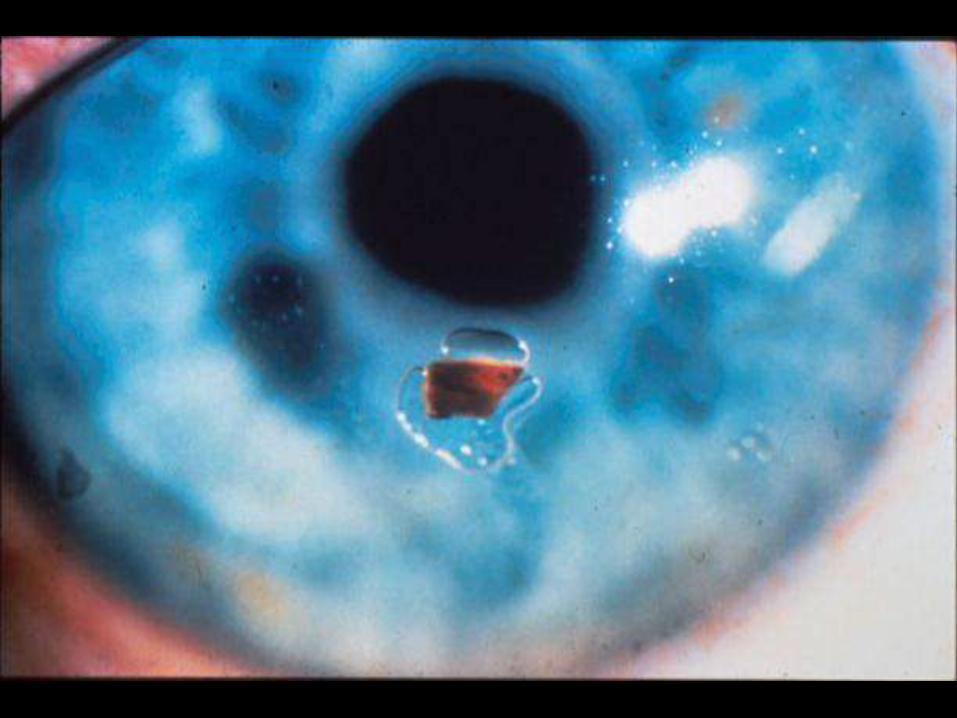
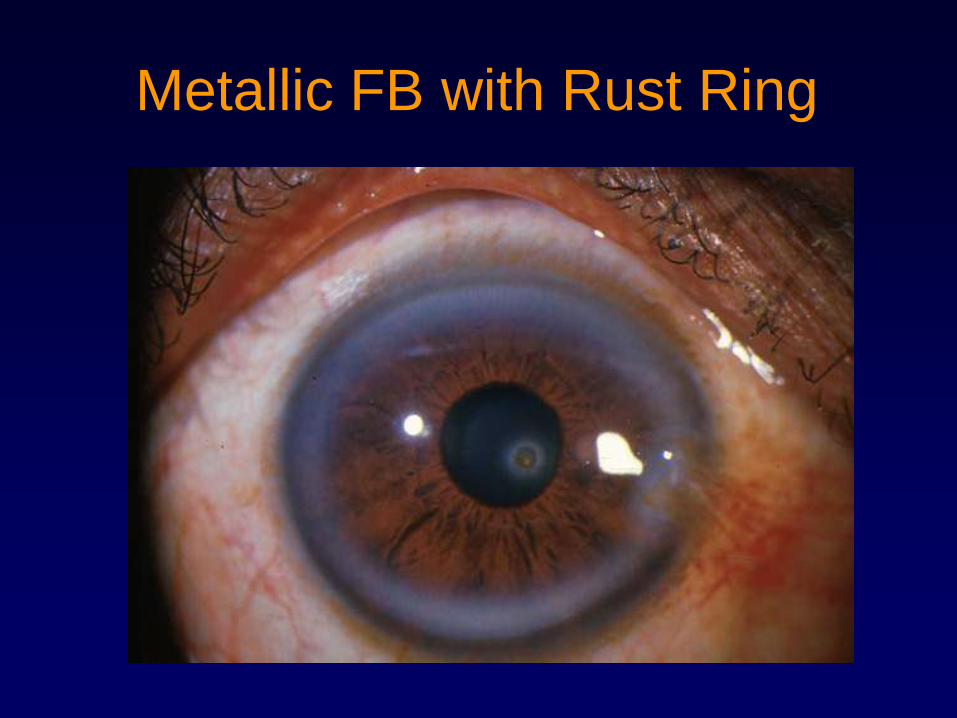
Metallic FB with Rust Ring
90
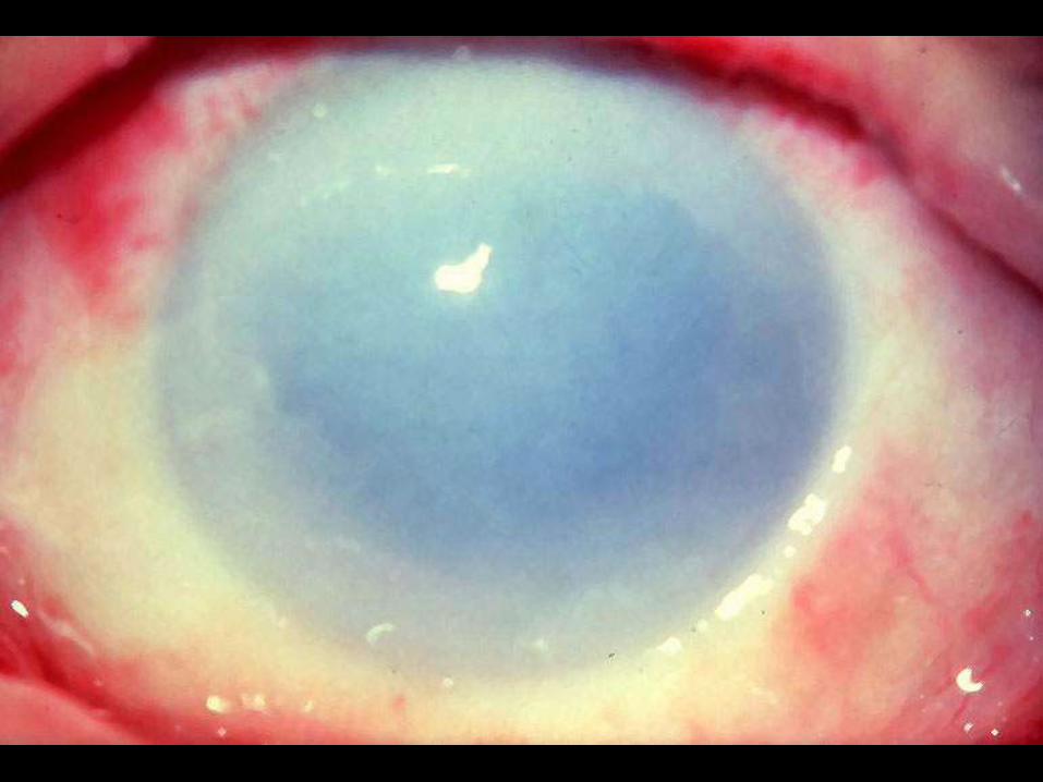
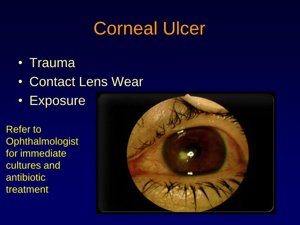
Corneal Ulcer
• Trauma
• Contact Lens Wear
• Exposure
90
Refer to
Ophthalmologist
for immediate
cultures and
antibiotic
treatment
91
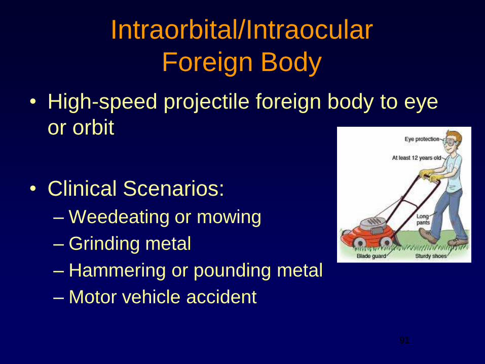
Intraorbital/Intraocular
Foreign Body
• High-speed projectile foreign body to eye
or orbit
• Clinical Scenarios:
– Weedeating or mowing
– Grinding metal
– Hammering or pounding metal
– Motor vehicle accident
91
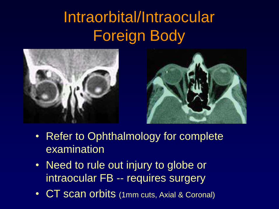
Intraorbital/Intraocular
Foreign Body
• Refer to Ophthalmology for complete
examination
• Need to rule out injury to globe or
intraocular FB -- requires surgery
• CT scan orbits (1mm cuts, Axial & Coronal)
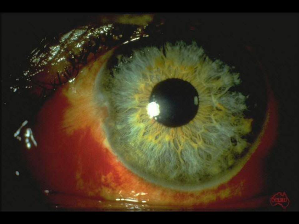
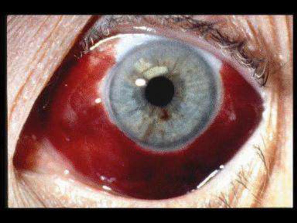
Subconjunctival Hemorrhage
• Very common after blunt trauma
• Superficial blood vessels broken
• May occur spontaneously (Coumadin,
aspirin, valsalva)
• Usually self-limited
• Treat with artificial tears and reassurance
• May be suggestive of ruptured globe
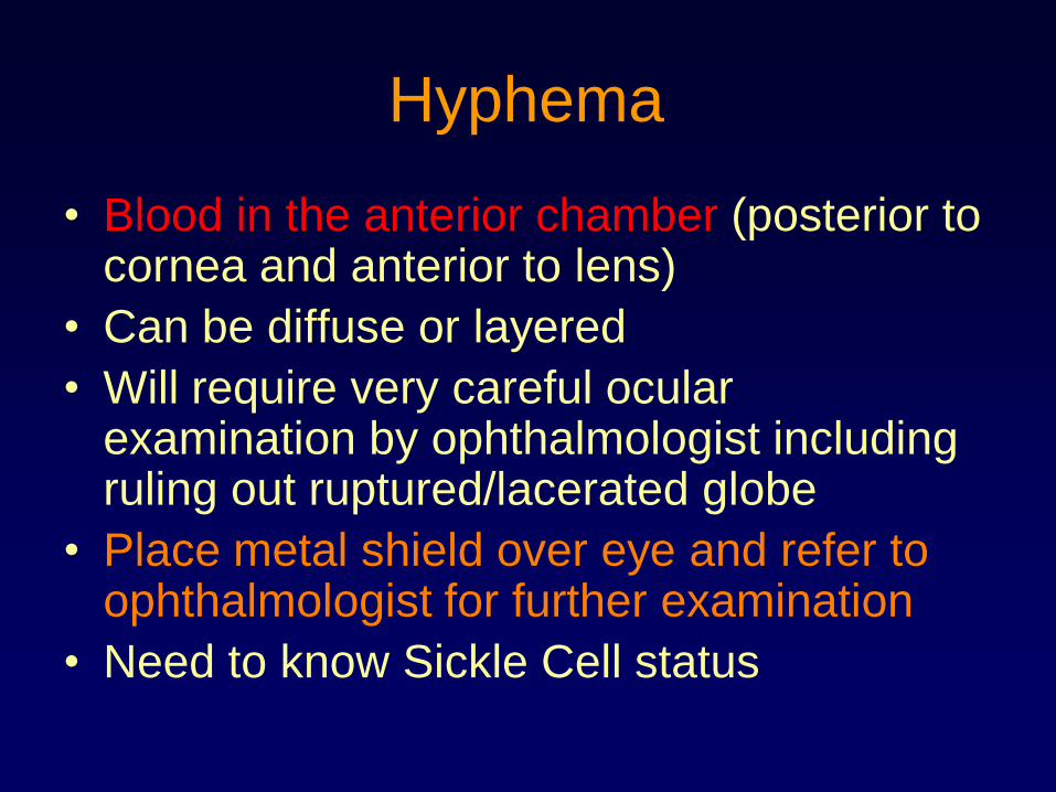
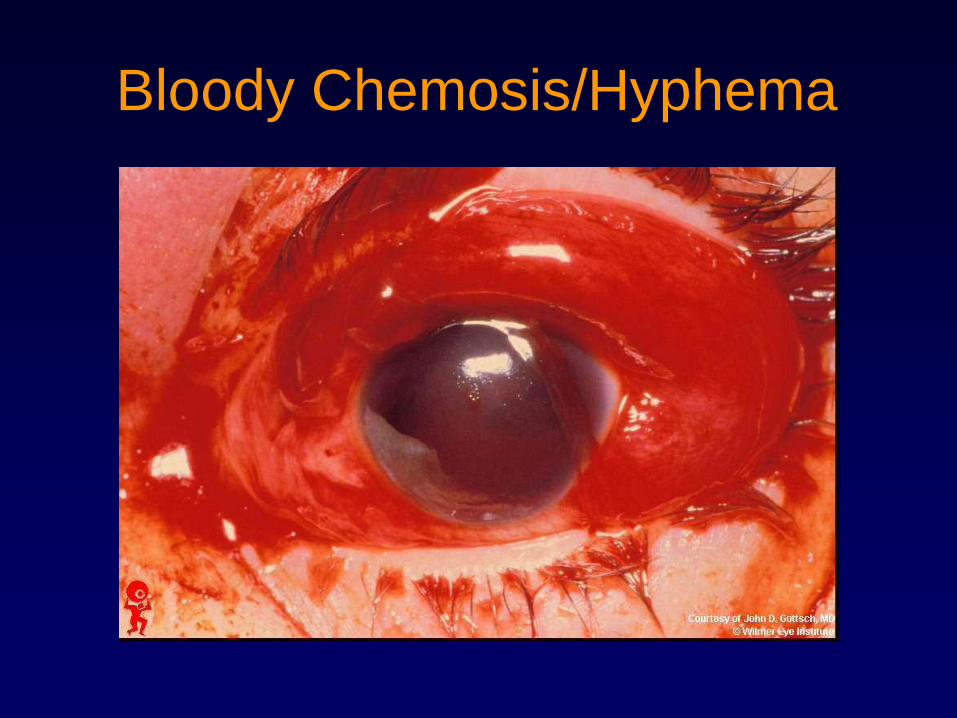
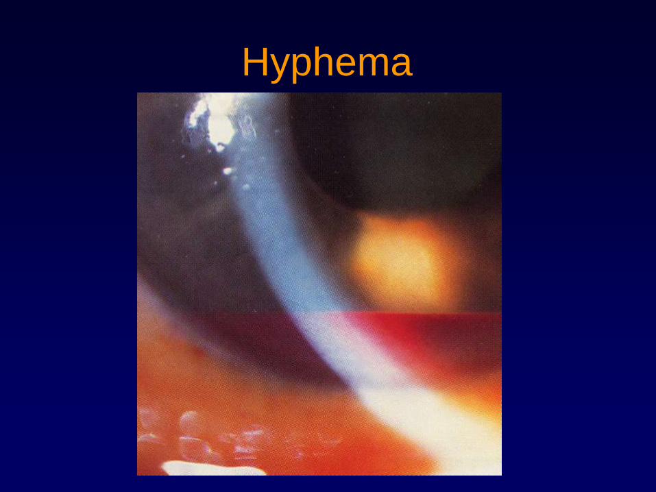
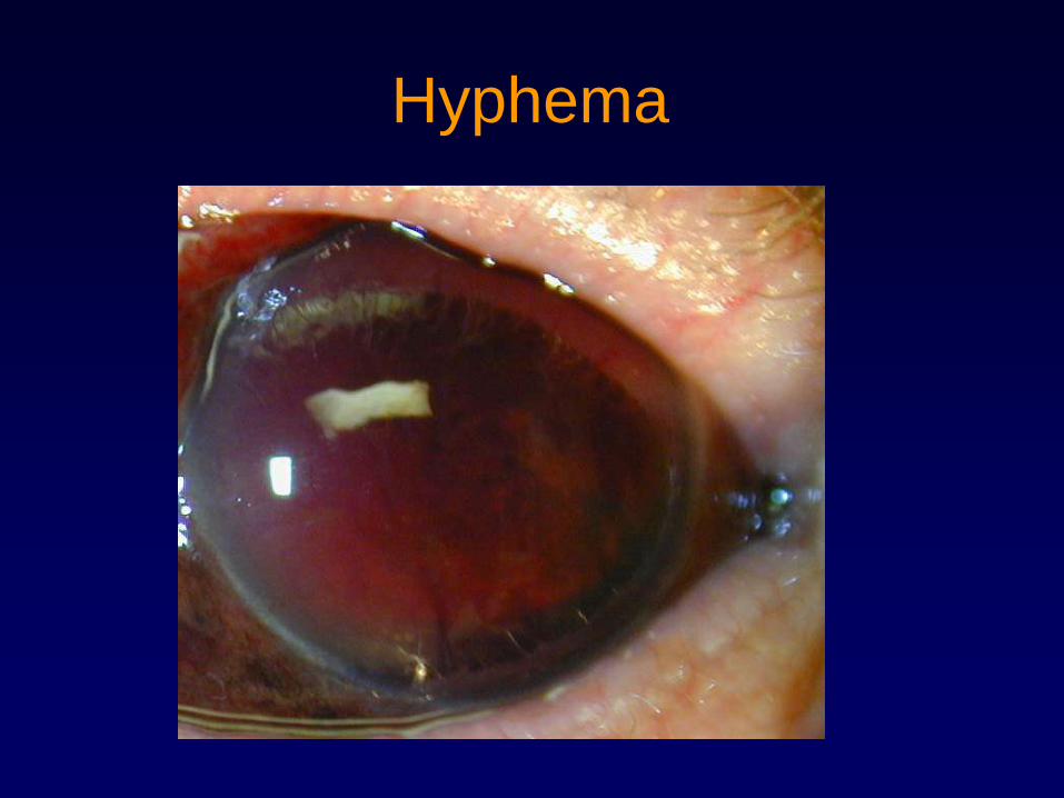
Hyphema
• Blood in the anterior chamber (posterior to cornea and anterior to lens)
• Can be diffuse or layered
• Will require very careful ocular examination by ophthalmologist including ruling out ruptured/lacerated globe
• Place metal shield over eye and refer to ophthalmologist for further examination
• Need to know Sickle Cell status
Hyphema
Hyphema
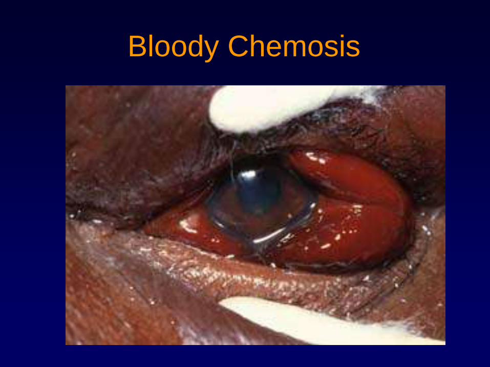
Bloody Chemosis
100
RETINAL & OPTIC NERVE
EMERGENCIES
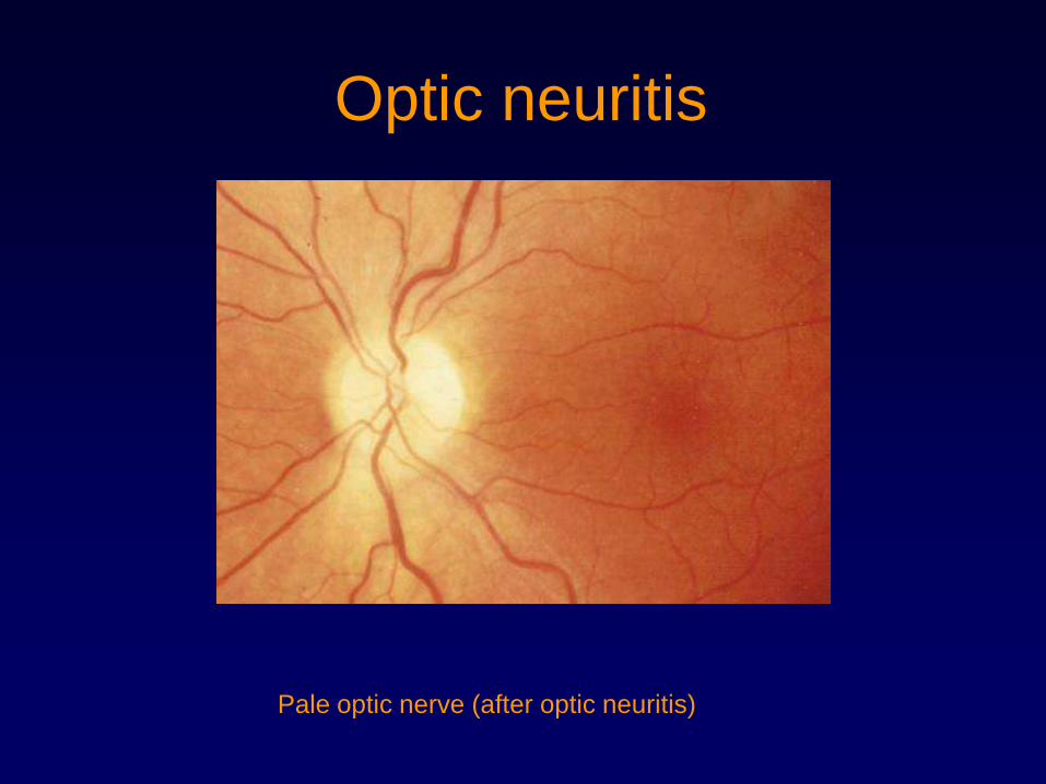
Optic Neuritis
• Inflammation of the optic nerve in young adults
• Symptoms: unilateral loss of vision over hours to days. Orbital pain with eye movement, acquired loss of color vision, reduced perception of light
• Signs: Relative afferent pupillary defect, decreased color, central, visual field defects, swollen or normal optic disc
• Tx: Ophthalmologic referral – will require MRI and possibly IV steroids
• Can be a risk for multiple sclerosis
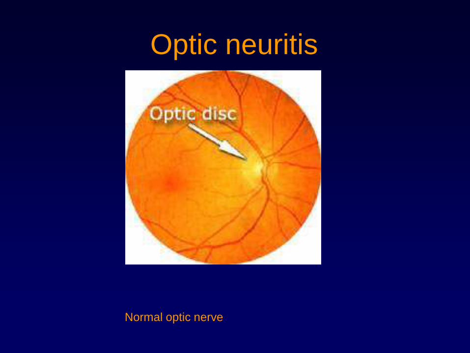
Optic neuritis
Normal optic nerve
103
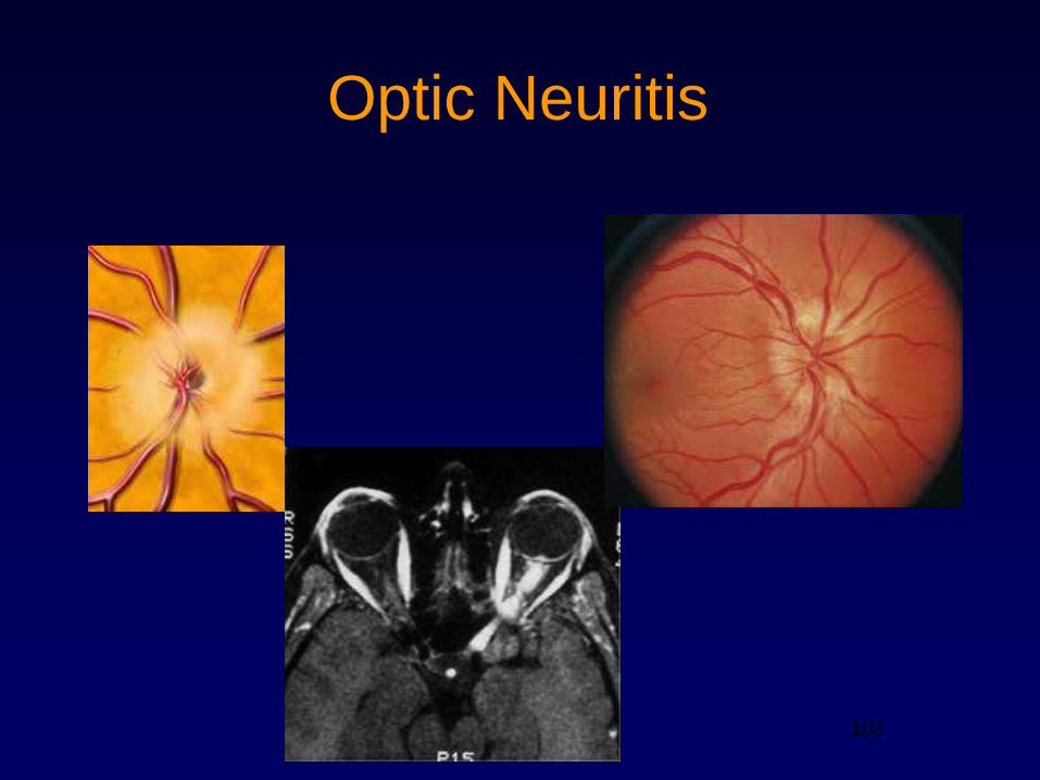
Optic Neuritis
Optic neuritis
Pale optic nerve (after optic neuritis)
Floaters/Photopsia
• Floaters and photopsias (flashes of light) can represent normal aging process or other pathologies.
• Normal:– Floaters in the vitreous as it becomes more liquid as
we age. Can cause posterior vitreous detachment (benign by itself but can lead to retinal tear and retinal detachment)
• Abnormal:– Posterior vitreous detachment leading to retinal tear
and possible retinal detachment
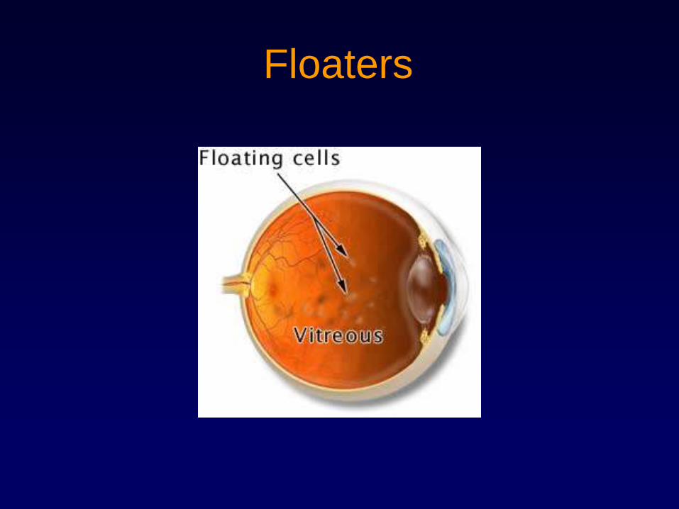
Floaters

Posterior Vitreous Detachment
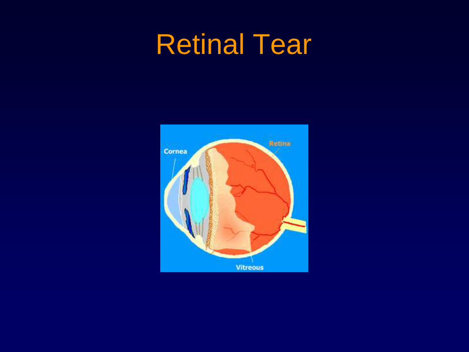
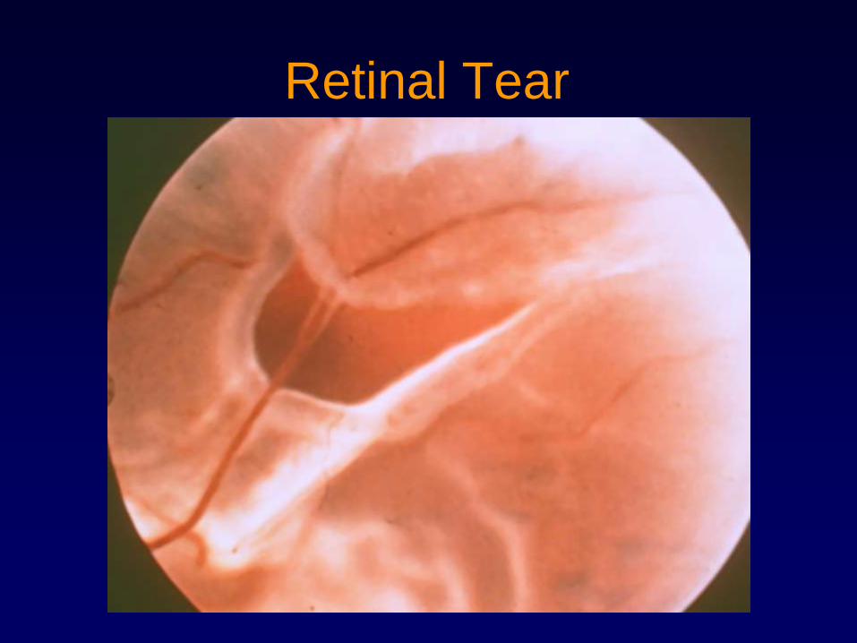
Retinal Tear
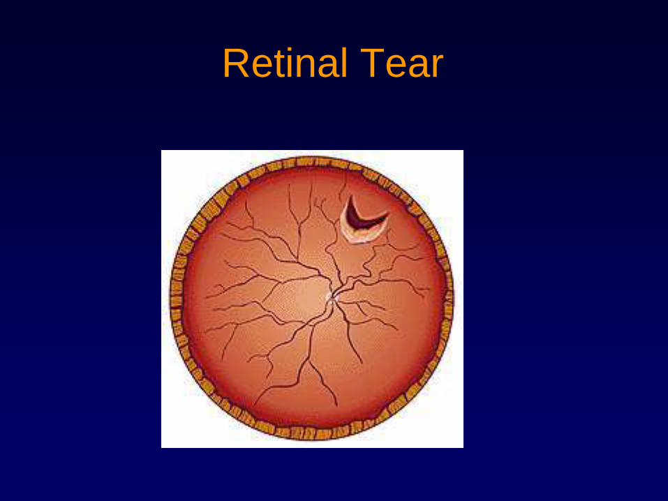
Retinal Tear
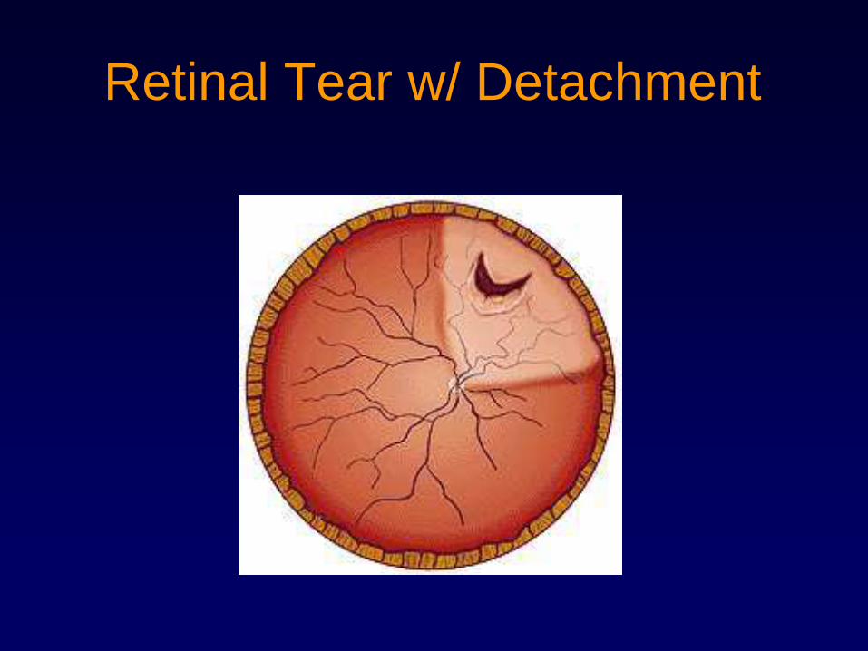
Retinal Tear w/ Detachment
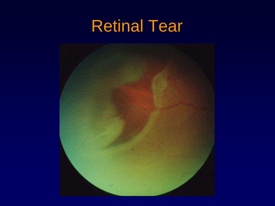
Retinal Tear
112
Retinal Tear
113
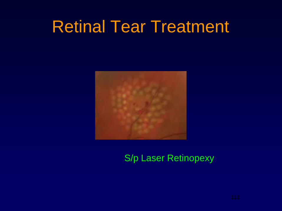
Retinal Tear Treatment
S/p Laser Retinopexy
Retinal Detachment
• Symptoms– Sudden onset of new floaters or flashes of
light in one eye
– Dark curtain ―moving over vision‖
– Blurred images in particular visual field in one eye
• Painless
• Increased risk in myopic patients (near sighted), patients with recent trauma.
Retinal Detachment
116
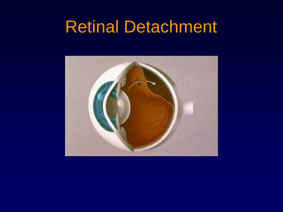
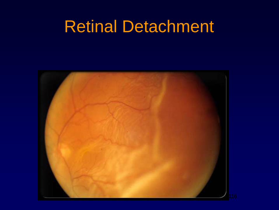
Retinal Detachment
116
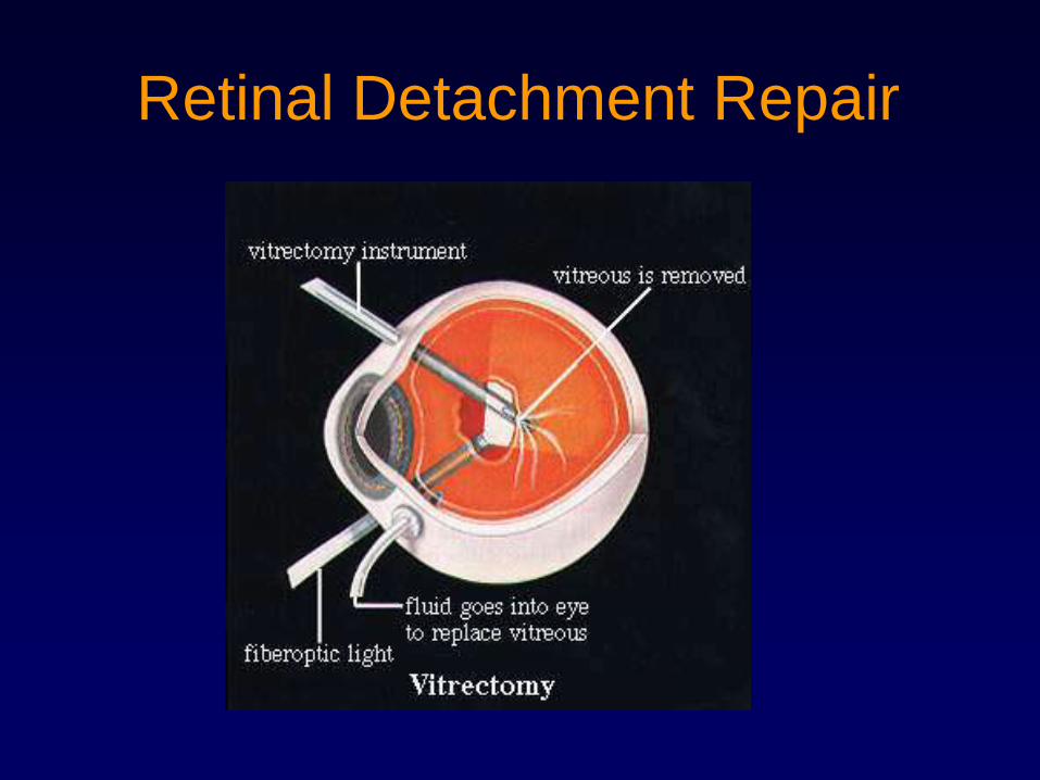
Retinal Detachment Repair
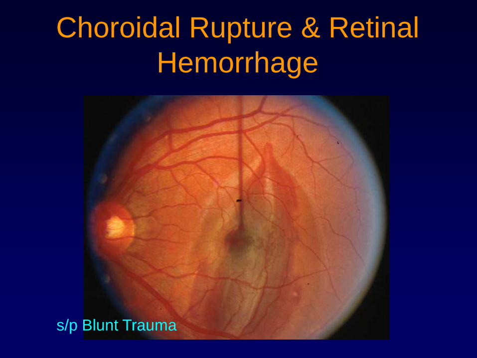
Choroidal Rupture & Retinal
Hemorrhage
s/p Blunt Trauma
119
Endophthalmitis
• Infection throughout the inside of the eye
cavity
• Pain, Decreased Vision, Red eye,
Hypopyon, Vitreous inflammation
• Etiology:
– Following trauma or surgery
– Endogenous (in setting of systemic illness --
e.g. sepsis, pneumonia, endocarditis)
• Requires urgent treatment with injection of
Antibiotics & sometimes surgery119
120
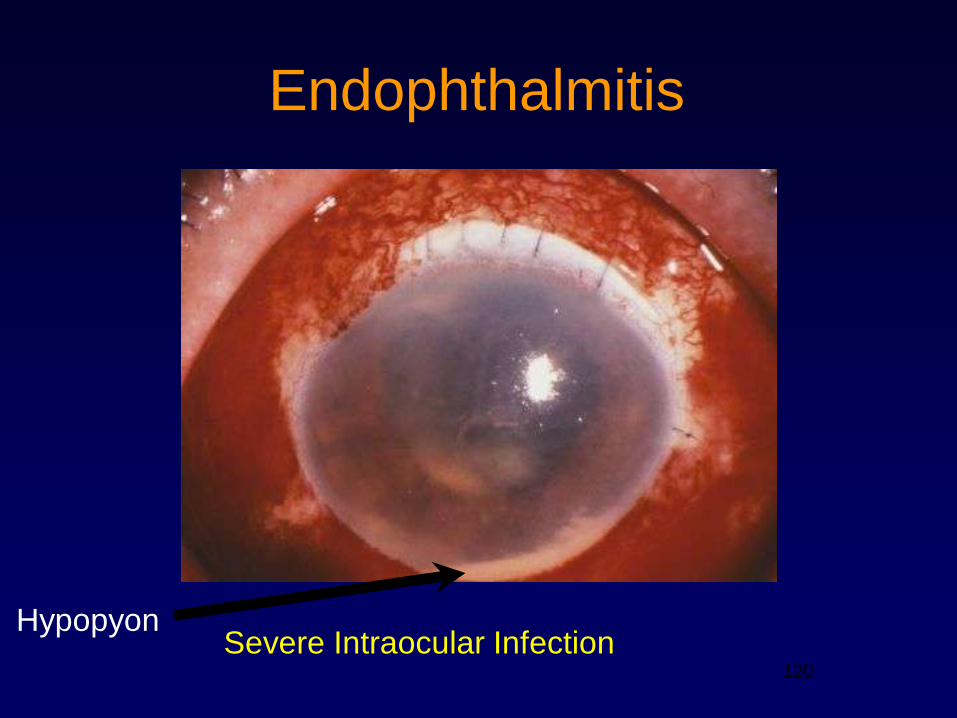
Endophthalmitis
HypopyonSevere Intraocular Infection
121
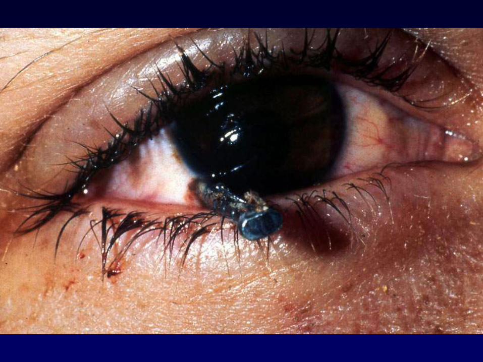
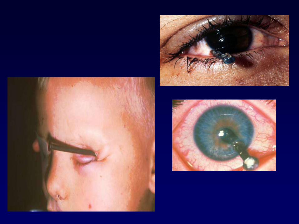
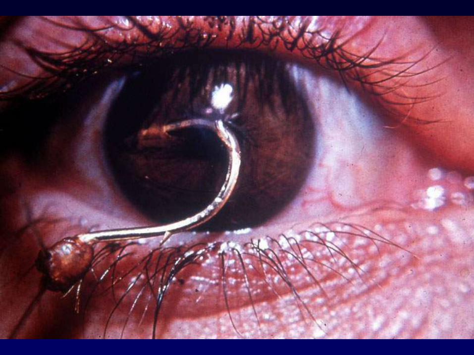
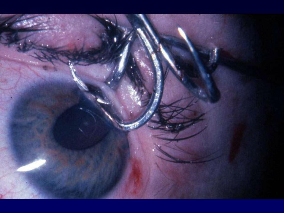
Ruptured or Lacerated Globe
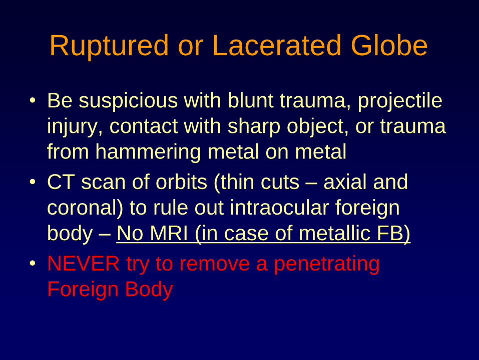
Ruptured or Lacerated Globe
• Be suspicious with blunt trauma, projectile
injury, contact with sharp object, or trauma
from hammering metal on metal
• CT scan of orbits (thin cuts – axial and
coronal) to rule out intraocular foreign
body – No MRI (in case of metallic FB)
• NEVER try to remove a penetrating
Foreign Body
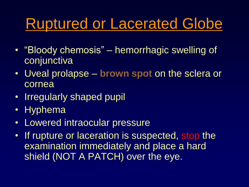
Ruptured or Lacerated Globe
• ―Bloody chemosis‖ – hemorrhagic swelling of conjunctiva
• Uveal prolapse – brown spot on the sclera or cornea
• Irregularly shaped pupil
• Hyphema
• Lowered intraocular pressure
• If rupture or laceration is suspected, stop the examination immediately and place a hard shield (NOT A PATCH) over the eye.

Bloody Chemosis
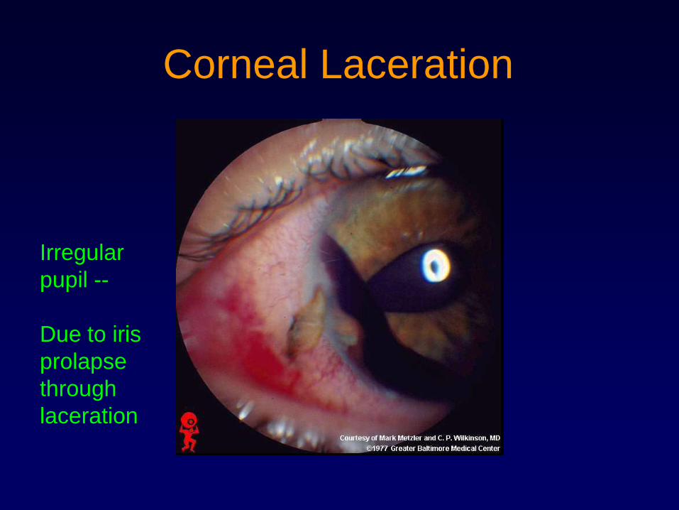
Corneal Laceration
Irregular
pupil --
Due to iris
prolapse
through
laceration
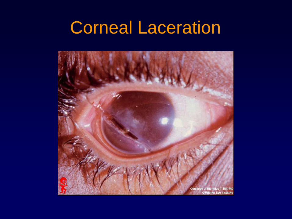
Corneal Laceration
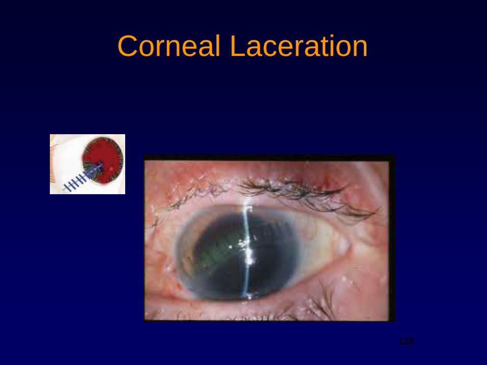
128
Corneal Laceration
128
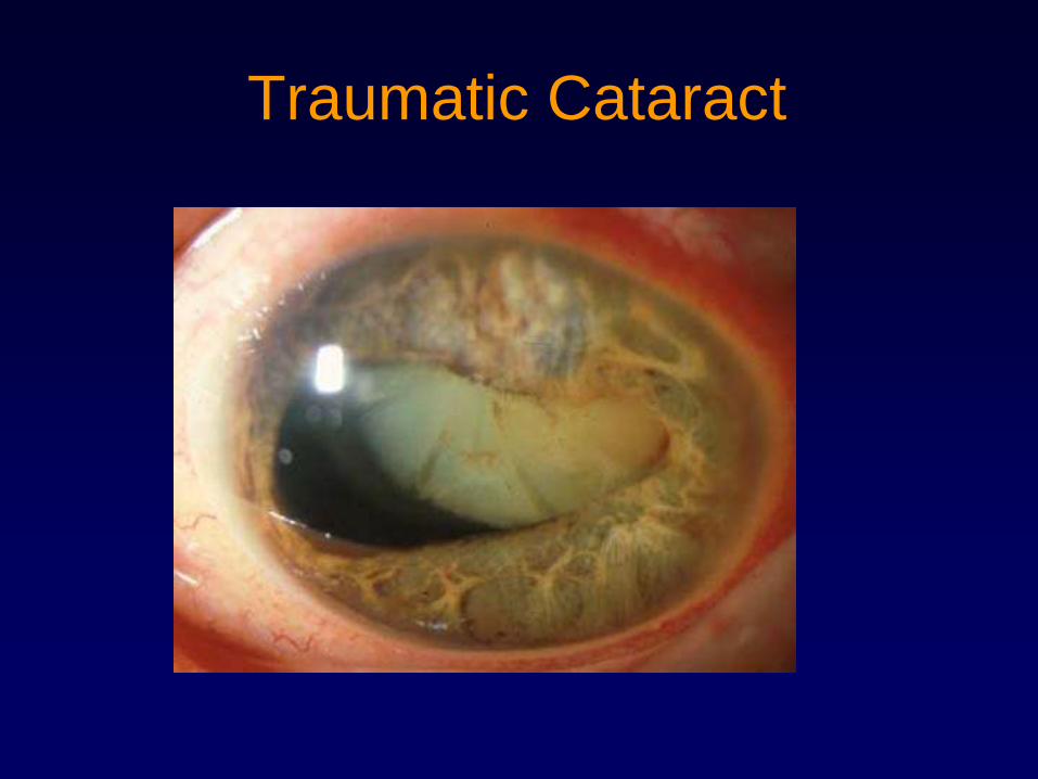
Traumatic Cataract
130
Penetrating Ocular Trauma
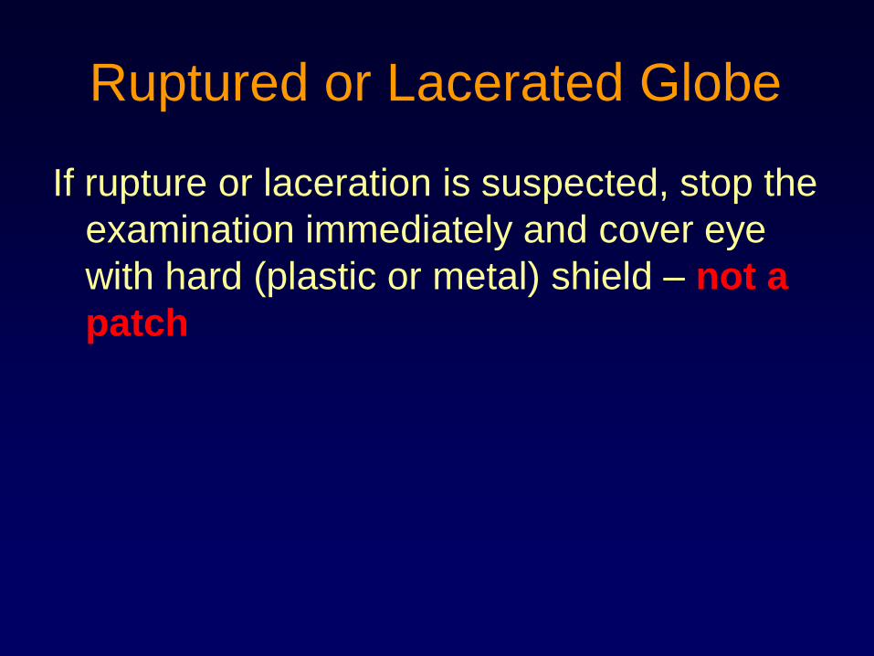
Ruptured or Lacerated Globe
If rupture or laceration is suspected, stop the
examination immediately and cover eye
with hard (plastic or metal) shield – not a
patch
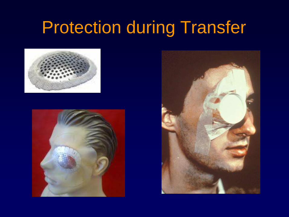
Protection during Transfer
137
Eye Injuries
PREVENTION
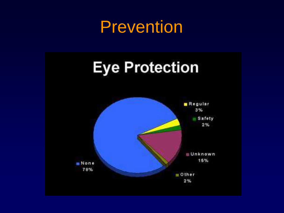
Prevention
Ocular Trauma and
Emergencies
Jacob J. Yunker, M.D.University of Kentucky College of Medicine
THANK YOU!
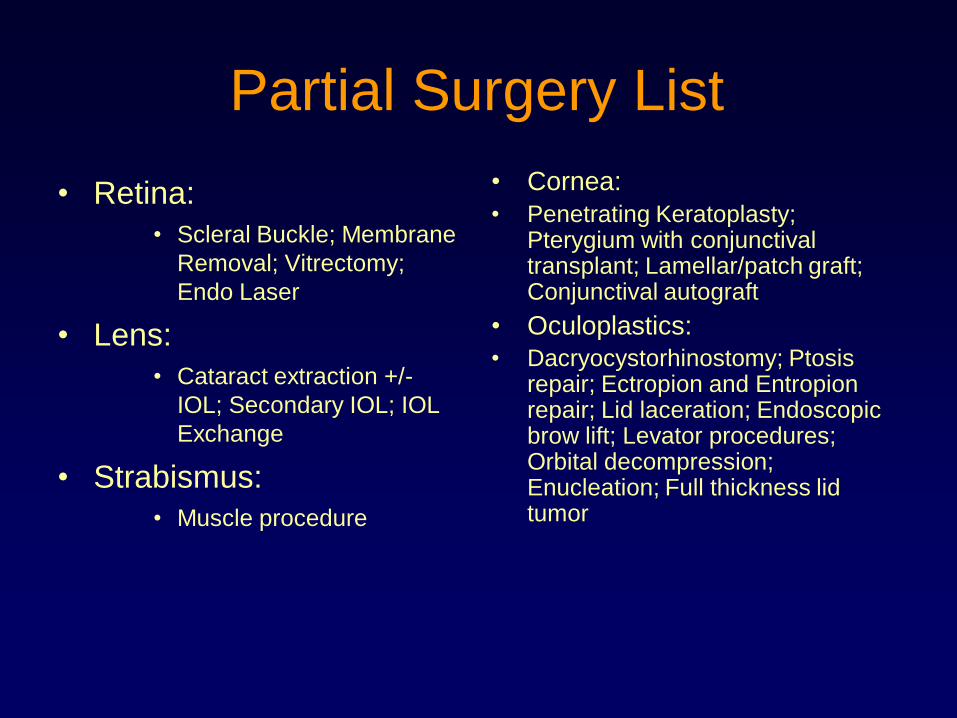
Partial Surgery List
• Retina:
• Scleral Buckle; Membrane
Removal; Vitrectomy;
Endo Laser
• Lens:
• Cataract extraction +/-
IOL; Secondary IOL; IOL
Exchange
• Strabismus:
• Muscle procedure
• Cornea:
• Penetrating Keratoplasty; Pterygium with conjunctival transplant; Lamellar/patch graft; Conjunctival autograft
• Oculoplastics:
• Dacryocystorhinostomy; Ptosis repair; Ectropion and Entropion repair; Lid laceration; Endoscopic brow lift; Levator procedures; Orbital decompression; Enucleation; Full thickness lid tumor
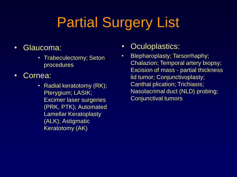
Partial Surgery List
• Glaucoma:
• Trabeculectomy; Seton
procedures
• Cornea:
• Radial keratotomy (RK);
Pterygium; LASIK;
Excimer laser surgeries
(PRK, PTK); Automated
Lamellar Keratoplasty
(ALK); Astigmatic
Keratotomy (AK)
• Oculoplastics:• Blepharoplasty; Tarsorrhaphy;
Chalazion; Temporal artery biopsy;
Excision of mass - partial thickness
lid tumor; Conjunctivoplasty;
Canthal plication; Trichiasis;
Nasolacrimal duct (NLD) probing;
Conjunctival tumors