OCULAR MANIFESTATIONS OF VON HIPPEL–LINDAU DISEASE ...
17
OCULAR MANIFESTATIONS OF VON HIPPEL–LINDAU DISEASE: CLINICAL AND GENETIC INVESTIGATIONS BY Emily Ying Chew MD ABSTRACT Purpose: To describe the clinical spectrum of lesions involving the visual system in von Hippel–Lindau (VHL) disease, the genetic alterations, and the molecular genetic properties of retinal hemangioblastomas. Methods: In this prospective case-series, 406 patients with VHL disease had systemic and ocular evaluations. Genetic mutations within six pathological specimens were evaluated, using microdissection and polymerase chain reaction amplification. Results: Half of the 406 patients (199 families) with VHL disease had ocular involvement. Visual acuity was 20/20 or better in 170 patients (84.5%) with hemangioblastomas; six (3%) were legally blind. Thirty-three (8.2%) had unilateral enucleations. Genetic mutations were detected in all VHL patients. The patients with complete deletion were less likely to have ocular VHL compared with those patients with partial deletion, missense, and nonsense mutation (9% versus 45%) (P < .0001), suggesting the importance of the gene or areas of genes on chromosome 3 for the development of retinal hemangioblastomas. The molecular genetic assessments of the pathology specimens showed that the foamy “stromal” cells were affected with the genetic mutations. There is an up-regulation of vascular endothelial growth factor, which was expressed in the ocular lesions. Conclusions: In this series, the largest of its kind, patients with ocular lesions of VHL disease are referred mostly from physicians. The systemic genetic mutation evaluations suggest that a certain gene or groups of genes in chromosome 3p are crucial for both the development and maintenance of the retinal tumor. This is the first series to find a difference between the phenotype and genotype. Trans Am Ophthalmol Soc 2005;103:495-511 INTRODUCTION The ocular manifestations of von Hippel–Lindau syndrome (VHL), consisting of retinal capillary hemangioblastomas, have a clinical appearance that is both characteristic and diagnostic of VHL. These ocular lesions have stimulated interest among ophthalmologists throughout the ages, as witnessed by the extensive literature on VHL. The intents of this report are twofold: (1) to describe the clinical manifestations of VHL from one institution in which patients do not present because of ocular symptoms but rather because of known systemic VHL involvement, and (2) to discuss genetic assessments of all patients with VHL and specific vascular lesions in selected cases. These data are integrated in an effort to better understand the development and evolution of the ocular lesions and to propose potential therapies for the potentially blinding ocular disease of VHL. In this thesis, the term “retinal hemangioblastoma” is used to denote the typical lesion seen in these patients. Various investigators have used other terms, including “capillary hemangioma” and “retinal angioma.” Although the term retinal angioma may be better known to both ophthalmologists and medical colleagues, hemangioblastoma may be more accurate, because it corresponds to the pathological aspects of the lesion. HISTORY OF VON HIPPEL-LINDAU DISEASE One hundred years ago, Eugen von Hippel (1867–1938), a German ophthalmologist, published descriptions of retinal hemangioblastomas transmitted through several generations of family members in a small number of kindreds. 1 A number of incomplete descriptions, as well as illustrations, of hemangioblastomas of the eye and vascular lesions in the cerebellum had been reported before 1904. In 1926, the Swedish pathologist Arvid Lindau established the link between retinal and cerebellar hemangioblastomas as well as the findings of cysts in the kidney, pancreas, and epididymis as part of a familial syndrome. 2 The landmark summary in 1964 by Melmon and Rosen established the first clinical diagnostic criteria for VHL. 3 Linkage of the VHL gene to the short arm of chromosome 3 was reported by Seizinger and colleagues in 1988. 4 Finally, in 1993, Latif and colleagues identified the VHL tumor suppressor gene. 5 EXTRAOCULAR CLINICAL FINDINGS OF VON HIPPEL-LINDAU DISEASE VHL is an autosomal dominant neoplastic disorder in which multiple benign or malignant tumors and cysts with specific histopathologic features develop in the tissues of the central nervous system, including brain, spinal cord, inner ear, and retina, and in visceral organs, including kidney, adrenal gland, pancreas, epididymis, and broad ligament. 6-17 The disease results from germline mutation in the VHL gene, which is located on the short arm of chromosome 3, 3p25-26. 5 VHL is a rare disorder (with an approximate incidence of 1 in 36,000 live births) and has a penetrance of over 90% by 65 years of age. 18,19 Prior to the development of routine comprehensive screening surveys, the median survival of patients with VHL was less than 50 years of age and the main causes of death were complications associated with renal cell carcinomas and central nervous system hemangioblastomas. 9,20-22 Improved surveillance, earlier diagnosis of lesions by modern imaging and laboratory studies, From the National Eye Institute, National Institutes of Health, Bethesda, Maryland. Trans Am Ophthalmol Soc / Vol 103/ 2005 495
Transcript of OCULAR MANIFESTATIONS OF VON HIPPEL–LINDAU DISEASE ...
OCULAR MANIFESTATIONS OF VON HIPPEL–LINDAU DISEASE: CLINICAL AND GENETIC INVESTIGATIONS BY Emily Ying Chew MD
ABSTRACT Purpose: To describe the clinical spectrum of lesions involving the visual system in von Hippel–Lindau (VHL) disease, the genetic alterations, and the molecular genetic properties of retinal hemangioblastomas.
Methods: In this prospective case-series, 406 patients with VHL disease had systemic and ocular evaluations. Genetic mutations within six pathological specimens were evaluated, using microdissection and polymerase chain reaction amplification.
Results: Half of the 406 patients (199 families) with VHL disease had ocular involvement. Visual acuity was 20/20 or better in 170 patients (84.5%) with hemangioblastomas; six (3%) were legally blind. Thirty-three (8.2%) had unilateral enucleations. Genetic mutations were detected in all VHL patients. The patients with complete deletion were less likely to have ocular VHL compared with those patients with partial deletion, missense, and nonsense mutation (9% versus 45%) (P < .0001), suggesting the importance of the gene or areas of genes on chromosome 3 for the development of retinal hemangioblastomas. The molecular genetic assessments of the pathology specimens showed that the foamy “stromal” cells were affected with the genetic mutations. There is an up-regulation of vascular endothelial growth factor, which was expressed in the ocular lesions.
Conclusions: In this series, the largest of its kind, patients with ocular lesions of VHL disease are referred mostly from physicians. The systemic genetic mutation evaluations suggest that a certain gene or groups of genes in chromosome 3p are crucial for both the development and maintenance of the retinal tumor. This is the first series to find a difference between the phenotype and genotype.
Trans Am Ophthalmol Soc 2005;103:495-511
INTRODUCTION
The ocular manifestations of von Hippel–Lindau syndrome (VHL), consisting of retinal capillary hemangioblastomas, have a clinical appearance that is both characteristic and diagnostic of VHL. These ocular lesions have stimulated interest among ophthalmologists throughout the ages, as witnessed by the extensive literature on VHL. The intents of this report are twofold: (1) to describe the clinical manifestations of VHL from one institution in which patients do not present because of ocular symptoms but rather because of known systemic VHL involvement, and (2) to discuss genetic assessments of all patients with VHL and specific vascular lesions in selected cases. These data are integrated in an effort to better understand the development and evolution of the ocular lesions and to propose potential therapies for the potentially blinding ocular disease of VHL.
In this thesis, the term “retinal hemangioblastoma” is used to denote the typical lesion seen in these patients. Various investigators have used other terms, including “capillary hemangioma” and “retinal angioma.” Although the term retinal angioma may be better known to both ophthalmologists and medical colleagues, hemangioblastoma may be more accurate, because it corresponds to the pathological aspects of the lesion.
HISTORY OF VON HIPPEL-LINDAU DISEASE One hundred years ago, Eugen von Hippel (1867–1938), a German ophthalmologist, published descriptions of retinal hemangioblastomas transmitted through several generations of family members in a small number of kindreds.1 A number of incomplete descriptions, as well as illustrations, of hemangioblastomas of the eye and vascular lesions in the cerebellum had been reported before 1904. In 1926, the Swedish pathologist Arvid Lindau established the link between retinal and cerebellar hemangioblastomas as well as the findings of cysts in the kidney, pancreas, and epididymis as part of a familial syndrome.2 The landmark summary in 1964 by Melmon and Rosen established the first clinical diagnostic criteria for VHL.3 Linkage of the VHL gene to the short arm of chromosome 3 was reported by Seizinger and colleagues in 1988.4 Finally, in 1993, Latif and colleagues identified the VHL tumor suppressor gene.5
EXTRAOCULAR CLINICAL FINDINGS OF VON HIPPEL-LINDAU DISEASE VHL is an autosomal dominant neoplastic disorder in which multiple benign or malignant tumors and cysts with specific histopathologic features develop in the tissues of the central nervous system, including brain, spinal cord, inner ear, and retina, and in visceral organs, including kidney, adrenal gland, pancreas, epididymis, and broad ligament.6-17 The disease results from germline mutation in the VHL gene, which is located on the short arm of chromosome 3, 3p25-26.5
VHL is a rare disorder (with an approximate incidence of 1 in 36,000 live births) and has a penetrance of over 90% by 65 years of age.18,19 Prior to the development of routine comprehensive screening surveys, the median survival of patients with VHL was less than 50 years of age and the main causes of death were complications associated with renal cell carcinomas and central nervous system hemangioblastomas.9,20-22 Improved surveillance, earlier diagnosis of lesions by modern imaging and laboratory studies,
From the National Eye Institute, National Institutes of Health, Bethesda, Maryland.
Trans Am Ophthalmol Soc / Vol 103/ 2005 495
Chew
improvements in treatment, and increased knowledge of this disease have improved prognosis and reduced the complications related to these tumors.
THE MOLECULAR GENETICS OF VON HIPPEL-LINDAU DISEASE The VHL gene, which was identified by Latif and coworkers in 1993,5 is located on the short arm of chromosome 3 (3p25-26) and is a tumor suppressor gene. Each person with VHL inherits a germline mutation of the VHL gene from the affected parent and a normal (wild type) gene from the unaffected parent. According to Knudson’s “two-hit” hypothesis of tumorigenesis, initiation of tumor formation follows when there is mutation of both alleles of the VHL gene.23,24 Germline mutations of the VHL gene are present in all of the cells of affected individuals. However, tumors develop only from those cells in the susceptible target organs (including the kidneys, adrenal glands, pancreas, reproductive adnexal organs, brain, spine, and eye) that also undergo a deletion, mitotic recombination, or mutation in the remaining wild type allele.
The VHL gene has three exons that encode for the VHL protein (pVHL).8 The VHL gene product (pVHL) binds with elongin C, which binds elongin B, Cul2, and RBX1 to form the VCB-CUL 2 complex.25,26 This VHL protein complex targets the hypoxia-inducible factors (HIFs), HIF1-a and HIF 2-a, for ubiquitin-dependent degradation.27 Under normoxic conditions, the complex degrades HIF. Under hypoxic conditions, HIF is not degraded, and it accumulates.28,29 Increased levels of HIF are associated with increased transcription of a number of downstream genes, including vascular endothelial growth factor (VEGF), erythropoietin, platelet-derived growth factor (PDGF-ß), and transforming growth factor-α.30-32 The vascular nature of the VHL tumors may result from the HIF-mediated increased levels of VEGF and PDGF-ß, which are known to support the proliferation of endothelial cells and pericytes, respectively. Moreover, increased vascular permeability of the tumor vessels resulting from increased levels of VEGF may also account for the edema seen with lesions of VHL.
Why do lesions occur only in these target lesions? Organ involvement varies considerably among families, and there may be variability in the severity of disease. Our colleagues who treat patients with renal cell carcinoma evaluated the genetic mutation of families with low prevalence of renal cell carcinoma and compared them with those families with high prevalence.33 These investigators found a higher prevalence of renal cell carcinoma in patients with partial germline VHL deletions relative to complete deletions (48.9% versus 22.6%, P = .007). A possible explanation is that the presence of a neighboring gene or group of genes located within the 30-kb region of chromosome 3p, immediately telomeric to VHL, may be critical to the development and maintenance of renal cell carcinoma. Thus patients with complete deletion will not develop or sustain such lesions. A major portion of this study was to further evaluate the genetics of patients with VHL, including changes associated with the ocular lesions.
PATHOLOGY OF OCULAR LESIONS The pathology of retinal hemangioblastoma has been proposed to be either a congenital, hamartomatous-type lesion or a benign vascular neoplastic process.15,34 Retinal hemangioblastomas have mainly thin-walled, capillary-like or slightly larger, blood vessels that are separated by plump, vacuolated, foamy, “stromal” cells.35 Such cells are strikingly similar to cerebellar hemangioblastoma and renal cell carcinoma cells found in VHL disease. In the past, ultrastructural and immunohistochemical studies have suggested that these “stromal” cells may represent lipidized fibrous astrocytes or glial cells.36,37 Other investigators had previously suggested that these were not glial cells but more likely “vasoformative stem cells.”38 A more recent study of cerebellar hemangioblastomas showed that these “stromal” cells may originate from developmentally arrested angioblasts.39
Previous investigations, using microdissection and molecular techniques, showed the “stromal” cells of the cerebellar hemangioblastomas and renal cell carcinomas to contain the VHL gene alterations.40,41 As stated by Knudson,23,24 it is believed that malignant neoplasms in VHL follow the “two-hit” theory, in which one allele is constitutionally inactivated (at the germline) while the other allele is subsequently inactivated (second hit) at the somatic level, the target organ level. The VHL lesions, the cerebellar and renal tumors, showed the gene deletion in the “stromal” cells. The loss of heterozygosity was seen in these cells, whereas the vascular component retained the heterozygosity.
CONVENTIONAL CURRENT TREATMENT OF OCULAR LESIONS Conventional treatment of the retinal hemangioblastomas with laser photocoagulation or cryotherapy depends on the location and size of the lesions. Small lesions are easy to treat successfully, whereas large lesions are notoriously difficult to treat. Photocoagulation with argon laser can eradicate small retinal hemangioblastomas in most locations.42-45 However, for those tumors that are too large or are located in the extreme periphery of the retina, cryotherapy may be indicated. Both photocoagulation and cryotherapy of peripheral tumors can often lead to massive retinal hard exudate accumulation and retinal edema in the macula, contributing to further decrease in vision following treatment. Although preoperative and postoperative therapy with corticosteroids, either locally or systemically, has not been assessed, it may be considered to decrease this side effect.
If the tumor is located on the optic nerve, the treatment is more difficult. Marked adverse side effects, including altitudinal defects and central scotomas in the visual fields, are associated with treatment of such tumors with laser photocoagulation.46 Photodynamic therapy has been used in the optic disc lesions without success.47
Rarely, radiation may be useful in cases that do not respond to conventional laser or cryotherapy.48 There are anecdotal reports of such success, but the benefits are not uniform. The usefulness of radiation and its role in management of retinal hemangioblastomas need to be further defined.
Vascular lesions often are accompanied by fibrous proliferation. Contraction of the fibrovascular lesion of the retinal
Trans Am Ophthalmol Soc / Vol 103/ 2005 496
Ocular Manifestations of Von Hippel-Lindau’s Disease
hemangioblastomas can lead to tractional retinal detachment. Vitrectomy can be performed on patients with either tractional or exudative retinal detachment.49 Vitreous hemorrhage from large tumors can also be treated by vitrectomy. However, the visual recovery following vitrectomy will be maintained only if the underlying tumors can be eradicated or their growth be interrupted. Enucleation may be necessary for irreversible glaucoma in which severe pain is associated with end-stage ocular hemangiomatosis.
MANAGEMENT OF PATIENTS WITH VON HIPPEL-LINDAU DISEASE The management of patients affected with VHL is complex because of the progressive and variable nature of multiple neoplasms in various organ systems. A multispecialty team is the best way to both evaluate and optimize treatment for these patients.
METHODS
THE STUDY POPULATION We report our experience of the largest series of patients diagnosed and treated for ocular lesions of VHL at any one medical center. These patients did not present for evaluation because of visual symptoms. Probands, identified in the community as affected with VHL, and their family members were invited to participate in a screening protocol that was approved by the institutional review board for human subjects. Informed consent was obtained from each patient. In our institute, patients with VHL are evaluated and treated by a team of experts that includes genetic counselors, medical geneticists, radiologists, pathologists, neurosurgeons, urologists, radiation oncologists, ophthalmologists, and otolaryngologists. Participants had complete systemic evaluations, including comprehensive eye examinations for the presence of VHL ocular lesions, using a standard protocol for all patients. Patients were referred from multiple sources throughout the United States and Canada, including the practices of ophthalmologists, internists, urologists, neurosurgeons, and patient support groups.
RESULTS
Over the period from 1988 to 2000, 406 affected individuals from 199 families were evaluated. Their ages ranged from 7 to 84 (mean ± SD, 36 ± 15 years), and 189 (47%) were male. In this population, 386 (95%) were white, seven (1.7%) were black, 10 (2.5%) were Hispanic, and three (0.7%) were Asian. Extensive physical examinations also included computed tomography (CT) and magnetic resonance imaging (MRI) of the target organs involved in VHL. These findings will not be presented in this report but were used to establish the clinical diagnosis of systemic VHL. Genetic evaluations performed on all patients showed mutations that are characteristic of VHL disease.
OCULAR FINDINGS All patients were evaluated at the eye clinic with a comprehensive eye examination that included the measurement of the best-corrected visual acuity, intraocular pressure, slit-lamp biomicroscopy, and dilated fundus examination and fundus photography. More recently, optical coherence tomography (OCT) was used to measure the macular thickening associated with peripheral or optic nerve hemangioblastomas. At the initial examination, 168 of the 406 patients (41.3%) presented with a past history of ocular VHL. An additional 37 (9%) of the asymptomatic patients were found to have characteristic ocular lesions at their examination. Therefore, more than 50% of the entire study population of patients affected with VHL was found to have retinal hemangioblastomas. Of these 205 patients with ocular VHL, 113 (55.1%) had bilateral retinal hemangioblastomas; 44 (21.5%) were in the right eye only and 48 (23.4%) were in the left eye only. In these patients, 90 (43.8%) had a single retinal hemangioblastoma in the affected eye at the screening examination, 80 (39.2%) had two to four lesions, and 35 (17.0%) had five or more lesions in each eye. Of the 205 patients with ocular lesions, 48 (23.4%) had optic nerve involvement, with four (2.0%) having bilateral lesions; 25 lesions (12.2%) were on the right optic nerve and 16 (7.8%) were on the left. Twenty-four (48.9%) of these optic nerve lesions were the sole lesion in the eye, whereas 25 (51.1%) were accompanied by peripheral retinal hemangioblastomas. Some patients had both optic nerve lesions and peripheral retinal lesions; thus these lesions are not mutually exclusive.
The measurement of visual acuities of the entire study cohort demonstrated a large proportion, 357 (88.1%), with 20/20 vision or better in their better-seeing eye (Table 1). Only eight (2%) had visual acuity of 20/200 or worse in their better-seeing eye. In those 205 patients affected with VHL ocular disease, 170 (84.6%) had visual acuity of 20/20 or better in their better-seeing eye (Table 2). Approximately 3% (n = 6) were legally blind (20/200 or worse in the better eye). However, when the visual acuity of each eye of the patients with ocular VHL was assessed, approximately 76 (8.4%) of the eyes had visual acuity of 20/200 or worse (Table 3). Thirty-three patients from the entire population with VHL (8.1%) had unilateral enucleation.
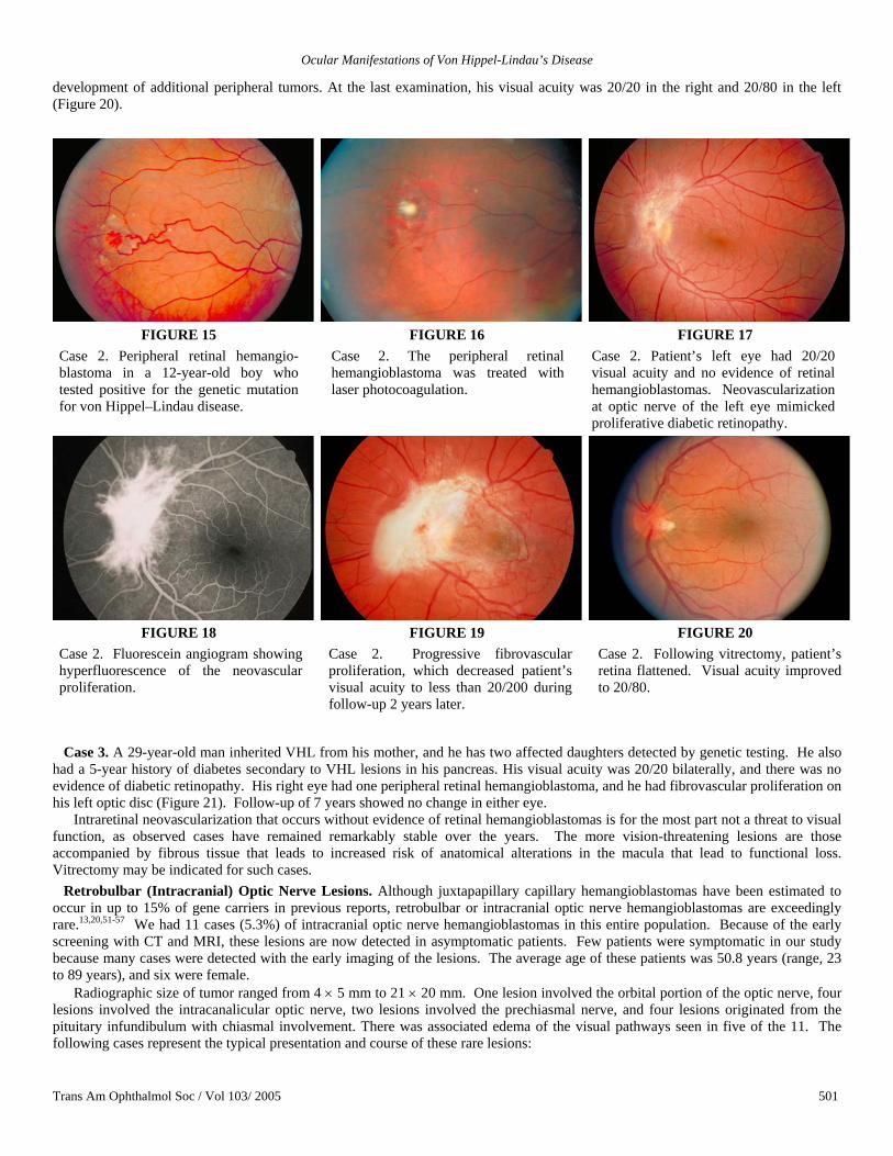
Retinal Hemangioblastomas Peripheral. The retinal hemangioblastomas, often the first manifestation of VHL, ranged from very small capillary abnormalities to large lesions that caused major visual impairment in this study population. The clinical appearance of these hemangioblastomas is typical and diagnostic of VHL. The initial appearance of a retinal hemangioblastoma is a subtle red or grayish dot no larger than a few hundred microns (Figure 1). These abnormalities can develop into a large nodule. As the proliferation of the vascular tumors progresses, secondary alterations occur to produce a distinctive clinical appearance. The blood vessels leading to and from the peripheral tumor become characteristically dilated with marked enlargement and tortuosity (Figure 2). Peripheral tumors can lead to leakage of retinal hard exudate and retinal edema surrounding the lesion (Figure 3). Peripheral hemangioblastomas can lead to marked retinal edema and hard exudates at the macula (Figure 4). Without treatment, and occasionally with treatment, these lesions
Trans Am Ophthalmol Soc / Vol 103/ 2005 497
Chew
can continue to grow and interfere with or displace the normal structures of the retina and affect the visual function by causing an exudative retinal detachment (Figure 5).
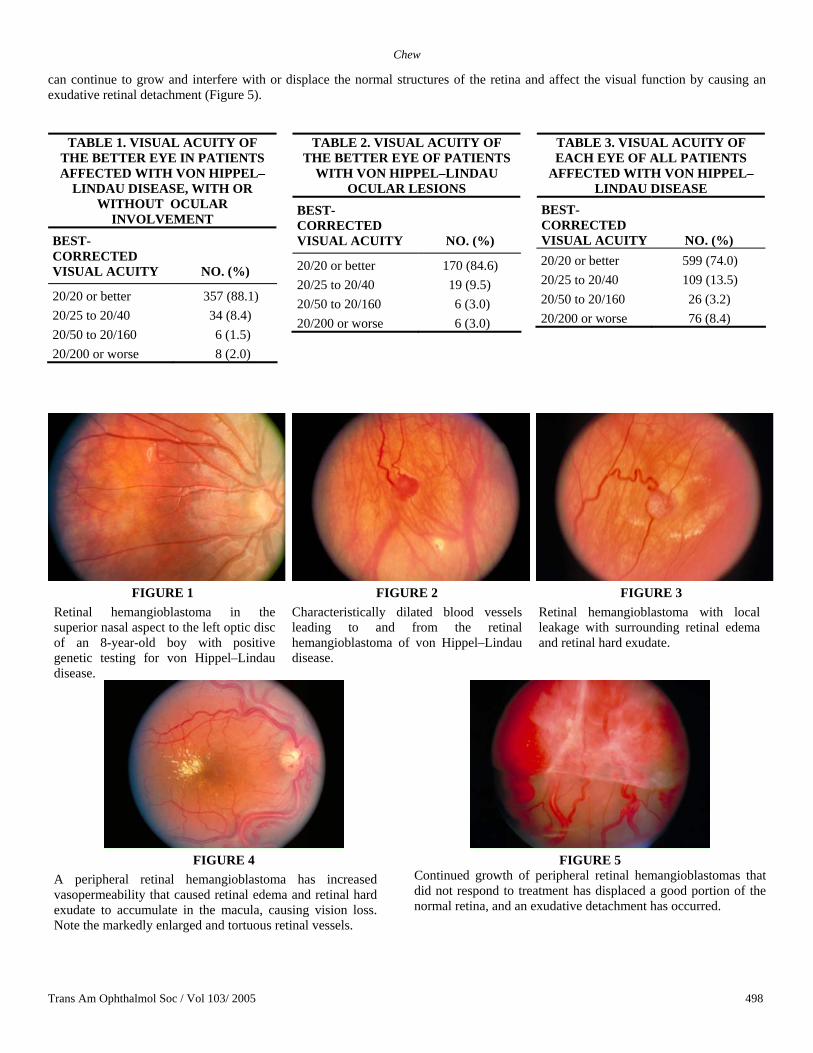
TABLE 1. VISUAL ACUITY OF
THE BETTER EYE IN PATIENTS AFFECTED WITH VON HIPPEL–
LINDAU DISEASE, WITH OR WITHOUT OCULAR
INVOLVEMENT BEST-CORRECTED VISUAL ACUITY NO. (%)
20/20 or better 357 (88.1) 20/25 to 20/40 34 (8.4) 20/50 to 20/160 6 (1.5) 20/200 or worse 8 (2.0)
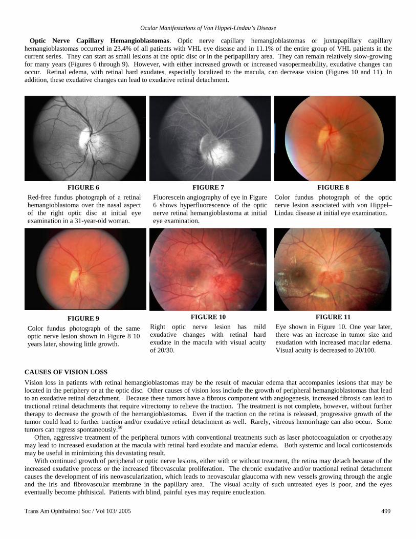
TABLE 2. VISUAL ACUITY OF THE BETTER EYE OF PATIENTS
WITH VON HIPPEL–LINDAU OCULAR LESIONS
BEST-CORRECTED VISUAL ACUITY NO. (%)
20/20 or better 170 (84.6) 20/25 to 20/40 19 (9.5) 20/50 to 20/160 6 (3.0) 20/200 or worse 6 (3.0)
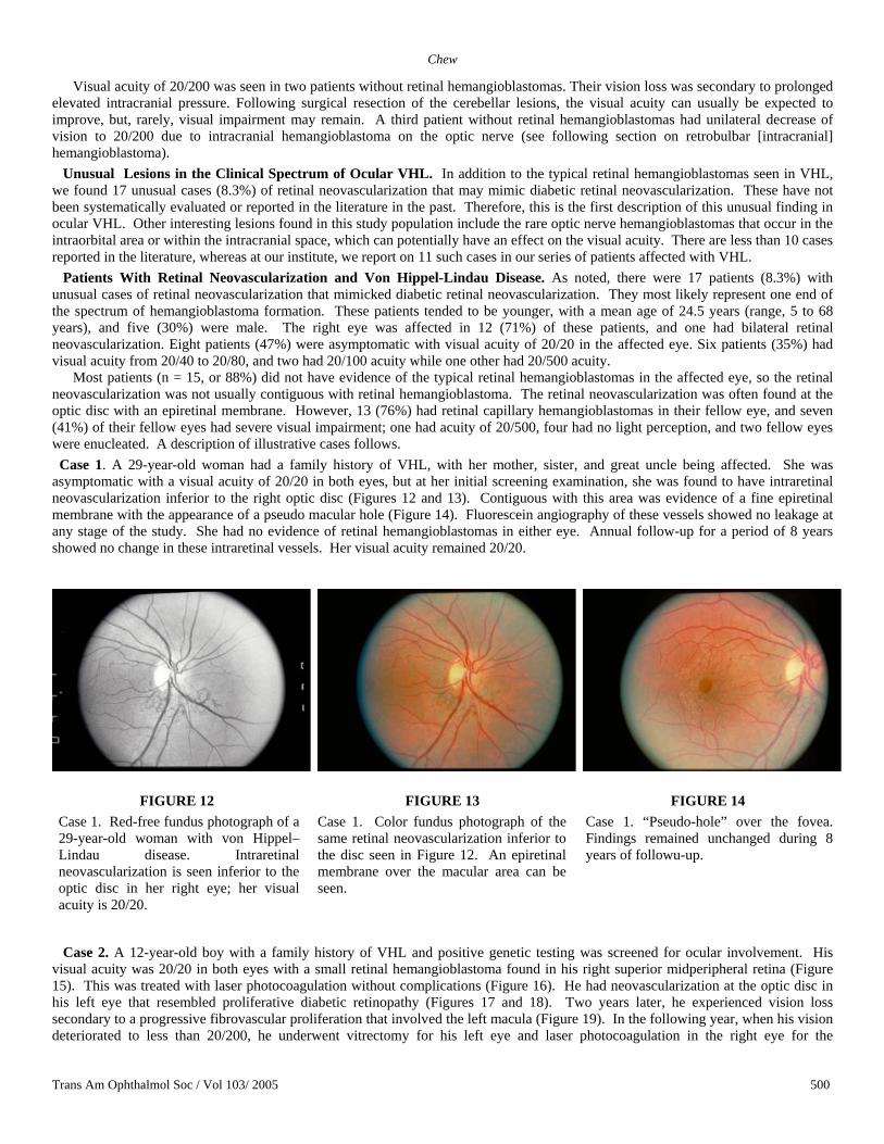
TABLE 3. VISUAL ACUITY OF EACH EYE OF ALL PATIENTS
AFFECTED WITH VON HIPPEL–LINDAU DISEASE
BEST-CORRECTED VISUAL ACUITY NO. (%) 20/20 or better 599 (74.0) 20/25 to 20/40 109 (13.5) 20/50 to 20/160 26 (3.2) 20/200 or worse 76 (8.4)
FIGURES 1 -3
FIGURE 1 Retinal hemangioblastoma in the superior nasal aspect to the left optic disc of an 8-year-old boy with positive genetic testing for von Hippel–Lindau disease.
FIGURE 2 Characteristically dilated blood vessels leading to and from the retinal hemangioblastoma of von Hippel–Lindau disease.
FIGURE 3 Retinal hemangioblastoma with local leakage with surrounding retinal edema and retinal hard exudate.
FIGURE 4
A peripheral retinal hemangioblastoma has increased vasopermeability that caused retinal edema and retinal hard exudate to accumulate in the macula, causing vision loss. Note the markedly enlarged and tortuous retinal vessels.
FIGURE 5
Continued growth of peripheral retinal hemangioblastomas that did not respond to treatment has displaced a good portion of the normal retina, and an exudative detachment has occurred.
Trans Am Ophthalmol Soc / Vol 103/ 2005 498
Ocular Manifestations of Von Hippel-Lindau’s Disease
Optic Nerve Capillary Hemangioblastomas. Optic nerve capillary hemangioblastomas or juxtapapillary capillary hemangioblastomas occurred in 23.4% of all patients with VHL eye disease and in 11.1% of the entire group of VHL patients in the current series. They can start as small lesions at the optic disc or in the peripapillary area. They can remain relatively slow-growing for many years (Figures 6 through 9). However, with either increased growth or increased vasopermeability, exudative changes can occur. Retinal edema, with retinal hard exudates, especially localized to the macula, can decrease vision (Figures 10 and 11). In addition, these exudative changes can lead to exudative retinal detachment.
FIGURE 6 Red-free fundus photograph of a retinal hemangioblastoma over the nasal aspect of the right optic disc at initial eye examination in a 31-year-old woman.
FIGURE 7 Fluorescein angiography of eye in Figure 6 shows hyperfluorescence of the optic nerve retinal hemangioblastoma at initial eye examination.
FIGURE 8 Color fundus photograph of the optic nerve lesion associated with von Hippel–Lindau disease at initial eye examination.
FIGURE 9 Color fundus photograph of the same optic nerve lesion shown in Figure 8 10 years later, showing little growth.
FIGURE 10
Right optic nerve lesion has mild exudative changes with retinal hard exudate in the macula with visual acuity of 20/30.
FIGURE 11
Eye shown in Figure 10. One year later, there was an increase in tumor size and exudation with increased macular edema. Visual acuity is decreased to 20/100.
CAUSES OF VISION LOSS Vision loss in patients with retinal hemangioblastomas may be the result of macular edema that accompanies lesions that may be located in the periphery or at the optic disc. Other causes of vision loss include the growth of peripheral hemangioblastomas that lead to an exudative retinal detachment. Because these tumors have a fibrous component with angiogenesis, increased fibrosis can lead to tractional retinal detachments that require vitrectomy to relieve the traction. The treatment is not complete, however, without further therapy to decrease the growth of the hemangioblastomas. Even if the traction on the retina is released, progressive growth of the tumor could lead to further traction and/or exudative retinal detachment as well. Rarely, vitreous hemorrhage can also occur. Some tumors can regress spontaneously.50
Often, aggressive treatment of the peripheral tumors with conventional treatments such as laser photocoagulation or cryotherapy may lead to increased exudation at the macula with retinal hard exudate and macular edema. Both systemic and local corticosteroids may be useful in minimizing this devastating result.
With continued growth of peripheral or optic nerve lesions, either with or without treatment, the retina may detach because of the increased exudative process or the increased fibrovascular proliferation. The chronic exudative and/or tractional retinal detachment causes the development of iris neovascularization, which leads to neovascular glaucoma with new vessels growing through the angle and the iris and fibrovascular membrane in the papillary area. The visual acuity of such untreated eyes is poor, and the eyes eventually become phthisical. Patients with blind, painful eyes may require enucleation.
Trans Am Ophthalmol Soc / Vol 103/ 2005 499
Chew
Visual acuity of 20/200 was seen in two patients without retinal hemangioblastomas. Their vision loss was secondary to prolonged elevated intracranial pressure. Following surgical resection of the cerebellar lesions, the visual acuity can usually be expected to improve, but, rarely, visual impairment may remain. A third patient without retinal hemangioblastomas had unilateral decrease of vision to 20/200 due to intracranial hemangioblastoma on the optic nerve (see following section on retrobulbar [intracranial] hemangioblastoma). Unusual Lesions in the Clinical Spectrum of Ocular VHL. In addition to the typical retinal hemangioblastomas seen in VHL, we found 17 unusual cases (8.3%) of retinal neovascularization that may mimic diabetic retinal neovascularization. These have not been systematically evaluated or reported in the literature in the past. Therefore, this is the first description of this unusual finding in ocular VHL. Other interesting lesions found in this study population include the rare optic nerve hemangioblastomas that occur in the intraorbital area or within the intracranial space, which can potentially have an effect on the visual acuity. There are less than 10 cases reported in the literature, whereas at our institute, we report on 11 such cases in our series of patients affected with VHL. Patients With Retinal Neovascularization and Von Hippel-Lindau Disease. As noted, there were 17 patients (8.3%) with unusual cases of retinal neovascularization that mimicked diabetic retinal neovascularization. They most likely represent one end of the spectrum of hemangioblastoma formation. These patients tended to be younger, with a mean age of 24.5 years (range, 5 to 68 years), and five (30%) were male. The right eye was affected in 12 (71%) of these patients, and one had bilateral retinal neovascularization. Eight patients (47%) were asymptomatic with visual acuity of 20/20 in the affected eye. Six patients (35%) had visual acuity from 20/40 to 20/80, and two had 20/100 acuity while one other had 20/500 acuity.
Most patients (n = 15, or 88%) did not have evidence of the typical retinal hemangioblastomas in the affected eye, so the retinal neovascularization was not usually contiguous with retinal hemangioblastoma. The retinal neovascularization was often found at the optic disc with an epiretinal membrane. However, 13 (76%) had retinal capillary hemangioblastomas in their fellow eye, and seven (41%) of their fellow eyes had severe visual impairment; one had acuity of 20/500, four had no light perception, and two fellow eyes were enucleated. A description of illustrative cases follows. Case 1. A 29-year-old woman had a family history of VHL, with her mother, sister, and great uncle being affected. She was asymptomatic with a visual acuity of 20/20 in both eyes, but at her initial screening examination, she was found to have intraretinal neovascularization inferior to the right optic disc (Figures 12 and 13). Contiguous with this area was evidence of a fine epiretinal membrane with the appearance of a pseudo macular hole (Figure 14). Fluorescein angiography of these vessels showed no leakage at any stage of the study. She had no evidence of retinal hemangioblastomas in either eye. Annual follow-up for a period of 8 years showed no change in these intraretinal vessels. Her visual acuity remained 20/20.
FIGURE 12 Case 1. Red-free fundus photograph of a 29-year-old woman with von Hippel–Lindau disease. Intraretinal neovascularization is seen inferior to the optic disc in her right eye; her visual acuity is 20/20.
FIGURE 13 Case 1. Color fundus photograph of the same retinal neovascularization inferior to the disc seen in Figure 12. An epiretinal membrane over the macular area can be seen.
FIGURE 14 Case 1. “Pseudo-hole” over the fovea. Findings remained unchanged during 8 years of followu-up.
Case 2. A 12-year-old boy with a family history of VHL and positive genetic testing was screened for ocular involvement. His visual acuity was 20/20 in both eyes with a small retinal hemangioblastoma found in his right superior midperipheral retina (Figure 15). This was treated with laser photocoagulation without complications (Figure 16). He had neovascularization at the optic disc in his left eye that resembled proliferative diabetic retinopathy (Figures 17 and 18). Two years later, he experienced vision loss secondary to a progressive fibrovascular proliferation that involved the left macula (Figure 19). In the following year, when his vision deteriorated to less than 20/200, he underwent vitrectomy for his left eye and laser photocoagulation in the right eye for the
Trans Am Ophthalmol Soc / Vol 103/ 2005 500
Ocular Manifestations of Von Hippel-Lindau’s Disease
development of additional peripheral tumors. At the last examination, his visual acuity was 20/20 in the right and 20/80 in the left (Figure 20).
FIGURE 15 Case 2. Peripheral retinal hemangio-blastoma in a 12-year-old boy who tested positive for the genetic mutation for von Hippel–Lindau disease.
FIGURE 16 Case 2. The peripheral retinal hemangioblastoma was treated with laser photocoagulation.
FIGURE 17 Case 2. Patient’s left eye had 20/20 visual acuity and no evidence of retinal hemangioblastomas. Neovascularization at optic nerve of the left eye mimicked proliferative diabetic retinopathy.
FIGURE 18 Case 2. Fluorescein angiogram showing hyperfluorescence of the neovascular proliferation.
FIGURE 19 Case 2. Progressive fibrovascular proliferation, which decreased patient’s visual acuity to less than 20/200 during follow-up 2 years later.
FIGURE 20 Case 2. Following vitrectomy, patient’s retina flattened. Visual acuity improved to 20/80.
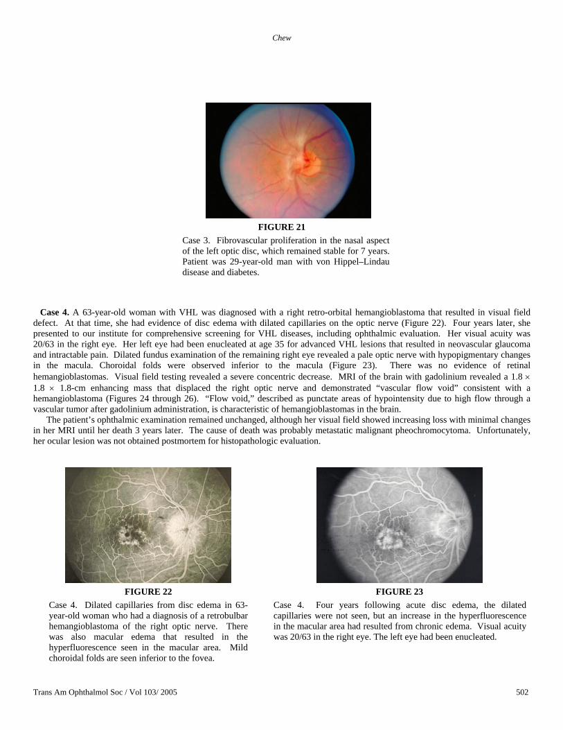
Case 3. A 29-year-old man inherited VHL from his mother, and he has two affected daughters detected by genetic testing. He also had a 5-year history of diabetes secondary to VHL lesions in his pancreas. His visual acuity was 20/20 bilaterally, and there was no evidence of diabetic retinopathy. His right eye had one peripheral retinal hemangioblastoma, and he had fibrovascular proliferation on his left optic disc (Figure 21). Follow-up of 7 years showed no change in either eye.
Intraretinal neovascularization that occurs without evidence of retinal hemangioblastomas is for the most part not a threat to visual function, as observed cases have remained remarkably stable over the years. The more vision-threatening lesions are those accompanied by fibrous tissue that leads to increased risk of anatomical alterations in the macula that lead to functional loss. Vitrectomy may be indicated for such cases. Retrobulbar (Intracranial) Optic Nerve Lesions. Although juxtapapillary capillary hemangioblastomas have been estimated to occur in up to 15% of gene carriers in previous reports, retrobulbar or intracranial optic nerve hemangioblastomas are exceedingly rare.13,20,51-57 We had 11 cases (5.3%) of intracranial optic nerve hemangioblastomas in this entire population. Because of the early screening with CT and MRI, these lesions are now detected in asymptomatic patients. Few patients were symptomatic in our study because many cases were detected with the early imaging of the lesions. The average age of these patients was 50.8 years (range, 23 to 89 years), and six were female.
Radiographic size of tumor ranged from 4 × 5 mm to 21 × 20 mm. One lesion involved the orbital portion of the optic nerve, four lesions involved the intracanalicular optic nerve, two lesions involved the prechiasmal nerve, and four lesions originated from the pituitary infundibulum with chiasmal involvement. There was associated edema of the visual pathways seen in five of the 11. The following cases represent the typical presentation and course of these rare lesions:
Trans Am Ophthalmol Soc / Vol 103/ 2005 501
Chew
FIGURE 21
Case 3. Fibrovascular proliferation in the nasal aspect of the left optic disc, which remained stable for 7 years. Patient was 29-year-old man with von Hippel–Lindau disease and diabetes.
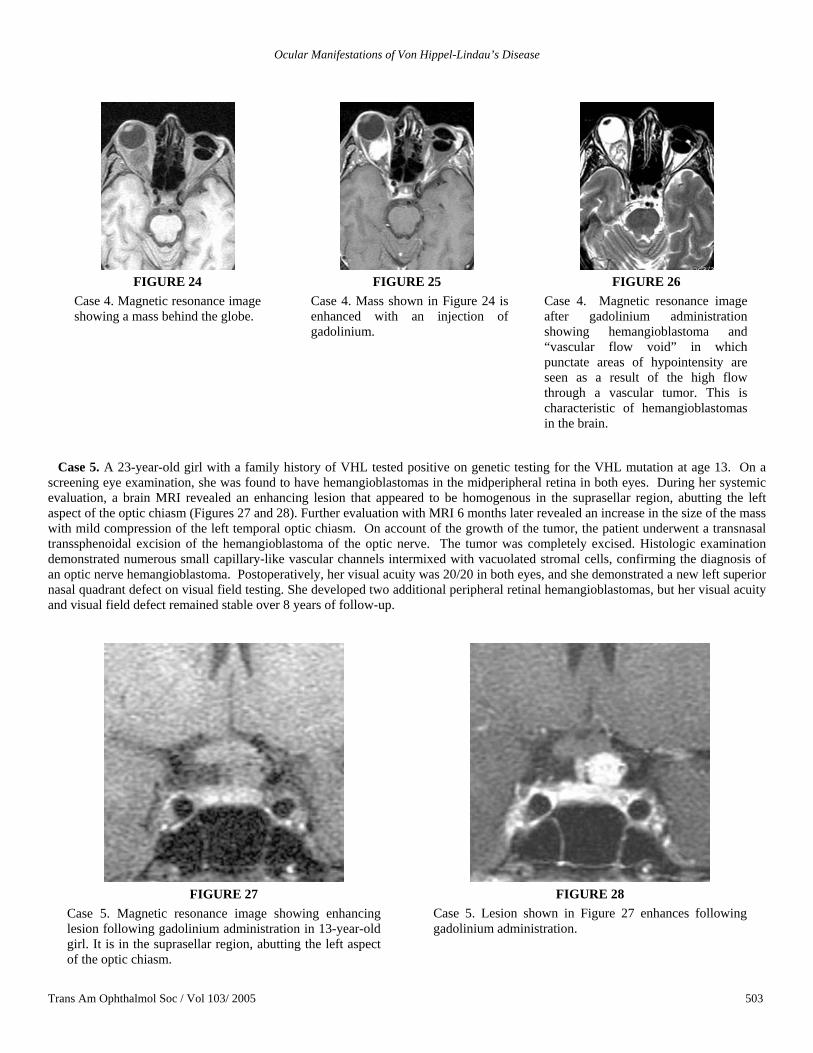
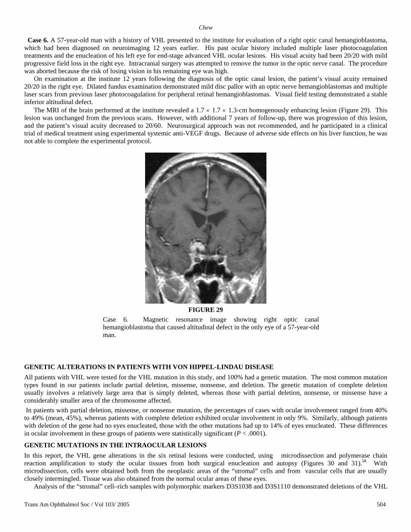
Case 4. A 63-year-old woman with VHL was diagnosed with a right retro-orbital hemangioblastoma that resulted in visual field defect. At that time, she had evidence of disc edema with dilated capillaries on the optic nerve (Figure 22). Four years later, she presented to our institute for comprehensive screening for VHL diseases, including ophthalmic evaluation. Her visual acuity was 20/63 in the right eye. Her left eye had been enucleated at age 35 for advanced VHL lesions that resulted in neovascular glaucoma and intractable pain. Dilated fundus examination of the remaining right eye revealed a pale optic nerve with hypopigmentary changes in the macula. Choroidal folds were observed inferior to the macula (Figure 23). There was no evidence of retinal hemangioblastomas. Visual field testing revealed a severe concentric decrease. MRI of the brain with gadolinium revealed a 1.8 × 1.8 × 1.8-cm enhancing mass that displaced the right optic nerve and demonstrated “vascular flow void” consistent with a hemangioblastoma (Figures 24 through 26). “Flow void,” described as punctate areas of hypointensity due to high flow through a vascular tumor after gadolinium administration, is characteristic of hemangioblastomas in the brain.
The patient’s ophthalmic examination remained unchanged, although her visual field showed increasing loss with minimal changes in her MRI until her death 3 years later. The cause of death was probably metastatic malignant pheochromocytoma. Unfortunately, her ocular lesion was not obtained postmortem for histopathologic evaluation.
FIGURE 22
Case 4. Dilated capillaries from disc edema in 63-year-old woman who had a diagnosis of a retrobulbar hemangioblastoma of the right optic nerve. There was also macular edema that resulted in the hyperfluorescence seen in the macular area. Mild choroidal folds are seen inferior to the fovea.
FIGURE 23
Case 4. Four years following acute disc edema, the dilated capillaries were not seen, but an increase in the hyperfluorescence in the macular area had resulted from chronic edema. Visual acuity was 20/63 in the right eye. The left eye had been enucleated.
Trans Am Ophthalmol Soc / Vol 103/ 2005 502
Ocular Manifestations of Von Hippel-Lindau’s Disease
FIGURE 24
Case 4. Magnetic resonance image showing a mass behind the globe.
FIGURE 25
Case 4. Mass shown in Figure 24 is enhanced with an injection of gadolinium.
FIGURE 26
Case 4. Magnetic resonance image after gadolinium administration showing hemangioblastoma and “vascular flow void” in which punctate areas of hypointensity are seen as a result of the high flow through a vascular tumor. This is characteristic of hemangioblastomas in the brain.
Case 5. A 23-year-old girl with a family history of VHL tested positive on genetic testing for the VHL mutation at age 13. On a screening eye examination, she was found to have hemangioblastomas in the midperipheral retina in both eyes. During her systemic evaluation, a brain MRI revealed an enhancing lesion that appeared to be homogenous in the suprasellar region, abutting the left aspect of the optic chiasm (Figures 27 and 28). Further evaluation with MRI 6 months later revealed an increase in the size of the mass with mild compression of the left temporal optic chiasm. On account of the growth of the tumor, the patient underwent a transnasal transsphenoidal excision of the hemangioblastoma of the optic nerve. The tumor was completely excised. Histologic examination demonstrated numerous small capillary-like vascular channels intermixed with vacuolated stromal cells, confirming the diagnosis of an optic nerve hemangioblastoma. Postoperatively, her visual acuity was 20/20 in both eyes, and she demonstrated a new left superior nasal quadrant defect on visual field testing. She developed two additional peripheral retinal hemangioblastomas, but her visual acuity and visual field defect remained stable over 8 years of follow-up.
FIGURE 27
Case 5. Magnetic resonance image showing enhancing lesion following gadolinium administration in 13-year-old girl. It is in the suprasellar region, abutting the left aspect of the optic chiasm.
FIGURE 28
Case 5. Lesion shown in Figure 27 enhances following gadolinium administration.
Trans Am Ophthalmol Soc / Vol 103/ 2005 503
Chew
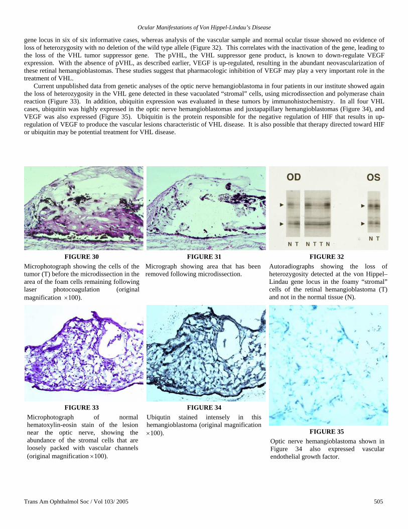
Case 6. A 57-year-old man with a history of VHL presented to the institute for evaluation of a right optic canal hemangioblastoma, which had been diagnosed on neuroimaging 12 years earlier. His past ocular history included multiple laser photocoagulation treatments and the enucleation of his left eye for end-stage advanced VHL ocular lesions. His visual acuity had been 20/20 with mild progressive field loss in the right eye. Intracranial surgery was attempted to remove the tumor in the optic nerve canal. The procedure was aborted because the risk of losing vision in his remaining eye was high.
On examination at the institute 12 years following the diagnosis of the optic canal lesion, the patient’s visual acuity remained 20/20 in the right eye. Dilated fundus examination demonstrated mild disc pallor with an optic nerve hemangioblastomas and multiple laser scars from previous laser photocoagulation for peripheral retinal hemangioblastomas. Visual field testing demonstrated a stable inferior altitudinal defect.
The MRI of the brain performed at the institute revealed a 1.7 × 1.7 × 1.3-cm homogenously enhancing lesion (Figure 29). This lesion was unchanged from the previous scans. However, with additional 7 years of follow-up, there was progression of this lesion, and the patient’s visual acuity decreased to 20/60. Neurosurgical approach was not recommended, and he participated in a clinical trial of medical treatment using experimental systemic anti-VEGF drugs. Because of adverse side effects on his liver function, he was not able to complete the experimental protocol.
FIGURE 29
Case 6. Magnetic resonance image showing right optic canal hemangioblastoma that caused altitudinal defect in the only eye of a 57-year-old man.
GENETIC ALTERATIONS IN PATIENTS WITH VON HIPPEL-LINDAU DISEASE All patients with VHL were tested for the VHL mutation in this study, and 100% had a genetic mutation. The most common mutation types found in our patients include partial deletion, missense, nonsense, and deletion. The genetic mutation of complete deletion usually involves a relatively large area that is simply deleted, whereas those with partial deletion, nonsense, or missense have a considerably smaller area of the chromosome affected. In patients with partial deletion, missense, or nonsense mutation, the percentages of cases with ocular involvement ranged from 40% to 49% (mean, 45%), whereas patients with complete deletion exhibited ocular involvement in only 9%. Similarly, although patients with deletion of the gene had no eyes enucleated, those with the other mutations had up to 14% of eyes enucleated. These differences in ocular involvement in these groups of patients were statistically significant (P < .0001).
GENETIC MUTATIONS IN THE INTRAOCULAR LESIONS In this report, the VHL gene alterations in the six retinal lesions were conducted, using microdissection and polymerase chain reaction amplification to study the ocular tissues from both surgical enucleation and autopsy (Figures 30 and 31).58 With microdissection, cells were obtained both from the neoplastic areas of the “stromal” cells and from vascular cells that are usually closely intermingled. Tissue was also obtained from the normal ocular areas of these eyes.
Analysis of the “stromal” cell–rich samples with polymorphic markers D3S1038 and D3S1110 demonstrated deletions of the VHL
Trans Am Ophthalmol Soc / Vol 103/ 2005 504
Ocular Manifestations of Von Hippel-Lindau’s Disease
gene locus in six of six informative cases, whereas analysis of the vascular sample and normal ocular tissue showed no evidence of loss of heterozygosity with no deletion of the wild type allele (Figure 32). This correlates with the inactivation of the gene, leading to the loss of the VHL tumor suppressor gene. The pVHL, the VHL suppressor gene product, is known to down-regulate VEGF expression. With the absence of pVHL, as described earlier, VEGF is up-regulated, resulting in the abundant neovascularization of these retinal hemangioblastomas. These studies suggest that pharmacologic inhibition of VEGF may play a very important role in the treatment of VHL.
Current unpublished data from genetic analyses of the optic nerve hemangioblastoma in four patients in our institute showed again the loss of heterozygosity in the VHL gene detected in these vacuolated “stromal” cells, using microdissection and polymerase chain reaction (Figure 33). In addition, ubiquitin expression was evaluated in these tumors by immunohistochemistry. In all four VHL cases, ubiquitin was highly expressed in the optic nerve hemangioblastomas and juxtapapillary hemangioblastomas (Figure 34), and VEGF was also expressed (Figure 35). Ubiquitin is the protein responsible for the negative regulation of HIF that results in up-regulation of VEGF to produce the vascular lesions characteristic of VHL disease. It is also possible that therapy directed toward HIF or ubiquitin may be potential treatment for VHL disease.
FIGURE 30 Microphotograph showing the cells of the tumor (T) before the microdissection in the area of the foam cells remaining following laser photocoagulation (original magnification ×100).
FIGURE 31 Micrograph showing area that has been removed following microdissection.
FIGURE 32 Autoradiographs showing the loss of heterozygosity detected at the von Hippel–Lindau gene locus in the foamy “stromal” cells of the retinal hemangioblastoma (T) and not in the normal tissue (N).
FIGURE 33 Microphotograph of normal hematoxylin-eosin stain of the lesion near the optic nerve, showing the abundance of the stromal cells that are loosely packed with vascular channels (original magnification ×100).
FIGURE 34 Ubiqutin stained intensely in this hemangioblastoma (original magnification ×100). FIGURE 35
Optic nerve hemangioblastoma shown in Figure 34 also expressed vascular endothelial growth factor.
Trans Am Ophthalmol Soc / Vol 103/ 2005 505
Chew
NOVEL THERAPY FOR VHL BASED UPON PROPOSED GENETIC FINDINGS The knowledge of the molecular genetics of both the systemic aspects of VHL disease and the tumor genetics may lead us toward the use of pharmacologic agents that could block at several points in the pathway. An example would be the inhibition of VEGF. In the following sections, the two cases in the literature are described, as well as the final two additional cases from our institution that had received intravitreal injection of an anti-VEGF drug for ocular VHL lesions that were not amenable to other treatments.
Systemic Anti-VEGF Treatment Previously Reported There are two reports of treatment with the systemic administration of the VEGF receptor inhibitor SU5416 in two patients with ocular lesions with VHL. The first case was a woman with optic nerve lesion.59 Although systemic administration of anti-VEGF did not change the size of the tumor, there was rapid reduction in the macular edema that persisted 18 months. The initial treatment was given over a period of 7 months. The second case was a woman who was refractory to the conventional treatments of laser, cryotherapy, and radiation.60 Again, the macular edema decreased, whereas the tumor did not change in size. However, 6 months following the administration of the same compound intravenously, the macular edema recurred. We believe that this treatment resulted in the decrease of the vasopermeability factor of the tumor, whereas the tumor itself did not change. The vessels in the tumor are already quite mature and may not be affected by such anti-VEGF treatment.
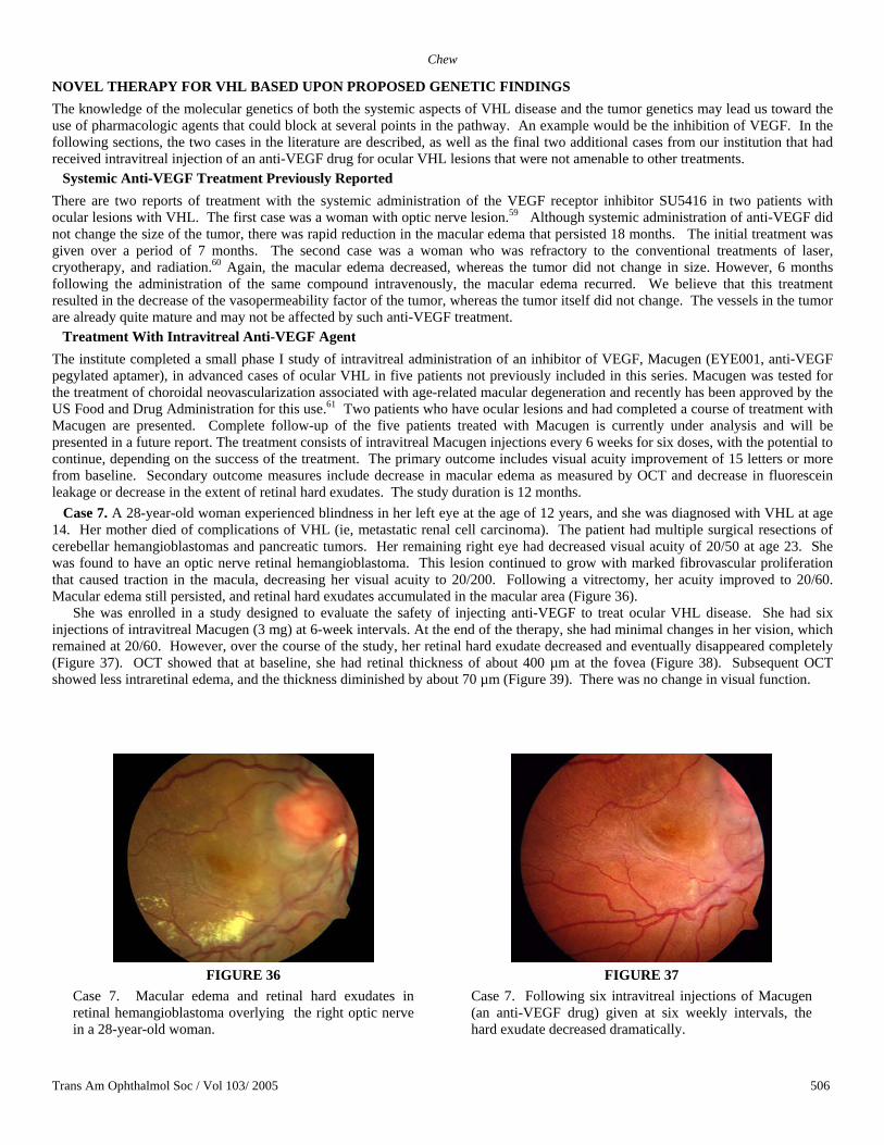
Treatment With Intravitreal Anti-VEGF Agent The institute completed a small phase I study of intravitreal administration of an inhibitor of VEGF, Macugen (EYE001, anti-VEGF pegylated aptamer), in advanced cases of ocular VHL in five patients not previously included in this series. Macugen was tested for the treatment of choroidal neovascularization associated with age-related macular degeneration and recently has been approved by the US Food and Drug Administration for this use.61 Two patients who have ocular lesions and had completed a course of treatment with Macugen are presented. Complete follow-up of the five patients treated with Macugen is currently under analysis and will be presented in a future report. The treatment consists of intravitreal Macugen injections every 6 weeks for six doses, with the potential to continue, depending on the success of the treatment. The primary outcome includes visual acuity improvement of 15 letters or more from baseline. Secondary outcome measures include decrease in macular edema as measured by OCT and decrease in fluorescein leakage or decrease in the extent of retinal hard exudates. The study duration is 12 months. Case 7. A 28-year-old woman experienced blindness in her left eye at the age of 12 years, and she was diagnosed with VHL at age 14. Her mother died of complications of VHL (ie, metastatic renal cell carcinoma). The patient had multiple surgical resections of cerebellar hemangioblastomas and pancreatic tumors. Her remaining right eye had decreased visual acuity of 20/50 at age 23. She was found to have an optic nerve retinal hemangioblastoma. This lesion continued to grow with marked fibrovascular proliferation that caused traction in the macula, decreasing her visual acuity to 20/200. Following a vitrectomy, her acuity improved to 20/60. Macular edema still persisted, and retinal hard exudates accumulated in the macular area (Figure 36).
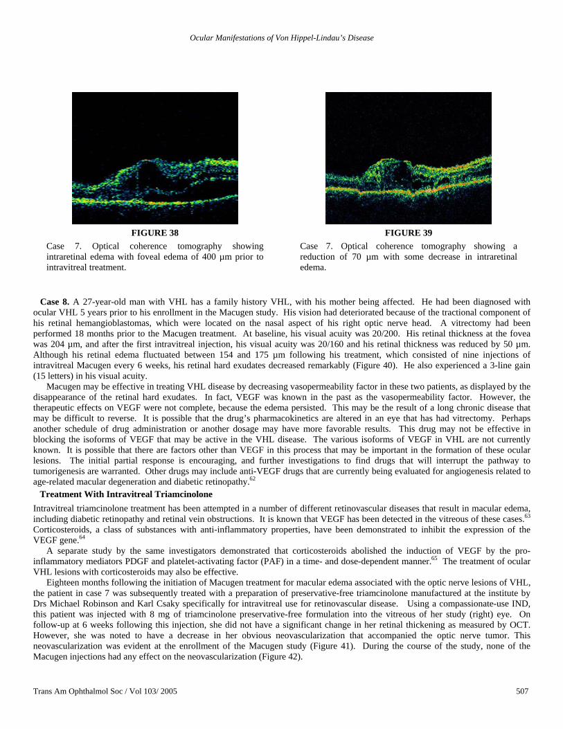
She was enrolled in a study designed to evaluate the safety of injecting anti-VEGF to treat ocular VHL disease. She had six injections of intravitreal Macugen (3 mg) at 6-week intervals. At the end of the therapy, she had minimal changes in her vision, which remained at 20/60. However, over the course of the study, her retinal hard exudate decreased and eventually disappeared completely (Figure 37). OCT showed that at baseline, she had retinal thickness of about 400 µm at the fovea (Figure 38). Subsequent OCT showed less intraretinal edema, and the thickness diminished by about 70 µm (Figure 39). There was no change in visual function.
FIGURE 36
Case 7. Macular edema and retinal hard exudates in retinal hemangioblastoma overlying the right optic nerve in a 28-year-old woman.
FIGURE 37
Case 7. Following six intravitreal injections of Macugen (an anti-VEGF drug) given at six weekly intervals, the hard exudate decreased dramatically.
Trans Am Ophthalmol Soc / Vol 103/ 2005 506
Ocular Manifestations of Von Hippel-Lindau’s Disease
FIGURE 38
Case 7. Optical coherence tomography showing intraretinal edema with foveal edema of 400 µm prior to intravitreal treatment.
FIGURE 39
Case 7. Optical coherence tomography showing a reduction of 70 µm with some decrease in intraretinal edema.
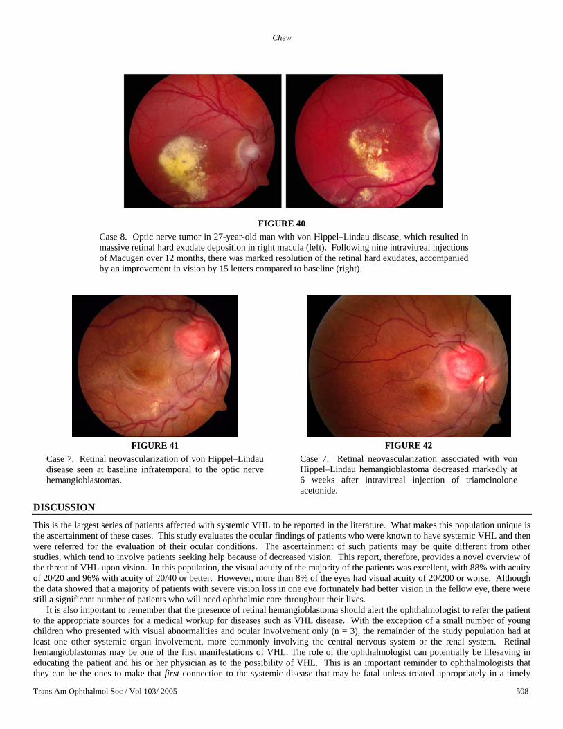
Case 8. A 27-year-old man with VHL has a family history VHL, with his mother being affected. He had been diagnosed with ocular VHL 5 years prior to his enrollment in the Macugen study. His vision had deteriorated because of the tractional component of his retinal hemangioblastomas, which were located on the nasal aspect of his right optic nerve head. A vitrectomy had been performed 18 months prior to the Macugen treatment. At baseline, his visual acuity was 20/200. His retinal thickness at the fovea was 204 µm, and after the first intravitreal injection, his visual acuity was 20/160 and his retinal thickness was reduced by 50 µm. Although his retinal edema fluctuated between 154 and 175 µm following his treatment, which consisted of nine injections of intravitreal Macugen every 6 weeks, his retinal hard exudates decreased remarkably (Figure 40). He also experienced a 3-line gain (15 letters) in his visual acuity.
Macugen may be effective in treating VHL disease by decreasing vasopermeability factor in these two patients, as displayed by the disappearance of the retinal hard exudates. In fact, VEGF was known in the past as the vasopermeability factor. However, the therapeutic effects on VEGF were not complete, because the edema persisted. This may be the result of a long chronic disease that may be difficult to reverse. It is possible that the drug’s pharmacokinetics are altered in an eye that has had vitrectomy. Perhaps another schedule of drug administration or another dosage may have more favorable results. This drug may not be effective in blocking the isoforms of VEGF that may be active in the VHL disease. The various isoforms of VEGF in VHL are not currently known. It is possible that there are factors other than VEGF in this process that may be important in the formation of these ocular lesions. The initial partial response is encouraging, and further investigations to find drugs that will interrupt the pathway to tumorigenesis are warranted. Other drugs may include anti-VEGF drugs that are currently being evaluated for angiogenesis related to age-related macular degeneration and diabetic retinopathy.62
Treatment With Intravitreal Triamcinolone Intravitreal triamcinolone treatment has been attempted in a number of different retinovascular diseases that result in macular edema, including diabetic retinopathy and retinal vein obstructions. It is known that VEGF has been detected in the vitreous of these cases.63 Corticosteroids, a class of substances with anti-inflammatory properties, have been demonstrated to inhibit the expression of the VEGF gene.64
A separate study by the same investigators demonstrated that corticosteroids abolished the induction of VEGF by the pro-inflammatory mediators PDGF and platelet-activating factor (PAF) in a time- and dose-dependent manner.65 The treatment of ocular VHL lesions with corticosteroids may also be effective.
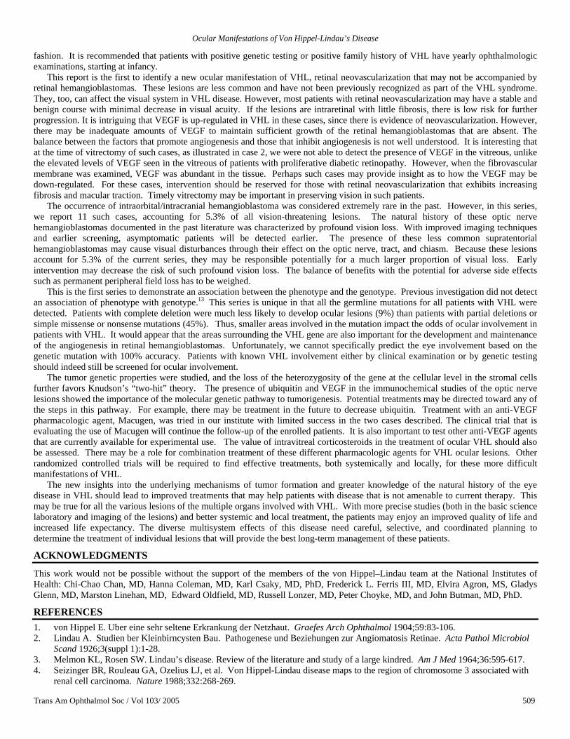
Eighteen months following the initiation of Macugen treatment for macular edema associated with the optic nerve lesions of VHL, the patient in case 7 was subsequently treated with a preparation of preservative-free triamcinolone manufactured at the institute by Drs Michael Robinson and Karl Csaky specifically for intravitreal use for retinovascular disease. Using a compassionate-use IND, this patient was injected with 8 mg of triamcinolone preservative-free formulation into the vitreous of her study (right) eye. On follow-up at 6 weeks following this injection, she did not have a significant change in her retinal thickening as measured by OCT. However, she was noted to have a decrease in her obvious neovascularization that accompanied the optic nerve tumor. This neovascularization was evident at the enrollment of the Macugen study (Figure 41). During the course of the study, none of the Macugen injections had any effect on the neovascularization (Figure 42).
Trans Am Ophthalmol Soc / Vol 103/ 2005 507
Chew
FIGURE 40
Case 8. Optic nerve tumor in 27-year-old man with von Hippel–Lindau disease, which resulted in massive retinal hard exudate deposition in right macula (left). Following nine intravitreal injections of Macugen over 12 months, there was marked resolution of the retinal hard exudates, accompanied by an improvement in vision by 15 letters compared to baseline (right).
FIGURE 41
Case 7. Retinal neovascularization of von Hippel–Lindau disease seen at baseline infratemporal to the optic nerve hemangioblastomas.
FIGURE 42
Case 7. Retinal neovascularization associated with von Hippel–Lindau hemangioblastoma decreased markedly at 6 weeks after intravitreal injection of triamcinolone acetonide.
DISCUSSION
This is the largest series of patients affected with systemic VHL to be reported in the literature. What makes this population unique is the ascertainment of these cases. This study evaluates the ocular findings of patients who were known to have systemic VHL and then were referred for the evaluation of their ocular conditions. The ascertainment of such patients may be quite different from other studies, which tend to involve patients seeking help because of decreased vision. This report, therefore, provides a novel overview of the threat of VHL upon vision. In this population, the visual acuity of the majority of the patients was excellent, with 88% with acuity of 20/20 and 96% with acuity of 20/40 or better. However, more than 8% of the eyes had visual acuity of 20/200 or worse. Although the data showed that a majority of patients with severe vision loss in one eye fortunately had better vision in the fellow eye, there were still a significant number of patients who will need ophthalmic care throughout their lives.
It is also important to remember that the presence of retinal hemangioblastoma should alert the ophthalmologist to refer the patient to the appropriate sources for a medical workup for diseases such as VHL disease. With the exception of a small number of young children who presented with visual abnormalities and ocular involvement only (n = 3), the remainder of the study population had at least one other systemic organ involvement, more commonly involving the central nervous system or the renal system. Retinal hemangioblastomas may be one of the first manifestations of VHL. The role of the ophthalmologist can potentially be lifesaving in educating the patient and his or her physician as to the possibility of VHL. This is an important reminder to ophthalmologists that they can be the ones to make that first connection to the systemic disease that may be fatal unless treated appropriately in a timely
Trans Am Ophthalmol Soc / Vol 103/ 2005 508
Ocular Manifestations of Von Hippel-Lindau’s Disease
fashion. It is recommended that patients with positive genetic testing or positive family history of VHL have yearly ophthalmologic examinations, starting at infancy.
This report is the first to identify a new ocular manifestation of VHL, retinal neovascularization that may not be accompanied by retinal hemangioblastomas. These lesions are less common and have not been previously recognized as part of the VHL syndrome. They, too, can affect the visual system in VHL disease. However, most patients with retinal neovascularization may have a stable and benign course with minimal decrease in visual acuity. If the lesions are intraretinal with little fibrosis, there is low risk for further progression. It is intriguing that VEGF is up-regulated in VHL in these cases, since there is evidence of neovascularization. However, there may be inadequate amounts of VEGF to maintain sufficient growth of the retinal hemangioblastomas that are absent. The balance between the factors that promote angiogenesis and those that inhibit angiogenesis is not well understood. It is interesting that at the time of vitrectomy of such cases, as illustrated in case 2, we were not able to detect the presence of VEGF in the vitreous, unlike the elevated levels of VEGF seen in the vitreous of patients with proliferative diabetic retinopathy. However, when the fibrovascular membrane was examined, VEGF was abundant in the tissue. Perhaps such cases may provide insight as to how the VEGF may be down-regulated. For these cases, intervention should be reserved for those with retinal neovascularization that exhibits increasing fibrosis and macular traction. Timely vitrectomy may be important in preserving vision in such patients.
The occurrence of intraorbital/intracranial hemangioblastoma was considered extremely rare in the past. However, in this series, we report 11 such cases, accounting for 5.3% of all vision-threatening lesions. The natural history of these optic nerve hemangioblastomas documented in the past literature was characterized by profound vision loss. With improved imaging techniques and earlier screening, asymptomatic patients will be detected earlier. The presence of these less common supratentorial hemangioblastomas may cause visual disturbances through their effect on the optic nerve, tract, and chiasm. Because these lesions account for 5.3% of the current series, they may be responsible potentially for a much larger proportion of visual loss. Early intervention may decrease the risk of such profound vision loss. The balance of benefits with the potential for adverse side effects such as permanent peripheral field loss has to be weighed.
This is the first series to demonstrate an association between the phenotype and the genotype. Previous investigation did not detect an association of phenotype with genotype.13 This series is unique in that all the germline mutations for all patients with VHL were detected. Patients with complete deletion were much less likely to develop ocular lesions (9%) than patients with partial deletions or simple missense or nonsense mutations (45%). Thus, smaller areas involved in the mutation impact the odds of ocular involvement in patients with VHL. It would appear that the areas surrounding the VHL gene are also important for the development and maintenance of the angiogenesis in retinal hemangioblastomas. Unfortunately, we cannot specifically predict the eye involvement based on the genetic mutation with 100% accuracy. Patients with known VHL involvement either by clinical examination or by genetic testing should indeed still be screened for ocular involvement.
The tumor genetic properties were studied, and the loss of the heterozygosity of the gene at the cellular level in the stromal cells further favors Knudson’s “two-hit” theory. The presence of ubiquitin and VEGF in the immunochemical studies of the optic nerve lesions showed the importance of the molecular genetic pathway to tumorigenesis. Potential treatments may be directed toward any of the steps in this pathway. For example, there may be treatment in the future to decrease ubiquitin. Treatment with an anti-VEGF pharmacologic agent, Macugen, was tried in our institute with limited success in the two cases described. The clinical trial that is evaluating the use of Macugen will continue the follow-up of the enrolled patients. It is also important to test other anti-VEGF agents that are currently available for experimental use. The value of intravitreal corticosteroids in the treatment of ocular VHL should also be assessed. There may be a role for combination treatment of these different pharmacologic agents for VHL ocular lesions. Other randomized controlled trials will be required to find effective treatments, both systemically and locally, for these more difficult manifestations of VHL.
The new insights into the underlying mechanisms of tumor formation and greater knowledge of the natural history of the eye disease in VHL should lead to improved treatments that may help patients with disease that is not amenable to current therapy. This may be true for all the various lesions of the multiple organs involved with VHL. With more precise studies (both in the basic science laboratory and imaging of the lesions) and better systemic and local treatment, the patients may enjoy an improved quality of life and increased life expectancy. The diverse multisystem effects of this disease need careful, selective, and coordinated planning to determine the treatment of individual lesions that will provide the best long-term management of these patients.
ACKNOWLEDGMENTS
This work would not be possible without the support of the members of the von Hippel–Lindau team at the National Institutes of Health: Chi-Chao Chan, MD, Hanna Coleman, MD, Karl Csaky, MD, PhD, Frederick L. Ferris III, MD, Elvira Agron, MS, Gladys Glenn, MD, Marston Linehan, MD, Edward Oldfield, MD, Russell Lonzer, MD, Peter Choyke, MD, and John Butman, MD, PhD.
REFERENCES 1. von Hippel E. Uber eine sehr seltene Erkrankung der Netzhaut. Graefes Arch Ophthalmol 1904;59:83-106. 2. Lindau A. Studien ber Kleinbirncysten Bau. Pathogenese und Beziehungen zur Angiomatosis Retinae. Acta Pathol Microbiol
Scand 1926;3(suppl 1):1-28. 3. Melmon KL, Rosen SW. Lindau’s disease. Review of the literature and study of a large kindred. Am J Med 1964;36:595-617. 4. Seizinger BR, Rouleau GA, Ozelius LJ, et al. Von Hippel-Lindau disease maps to the region of chromosome 3 associated with
renal cell carcinoma. Nature 1988;332:268-269.
Trans Am Ophthalmol Soc / Vol 103/ 2005 509
Chew
5. Latif F, Tory K, Gnarra J, et al. Identification of the von Hippel-Lindau disease tumour suppressor gene. Science 1993;260:1317-1320.
6. Lonser RR, Glenn GM, Walther M, et al. von Hippel-Lindau disease. Lancet 2003;361:2059-2067. 7. Maher ER, Kaelin WG Jr. von Hippel-Lindau disease. Medicine (Baltimore) 1997;76:381-391. 8. Linehan WM, Lerman MI, Zbar B. Identification of the von Hippel-Lindau (VHL) gene. Its role in renal cancer. JAMA
1995;273:564-570. 9. Richard S, Campello C, Taillandier L, et al. Haemangioblastoma of the central nervous system in von Hippel-Lindau disease.
French VHL Study Group. J Intern Med 1998;243:547-553. 10. Richard S, David P, Marsot-Dupuch K, et al. Central nervous system hemangioblastomas, endolymphatic sac tumors, and von
Hippel-Lindau disease. Neurosurg Rev 2000;23:1-22; discussion 23-24. 11. Wanebo JE, Lonser RR, Glenn GM, et al. The natural history of central nervous system hemangioblastomas in patients with von
Hippel-Lindau disease. J Neurosurg 2003;98:82-94. 12. Hammel PR, Vilgrain V, Terris B, et al. Pancreatic involvement in von Hippel-Lindau disease. The Groupe Francophone
d’Etude de la Maladie de von Hippel-Lindau. Gastroenterology 2000;119:1087-1095. 13. Webster AR, Maher ER, Moore AT. Clinical characteristics of ocular angiomatosis in von Hippel-Lindau disease and correlation
with germline mutation. Arch Ophthalmol 1999;117:371-378. 14. Dollfus H, Massin P, Taupin P, et al. Retinal hemangioblastoma in von Hippel-Lindau disease: a clinical and molecular study.
Invest Ophthalmol Vis Sci 2002;43:3067-3074. 15. Welch RB. von Hippel-Lindau disease: the recognition and treatment of early angiomatosis retinae and the use of cryosurgery
as an adjunct to therapy. Trans Am Ophthalmol Soc 1970;68:367-424. 16. Singh A, Shields CL, Shields JA. von Hippel-Lindau disease. Surv Ophthalmol 2001;46:117-142. 17. Ridley M, Green J, Johnson G. Retinal angiomatosis: the ocular manifestations of von Hippel-Lindau Disease. Can J
Ophthalmol 1986;21:276-283. 18. Maher ER, Iselius L, Yates JR, et al. Von Hippel-Lindau disease: a genetic study. J Med Genet 1991;28:443-447. 19. Neumann HP, Wiestler OD. Clustering of features of von Hippel-Lindau syndrome: evidence for a complex genetic locus.
Lancet 1991;337:1052-1054. 20. Maher ER, Yates JR, Harries R, et al. Clinical features and natural history of von Hippel-Lindau disease. Q J Med 1990;77:1151-
1163. 21. Neumann HP, Eggert HR, Scheremet R, et al. Central nervous system lesions in von Hippel-Lindau syndrome. J Neurol
Neurosurg Psychiatry 1992;55:898-901. 22. Lamiell JM, Salazar FG, Hsia YE. von Hippel-Lindau disease affecting 43 members of a single kindred. Medicine 1989;68:1-29. 23. Knudson AG Jr, Strong LC. Mutation and cancer: neuroblastoma and pheochromocytoma. Am J Hum Genet 1972;24:514-532. 24. Knudson AGJ. Genetics of human cancer. Ann Rev Genet 1986;20:231-251. 25. Duan DR, Pause A, Burgess WH, et al. Inhibition of transcription elongation by the VHL tumour suppressor protein. Science
1995; 269:1402–1406. 26. Pause A, Lee S, Worrell RA, et al. The von Hippel-Lindau tumour-suppressor gene product forms a stable complex with human
CUL-2, a member of the Cdc53 family of proteins. Proc Natl Acad Sci U S A 1997; 94: 2156–2161. 27. Maxwell PH, Wiesener MS, Chang GW, et al. The tumour suppressor protein VHL targets hypoxia-inducible factors for oxygen-
dependent proteolysis. Nature 1999;399:271-275. 28. Kaelin WG Jr. Molecular basis of the VHL hereditary cancer syndrome. Nat Rev Cancer 2002;2:673-682. 29. Kondo K, Klco J, Nakamura E, et al. Inhibition of HIF is necessary for tumour suppression by the von Hippel-Lindau protein.
Cancer Cell 2002;1:237-246. 30. Bohling T, Hatva E, Kujala M, et al. Expression of growth factors and growth factor receptors in capillary hemangioblastoma. J
Neuropathol Exp Neurol 1996;55:522-527. 31. Reifenberger G, Reifenberger J, Bilzer T, et al. Coexpression of transforming growth factor-alpha and epidermal growth factor
receptor in capillary hemangioblastomas of the central nervous system. Am J Pathol 1995;147:245-250. 32. Carmeliet P, Dor Y, Herbert JM, et al. Role of HIF-1alpha in hypoxia-mediated, apoptosis, cell proliferation and tumour
angiogenesis. Nature 1998;394:485-490. 33. Maranchie JK, Afonso A, Albert PS, et al. Solid renal tumor severity in von Hippel Lindau disease is related to germline
deletion length and location. Hum Mutat 2004;23:40-46. 34. Font RL, Ferry AP. The phakomatoses. Int Ophthalmol Clin 1972;12:1-50. 35. Green WR. Retina: capillary hemangioma. In: Spencer WH, ed. Ophthalmic Pathology: An Atlas and Textbook. Philadelphia:
WB Saunders; 1996:709-718. 36. Jakobiec FA, Font RL, Johnson FB. Angiomatosis retinae: an ultrastructural study and lipid analyses. Cancer 1976;38:2042-
2056. 37. Grossniklaus HE, Thomas JW, Vigneswaran N, et al. Retinal hemangioblastoma: a histologic, immunohistochemical, and
ultrastructural evaluation. Ophthalmology 1992;99:140-145. 38. Mottow-Lippa L, Tso MO, Peyman GA, et al. von Hippel angiomatosis. A light, electron microscopic, and immunoperoxidase
characterization. Ophthalmology 1983;90:848-855.
Trans Am Ophthalmol Soc / Vol 103/ 2005 510
Ocular Manifestations of Von Hippel-Lindau’s Disease
39. Vortmeyer AO, Frank S, Jeong SY, et al. Developmental arrest of angioblastic lineage initiates tumorigenesis in von Hippel-Lindau disease. Cancer Res 2003;63:7051-7055.
40. Lubensky IA, Gnarra JR, Bertheau P, et al. Allelic deletions of the VHL gene detected in multiple microscopic clear cell renal lesions in von Hippel-Lindau disease patients. Am J Pathol 1996;149:2089-2094.
41. Vortmeyer AO, Gnarra JR, Emmert-Buck MR, et al. von Hippel-Lindau gene deletion detected in stromal cell component of a cerebellar hemangioblastoma associated with von Hippel-Lindau disease. Hum Pathol 1997;28:540-543.
42. Singh AD, Nouri M, Shields CL, et a;. Treatment of retinal capillary hemangioma. Ophthalmology 2002;109:1799-1806. 43. Annesley WH, Leonard BC, Shields JA, et al. Fifteen year review of treated cases of retinal angiomatosis. Trans Sect
Ophthalmol Am Acad Ophthalmol Otolaryngol 1977;83:446-453. 44. Rosa RH, Goldberg MF, Green WR. Clinicopathologic correlation of argon laser photocoagulation of retinal angiomas in a
patient with von Hippel-Lindau disease followed for more than 20 years. Retina 1996;16:145-156. 45. Schmidt D, Natt E, Neumann HP. Long-term results of laser treatment for retinal angiomatosis in von Hippel-Lindau disease.
Eur J Med Res 2000;5:47-58. 46. Garcia-Arumí J, Sararols LH, Cavero L, et al. Therapeutic options for capillary papillary angiomas. Ophthalmology
2000;107:48-54. 47. Schmidt-Erfurth UM, Kusserow C, Barbazetto IA, et al. Benefits and complications of photodynamic therapy of papillary
capillary angiomas. Ophthalmology 2002;109:1256-1266. 48. Palmer JD, Gragoudas ES. Advances in treatment of retinal angiomas. Int Ophthalmol Clin 1997;37:150-170. 49. 49 McDonald HR, Schatz H, Johnson RN, et al. Vitrectomy in eyes with peripheral retinal angioma associated with traction
macular detachment. Ophthalmology 1996;103:329-335. 50. 50 Whitson JT, Welch RB, Green WR. Von Hippel-Lindau disease: case report of a patient with spontaneous regression of a
retinal angioma. Retina 1986;6:253-259. 51. Kupersmith MJ, Berenstein A. Visual disturbances in von Hippel-Lindau disease. Ann Ophthalmol 1981;2:195-197. 52. In S, Miyagi J, Kojho N, et al. Intraorbital optic nerve hemangioblastoma with von Hippel-Lindau disease. J Neurosurg
1982;56:426-429. 53. O’Reilly GV, Rumbaugh CL, Bowens M, et al. Supratentorial haemangioblastoma: the diagnostic roles of computed
tomography and angiography. Clin Radiol 1980;32:389-392. 54. Nerad JA, Kersten RC, Anderson RL. Hemangioblastoma of the optic nerve. Report of a case and review of literature.
Ophthalmology 1988;95;398-402. 55. Rubio A, Meyers SP, Powers JM, et al. Hemangioblastoma of the optic nerve. Hum Pathol 1994;25:1249-1251. 56. Kerr DJ, Scheithauer BW, Miller GM, et al. Hemangioblastoma of the optic nerve: case report. Neurosurgery 1995;36:573-
581. 57. Wasenko JJ, Rodziewicz GS. Suprasellar hemangioblastoma in Von Hippel-Lindau disease: a case report. Clin Imaging
2003;27:18-22. 58. Chan CC, Vortmeyer AO, Chew EY, et al. VHL gene deletion and enhanced VEGF gene expression detected in the stromal
cells of retinal angioma. Arch Ophthalmol 1999;117:625-630. 59. Aiello LP, George DJ, Cahill MT, et al. Rapid and durable recovery of visual function in a patient with von Hippel-Lindau
syndrome after systemic therapy with vascular endothelial growth factor receptor inhibitor SU5416. Ophthalmology 2002;109:1745-1751.
60. Girmens JF, Erginay A, Massin P, et al. Vascular endothelial growth factor receptor inhibitor SU5416 is more effective for associated macular edema than for hemangioblastomas. Am J Ophthalmol 2003;136:194-196.
61. Eyetech Study Group. Anti-vascular endothelial growth factor therapy for subfoveal choroidal neovascularization secondary to age-related macular degeneration: phase II study results. Ophthalmology 2003;110:979-986.
62. Krzystolik MG, Afshari MA, Adamis AP, et al. Prevention of experimental choroidal neovascularization with intravitreal anti-vascular endothelial growth factor antibody fragment. Arch Ophthalmol 2002;120:338-346.
63. Aiello LP, Bursell SE, Clermont A, et al. Vascular endothelial growth factor-induced retinal permeability is mediated by protein kinase C in vivo and suppressed by an orally effective beta-isoform-selective inhibitor. Diabetes 1997;46:1473-1480.
64. Nauck M, Karakiulakis G, Perruchoud A, et al. Corticosteriods inhibit the expression of the vascular endothelial growth factor gene in human vascular smooth muscle cells. Eur J Pharmacol 1998;341:309-315.
65. Nauck M, Roth M, Tamm M, et al. Induction of vascular endothelial growth factor by platelet-activating factor and platelet-derived growth factor is downregulated by corticosteroids. Am J Respir Cell Mol Biol 1997;16:398-406.
Trans Am Ophthalmol Soc / Vol 103/ 2005 511

![Von Hippel Lindau Disease [VHL]: Magnetic Resonance Imaging](https://static.fdocuments.net/doc/165x107/620636d38c2f7b17300580b0/von-hippel-lindau-disease-vhl-magnetic-resonance-imaging.jpg)


![Von Hippel Lindau Disease [VHL]: Magnetic Resonance Imaging ...](https://static.fdocuments.net/doc/165x107/587759fb1a28ab13448b6a8e/von-hippel-lindau-disease-vhl-magnetic-resonance-imaging-.jpg)