Ocular involvement in toxoplasmosis - British Journal...
7
BritishJournal ofOphthalmology 1993; 77: 371-377 PERSPECTIVE Ocular involvement in toxoplasmosis Aniki Rothova Toxoplasmosis is a major and preventable cause of severe visual handicap and blindness in young people. Congenital toxoplasmosis, as well as toxoplasmosis in immuno- compromised patients, is a serious, sometimes fatal disease. I 2 Toxoplasmic encephalitis has gained importance as a leading cause of AIDS related mortality. Toxoplasma gondii, the aetiological agent of human and animal toxoplasmosis, is ubiquitous in nature. The organism is an obligate intracellular parasite and its definitive host is the cat. Oocysts, which are excreted in cat faeces, are highly resistant and may remain infective for more than 1 year. Usually only young cats are infected for a short period of time. The infected cat is not the primary source of infection for humans but is a very important cause of contamination of the environment (soil, fruit, and vegetables and thus other infected animals which become intermediate hosts). Toxoplasma gondii is the most important protozoan cause of intraocular inflammation owing to its widespread distri- bution throughout the world, the frequency of ocular involvement, and the seriousness of the symptoms it may cause. Toxoplasmosis was first recognised in patients with congenital disease: in 1923 Janku' in Prague presented the first report on a boy who became blind at the age of 3 months and died of hydrocephalus; a granulomatous inflammation of the eye with protozoa in the retina was found.3 Wolf subsequently verified the aetiology by recovering the Toxoplasma and transmitting the infection to animals.4 Retrospective analysis of specimens has demonstrated the disease in a large number of patients with neonatal encephalitis, thus emphasising the possibility of toxo- plasmosis in such cases.5 The major problem in diagnosing ocular toxoplasmosis in adults was the difficulty of establishing the presence of the parasite in ocular lesions. Helenor Wilder provided proof by demonstrating organisms with the morphological character- istics of Toxoplasma gondii in the eyes of patients with chorioretinitis, who had undergone enucleation because of blindness and pain.6 Human infection may occur by either the congenital or the acquired route. Acquired disease usually develops after ingestion of oocysts or tissue cysts and almost all cases (in immunocompetent individuals) are asymptomatic. The prevalence of seropositivity due to a previous Toxoplasma infection varies among different populations, depending on eating habits, climate, etc. In the Netherlands the majority of the population has already been infected by the age of 40 years.7 Congenital infection can occur when a pregnant woman becomes infected; during the parasitaemia, Toxoplasma crosses the placenta and invades the tissues of the developing fetus. The risk and severity of infection in the child depend on the time of gestation in which the mother acquires the infection.8 The severity of congenital infection is greatest when the disease is acquired during the early stages of pregnancy. The frequency of transmission to the fetus is highest during the third trimester, when contact between the maternal and fetal circulations is more likely to occur.8 Overall, about 40% of children of infected mothers will also become infected. Of these infected children only a small proportion will exhibit clinical symptoms directly after birth. However, long term follow up has shown that about 80% of these infected children will eventually develop neurological and/or ocular sequelae.111 Once maternal immunity has developed, it is believed that all subsequent fetuses will be protected against congenital toxoplasmosis. However, a case of repeated fetal infections acquired from the same mother with presumed chronic toxoplasmic endometritis has been reported.'2 Toxoplasmic scars present in multiple siblings have also been documented, but whether these scars were caused by acquired or congenital disease remains unclear.'3 Ocular recurrence during pregnancy in three patients did not cause an infection of the fetus. 14 Until recently it has been assumed that ocular toxo- plasmosis represents a (frequently late) manifestation of congenital disease.'5 In 1973 Perkins, who performed a retrospective review of patients with acquired toxoplasmosis, showed that only 3% had associated ocular disease. Further- more, an epidemiological study on a South Pacific island, where 90% of the population over 22 years of age had already been infected by Toxoplasma, revealed virtually no cases of either congenital or ocular disease.'6 Population surveys showed that the number of acquired infections increased with age, while the ocular disease affected mainly young individuals - the age of onset lying between 10 and 20 years of age. In contrast, recent reports support the view that acquired infection may lead to ocular disease and may even be a more important cause of ocular involvement than previously thought.'4 17-21 Although absence of retinal lesions was noted in patients with an acute stage of acquired toxoplasmosis, ocular involvement may not become manifest until several years after initial seroconversion.'8 In a large- population based household study in southern Brazil, the incidence of ocular toxoplasmosis was 18%, which is extremely high.2' The authors argued that ocular toxoplasmosis in the affected area was not of congenital origin, since systemic congenital toxoplasmosis is uncommon there. Furthermore, in the same area, IgM antibodies were found in fewer than 1% of cord blood specimens and the ocular lesions were also documented in multiple siblings. 14 21 The epidemiological pattern revealed a paucity of ocular lesions in young children and an increase in the prevalence of ocular lesions during adolescence, a phenomenon which has also been noted in congenital disease.9'0 Several hypotheses have been presented on the role of acquired toxoplasmosis in ocular disease, addressing the importance of infection at an early age, long term antigen exposure and the role of reinfections, strain differences between parasites, as well as genetic differences between hosts. Immunocompromised patients may develop lethal disseminated toxoplasmosis and/or toxoplasmic encephalitis. 371 on 23 September 2018 by guest. Protected by copyright. http://bjo.bmj.com/ Br J Ophthalmol: first published as 10.1136/bjo.77.6.371 on 1 June 1993. Downloaded from
-
Upload
truongdiep -
Category
Documents
-
view
215 -
download
0
Transcript of Ocular involvement in toxoplasmosis - British Journal...
BritishJournal ofOphthalmology 1993; 77: 371-377
PERSPECTIVE
Ocular involvement in toxoplasmosis
Aniki Rothova
Toxoplasmosis is a major and preventable cause of severevisual handicap and blindness in young people. Congenitaltoxoplasmosis, as well as toxoplasmosis in immuno-compromised patients, is a serious, sometimes fataldisease. I2 Toxoplasmic encephalitis has gained importance asa leading cause ofAIDS related mortality.
Toxoplasma gondii, the aetiological agent of human andanimal toxoplasmosis, is ubiquitous in nature. The organismis an obligate intracellular parasite and its definitive host isthe cat. Oocysts, which are excreted in cat faeces, are highlyresistant and may remain infective for more than 1 year.Usually only young cats are infected for a short period oftime. The infected cat is not the primary source of infectionfor humans but is a very important cause of contamination ofthe environment (soil, fruit, and vegetables and thus otherinfected animals which become intermediate hosts).
Toxoplasma gondii is the most important protozoan causeof intraocular inflammation owing to its widespread distri-bution throughout the world, the frequency of ocularinvolvement, and the seriousness of the symptoms it maycause. Toxoplasmosis was first recognised in patients withcongenital disease: in 1923 Janku' in Prague presented thefirst report on a boy who became blind at the age of 3 monthsand died ofhydrocephalus; a granulomatous inflammation ofthe eye with protozoa in the retina was found.3 Wolfsubsequently verified the aetiology by recovering theToxoplasma and transmitting the infection to animals.4Retrospective analysis of specimens has demonstrated thedisease in a large number of patients with neonatalencephalitis, thus emphasising the possibility of toxo-plasmosis in such cases.5The major problem in diagnosing ocular toxoplasmosis in
adults was the difficulty of establishing the presence of theparasite in ocular lesions. Helenor Wilder provided proof bydemonstrating organisms with the morphological character-istics of Toxoplasma gondii in the eyes of patients withchorioretinitis, who had undergone enucleation because ofblindness and pain.6Human infection may occur by either the congenital or the
acquired route. Acquired disease usually develops afteringestion of oocysts or tissue cysts and almost all cases (inimmunocompetent individuals) are asymptomatic. Theprevalence of seropositivity due to a previous Toxoplasmainfection varies among different populations, depending oneating habits, climate, etc. In the Netherlands the majority ofthe population has already been infected by the age of 40years.7
Congenital infection can occur when a pregnant womanbecomes infected; during the parasitaemia, Toxoplasmacrosses the placenta and invades the tissues of the developingfetus. The risk and severity of infection in the child dependon the time of gestation in which the mother acquires theinfection.8 The severity of congenital infection is greatestwhen the disease is acquired during the early stages ofpregnancy. The frequency of transmission to the fetus is
highest during the third trimester, when contact between thematernal and fetal circulations is more likely to occur.8Overall, about 40% of children of infected mothers will alsobecome infected. Of these infected children only a smallproportion will exhibit clinical symptoms directly after birth.However, long term follow up has shown that about 80% ofthese infected children will eventually develop neurologicaland/or ocular sequelae.111 Once maternal immunity hasdeveloped, it is believed that all subsequent fetuses will beprotected against congenital toxoplasmosis. However, a caseof repeated fetal infections acquired from the same motherwith presumed chronic toxoplasmic endometritis has beenreported.'2 Toxoplasmic scars present in multiple siblingshave also been documented, but whether these scars werecaused by acquired or congenital disease remains unclear.'3Ocular recurrence during pregnancy in three patients did notcause an infection of the fetus. 14
Until recently it has been assumed that ocular toxo-plasmosis represents a (frequently late) manifestation ofcongenital disease.'5 In 1973 Perkins, who performed aretrospective review of patients with acquired toxoplasmosis,showed that only 3% had associated ocular disease. Further-more, an epidemiological study on a South Pacific island,where 90% of the population over 22 years of age had alreadybeen infected by Toxoplasma, revealed virtually no cases ofeither congenital or ocular disease.'6 Population surveysshowed that the number of acquired infections increasedwith age, while the ocular disease affected mainly youngindividuals - the age ofonset lying between 10 and 20 years ofage. In contrast, recent reports support the view thatacquired infection may lead to ocular disease and may even beamore important cause ofocular involvement than previouslythought.'4 17-21 Although absence of retinal lesions was notedin patients with an acute stage of acquired toxoplasmosis,ocular involvement may not become manifest until severalyears after initial seroconversion.'8 In a large- populationbased household study in southern Brazil, the incidence ofocular toxoplasmosis was 18%, which is extremely high.2'The authors argued that ocular toxoplasmosis in the affectedarea was not of congenital origin, since systemic congenitaltoxoplasmosis is uncommon there. Furthermore, in the samearea, IgM antibodies were found in fewer than 1% of cordblood specimens and the ocular lesions were also documentedin multiple siblings. 14 21 The epidemiological pattern revealeda paucity of ocular lesions in young children and an increasein the prevalence of ocular lesions during adolescence, aphenomenon which has also been noted in congenitaldisease.9'0 Several hypotheses have been presented on therole of acquired toxoplasmosis in ocular disease, addressingthe importance of infection at an early age, long term antigenexposure and the role of reinfections, strain differencesbetween parasites, as well as genetic differences betweenhosts.
Immunocompromised patients may develop lethaldisseminated toxoplasmosis and/or toxoplasmic encephalitis.
371 on 23 S
eptember 2018 by guest. P
rotected by copyright.http://bjo.bm
j.com/
Br J O
phthalmol: first published as 10.1136/bjo.77.6.371 on 1 June 1993. D
ownloaded from

Rothova
Figure I (Left and right) Inactive old toxoplasmosis scarwith typical atrophy and hyperpigmentation. Preretinalvitreous detachment is also visible (right).
Figure 2 (Left and right) Active retinal toxoplasmosislesion adjacent to an old retinal scar. The scar is hazy becauseofthe vitreal inflammatory reaction and can be recognised bythe hyperpigmentation. Preretinal vitreous membrane withcentral hole (right).
Figure 3 (Left and right) Satellite lesions adjacent to oldtoxoplasmosis scars.
Figure 4 (Left) Vitreous opacities present in the eye withtoxoplasmic chorioretinitis. (Right) Retinal haemorrhageencircling the active lesion and old scars.
Ocular disease, which is not common in such cases, may beatypical with such features as bilateral multifocal lesions.AIDS patients with (ocular) toxoplasmosis usually requirecontinuous, lifelong treatment.22 23 Toxoplasmic retino-choroiditis in AIDS is probably due to (reactivation of)acquired disease, although in some cases it may result frompre-existing congenital ocular lesions.22
Clinical manifestationsThe ocular lesions primarily affect the retina. The hallmarkof ocular toxoplasmosis is focal necrotising retinitis,ultimately resulting in characteristic atrophic scars. Old
inactive atrophic scars sometimes help to establish the correctdiagnosis of a former ocular disease, but such a finding doesnot present a therapeutic dilemma (Fig 1). It is active retinitiswhich poses diagnostic and therapeutic problems. Fundu-scopy may be difficult because of the inflammatory reactionin the vitreous - the lesions resemble 'headlights in the fog'(Fig 2). The active lesions may vary in size but are usuallyoval or circular. Frequently reactivation is situated adjacentto an old atrophic scar with hyperpigmentation along theborders, indicating an old infection (satellite formation; Fig3). During active inflammation the retina is thickened andcream-coloured due to necrosis and associated oedema.Overlying inflammatory cells and sometimes dense infiltrates
372 on 23 S
eptember 2018 by guest. P
rotected by copyright.http://bjo.bm
j.com/
Br J O
phthalmol: first published as 10.1136/bjo.77.6.371 on 1 June 1993. D
ownloaded from
Ocular involvement in toxoplasmosis
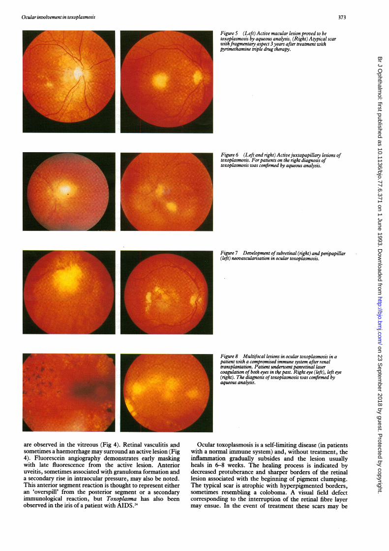
Figure 5 (Left) Active macular lesion proved to betoxoplasmosis by aqueous analysis. (Right) Atypical scarwith fragmentary aspect 3years after treatment withpyrimethamine triple drug therapy.
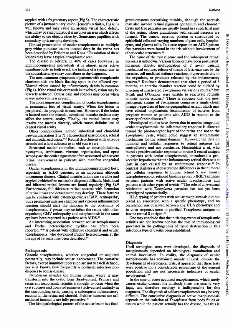
Figure 6 (Left and right) Activejuxtapapillary lesions oftoxoplasmosis. For patients on the right diagnosis oftoxoplasmosis was confirmed by aqueous analysis.
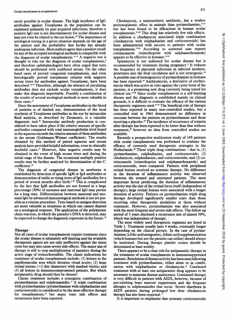
Figure 7 Development ofsubretinal (right) and peripapillar(left) neovascularisation in ocular toxoplasmosis.
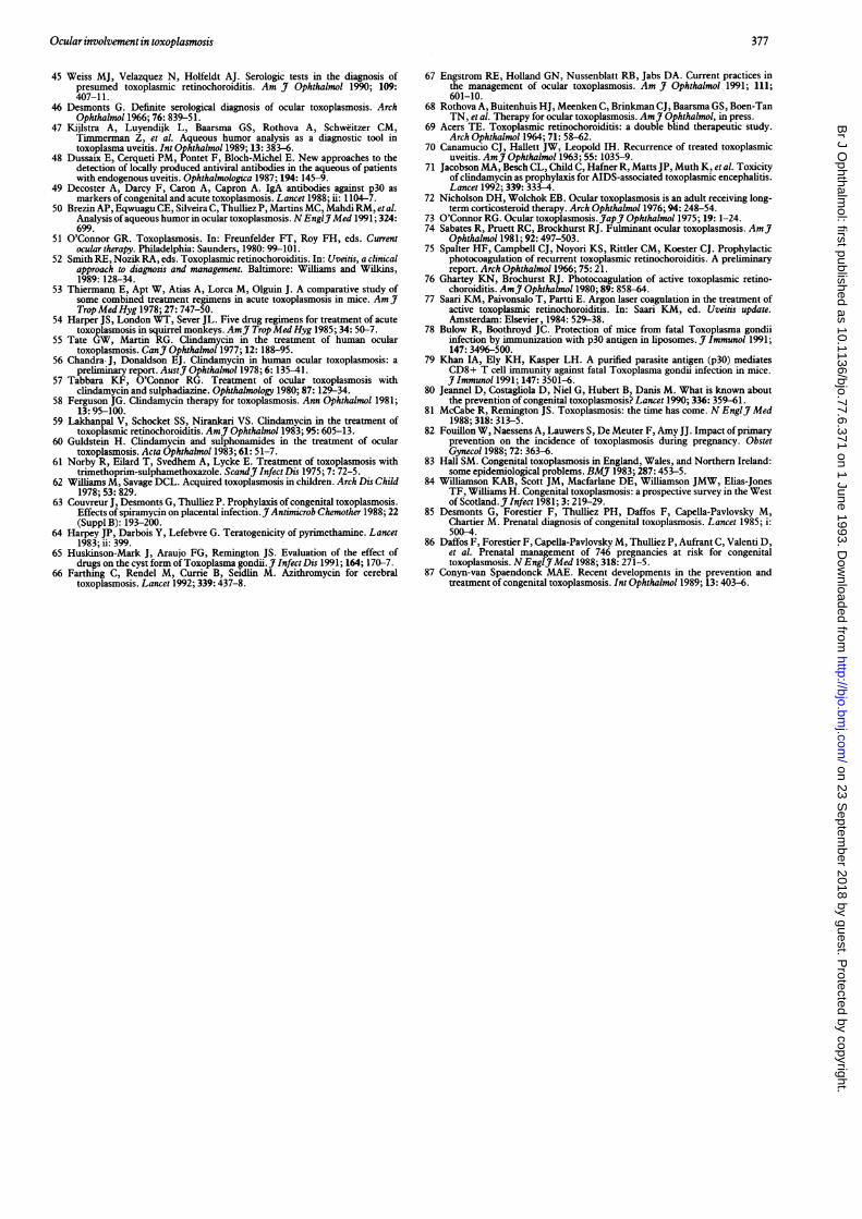
Figure 8 Multifocal lesions in ocular toxoplasmosis in apatient with a compromised immune system after renaltransplantation. Patient underwent panretinal lasercoagulation ofboth eyes in the past. Right eye (left), left eye(right). The diagnosis oftoxoplasmosis was confirmed byaqueous analysis.
are observed in the vitreous (Fig 4). Retinal vasculitis andsometimes a haemorrhage may surround an active lesion (Fig4). Fluorescein angiography demonstrates early maskingwith late fluorescence from the active lesion. Anterioruveitis, sometimes associated with granuloma formation anda secondary rise in intraocular pressure, may also be noted.This anterior segment reaction is thought to represent eitheran 'overspill' from the posterior segment or a secondaryimmunological reaction, but Toxoplasma has also beenobserved in the iris of a patient with AIDS.24
Ocular toxoplasmosis is a self-limiting disease (in patientswith a normal immune system) and, without treatment, theinflammation gradually subsides and the lesion usuallyheals in 6-8 weeks. The healing process is indicated bydecreased protuberance and sharper borders of the retinallesion associated with the beginning of pigment clumping.The typical scar is atrophic with hyperpigmented borders,sometimes resembling a coloboma. A visual field defectcorresponding to the interruption of the retinal fibre layermay ensue. In the event of treatment these scars may be
373
on 23 Septem
ber 2018 by guest. Protected by copyright.
http://bjo.bmj.com
/B
r J Ophthalm
ol: first published as 10.1136/bjo.77.6.371 on 1 June 1993. Dow
nloaded from
Rothova
atypical with a fragmentary aspect (Fig 5). The characteristicpicture of a juxtapapillary lesion (Jensen's retinitis, Fig 6) iswell known and may cause an arcuate visual field defect,which may be symptomatic if it involves an area which affectsthe ability to see objects close by. Sometimes papillitis withsecondary optic atrophy develops.
Clinical presentation of ocular toxoplasmosis as multiplegrey-white punctate lesions located deep in the retina hasbeen described by Friedman and Knox.25 Resolution of theselesions may leave a typical toxoplasmic scar.The disease is bilateral in 40% of cases (however, in
immunocompetent individuals it is almost never activesimultaneously in both eyes); the finding of a typical scar inthe contralateral eye may contribute to the diagnosis.The most common symptoms ofpatients with toxoplasmic
chorioretinitis are black floating spots in the affected eye.Blurred vision caused by inflammatory debris is common(Fig 4). If the visual axis or macula is involved, vision may beseverely reduced. Ocular pain and redness may occur whensevere iridocyclitis is present.The most important complication of ocular toxoplasmosis
is permanent loss of visual acuity. When the lesion isperipheral, the prognosis is excellent. However, if the lesionis located near the macula, associated macular oedema mayaffect the central acuity. Finally, the retinal lesion mayinvolve the macula directly, which causes definite loss ofcentral visual acuity.
Other complications include subretinal and choroidalneovascularisation (Fig 7), chorioretinal anastomoses, retinaland choroidal occlusion.2026 Retinal detachment with vitreousstrands and a hole adjacent to an old scar is rare.
Structural ocular anomalies, such as microphthalmos,nystagmus, strabismus, vitreous organisation, and opticatrophy are the ocular signs most often associated with severeretinal involvement in patients with manifest congenitaldisease.27
Ocular toxoplasmosis in the immunocompromised host,especially in AIDS patients, is an important althoughuncommon disease. Clinical manifestations are variable andatypical, which often makes the diagnosis difficult. Multifocaland bilateral retinal lesions are found regularly (Fig 8).22Furthermore, full thickness retinal necrosis with formationof retinal tears and detachment may develop. These featurescan be confused with cytomegalovirus (CMV) retinopathy,but a prominent anterior chamber and vitreous inflammatoryreaction should alert the clinician to the possibility oftoxoplasmosis. T gondii may co-infect the retina with otherorganisms; CMV retinopathy and toxoplasmosis in the sameeye have been reported in a patient with AIDS.'An interesting association between ocular toxoplasmosis
and Fuchs' heterochromic cyclitis has often beenreported.2'3- A patient with definitive congenital and oculartoxoplasmosis, who developed Fuchs' heterochromia at thethe age of 13 years, has been described.3'
PathogenesisChronic toxoplasmosis, whether congenital or acquiredpostnatally, may include ocular involvement. The causativefactors, except immunosuppression are not yet well defined,nor is it known how frequently a postnatal infection pre-disposes to ocular disease.
Toxoplasma invades the human retina, where it maytransform into the cystic form (bradyzoites). Primary andrecurrent toxoplasmic retinitis is thought to occur when thecyst ruptures and liberated parasites (tachyzoites) multiply inthe surrounding cells, causing a subsequent inflammatoryreaction in the retina and choroid. Neither humoral nor cellmediated immunity are fully protective.32The histopathological pattern of the ocular lesion is a focal
granulomatous necrotising retinitis, although the necrosismay also involve retinal pigment epithelium and choroid.6Cysts and free parasites are usually found in a superficial partof the retina, where granulomas with central necrosis areformed. The central necrotic portion is surrounded byepithelioid cells and varying numbers of giant cells, lympho-cytes, and plasma cells. In a case report on an AIDS patientfree parasites were found in the iris without involvement ofother ocular structures.24The cause of the cyst rupture and the subsequent retinal
necrosis is unknown. Various theories have been postulated:hormonal effects, multiplication of T gondii causingmechanical rupture, release of toxins of lytic enzymes by theparasite, cell mediated defence reactions, hypersensitivity tothe organism, or products released by' the inflammatoryprocess.33 Kaufman demonstrated that after a period of 3months, an anterior chamber reaction could be elicited byinjection of inactivated Toxoplasma via various routes,34 butNozik and O'Connor were unable to reactivate retinitisin their rabbit studies.35 There is evidence that the mostpathogenic strains of Toxoplasma comprise a single clonallineage, regardless of host or geographical origin, which mayhave clinical implications (examination of isolates frompregnant women or patients with AIDS in relation to theseverity of their disease). 6
Histological studies have shown that in murine congenitalocular toxoplasmosis the inflammatory response is directedtoward the photoreceptor layer of the retina and not to theToxoplasma cysts, which could suggest an autoimmunemechanism for the retinal damage.37 However, the data onhumoral and cellular responses to retinal antigens arecontradictory and not conclusive. Nussenblatt et al, whofound a positive cellular response to bovine S retinal antigenin patients with ocular toxoplasmosis, introduced a pro-vocative hypothesis that the inflammatory retinal disease is atleast in part caused by an autoimmune response.38 Incontrast, Kijlstra et al observed no differences in the humoraland cellular responses to human retinal S and humaninterphotoreceptor retinoid binding protein (IRBP) antigensbetween patients with active ocular toxoplasmosis andpatients with other types of uveitis.39 The role of an eventualreinfection with Toxoplasma parasites has not yet beeninvestigated systematically.HLA typing of patients with ocular toxoplasmosis did not
reveal an association with a specific phenotype, and nocorrelation was observed between any HLA phenotype andin vitro responsiveness to purified Toxoplasma antigen orbovine retinal S antigen.38One may conclude that the initiating events of toxoplasmic
retinitis are not known nor has the role of immunologicalprocesses in the pathogenesis of tissue destruction in thisinfectious type of uveitis been established.
DiagnosisUntil serological tests were developed, the diagnosis oftoxoplasmosis depended on histological examination andanimal inoculation. In reality, the diagnosis of oculartoxoplasmosis has remained mainly clinical, despite thedevelopment of serological tests; it appeared that these testswere positive for a considerable percentage of the generalpopulation and are not necessarily indicative of ocularinvolvement.7 40
In the case of acute acquired toxoplasmosis, which rarelycauses ocular disease, the antibody titres are usually veryhigh, and therefore serology is indispensable for thisdiagnosis. The diagnosis of ociular toxoplasmosis may be verydifficult. The conclusive diagnosis of active toxoplasmosisdepends on the isolation of Toxoplasma from body fluids ortissues while the patient actually has the disease, but this is
374 on 23 S
eptember 2018 by guest. P
rotected by copyright.http://bjo.bm
j.com/
Br J O
phthalmol: first published as 10.1136/bjo.77.6.371 on 1 June 1993. D
ownloaded from
375Ocular involvement in toxoplasmosis
rarely possible in ocular disease. The high incidence of IgGantibodies against Toxoplasma in the population can beexplained primarily by past acquired infections; therefore apositive IgG test is not discriminatory for ocular disease andmay not even be related to the eye lesion.4' The importance ofserological testing in a given situation depends on the age ofthe patient and the probability that he/she has alreadyundergone infection. Most authors agree that a positive resultby any of the accepted serological methods is compatible withthe diagnosis of ocular toxoplasmosis.41A5 A negative test isthought to rule out the diagnosis of ocular toxoplasmosis,8and therefore ophthalmologists have often urged that testsshould be performed with undiluted serum. On the otherhand cases of proved congenital toxoplasmosis, and even
histologically proved toxoplasmic retinitis with negativeserum titres for antibodies against Toxoplasma, have beendescribed."" Therefore, although the absence of circulatingantibodies does not exclude ocular toxoplasmosis, it doesmake this diagnosis improbable. Possibly a combination ofthe results of several serological methods is more valuable inthese cases.45
Since the assessment of Toxoplasma antibodies in the bloodof patients is of limited use, demonstration of the localsynthesis of Toxoplasma antibodies in the eye by intraocularfluid analysis, as described by Desmonts, is a valuablediagnostic tool.'6 Intraocular antibody production is con-
sidered to have taken place if the relative amount of specificantibodies compared with total immunoglobulin level foundin the aqueous exceeds the relative amount ofthese antibodiesin the serum (Goldmann-Witmer coefficient). The resultsobtained by this method of paired aqueous and serum
analysis have provided helpful information, even in clinicallydoubtful cases.47 However, false negative results may beobtained in the event of high serum titres and during theinitial stage of the disease. The occasional multiply positiveresults may be further analysed by determination of the C'coefficient.'6The diagnosis of congenital disease in newborns is
established by detection of specific IgM or IgA antibodies or
demonstration of stable or rising titres of IgG antibodies for a
period of several months after birth.849 This is complicatedby the fact that IgM antibodies are not formed in a largepercentage (30%) of neonates and maternal IgG may persistfor a long time. Differentiation between maternal and neo-
natal IgG by advanced immunological methods is not yet pos-sible as a routine procedure. Tests based on antigen detectionare more valuable in situations in which one cannot dependon a serological response. The application of the polymerasechain reaction, in which the parasite's DNA is detected, maybe expected to change the diagnostic repertoire in the future.50
TherapyNot all cases of ocular toxoplasmosis require treatment sincethe ocular disease is ultimately self-limiting and the availabletherapeutic agents are not only ineffective against the tissuecysts but may also cause severe side effects. The major aim oftherapy is still to stop multiplication of parasites during theactive stage of retinochoroiditis. The classic indications fortreatment of ocular toxoplasmosis include: (1) lesions in thepapillomacular area which threaten visual acuity; (2) largeretinal lesions (>2 disc diameters) with marked vitritis; and(3) all lesions in immunocompromised patients. But whichantiparasitic drug should then be chosen?
Classic treatment includes a synergistic combination ofpyrimethamine and sulphonamides.5 A triple combinationwith pyrimethamine (pyrimethamine with sulphadiazine andcorticosteroids) is considered to be the most effective therapyfor toxoplasmosis,52 but many toxic side effects andrecurrences have been reported.
Clindamycin, a semisynthetic antibiotic, has a weakerantitoxoplasmic effect in animals than pyrimethamine,5354but has been found to be efficacious in human oculartoxoplasmosis."4-'4 This drug has relatively few side effects.In addition a clindamycin associated triple combination(clindamycin with sulphadiazine and corticosteroids) hasbeen administered with success to patients with oculartoxoplasmosis.571 According to scattered case reportscotrimoxazole (trimethoprim with sulphamethoxazole)appears to be effective in humans.6 162
Spiramycin is not indicated for ocular disease but isrecommended for treatment during pregnancy.8 It reducesthe frequency of placental infections in infected mothers,penetrates into the fetal circulation and is not teratogenic.63A possible case ofteratogenicity ofpyrimethamine in humanshas been reported.' Azithromycin, a derivative of erythro-mycin which was active in vitro against the cystic form of theparasite, is a promising new drug currently being tested forclinical use.6566 Since ocular toxoplasmosis is a self-limitingdisease and the diagnosis is established mainly on clinicalgrounds, it is difficult to evaluate the efficacy of the varioustherapeutic regimens used.67 6 The beneficial role of therapyhas been reported in many non-controlled studies, but acontrolled trial in 1964 demonstrated no difference inoutcome between the patients on pyrimethamine and thosereceiving a placebo.69 The incidence of recurrence of retinitisafter therapy has been reported to be lower than that withouttreatment,70 however no data from controlled studies areavailable.
Recently a prospective multicentre study of 149 patientswith ocular toxoplasmosis was performed to evaluate theefficacy of currently used therapeutic strategies in theNetherlands.6" Three triple drug combinations - that is, (1)pyrimethamine, sulphadiazine, and corticosteroids; (2)clindamycin, sulphadiazine, and corticosteroids; and (3) co-trimoxazole (trimethoprim and sulphamethoxazole) andcorticosteroids, were compared. Patients with peripheralretinal lesions received no systemic therapy. No differencein the duration of inflammatory activity was observedbetween the treated and untreated patients. The mostimportant factor predicting the duration of inflammatoryactivity was the size of the retinal focus itself (independent oftherapy); large retinal lesions were associated with a longerduration of activity. Patients on pyrimethamine triple drugtherapy developed significantly smaller scars than thosereceiving other therapeutic modalities or those withouttreatment. However, pyrimethamine was also associatedwith the most frequent and severe side effects. The follow upperiod of 3 years disclosed a recurrence rate of almost 50%,which was independent of therapy. -
The most widely used therapeutic regimens are listed inTable 1. Treatment usually lasts 4 weeks, eventually longerdepending on the clinical picture. In the case of pyrime-thamine (a folic acid antagonist), folinic acid supplementation(which humans but not the parasite can utilise) should alwaysbe instituted. During therapy platelet counts should bedetermined at least weekly.There appears to be a clear role for antiparasitic therapy in
the treatment of ocular toxoplamosis in immunosuppressedpatients. Resolution ofdisease activityhas been seen followingtreatment with pyrimethamine, either alone or in combi-nation with sulphadiazine or clindamycin." Continuedtreatment with at least one antiparasitic drug appears to benecessary to maintain disease quiescence. Continued therapyis very difficult in patients with AIDS, however, because ofpre-exisiting bone marrow suppression and the frequentallergies to sulphonamides that occur. Severe diarrhoea inAIDS patients during prolonged low dose clindamycintherapy has also been reported.7'
It is important to emphasise that systemic corticosteroids
on 23 Septem
ber 2018 by guest. Protected by copyright.
http://bjo.bmj.com
/B
r J Ophthalm
ol: first published as 10.1136/bjo.77.6.371 on 1 June 1993. Dow
nloaded from

Rothova
Table I Therapyfor ocular toxoplasmosis
Pyrimethamine triple drug therapypyrimethamine: loading dose 100 mg first day, followed by 2 x 25 mg/daysulphadiazine: 4x 1 g/daycorticosteroids: 60 mg/day from third to seventh day, then taper off graduallyfolinic acid: 3 mg twice a week
Clindamycin triple drug therapyclindamycin: 4x 300 mg/daysulphadiazine: 4x 1 g/daycorticosteroids: 60 mg/day from third to seventh day, then taper off gradually
Co-trimoxazole triple dnrg therapyco-trimoxazole: 2 x 960 mg/day during the first 2 weeks, then 2x 380 mg/day
(trimethoprimand sulpha-methoxazole)
corticosteroids: 60 mg/day from third to seventh day, then taper off gradually
should never be used exclusively, only in therapeuticcombination with a specific antiparasitic therapy, sinceseveral cases of fulminant ocular toxoplasmosis have beendescribed after the use of corticosteroids without anti-parasitic drugs.7273 Periocular injections of corticosteroids arecontraindicated since the disease may be exacerbated.74
It has been reported that cryotherapy (as well as photo-coagulation of the retinal lesions) can be of benefit since itdecreases the number of recurrences. However the numberof patients are too small to be able to draw definite con-
clusions.7"77In view of these findings it seems that an important means
ofcontrolling ocular toxoplasmosis is prevention ofcongenitalinfection. This is achieved by preventing maternal infectionduring pregnancy. Induction of protective immunity againsttoxoplasmosis using purified parasite antigen (p30) vaccinehas been achieved in mice and is an important step forward inthe development of an effective vaccine to protect humans ordomestic animals (thus blocking an important route oftransmission).78 79 Since vaccine is not yet available, the majorapproach to prevention is the dissemination of adequateinformation about how to avoid the infection (for example,cooking meat and not tasting it until it is well done, washingfruit and vegetables, wearing gloves when working in thegarden, and not touching the cat litter). Secondary preventionincludes the detection of active infection during pregnancyand its treatment. This requires repeated serological screeningof pregnant women by means of uniform and quick testsperformed in reliable laboratories that provide a rapid andaccurate interpretation; here the problems of cost benefitanalysis are obvious.811 The recent possibility of antenataldiagnosis will lead to improved decision making regardingtreatment of the mother and eventual termination of thepregnancy in antenatally confirmed cases.85 8There are several examples in Europe (for example,
France, Austria) where the preventive strategy has beenincorporated in a general programme for all women ofchildbearing age. It should be kept in mind that for a
preventive programme to be successful it is necessary thatdiagnosis of the maternal infection should be reliable and alsothat eventual treatment be effective. Unfortunately, theseaims have not yet been achieved. In the Netherlands, thepreventive measures were analysed in a Toxoplasma infectionprevention study. It was provisionally concluded that primaryprevention (information about the sources of the disease,hygienic measures) contributed markedly to the success ofthe preventive programme.87 The secondary prevention(detection of recent infection - that is, seroconversion -
during pregnancy) was of limited value because of the abovementioned problems and the low incidence of infection in thestudy. Only large scale studies will be able to provideconclusive evidence on the best way to assess this serious butpreventable ocular disease.
Department ofOphthalmology,Academic Medical Centre, andDepartment of Ophthalmo-Immunology,Netherlands Ophthalmic Research Institute,1100 AC, Amsterdam, The Netherlands
ANIKI ROTHOVA
1 Sever JL, Ellenberg JH, Ley AC, Madden DL, Fucillo DA, Tzan NR, et al.Toxoplasmosis: maternal and pediatric findings in 23,000 pregnancies.Pediatrics 1988; 82: 181-92.
2 Ruskin J, Remington JS. Toxoplasmosis in the compromised host. Ann InternMed 1976; 84: 193-9.
3 Janku J. Pathogenesa a patologicka anatomie tak nazvaneho vrozenehokolobomu zlute skvrny v oku normalne velikem a microphthalmickem snalezem parasitu v sitnici. Cas Lek Ces 1923; 62: 1021, 1054, 1081, 1111,1138.
4 Wolf A, Cowen D, Paige BH. Human toxoplasmosis: occurrence in infants asan encephalomyelitis verification by transmission to animals. Science 1939;89: 226-7.
5 Wolf A, Cowen D, Paige BH. Toxoplasmic encephalomyelitis. III. A new caseof granulomatous encephalomyelitis due to a protozoon. Amj Pathol 1939;15: 657-95.
6 Wilder H. Toxoplasma chorioretinitis in adults. Arch Ophthalmol 1952; 48:127-36.
7 van der Veen J, Polak MF. Prevalence of Toxoplasma antibodies according toage with comments on the risk of prenatal infection.J Hyg (Lond) 1980; 85:165-74.
8 Desmonts' G, Remington JS, Couvreur J. Congenital toxoplasmosis. In: SternL, Vert P, eds. Neonatal medicine. New York: Masson 1987; 662-78.
9 Loewer-Sieger DH, Rothova A, Koppe JG, Kijlstra A. Congenitaltoxoplasmosis: a prospective study based on 1821 pregnant women. In:Saari KM, ed. Uveitis update. Amsterdam: Elsevier, 1984; 203-7.
10 Wilson CB, Remington JS, Stagno S, Reynold DW. Development of adversesequelae in children born with subclinical toxoplasma infection. Pediatrics1980; 66: 767-74.
11 Desmonts G, Couvreur J. Congenital toxoplasmosis: a prospective study of 378pregnancies. NEnglJMed 1974; 290: 1110-6.
12 Langer H. Repeated congenital infection with Toxoplasma gondii. ObstetGynecol 1963; 21: 318-29.
13 Silveira C, Belfort Jr R, Burnier Jr M, Nussenblatt R. Acquired toxoplasmicinfection as a cause of toxoplasmic retinochoroiditis in families. Am JOphthalmol 1988; 106: 362-4.
14 Oniki S. Prognosis of pregnancy in patients with toxoplasmic retinochoroiditis.JpnJ Ophthalmol 1983; 27: 166-74.
15 Perkins ES. Ocular toxoplasmosis. BrJ Ophthalmol 1973; 57: 1-17.16 Darrell RW, Pieper S, Kurland LT, Jacobs L. Chorioretinopathy and
toxoplasmosis. Arch Ophthalmol 1964; 71: 63-8.17 Stagno S, Dykes AC, Amos CS, Head RA, Juranek DD, Walls K. An outbreak
of toxoplasmosis linked to cats. Pediatrics 1980; 65: 706-12.18 Akstein RB, Wilson LA, Teutsch SM. Acquired toxoplasmosis. Ophthalmology
1982; 89: 1299-302.19 Michelson JB, Shields JA, McDonald R, Manko MA, Abraham AA, Federman
JL. Retinitis secondary to acquired systemic toxoplasmosis with isolation ofparasite. Amj Ophthalmol 1978; 86: 548-52.
20 Hausmann N, Richard G. Acquired ocular toxoplasmosis. Ophthalmology 1991;98:1647-51.
21 Glasner PD, Silveira C, Kruszon-Moran D, Martins MC, Burnier Jr M,Silveira S, et al. An unusually high prevalence of ocular toxoplasmosis inSouthern Brazil. AmJ Ophthalmol 1992; 114: 136-44.
22 Holland GN, Engstrom RE, Glasgow BJ, Berger BB, Daniels SA, Sidikaro Y,et al. Ocular toxoplasmosis in patients with the acquired immunodeficiencysyndrome. AmJ Ophthalmol 1988; 106: 653-67.
23 Cochereau-Massin I, LeHoang P, Lautier-Frau M, Zerdoun E, Zazoun L,Robinet M, et al. Ocular toxoplasmosis in human immunodeficiency virus-infected patients. AmJ Ophthalmol 1992; 114: 130-5.
24 Rehder JR, Burnier M, Pavesio CE, Kim MK, Rigueiro M, Petrilli AM, et al.Acute unilateral toxoplasmic iridocyclitis in an AIDS patient. Am JOphthalmol 1988; 106: 740-1.
25 Friedman CT, Knox DL. Variations in recurrent active toxoplasmic retino-choroiditis. Arch Ophthalmol 1969; 81: 481-93.
26 Braunstein RA, Gass JDM. Branch artery obstruction caused by acutetoxoplasmosis. Arch Ophthalmol 1980; 98: 512-3.
27 De Jong PTVM. Ocular toxoplasmosis; common and rare symptoms and signs.IntOphthalmol 1989; 13: 391-7.
28 Schwab IR. TheepidemiologicassociationofFuchs' heterochromiciridocyclitisand'oculartoxoplasmosis. AmJrOphthalmol 1991;111: 356-62.
29 De Abreu ML, Belfort R, Hirata PS. Fuchs' heterochromic cyclitis and oculartoxoplasmosis. AmJ Ophthalmol 1982; 93: 739-44.
30 Arffa RC, Schlaegel TF. Chorioretinal scars in Fuchs' heterochromic irido-cyclitis. Arch Ophthalmol 1984; 102: 1153-5.
31 La Heij E, Rothova A. Fuchs's heterochromic cycitis in congenital oculartoxoplasmosis. BrJ7 Ophthalmol 1991; 75: 372-3.
32 Pavia CS. Protection against experimental toxoplasmosis by adoptive immuno-therapy.J Immunol 1986; 137: 2985-90.
33 Schlaegel TF Jr, ed. Pathogenesis of ocular toxoplasmosis. In: Oculartoxoplasmosis and pars planitis. New York: Grune and Stratton 1978: 127-34.
34 Kaufman HE. Experimental ocular toxoplasmosis. Surv Ophthalmol 1961; 6(part 2): 877-82.
35 Nozik RA, O'Connor GR. Studies on experimental ocular toxoplasmosis in therabbit. I. The effect of antigenic stimulation. Arch Ophthalmol 1970; 83:724-8.
36 Sibley LD, Boothroyd JC. Virulent strains of Toxoplasma gondii comprise asingle clonal lineage. Nature 1992; 359: 82-5.
37 Dutton GN, McMenamin PG, Hay J, Cameron S. The ultrastructuralpathology of congenital murine toxoplasmic chorioretinitis; II. Themorphology of the inflammatory changes. Exp Eye Res 1986; 43: 545-60.
38 Nussenblatt RB, Mittal KK, Fuhrman S, Sharma SD, Palestine AG. Lympho-cyte proliferative responses of patients with ocular toxoplasmosis to parasiteand retinal antigens. AmJ7 Ophthalmol 1989; 107: 632-41.
39 Kijlstra A, Hoekzema R, van der Lelij A, Doekes G, Rothova A. Humoral andcellular immune reactions against retinal antigens in clinical disease. CurrEyeRes 1990; 9 (suppl): 85-9.
40 Rothova A, van Knapen F, Baarsma GS, Kruit PJ, Loewer-Sieger DH, KijlstraA. Serology in ocular toxoplasmosis. BrJ Ophthalmol 1986; 70: 615-22.
41 Scott EH. New concepts in toxoplasmosis. Surv Ophthalmol 1974; 18: 255-74.42 Schlaegel TF Jr, ed. Diagnosis and differential diagnosis of toxoplasmosis. In:
Ocular toxoplasmosis and pars planitis. New York: Grune and Stratton, 1978:138-72.
43 Nussenblatt RB, Palestine AG. Ocular toxoplasmosis In: Uveitis. Fundamentalsand clinical practice. Chicago: Year Book, 1989: 336-55.
44 Ikuy H, Nonaka M, Miyazaki I, Yamamoto I. Adult ocular toxoplasmosis.JpnJ Ophthalmol 1960; 4: 44-82.
376 on 23 S
eptember 2018 by guest. P
rotected by copyright.http://bjo.bm
j.com/
Br J O
phthalmol: first published as 10.1136/bjo.77.6.371 on 1 June 1993. D
ownloaded from
Ocular involvement in toxoplasmosis
45 Weiss MJ, Velazquez N, Holfeldt AJ. Serologic tests in the diagnosis ofpresumed toxoplasmic retinochoroiditis. Am J Ophthalmol 1990; 109:407-11.
46 Desmonts G. Definite serological diagnosis of ocular toxoplasmosis. ArchOphthalmol 1966; 76: 839-51.
47 Kijlstra A, Luyendijk L, Baarsma GS, Rothova A, Schweitzer CM,Timmerman Z, et al. Aqueous humor analysis as a diagnostic tool intoxoplasma uveitis. Int Ophthalmol 1989; 13: 383-6.
48 Dussaix E, Cerqueti PM, Pontet F, Bloch-Michel E. New approaches to thedetection of locally produced antiviral antibodies in the aqueous of patientswith endogenous uveitis. Ophthalmologica 1987; 194: 145-9.
49 Decoster A, Darcy F, Caron A, Capron A. IgA antibodies against p30 asmarkers of congenital and acute toxoplasmosis. Lancet 1988; ii: 1104-7.
50 Brezin AP, Eqwuagu CE, Silveira C, Thulliez P, Martins MC, Mahdi RM, et al.Analysis ofaqueous humor in ocular toxoplasmosis. NEnglJMed 1991; 324:699.
51 O'Connor GR. Toxoplasmosis. In: Freunfelder FT, Roy FH, eds. Currentocular therapy. Philadelphia: Saunders, 1980: 99-101.
52 Smith RE, Nozik RA, eds. Toxoplasmic retinochoroiditis. In: Uveitis, a clinicalapproach to diagnosis and management. Baltimore: Williams and Wilkins,1989: 128-34.
53 Thierman4 E, Apt W, Atias A, Lorca M, Olguin J. A comparative study ofsome combined treatment regimens in acute toxoplasmosis in mice. Am JTrop MedHyg 1978; 27: 747-50.
54 Harper JS, London WT, Sever JL. Five drug regimens for treatment of acutetoxoplasmosis in squirrel monkeys. AmJ Trop Med Hyg 1985; 34: 50-7.
55 Tate GW, Martin RG. Clindamycin in the treatment of human oculartoxoplasmosis. CanJ Ophthalmol 1977; 12: 188-95.
56 Chandra J, Donaldson EJ. Clindamycin in human ocular toxoplasmosis: apreliminary report. AustJ Ophthalmol 1978; 6: 135-41.
57 Tabbara KF, O'Connor RG. Treatment of ocular toxoplasmosis withclindamycin and sulphadiazine. Ophthalmology 1980; 87: 129-34.
58 Ferguson JG. Clindamycin therapy for toxoplasmosis. Ann Ophthalmol 1981;13: 95-100.
59 Lakhanpal V, Schocket SS, Nirankari VS. Clindamycin in the treatment oftoxoplasmic retinochoroiditis. AmJ Ophthalmol 1983; 95: 605-13.
60 Guldstein H. Clindamycin and sulphonamides in the treatment of oculartoxoplasmosis. Acta Ophthalmol 1983; 61: 51-7.
61 Norby R, Eilard T, Svedhem A, Lycke E. Treatment of toxoplasmosis withtrimethoprim-sulphamethoxazole. ScandJ Infect Dis 1975; 7: 72-5.
62 Williams M, Savage DCL. Acquired toxoplasmosis in children. Arch Dis Child1978; 53: 829.
63 Couvreur J, Desmonts G, Thulliez P. Prophylaxis of congenital toxoplasmosis.Effects of spiramycin on placental infection. Antimicrob Chemother 1988; 22(Suppl B): 193-200.
64 Harpey JP, Darbois Y, Lefebvre G. Teratogenicity of pyrimethamine. Lancet1983; ii: 399.
65 Huskinson-Mark J, Araujo FG, Remington JS. Evaluation of the effect ofdrugs on the cyst form ofToxoplasma gondii. J Infect Dis 1991; 164; 170-7.
66 Farthing C, Rendel M, Currie B, Seidlin M. Azithromycin for cerebraltoxoplasmosis. Lancet 1992; 339: 437-8.
67 Engstrom RE, Holland GN, Nussenblatt RB, Jabs DA. Current practices inthe management of ocular toxoplasmosis. Am J Ophthalmol 1991; 111;601-10.
68 Rothova A, Buitenhuis HJ, Meenken C, Brinkman CJ, Baarsma GS, Boen-TanTN, et al. Therapy for ocular toxoplasmosis. AmJ Ophthalmol, in press.
69 Acers TE. Toxoplasmic retinochoroiditis: a double blind therapeutic study.Arch Ophthalmol 1964; 71: 58-62.
70 Canamucio CJ, Hallett JW, Leopold IH. Recurrence of treated toxoplasrnicuveitis. AmJ Ophthalmol 1963; 55: 1035-9.
71 Jacobson MA, Besch CL, Child C, Hafner R, Matts JP, Muth K, et al. Toxicityof clindamycin as prophylaxis for AIDS-associated toxoplasmic encephalitis.Lancet 1992; 339: 333-4.
72 Nicholson DH, Wolchok EB. Ocular toxoplasmosis is an adult receiving long-term corticosteroid therapy. Arch Ophthalmol 1976; 94: 248-54.
73 O'Connor RG. Ocular toxoplasmosis.JapJ Ophthalmol 1975; 19:1-24.74 Sabates R, Pruett RC, Brockhurst RJ. Fulminant ocular toxoplasmosis. AmJ
Ophthalmol 1981; 92: 497-503.75 Spalter HF, Campbell CJ, Noyori KS, Rittler CM, Koester CJ. Prophylactic
photocoagulation of recurrent toxoplasmic retinochoroiditis. A preliminaryreport. Arch Ophthalmol 1966; 75: 21.
76 Ghartey KN, Brochurst RJ. Photocoagulation of active toxoplasmic retino-choroiditis. AmJ Ophthalmol 1980; 89: 858-64.
77 Saari KM, Paivonsalo T, Partti E. Argon laser coagulation in the treatment ofactive toxoplasmic retinochoroiditis. In: Saari KM, ed. Uveitis update.Amsterdam: Elsevier, 1984: 529-38.
78 Bulow R, Boothroyd JC. Protection of mice from fatal Toxoplasma gondiiinfection by immunization with p30 antigen in liposomes. J7 Immunol 1991;147:3496-500.
79 Khan IA, Ely KH, Kasper LH. A purified parasite antigen (p30) mediatesCD8+ T cell immunity against fatal Toxoplasma gondii infection in mice.J Immunol 1991; 147: 3501-6.
80 Jeannel D, Costagliola D, Niel G, Hubert B, Danis M. What is known aboutthe prevention of congenital toxoplasmosis? Lancet 1990; 336: 359-61.
81 McCabe R, Remington JS. Toxoplasmosis: the time has come. N EnglJ Med1988; 318:313-5.
82 Fouillon W, Naessens A, Lauwers S, De Meuter F, Amy JJ. Impact of primaryprevention on the incidence of toxoplasmosis during pregnancy. ObstetGynecol 1988; 72: 363-6.
83 Hall SM. Congenital toxoplasmosis in England, Wales, and Northern Ireland:some epidemiological problems. BMJ 1983; 287: 453-5.
84 Williamson KAB, Scott JM, Macfarlane DE, Williamson JMW, Elias-JonesTF, Williams H. Congenital toxoplasmosis: a prospective survey in the Westof Scodand.J Infect 1981; 3: 219-29.
85 Desmonts G, Forestier F, Thulliez PH, Daffos F, Capella-Pavlovsky M,Chartier M. Prenatal diagnosis of congenital toxoplasmosis. Lancet 1985; i:500-4.
86 Daffos F, Forestier F, Capella-Pavlovsky M, Thulliez P, Aufrant C, Valenti D,et al. Prenatal management of 746 pregnancies at risk for congenitaltoxoplasmosis. NEngl7Med 1988; 318: 271-5.
87 Conyn-van Spaendonck MAE. Recent developments in the prevention andtreatment of congenital toxoplasmosis. Int Ophthalmol 1989; 13: 403-6.
377
on 23 Septem
ber 2018 by guest. Protected by copyright.
http://bjo.bmj.com
/B
r J Ophthalm
ol: first published as 10.1136/bjo.77.6.371 on 1 June 1993. Dow
nloaded from