Ocular Anatomy and Physiology 3 FO OPTOMETIC CLINICAL ASSISTANTS 5.1 201 4 Distance Learning Ltd...
86
CERTIFICATE 3 FOR OPTOMETRIC CLINICAL ASSISTANTS 5.1 © 2014 Distance Learning Ltd Chapter 5 CERT 3 CL OCA Semester 1 2014 Ocular Anatomy and Physiology Introduction By the end of this chapter you will be expected to possess knowledge and understanding: • Of the gross anatomy of the human eye • Of the physiology of the human eye (how the structures function) • Of the relation to the eyes and function of extraocular structures • Of the function of tears • Of the eye’s refractive components • Of the basic principles that enable the eye to see detail and colour • About how visual information is relayed to the brain 5.1 The Orbit The two orbital cavities are depressions within the skull which contain the eyeballs. The walls of the orbit are composed of seven different bones. Within the bony orbits are the eyeballs, the muscles responsible for eye movements, which are called the extraocular muscles, as well as blood vessels, nerves and fat. Holes, called canals, within the orbits enable nerves and blood vessels to pass to and from the eyes. The optic nerve, which passes from the back of the eye to the brain, leaves the orbit through the optic canal. The eye is supplied with nerves from different parts of the nervous system. Different nerves perform different functions in the body. Throughout this course, you will see reference to the following functional groups: • sensory nerves • motor nerves • sympathetic nerves • parasympathetic nerves
Transcript of Ocular Anatomy and Physiology 3 FO OPTOMETIC CLINICAL ASSISTANTS 5.1 201 4 Distance Learning Ltd...

CERTIFICATE 3 FOR OPTOMETRIC CLINICAL ASSISTANTS
5.1© 2014 Distance Learning Ltd
Chapter 5
CERT 3 CL OCA Semester 1 2014
Ocular Anatomy and PhysiologyIntroduction
By the end of this chapter you will be expected to possess knowledge and understanding:
• Of the gross anatomy of the human eye
• Of the physiology of the human eye (how the structures function)
• Of the relation to the eyes and function of extraocular structures
• Of the function of tears
• Of the eye’s refractive components
• Of the basic principles that enable the eye to see detail and colour
• About how visual information is relayed to the brain
5.1 The Orbit
The two orbital cavities are depressions within the skull which contain the eyeballs. The walls of the orbit are composed of seven different bones. Within the bony orbits are the eyeballs, the muscles responsible for eye movements, which are called the extraocular muscles, as well as blood vessels, nerves and fat. Holes, called canals, within the orbits enable nerves and blood vessels to pass to and from the eyes. The optic nerve, which passes from the back of the eye to the brain, leaves the orbit through the optic canal.
The eye is supplied with nerves from different parts of the nervous system. Different nerves perform different functions in the body.
Throughout this course, you will see reference to the following functional groups:
• sensory nerves
• motor nerves
• sympathetic nerves
• parasympathetic nerves

CERTIFICATE 3 FOR OPTOMETRIC CLINICAL ASSISTANTS
5.2© 2014 Distance Learning Ltd
There are two basic muscle types, smooth and striated, which differ in nerve supply, mode of contraction and structural features.
Smooth-muscle action is largely coordinated by sympathetic and parasympathetic nerves.
The orbit is very important in providing protection for the eyeballs. However some parts of the orbital walls are particularly thin and vulnerable, and can break if the eye is hit by a hard blunt object. The eye may be displaced and left with very limited movement if the extraocular muscles are trapped as a result of a fracture.
Blow-out fractures, where part of the contents of the orbit break through the wall due to pressure, may result from the impact of a squash ball, which is the same size as the eye and fits perfectly into the orbit. This is why eye protection is recommended for ball sports, particularly squash.
5.2 The Extraocular Muscles
There are seven extraocular muscles. Six of these are responsible for eye movement, and are also known as oculorotatory muscles.
The muscles cause rotation of the globe of the eye around a central point. The seventh lifts the upper eyelid (section 5.3).
Abnormality of one or more of these muscles or the nerves that supply them may result in a squint (strabismus). In a squint, the line of sight of the two eyes is not perfectly coordinated for binocular vision, and the eyes fixate on different points. In a congenital squint (ie squint from birth) the brain learns to suppress or ignore the image from the squinting eye in order to avoid diplopia (double vision), and that eye may become amblyopic (‘lazy’ eye). In squint plural acquired after infancy, due to trauma or pathology, the brain is unable to suppress the image from the deviating eye, and the patient will experience diplopia. Any double vision of sudden onset is therefore thoroughly and urgently investigated, as it may indicate serious pathology such as a tumour or stroke.
5.3 The Eyelids
Humans have an upper and lower eyelid which protect each eye, and are similar but not identical in structure. The lower eyelid is much shorter and stumpier than the upper eyelid.
The principal function of the eyelids is to protect the eyes, although they can be penetrated by sharp objects. In this the eyelids are assisted by the eyelashes, which help protect the eyes from dust and other foreign bodies. The lids also assist in moving the tears over the eyes, to keep them moist, and in draining the tears. There are specialised glands within the eyelids that are associated with tear production and keep the eyes moist.
Eyelids also help remove superficial foreign bodies, block light from the eyes when we close our eyelids and maintain the tear film.
CHAPTER 5 - OCULAR ANATOMY AND PHYSIOLOGY
5.3© 2014 Distance Learning Ltd
upper eyelid
outercanthus
eyelasheslower eyelid
plicasemilunaris
innercanthus
caruncle
palpebralfissure
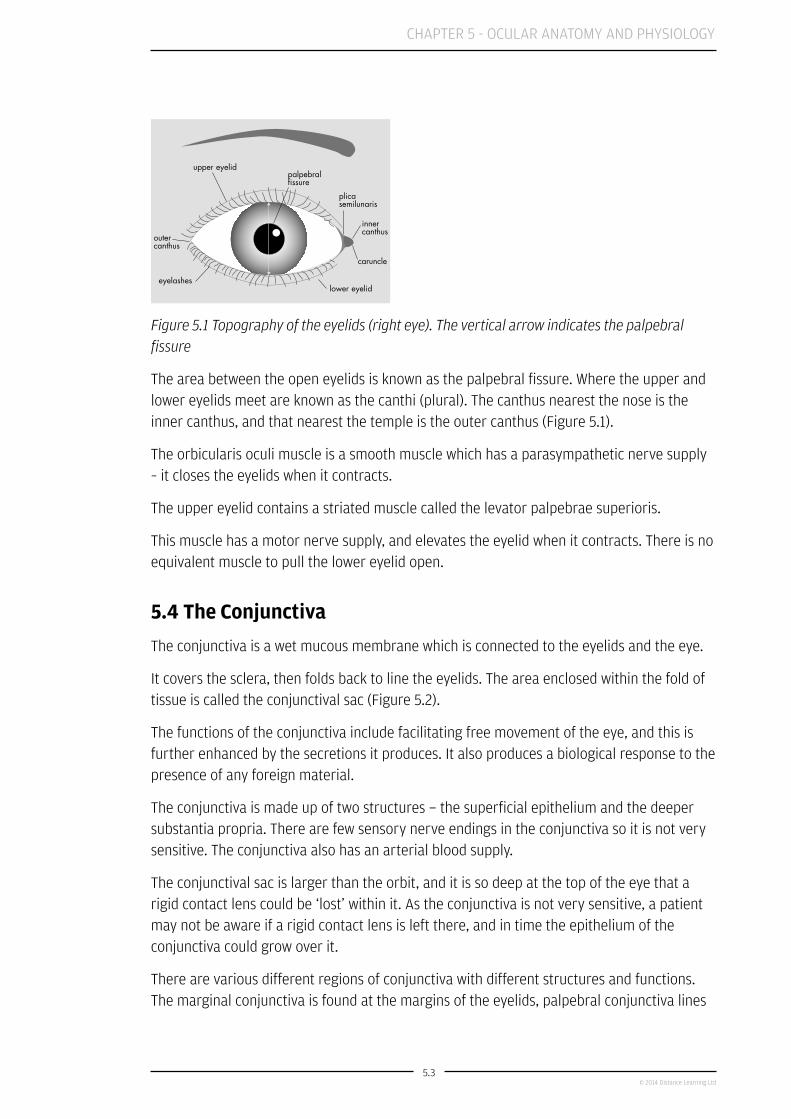
Figure 5.1 Topography of the eyelids (right eye). The vertical arrow indicates the palpebral fissure
The area between the open eyelids is known as the palpebral fissure. Where the upper and lower eyelids meet are known as the canthi (plural). The canthus nearest the nose is the inner canthus, and that nearest the temple is the outer canthus (Figure 5.1).
The orbicularis oculi muscle is a smooth muscle which has a parasympathetic nerve supply – it closes the eyelids when it contracts.
The upper eyelid contains a striated muscle called the levator palpebrae superioris.
This muscle has a motor nerve supply, and elevates the eyelid when it contracts. There is no equivalent muscle to pull the lower eyelid open.
5.4 The Conjunctiva
The conjunctiva is a wet mucous membrane which is connected to the eyelids and the eye.
It covers the sclera, then folds back to line the eyelids. The area enclosed within the fold of tissue is called the conjunctival sac (Figure 5.2).
The functions of the conjunctiva include facilitating free movement of the eye, and this is further enhanced by the secretions it produces. It also produces a biological response to the presence of any foreign material.
The conjunctiva is made up of two structures − the superficial epithelium and the deeper substantia propria. There are few sensory nerve endings in the conjunctiva so it is not very sensitive. The conjunctiva also has an arterial blood supply.
The conjunctival sac is larger than the orbit, and it is so deep at the top of the eye that a rigid contact lens could be ‘lost’ within it. As the conjunctiva is not very sensitive, a patient may not be aware if a rigid contact lens is left there, and in time the epithelium of the conjunctiva could grow over it.
There are various different regions of conjunctiva with different structures and functions. The marginal conjunctiva is found at the margins of the eyelids, palpebral conjunctiva lines

CERTIFICATE 3 FOR OPTOMETRIC CLINICAL ASSISTANTS
5.4© 2014 Distance Learning Ltd
the lids, fornical conjunctiva lines the cul de sac of the eyelid (the fornix), bulbar conjunctiva covers the ‘white of the eye’ (the sclera and episclera) and the limbal conjunctiva stops at the cornea. The fornical conjunctiva contains goblet cells which produce mucus that forms the mucin layer of the tears.
fornix
palpebral conjunctiva
cornea
marginal conjunctiva
tarsal gland
tarsal plate
skin
eyelashes
limbal conjunctiva
bulbar conjunctiva
orbital conjunctiva
Figure 5.2 Sagittal section of upper eyelid and anterior eye demonstrating the various regions of the conjunctiva
5.5 Tears and the Lacrimal Apparatus
The lacrimal apparatus consists of the lacrimal gland, the accessory lacrimal glands and the lacrimal drainage structures. The lacrimal gland is about 15mm wide, 20mm deep and 5mm thick and is located in the upper temporal (nearest the temple) region of the orbit, between the eyeball and the wall of the orbit. The lacrimal gland is almost split in two by the levator palpebrae superioris muscle of the upper eyelid (Figure 5.3). The upper portion of the lacrimal gland is known as the orbital portion and it is about twice as large as the lower, palpebral, portion.
The lacrimal gland contains structures called acini which secrete a fluid into ducts. Four to five ducts connect the orbital portion to the palpebral portion of the lacrimal gland, and these ducts, along with another six to eight from the palpebral portion, deliver the lacrimal fluid, or tears, into the conjunctival sac.
As the lacrimal gland is highly active, it has a rich blood supply from the lacrimal artery.

CHAPTER 5 - OCULAR ANATOMY AND PHYSIOLOGY
5.5© 2014 Distance Learning Ltd
Figure 5.3 Sagittal section showing location of lacrimal gland
Tears provide defence against bacteria, lubricate the eye, and provide an even and regular surface for refraction of light into the eye. Tears are composed of three layers;
1. The thin, outer lipid layer is produced by the meibomian (tarsal) glands of the eyelids, and the glands of Moll and Zeis positioned near the eyelash follicles. This outer lipid layer helps to minimise evaporation and overflow of the tears.
2. The middle, aqueous layer (do not confuse this with the aqueous humour in the anterior chamber) is produced by the lacrimal and accessory glands and provides the bulk of the tears.
3. The conjunctival goblet cells produce the inner, thin mucin layer which helps the tear film adhere to the cornea.
Tear constituents also include an antibacterial enzyme (lysozyme) and immunoglobulin A, which helps provide defence against micro-organisms.
The tears drain through small holes called puncta, one of which is found at the nasal end of the upper eyelid margin, and another at the nasal end of the lower lid margin (Figure 5.4). The puncta siphon the tears away to allow a constant turnover of tears.
The fluid is drawn into tubes called canaliculi which drain into the lacrimal sac. This drains into the nasolacrimal duct, which opens up into the nose.

CERTIFICATE 3 FOR OPTOMETRIC CLINICAL ASSISTANTS
5.6© 2014 Distance Learning Ltd
canaliculus
direction of tear flow
bone
lacrimal gland
lacrimal punctum
lacrimal sac
lacrimal punctum
nasolacrimal duct
Figure 5.4 Tear drainage structures and routes
5.6 The Cornea
anterior surface
posterior surface
cornea
anterior chamber
iris root
Fig 5.5 Comparison of anterior and posterior radius of curvature of the corneal sagittal section. Actual radii vary between individuals
5.6.1 Nutrition and Oxygen Supply
In order to be transparent and allow maximum light to reach the retina, a healthy cornea is completely avascular (has no blood vessels).
E-learning
The internal ocular structures will be introduced to you via e learning. Please log on to iLearn (www.specsaverspeople.com) and complete the e-learning for Chapter 5 titled Anatomy of the Eye.

CHAPTER 5 - OCULAR ANATOMY AND PHYSIOLOGY
5.7© 2014 Distance Learning Ltd
It therefore needs an alternative supply of oxygen and nutrients. The aqueous humour in the anterior chamber (see section 5.7, below) is the major source of nutrients for the cornea and, when the eye is closed, of oxygen. When the eye is open, the aqueous humour provides the corneal endothelium with oxygen, but the tears supply most oxygen to the cornea, as oxygen diffuses through from the atmosphere.
Contact lens wear decreases the amount of oxygen available to the cornea from the atmosphere, and overwearing contact lenses can cause oxygen deprivation of the cornea.
Ongoing oxygen deprivation may cause existing blood vessels to invade the cornea to restore its oxygen supply. This is called neovascularisation. Any such vessel growth should be avoided and, in the case of contact lens overwear, the patient may have to cease lens wear.
When the eye is closed, no atmospheric oxygen supply is available, so the vessels of the conjunctiva of the eyelid supply the corneal epithelial surface, while the aqueous humour supplies the majority of the oxygen required by the cornea.
5.6.2 Sensory Nerve Supply
The cornea has a very high sensory nerve innervation, making it one of the most sensitive tissues in the human body. Corneal sensitivity is greatest in blue-eyed people, in the central cornea, and decreases with age and with contact lens wear.
5.7 The Anterior Chambersclera
ciliary body
cornea
anterior chamber angle
canal of Schlemm
trabecular meshwork
iris
anterior chamber
Fig 5.6 Sagittal section showing the anterior chamber angle and aqueous humour drainage structures
The refractive index of aqueous humour in the normal healthy eye is between 1.333 and 1.337.

CERTIFICATE 3 FOR OPTOMETRIC CLINICAL ASSISTANTS
5.8© 2014 Distance Learning Ltd
Aqueous humour is produced by the ciliary body, from where it flows from the posterior chamber, between the iris and lens, through the pupil into the anterior chamber.
5.8 Uveal Tract
Collectively the iris, the ciliary body and the choroid are known as the uvea (uveal tract).
Clinically we can refer to the anterior uvea, which comprises the iris and ciliary body, and the posterior uvea or choroid. These three structures have similar properties in that they are highly vascular (contain many blood vessels) and they contain many melanocytes (cells that contain the pigment melanin). These pigment cells are important in absorbing any scattered light.
5.8.1 The Iris
Pigmented cells called melanocytes are found in the iris. The number of melanocytes determines the colour of an individual’s iris.
The more melanocytes there are, the darker the iris will be, hence a brown iris will contain more melanocytes in this layer than a blue iris. Flat clumps of pigment granules can be seen in some normal eyes, and these are usually iris naevi (plural) or freckles. Very occasionally a pigmented mass on the iris may be malignant (cancerous); this is a likely diagnosis if the mass is raised and has a blood vessel supply.
5.8.1.1 Pupil control
In the pupillary zone of the iris is a ring (annulus) of muscle − the sphincter pupillae muscle. This muscle is innervated by parasympathetic nerves. When these nerves are stimulated, the muscle contracts to constrict the pupil.
There is a muscle which runs the length of the iris, called the dilatator (or dilator) pupillae muscle. Sympathetic nerves supply this muscle, and nerve stimulation causes the muscle to contract and dilate the pupil.
Therefore the two muscles of the iris − the sphincter pupillae and the dilator pupillae muscles − work in opposition to each other.
Whichever muscle receives the greater nerve stimulation at a given moment will dictate whether the pupil is dilated or constricted. For example, parasympathetic nervous impulses combined with minimal sympathetic nervous activity will result in unopposed sphincter muscle contraction, constricting the pupil.
The technical term for a constricted pupil is a ‘miosed’ pupil.
Many factors result in constriction of the pupils, including bright light, focusing on close objects, eg reading material (accommodating), and certain ocular drugs called miotics, as they induce pupil miosis.

CHAPTER 5 - OCULAR ANATOMY AND PHYSIOLOGY
5.9© 2014 Distance Learning Ltd
Sympathetic nervous stimulation of the dilator muscle, unopposed by the sphincter pupillae muscle, will result in the pupil dilating. The technical term for pupil dilation is ‘mydriasis’.
The sympathetic nervous system is stimulated by frightening or threatening situations, and produces the classic ‘fright, flight or fight’ response throughout the body, including pupil dilation. Ophthalmic drugs such as mydriatics and cycloplegics result in pupil dilation.
5.8.2 The Ciliary Body
choroid
retina
ora serrata
stroma
pars plicata
pigmented epithelium
unpigmented epithelium
suspensory ligaments
crystalline lens
pars plana
ciliary muscle fibres
sclera
capillaries
cornea
iris
Fig 5.7 Ciliary body
The suspensory ligaments (or zonular fibres) insert between the ciliary processes and attach to the crystalline lens. The ciliary body is a layered structure composed of:
• The ciliary epithelium
• The stroma
• The ciliary muscle (Figure 5.7).
The ciliary muscle is supplied by the parasympathetic nerves. The ciliary muscle is stimulated during accommodation (adjusting one’s focus from a distant object to a near object).
5.8.3 The Choroid
sclera
suprachoroid
Haller’s layer
Sattler’s layer
choriocapillaris
pigment epithelium of retina
melanocytes
stroma
Fig 5.8 The choroid

CERTIFICATE 3 FOR OPTOMETRIC CLINICAL ASSISTANTS
5.10© 2014 Distance Learning Ltd
There are three main vessel beds in the choroid (Figure 5.8):
1. An outer layer of large-diameter veins, known as Haller’s layer
2. A middle layer of mainly smaller-diameter arterioles, known as Sattler’s layer
3. An inner layer of closely packed permeable capillaries, known as the choriocapillaris.
This inner layer of choroidal capillaries is responsible for providing the outer layers of the retina with nutrients and removing their waste products. Melanocytes are also found in this layer.
Bruch’s membrane is the innermost layer of the choroid, is unpigmented, and separates the choriocapillaris of the choroid from the retina.
5.9 The Lens
The lens itself is within the lens capsule, a sac-like structure suspended from the ciliary body by the suspensory ligaments (see above).
The thickness of the lens capsule varies in different regions. It keeps growing throughout the lifespan.
ciliary body
suspensory ligaments
capsule
new lens fibres
nucleus
cornea
anterior pole
posterior pole
cortex
epithelium
equator
pupil
Fig 5.9 Crystalline lens structure and location (sagittal section)
The refractive index varies in different regions of the lens.
In accommodation, the parasympathetic nervous system stimulates the ciliary muscle in the ciliary body (Figure 5.10). When the ciliary muscle contracts, tension is reduced in the suspensory ligaments, and the lens assumes its steeper convex shape, resulting in the refractive power of the lens becoming more powerful.

CHAPTER 5 - OCULAR ANATOMY AND PHYSIOLOGY
5.11© 2014 Distance Learning Ltd
Conversely, when observers move their point of focus from near to distance, parasympathetic stimulation of the ciliary muscle ceases, and the muscle relaxes. As the muscle is annular, when it relaxes tension increases in the suspensory ligaments. This tension results in the lens being flattened, and hence becoming less refractive.
suspensory ligaments under tension
t1 = thickness of lens in unaccommodated state
lens in flattened unaccommodated state
ciliary muscle relaxed
Accommodated eye
Unaccommodated eye
t1
suspensory ligaments relaxed
t2 = thickness of lens in accommodated state
lens in steeper accommodated state
ciliary muscle contracts
t2
Fig 5.10 How the eye accommodates

CERTIFICATE 3 FOR OPTOMETRIC CLINICAL ASSISTANTS
5.12© 2014 Distance Learning Ltd
Exercise 5.1 : Slit-lamp observations
The aim of this exercise is to view certain ocular anatomical structures with the use of a slit-lamp.
You are not expected to become proficient at using the slit-lamp.
You will require the assistance of an optometrist, a willing patient (preferably a member of staff) and the use of a slit-lamp. Ask the optometrist to focus the slit-lamp on the following structures:
• Eyelids
• The cornea and tear film
• The iris
• The front surface of the lens
With the aid of the course notes, observe and make annotated diagrams of as many of these structures that are visible on the slit-lamp as possible. Some details of certain structures (such as the individual layers of the cornea) mentioned in your course notes will not be visible, due to the limited magnification obtainable with a slit-lamp, whereas other details of structures (such as the crypts of Fuchs of the iris) should be easily identified on most patients.
5.10 The Retina
By using an ophthalmoscope or fundus camera, an optometrist can view the various structures of the retina, including arteries and veins, the macula and the optic nerve head (Figure 5.11). The blood vessels branch: veins branch into smaller venules and then capillaries, and arteries branch into smaller arterioles and then capillaries. Capillaries are vessels with a very small diameter and walls only one cell thick. Certain diseases, such as high blood pressure and diabetes, affect blood vessels. As the retina is the only place in the body where blood vessels can be seen directly, ophthalmoscopic examination of the retina is a useful tool to detect pathological blood vessel changes.

CHAPTER 5 - OCULAR ANATOMY AND PHYSIOLOGY
5.13© 2014 Distance Learning Ltd
optic disc
branch of central retinal vein
branch of central retinal artery
arteriole
venule
capillary
foveola
Fig 5.11 Retina of right eye as seen with an ophthalmoscope
5.10.1 Photoreceptors
The photoreceptors are specialised cells which have an inner and an outer segment. The outer segments contain discs, within which are the visual pigments which absorb light. When the visual pigments absorb light of the appropriate wavelength, they become ‘bleached’, and the neural process of vision begins.
There are two types of photoreceptor in the human retina, called rods and cones. Their names are derived from the shape of the outer segment of the two types of cell. The outer segments of the rods are elongated and rod-shaped, whereas the outer segments of the cones are roughly conical. There are more rods than cones, and the density of each receptor types varies in different regions of the retina.
The central region of the retina is known as the macula, and at the centre of the macula is a pit called the foveola which has the highest density of cones and no rods (Figure 5.11). Here the cones are closely packed and modified in shape.
Away from the macula towards the peripheral retina, the density of cones decreases and the density of rods increases.
Cones and rods have different functions. Cone receptor cells are used in resolving fine detail, whereas rods are poor at resolving detail.
Cones operate in high light levels (photopic light conditions), whereas rods operate in low light (scotopic) conditions.
There are three different cone photoreceptors in a normal human eye. Each contains a different photopigment, sensitive to light of different wavelengths. If one or more type of cone photoreceptor is missing, or contains an abnormal photopigment, this results in a

CERTIFICATE 3 FOR OPTOMETRIC CLINICAL ASSISTANTS
5.14© 2014 Distance Learning Ltd
colour deficiency. Note that a patient is described as being ‘colour-deficient’ rather than ‘colour-blind’, because it is very rare for a patient to be totally blind to all colours, ie to have no functioning cone receptors. Usually any abnormality results in a confusion of only certain colours, such as reds and greens.
A common type is a red−green colour deficiency, which affects approximately 8% of the male population but only about 0.4% of the female population. This type of defect is inherited, and a male only needs to inherit one faulty colour vision gene either from his father or mother to exhibit this type of deficiency. In contrast, a female has to inherit two faulty colour vision genes, one from both her father and her mother, in order to have a red−green colour deficiency, hence the fact that far fewer females have this type of deficiency Blue−yellow colour vision deficiencies are much rarer than the red−green type, and tend to be acquired rather than inherited (that is, the person is not born with the deficiency, but acquires it later in life). Certain drugs can cause a blue−yellow deficiency, as can certain diseases such as multiple sclerosis. There are numerous colour vision tests available.
Exercise 5.2: Fundus observations
The aim of this exercise is to view certain ocular anatomical structures with the use of a fundus camera.
You will require the assistance of an optometrist and a selection of fundus photographs. Ask your supervising optometrist to explain the fundus photograph and point out the following structures:
• The optic disc
• The retinal blood vessels
• The macula
If available compare a healthy fundus with an abnormal fundus and have the Optometrist explain any pathology.
5.11 The Optic Nerve
The ganglion cell axons form the optic nerve.
As they leave the eye through the posterior scleral foramen, they pass through a sieve-like structure, called the lamina cribrosa, which bows slightly backwards (Figure 5.12). The lamina cribrosa is composed of collagen, and is an extension of part of the sclera. It contains approximately 200 pores (holes) which allow bundles of nerve fibres to pass through. Its function is to provide protection for the delicate nerve fibres as they pass out of the eye.

CHAPTER 5 - OCULAR ANATOMY AND PHYSIOLOGY
5.15© 2014 Distance Learning Ltd
thickening of nerve fibre layer
unmyelinated nerve fibres
central retinal vein
central retinal artery
bundles of myelinated optic nerve fibres
pia materarachnoid materdura mater
themeninges
lamina cribrosa
sclera
retina choroid
Fig 5.12 Optic nerve head - sagittal section)
As the nerve fibres pass beyond the lamina cribrosa, they become myelinated (insulated by a white coating called myelin). The function of myelin is to prevent nerve impulses ‘short-circuiting’ between adjacent nerve fibres, restricting the nerve impulse to pass along the nerve fibre.
The optic nerves are surrounded by meningeal sheaths which are continuous with the meninges (membranes) of the brain. The meninges are made up of three layers: the dura mater, arachnoid mater and pia mater (Figure 5.12).
5.12 The Visual Pathway
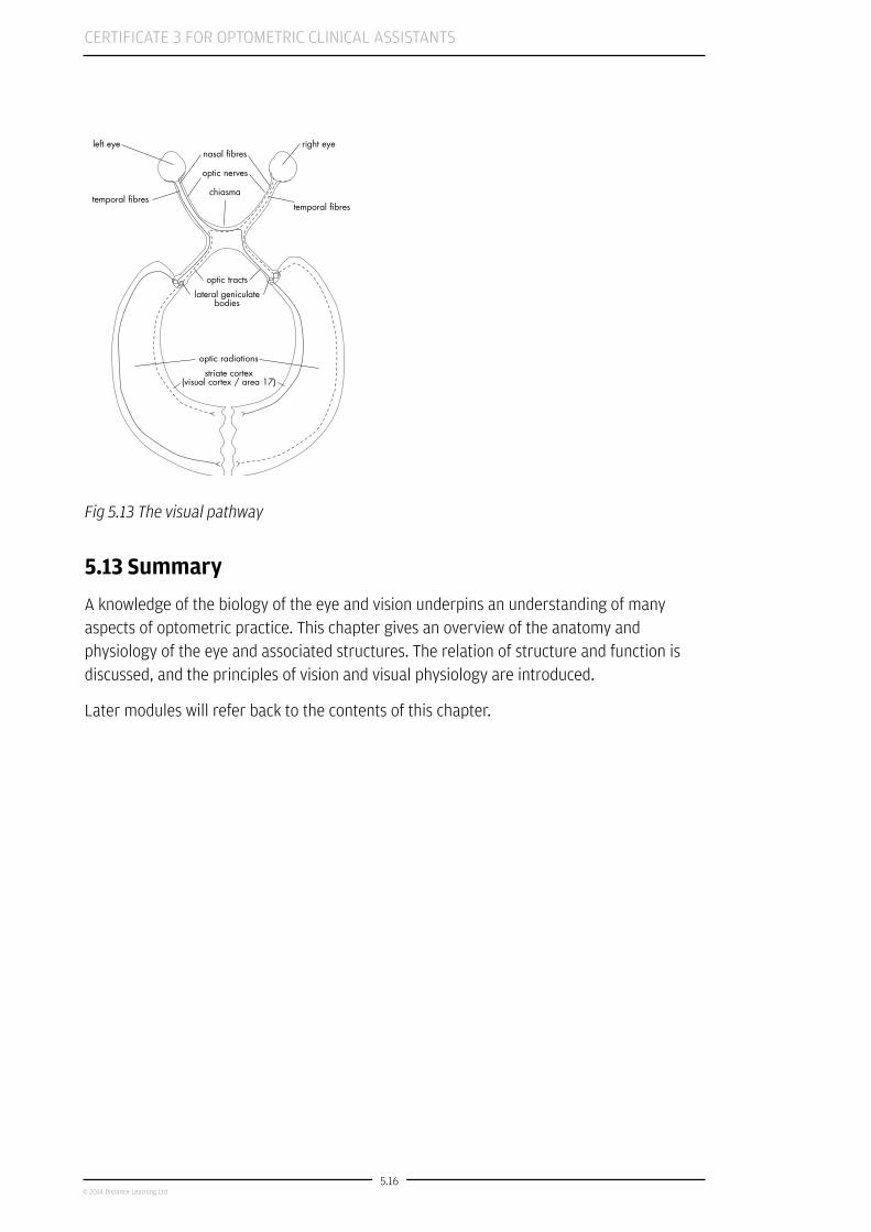
Each optic nerve passes away from the eye and through the optic canal in the bony orbit. Both optic nerves meet at the chiasma (Figure 5.13). Here the ganglion cell axons from the temporal retina of both eyes remain on the same side, and continue into the optic tracts. That is, the temporal ganglion cell axons of the right eye pass down the right optic tract, and the temporal ganglion cell axons of the left eye pass along the left optic tract.
In contrast, the ganglion cell axons from the nasal regions of the retinas of both eyes cross over at the chiasma. Therefore, only half of the nerve fibres from the eyes cross over at the chiasma.
The nerve fibres continue down the optic tracts until they reach the lateral geniculate nuclei, also known as the lateral geniculate bodies. Here, the nerve fibres synapse with other nerves that form the optic radiations.
The nerve fibres in these optic radiations pass through the brain until they reach the visual cortex of the brain. This region is also known as the striate cortex or area 17 (Figure 5.13).
This is where visual information is processed, and is found at the back of the cortex of the brain.
CERTIFICATE 3 FOR OPTOMETRIC CLINICAL ASSISTANTS
5.16© 2014 Distance Learning Ltd
left eye right eye
temporal fibrestemporal fibres
optic tractslateral geniculate
bodies
optic radiationsstriate cortex
(visual cortex / area 17)
nasal fibres
optic nerves
chiasma
Fig 5.13 The visual pathway
5.13 Summary
A knowledge of the biology of the eye and vision underpins an understanding of many aspects of optometric practice. This chapter gives an overview of the anatomy and physiology of the eye and associated structures. The relation of structure and function is discussed, and the principles of vision and visual physiology are introduced.
Later modules will refer back to the contents of this chapter.

CERTIFICATE 3 FOR OPTOMETRIC CLINICAL ASSISTANTS
6.1© 2014 Distance Learning Ltd
Chapter 6
CERT 3 CL OCA Semester 1 2014
Common Pathological Ocular ConditionsIntroduction
• By the end of this module you will be expected to possess the ability and understanding:
• To recognise when a patient may have an ocular emergency
• To know the correct procedure if an ocular emergency is suspected
• To communicate appropriately with a patient if an ocular emergency is suspected
• About the basic principles of common eye diseases
• To understand the significance of intraocular pressure
• To be aware of risk factors in certain eye diseases
• To understand common causes of blindness and partial sight
• To be aware that you must never to attempt to make a diagnosis when dealing with a patient
• To understand the ocular effects of common systemic conditions
E-learning
This chapter will be delivered in the form of E-learning. Please log on to iLearn (www.specsaverspeople.com) and complete the e-learning for Chapter 6 entitled Ocular Conditions. Be sure to take notes as you work through the module.

CERTIFICATE 3 FOR OPTOMETRIC CLINICAL ASSISTANTS
6.2© 2014 Distance Learning Ltd
6.1 Additional Information on Ocular Conditions
6.1.1 Red Eye
Red eye is the cardinal sign of ocular inflammation. The condition is usually benign and can be managed by optometrists or general practitioners. Conjunctivitis is the most common cause of red eye. Other common causes include corneal abrasion, foreign body, subconjunctival hemorrhage, keratitis, iritis, glaucoma, and scleritis.
Signs and symptoms of red eye include eye discharge, redness, pain, sensitivity to light, itching, and visual changes. Generally, viral and bacterial conjunctivitis are self-limiting conditions, and serious complications are rare. Because there is no specific diagnostic test to differentiate viral from bacterial conjunctivitis, most cases are treated using broad-spectrum antibiotics. Allergies or irritants also may cause conjunctivitis. The cause of red eye can be diagnosed through a detailed patient history and careful eye examination, and treatment is based on the underlying cause.
Recognising the need for emergency referral to an ophthalmologist is key in the optometric management of red eye. Referral is necessary when the patient is in severe pain, topical steroids may be needed, or the patient has vision loss. A significant thick discharge, corneal involvement, trauma to the eye, recent eye surgery, or recurrent infections would also warrant emergency evaluation.
6.1.2 Cataract
Cataracts are a very common eye condition. As we get older the lens inside our eye gradually changes and becomes less transparent (clear). A lens that has turned misty, or cloudy, is said to have a cataract. Over time a cataract can get worse, gradually making your vision mistier. A straightforward operation can usually remove the misty lens and replace it with an artificial lens to enable you to see more clearly again.
Cataracts result from changes in the way the cells of the lens are arranged. When this happens, light cannot pass directly through the lens and you may notice problems with your vision. A cataract is not a growth or a film growing over the eye, it is simply the lens becoming cloudy.
6.1.2.1 Causes of cataract
Cataracts can be caused by a number of things, but by far the most common reason is growing older. Most people over the age of 65 have some changes in their lens and most of us will develop a cataract in time. Apart from getting older, the other common causes of cataract include:
• diabetes
• trauma
• medications, such as steroids
CHAPTER 6 – COMMON PATHOLOGICAL OCULAR CONDITIONS
6.3© 2014 Distance Learning Ltd
• eye surgery for other eye conditions
• other eye conditions.
6.1.2.2 Symptoms
When a cataract is starting to develop a patient will usually say that their vision is becoming less clear.
Another common symptom of a cataract is a problem with bright lights. Lights can seem to glare, or you may find that the headlights of a car dazzle you more than they used to. You may also notice a slight change in your colour vision - things may appear more yellow than before. This often happens if one eye develops a cataract first and colours look different when you compare one eye with the other.
6.1.2.3 Treatment
The operation to remove a cataract can be performed at any stage of their development. There is no longer a reason to wait until a cataract is “ripe” before removing it. However, because any surgery involves some risk, it is usually worth waiting until there is some change in vision before removing the cataract.
Cataracts are not ocular emergencies and are usually referred routinely by a optometrist to an eye specialist. The optometrist will discuss with the patient the nature of their symptoms and the affect they are having on day to day activities like driving and reading. They will then have a consultation with a surgeon who will discuss the procedure itself as well as the pros and cons of surgery.
To remove the cataract, the eye specialist removes the natural lens from the eye and replaces it with a plastic lens (implant). The most common way to remove cataracts is called phacoemulsification. This technique uses high frequency sound energy to break up your natural lens with the cataract. Very small incisions are used, so no stitches are required.
The lens inside the eye is made up of different layers and the outside layer is called the lens capsule. During the operation, the eye specialist cuts through the front of the lens capsule so they can reach the lens inside. Using the same instrument, the eye specialist can break up the lens and the cataract inside the eye, and remove it using suction. The lens capsule is kept in place so that the artificial lens implant can be placed inside it. The tiny implant is folded so that it can be put into the eye through the same instrument that is used to remove the cataract. Once it reaches the right position, the eye specialist unfolds the artificial lens so that it sits in the right place inside the lens capsule.
Cataract surgery is generally very successful. Only about three per cent of people who have cataracts experience complications. The most common complications can be dealt with and usually don’t affect sight in the long term.
One of the most common complications is clouding of the capsule which holds the implant in place. This may occur a couple of months or even years after the original operation. If

CERTIFICATE 3 FOR OPTOMETRIC CLINICAL ASSISTANTS
6.4© 2014 Distance Learning Ltd
this happens, your sight will become cloudy again, as though the cataract has come back. Optometrists call this complication posterior capsule opacification and it is treated using a laser.
More serious complications are much rarer and include:
• retinal detachment
• problems with the lens implant, the wrong lens implant or problems with its position
• a break in the lens capsule
• infection
6.1.3 Glaucoma
Glaucoma is a disease in which the optic nerve slowly dies, if untreated it will eventually result in blindness.
One of the tests we use to help us detect glaucoma is checking patients’ eye pressures but it may surprise you to hear that in half of all newly diagnosed cases of glaucoma the eye pressure is normal. For this reason, the optometrists in your store will use a combination of eye pressure, visual field results and examination of the optic nerves to try to identify patients with the disease. Even when combining the results of all three tests, early glaucoma is very difficult to detect, it is the most difficult part of an optometrists job on a day to day basis.
Glaucoma tends to affect elderly patients but it is not a very common disease affecting only 2% of patients over the age of 40. We know it is more common in those with a family history (particularly if a sibling has the disease). It is also more common in afro-Caribbeans and patients with high eye pressures.
The most effective way to diagnose glaucoma is to examine the optic nerve head closely. In patients with glaucoma the central, white, portion of the nerve which we call the optic cup tends to get larger. Hopefully this was apparent on the images you looked at in the presentation.
To complicate things further it is not uncommon for some patients to have what we call physiological cupping. This is the appearance of a large white area in the middle of the nerve without any disease being present.
I have already mentioned that half of all newly diagnosed glaucoma patients have normal pressure, this is taken to be a pressure reading less than 21mm Hg. Having said that, patients with high pressures are still known to be at an increase risk of developing the disease. This may sound confusing but what we are basically saying is that high eye pressures are bad but low pressures are not necessarily good. Under NICE guidelines optometrists must refer to an eye specialist all patients under the age of 65 with eye pressures greater than 21mm Hg. This has caused significant problems to the hospital eye service who struggle to cope with the high volume of referrals.

CHAPTER 6 – COMMON PATHOLOGICAL OCULAR CONDITIONS
6.5© 2014 Distance Learning Ltd
Most of you will have carried out visual field tests on patients with high pressures or suspicious optic nerve heads. Glaucoma causes visual field loss close to but just outside the central region. Most glaucoma field tests screen in the central 30 degrees of vision. Patients find visual field tests very difficult and the results are often poor. For this reason an optometrist will often ask for a field test to be repeated to see if any defect detected is still present on re-testing.
There is only one effective way of treating glaucoma and that is by using eye pressure lowering drops. Even if the patient had normal pressures to begin with the goal of the doctor will still be to lower the pressure even further. Most modern glaucoma drugs are designed to be instilled only once a day but if these are ineffective patients often end up taking a number of different drops. Laser treatments and surgical options are also available.
6.2.4 Retinal Detachment
Retinal detachment occurs when the thin lining at the back of your eye called the retina begins to pull away from the blood vessels that supply it with oxygen and nutrients.
Symptoms of retinal detachment can include the following:
• floaters – black dots that float across your field of vision. There can be multiple small floaters or a single large one.
• flashes of light. These last no more than a second.
• blurring or distortion of vision.
• a shadow or “black curtain”
The most common cause of retinal detachment is when tiny holes or tears develop.
The holes allow the fluid found in the vitreous chamber to leak underneath the retina.
If too much fluid builds up it can cause the retina to pull away from the blood vessels that supply it with blood and the nerve cells inside the retina will begin to die.
Myopia is a major risk factor for age-related retinal detachment (though in relative terms the risk is still very small) because myopes tend to have thinner retinas.
In some cases, tears can develop if the eye is suddenly injured.
If an optometrist suspects a retinal detachment, it is normal for the patient to be referred to an eye specialist urgently.
6.1.4.1 Treatment
The quicker retinal detachment is treated, the less risk there is of the patient permanently losing some or all of their vision in the affected eye.

CERTIFICATE 3 FOR OPTOMETRIC CLINICAL ASSISTANTS
6.6© 2014 Distance Learning Ltd
Most detached retinas can be successfully reattached with surgery. There are a number of different types of surgery available.
• Pneumatic retinopexy
If the detachment is relatively small and uncomplicated, a procedure called pneumatic retinopexy may be used. This involves injecting a small bubble of gas into the eye, which presses the retina back into place.
Laser or freezing treatment is often then used to create scar tissue that keeps the retina in the correct place. The bubble is slowly absorbed into the eye over the following weeks.
After the procedure, you will be asked to keep your head in a certain position for a while, known as “posturing”, so the bubble settles in the correct position.
If the patient has had a gas bubble put in their eye, they not be able to travel by air for a while.
• Scleral buckling
Scleral buckling involves fine bands of silicone rubber or sponge that are stitched onto the sclera in the area where the retina has detached. The bands act like a buckle and press the sclera in towards the middle of the eye, so the torn retina can lie against the wall of the eye.
Laser or freezing treatment is used to scar the tissue around the retina, which creates a seal between the retina and the wall of the eye and closes up the tear or hole.
The bands can be left on the eye and should not be noticeable after the operation.
• Vitrectomy
Vitrectomy works by removing the fluid from the inside of the eye and replacing it with either a gas or silicone bubble. This holds the retina in position from the inside.
A vitrectomy may be recommended if the fluid in front of the eye is unusually thick and dense and is pulling the retina away from the underlying blood vessels.
As with a pneumatic retinopexy, posturing will be necessary to ensure the bubble is in the right place. The same restrictions on flying and precautions for further surgery that apply to pneumatic retinopexy also apply to vitrectomy.
6.1.5 Diabetic Retinopathy
Diabetic retinopathy is a common complication of diabetes. It occurs when high blood sugar levels damage the cells at the back of the eye, known as the retina. If it is not treated, it can lead to blindness.

CHAPTER 6 – COMMON PATHOLOGICAL OCULAR CONDITIONS
6.7© 2014 Distance Learning Ltd
Therefore, it is important for people with diabetes to keep their blood sugar levels under control. Everyone with diabetes who is 12 years old or over should have their eyes examined once a year for signs of damage (see below).
To work effectively, the retina needs a constant supply of blood, which it receives through a network of tiny blood vessels.
Over time, a continuously high blood sugar level can cause the blood vessels to become blocked or to leak. This damages the retina and stops it from working
Over the course of many years, the blood vessels can be damaged by high blood sugar (glucose) levels that may be present in people with poorly controlled diabetes.
During the initial stages of retinopathy, the damage is limited to tiny bulges (microaneurysms) in the blood vessel walls. Although these can leak blood and fluid, they do not usually affect your vision.
Gradually over time, the blood vessels that supply the most sensitive part of the retina, called the macula, can become damaged. The macula enables you to distinguish colours and focus your eyes for tasks such as reading and writing.
If fluid leaks into the macula, it can cause swelling leading to some loss of vision.
When retinopathy reaches its most advanced stage, some of the blood vessels that supply the retina will become blocked. To compensate for this, new blood vessels will start to form in an attempt to restore the supply of blood.
However, as the new blood vessels are unstable and prone to bleeding, they can lead to blurred and patchy vision because the bleeding obscures your sight.
Over time, the bleeding can lead to the formation of scar tissue which can cause a retinal detachment
6.1.5.1 Risk factors
Several factors increase your risk of developing diabetic retinopathy.
1. Length of time you have had diabetes The longer you have had diabetes, the greater your chance of developing retinopathy.
About 90% of people with type 1 diabetes will have some degree of retinopathy after 10 years of having diabetes symptoms.
For people with type 2 diabetes who do not need to take insulin, about 67% will have some degree of retinopathy after 10 years of having diabetes symptoms.
2. Blood glucose level If you have diabetes and your blood glucose level is high, you have a higher risk of developing retinopathy.

CERTIFICATE 3 FOR OPTOMETRIC CLINICAL ASSISTANTS
6.8© 2014 Distance Learning Ltd
3. High blood pressure If you have diabetes and high blood pressure, your risk of developing advanced retinopathy is increased.
Therefore, taking steps to prevent high blood pressure, such as giving up smoking and cutting down the amount of salt in your diet, can help reduce your risk of developing retinopathy.
6.1.5.2 Symptoms of diabetic retinopathy
During the initial stages, retinopathy does not cause any noticeable symptoms. The patient may not realise that their retina is damaged until the later stages, when their vision becomes affected.
Possible symptoms of late-stage retinopathy include:
• shapes floating in your field of vision (floaters)
• blurred vision
• reduced night vision
• sudden blindness
6.1.5.3 Screening for diabetic retinopathy
As retinopathy can cause blindness, it is very important that it is identified and treated as early as possible.
The NHS Diabetic Eye Screening Programme aims to reduce the risk of vision loss in people with diabetes. This is done by identifying retinopathy at an early stage and, if necessary, ensuring that appropriate treatment is given.
Everyone with diabetes who is 12 years of age or over is invited for screening once a year.
6.1.5.4 Treating diabetic retinopathy
Treatment for retinopathy will depend on the stage the condition has reached.
For example, if retinopathy is identified in its early stages, it may be possible to treat it by controlling your diabetes more effectively.
If you have more advanced retinopathy, you may need to have laser surgery to prevent further damage to your eyes.
6.1.5.5 Preventing diabetic retinopathy
To reduce your risk of developing retinopathy, it is important to control your blood sugar level and keep your blood pressure as close to normal as possible.
Other steps that you can take to help prevent retinopathy include:
CHAPTER 6 – COMMON PATHOLOGICAL OCULAR CONDITIONS
6.9© 2014 Distance Learning Ltd
• attending your annual screening appointment
• informing your GP if you notice any changes to your vision (do not wait until your next screening appointment)
• taking your medication as prescribed
• losing weight (if you’re overweight) and eating a healthy, balanced diet
• exercising regularly
• giving up smoking (if you smoke)
6.1.6 Age Related Macular Degeneration
Age related macula degeneration (AMD) is a very common and potentially blinding condition that affects a tiny part of the retina at the back of your eye, which is called the macula. AMD causes problems with central vision, but does not lead to total loss of sight and is not painful. Traditionally it has been divided into two types, dry and wet.
6.1.6.1 Types of AMD
Dry AMD is characterised by the presence of small yellow/white lesions at the macula we call drusen. Hopefully you have seen these in the images in the presentation. These drusen result from a build up of waste products from the retinas light receiving cells. Over time they get larger and eventually cause significant scarring and severe visual loss. About 85-90% of AMD is dry the remaining 10-15% is the wet type.
Wet AMD is characterised by the presence of new blood vessels which grow inside and beneath the retina. These vessels have very weak walls and can leak causing devastating rapid visual loss. Hopefully you will have seen how this appears in the image in the presentation.
6.1.6.2 Risk factors for AMD
The following are the most common risk factors for AMD:
• Increasing age: it occurs in 0.2% of the population aged 55-64 years increasing to 13% of those aged 85 or older.
• Gender: more women have AMD than men, probably because women tend to live longer than men.
• Genes: some genes have been identified which seem to be linked to the development of AMD in some people. This has been discovered by looking at families with more than one member who has AMD, but not all AMD is thought to be inherited.
• Smoking: smoking greatly increases your risk of developing AMD. Studies also show that stopping smoking can reduce your risk of developing AMD.

CERTIFICATE 3 FOR OPTOMETRIC CLINICAL ASSISTANTS
6.10© 2014 Distance Learning Ltd
• UV radiation: some studies have shown that exposure to high levels of UV throughout your life may increase your risk of developing AMD.
• Diet: a number of studies have looked at diet as a risk factor for someone developing AMD. At the moment there isn’t agreement on how much of a risk factor diet is. There is some evidence that vitamins A, C and E and zinc may help to slow the progression of AMD in people who already have the condition.
6.1.6.3 Signs and symptoms of AMD
Symptoms of AMD can vary from person to person, but usually the first problems people notice are with their ability to see detail. This especially affects a person’s ability to read small print, watch television and recognise faces. Some patients will describe their vision as being distorted and straight lines appearing wavy.
With the dry type of the disease visual loss is gradual over a number of years, with the wet type it can be lost very quickly, in a matter of weeks.
6.1.6.4 Treatment
A number of treatments are available for wet AMD. These mainly work by stopping the growth of new blood vessels. This means that treatments usually need to be given fairly quickly once the blood vessels start to grow in your eye. If the blood vessels are allowed to grow for too long the blood vessels may scar the retina and this scarring cannot be treated.
At the moment there is no treatment for dry AMD. This is because dry AMD doesn’t involve new blood vessels growing. Although research is continuing to find a treatment for dry AMD, nothing is available yet.
There is some evidence that high doses of vitamin A, C, E and the minerals zinc and copper when taken together may help slow down the progression of dry AMD, particularly if someone already has changes to their vision because of AMD in one eye.
There are a number of vitamin products available which have been designed for people with dry AMD and you can usually buy these over the counter from your pharmacist. However, there is no evidence that taking high doses of these vitamins can prevent you developing AMD in the first place. A balanced diet with plenty of fresh fruit and vegetables is good for your general health and may also help your eye health.
Patients with wet or dry AMD may benefit from consulting a low vision practitioner who will discuss magnifiers and other low vision aids designed to maximize their remaining vision.
6.1.7 Retinitis Pigmentosa
Retinitis pigmentosa (RP) is an inherited, degenerative eye disease that causes severe vision impairment and often blindness The progress of RP is not consistent. Some people will exhibit symptoms from infancy, others may not notice symptoms until later in life. Generally, the later the onset, the more rapid is the deterioration in sight.

CHAPTER 6 – COMMON PATHOLOGICAL OCULAR CONDITIONS
6.11© 2014 Distance Learning Ltd
All types of RP affect the retina. The retinal cells gradually stop working and eventually die. In most cases, the peripheral rod cells are affected first and RP later affects the central cone cells. The symptoms you experience depend on the way your retina is affected by RP and can be very different from person to person.
Almost all types of RP are inherited, caused by a fault in the genetic information passed down from a parent. In RP, the faulty genes cause the retinal cells to stop working and eventually die.
As there are many genes that can cause the retinal cells to stop working, there are many different types of RP. This is why RP is described as a group of inherited retinal disorders..
RP can also be associated with other problems such as hearing loss. These rare conditions are referred to as RP syndromes.
6.1.7.1 Modes of inheritance
• Autosomal dominant inheritance Autosomal dominant RP affects men and women equally and there tends to be a known history of the condition in the family. This form of RP is less severe than the other two listed below and the first signs of it tend to appear at around 30 years of age.
• Autosomal recessive inheritance Autosomal recessive RP also affects men and women equally but there may be little or no known history of the condition in either family in the past. This form of RP tends to show first signs between 30 and 40 years and tends to cause more severe sight loss.
• X-linked inheritance This is a pattern of inheritance that affects mostly men. Female members of a family are carriers of the faulty gene but rarely develop the full condition, although some carriers can develop a mild form of RP. If there have been no boys in the family in the last few generations then there may be no history of the condition. This type of RP affects vision severely and can result in very poor vision by the age of 40.
• No known relative In about half of diagnosed cases of RP there does not seem to be any previously affected relatives. Relatives will have passed on the faulty genetic information but may have not developed symptoms themselves. In such cases it may not be possible to determine which of the three types of inheritance have caused the RP.
6.1.7.2 Symptoms
There might be some difficulty seeing in low light such as outdoors at dusk or in a dimly lit room. The visual field is also reduced and sight loss can be from above and below. This is often referred to as tunnel vision it means that the rod cells and some of the outer cone cells have been affected first. In some RP related conditions central vision is lost first and the person affected can have difficulty reading print or doing detailed work. In many types

CERTIFICATE 3 FOR OPTOMETRIC CLINICAL ASSISTANTS
6.12© 2014 Distance Learning Ltd
of RP the glare from bright lights can cause a problem although some people do not suffer from this until the condition has developed.
How is it diagnosed?
Two tests are essential in the diagnosis:
Visual field testing will find defects in the peripheral vision, over time, the visual field may reduce to a small central island of vision causing “tunnel vision.” The final progression may be the complete loss of the remaining central vision.
Electrophysiological testing by an ophthalmologist is often diagnostic. Responses to flashes of light are measured via electrodes placed on the surface of the eye. It is a painless test. The electroretinogram (ERG), in conjunction with the visual field exam, will usually confirm the diagnosis. This will also determine if there is any cone involvement.
Recently, gene testing for defects is being done to clarify the basic cause for RP and assist in ultimately finding a treatment.
6.1.7.3 Treatment
There is no specific cure for retinitis pigmentosa though some eye specialists recommend particular dietary supplements. Research is currently being conducted into the use of stem cells from mice to restore some retinal function.
Low vision services may be beneficial to help patients to maximize the remaining vision.
6.1.8 Dry Eye
Dry eye disease occurs when the eyes do not make enough tears, or the tears evaporate too quickly. This leads to the eyes drying out and becoming inflamed and irritated.
The symptoms of dry eye syndrome can be mild or severe. They include:
• dry or sore eyes
• blurred vision
• the feeling of something in your eye
• burning
• watering
• Dry eye disease can have a number of causes, including:
• being in a hot or windy climate
• certain chronic diseases
• side effects of medicines
CHAPTER 6 – COMMON PATHOLOGICAL OCULAR CONDITIONS
6.13© 2014 Distance Learning Ltd
• hormonal changes
• getting older (up to a third of people aged 65 or older may have dry eye syndrome)
• contact lenses
6.1.8.1 Diagnosis
There are a number of tests which an optometrist may want to do to work out if you have dry eye and if you do, how dry your eyes are. These tests help them decide how to treat your eyes. The tests check how many tears you produce and detect any areas on the front of your eye that don’t have enough tears.
As well as examining the front of your eyes and the quality of the tears with a slit lamp, there are three other tests your optometrist or ophthalmologist may want to do:
1. Tear film break-up time This test finds out how long after blinking your eye starts to dry out. The optometrist first instills fluorescein dye which makes the tears easier to observe. They then ask you to blink a number of times to make sure that the dye is in your tears properly. They will then ask you to stop and keep your eyes open without blinking.
The optometrist then uses a blue light to see the dye and times the period between your last blink and the formation of dry patches. The dry patches are shown up by the dye. If your eyes start to show patches of dryness before ten seconds it usually means that there is some evidence of a dry eye. The dye does not change the colour of your eye and only stays in your eye for a short while.
2. Lissamine green staining This test uses a different dye, which makes damaged tissue on the front of your eye easier to see. Sometimes the front of your eye can be slightly damaged in the dry patches.
3. Phenol red test This is a test in which a special thread is used to measure tear volume. The thread is hooked over the lower eyelid and left in the eye for about fifteen seconds. As the tears flow down the thread it changes colour from red to yellow. After this the optometrist is able to see how much tears the eye produces in that period.
6.1.8.2 Complications of dry eyes
Generally, dry eyes don’t cause serious problems. However, possible complications include:
1. More-frequent eye infections. Your tears protect the surface of your eyes from infection. Without adequate tears, you may have an increased risk of eye infection.
2. Scarring on the surface of your eyes. If left untreated, severe dry eyes may lead to eye inflammation, scarring on the surface of your corneas and vision problems.

CERTIFICATE 3 FOR OPTOMETRIC CLINICAL ASSISTANTS
6.14© 2014 Distance Learning Ltd
3. Decreased quality of life. Dry eyes can make it difficult to perform everyday activities.
6.1.8.3 Treatment
There are three main ways to help your dry eye:
1. Making the most of your natural tears There are things that you can do yourself that may help reduce the symptoms of dry eye. You can often lower the temperature in a room because high temperatures and central heating can make tears evaporate more quickly. However, you need to make sure that you keep yourself comfortable. A humidifier is a small machine that helps put more water into the air, which may help slow down the evaporation of your tears.
Many people find that their dry eye is more uncomfortable when they’re reading or using a computer. This is usually because you tend to blink less when you are doing this sort of thing, which gives the tears more chance to evaporate. You can try to blink more when you’re doing these tasks or use eye drops before you do anything, like reading, as this may help to keep your eyes comfortable.
2. Using eye drops Most people with dry eye need to use some form of eye drops, also known as “artificial tears”. Eye drops aim to supplement and replace your natural tears and make the eye more comfortable. They can also prevent any damage to the front of your eye, which can happen if the eye is dry for a long time.
Eye drops don’t contain any drugs, they are just replacement tears. This means that they can be used frequently, or as much as you need them. However if you are having to use your drops more than 4 or 6 times a day then you should let your ophthalmologist or optometrist know as you may need a different treatment to the drops you’re using.
There are three main types of eye drops which an optometrist may recommend:
a) Artificial tears
Many different companies make artificial tears. Most artificial tears can be bought over the counter from the pharmacist. If you’re entitled to free prescriptions, or have a prepayment certificate, you can ask your doctor to prescribe them. Some people develop sensitivity to the preservative used in the drops, especially if they’re using them a lot. This can make your eyes sore. Preservative-free drops are available.
b) Gels
If your standard eye drops aren’t helping, the optometrist may suggest thicker gel-like drops which are made from different chemicals and may last longer in the eye. They do the same thing as the ordinary drops but you don’t have to put them in as often.

CHAPTER 6 – COMMON PATHOLOGICAL OCULAR CONDITIONS
6.15© 2014 Distance Learning Ltd
c) Ointments
Ointments are also available to help keep your eye moist overnight. When you sleep, sometimes your eyes aren’t fully closed, so tears can evaporate and leave your eyes very dry when you wake up. Ointments help stop the eyes drying out overnight so that they feel more comfortable in the morning. Ointments tend to be used overnight because they are sticky and cause blurry vision. Ointments are usually used as well as eye drops during the day.
3. Reducing the draining away of tears It is possible to help dry eye by blocking up the two drainage holes in your lower eyelids. Stopping the tears from draining away may help your tears to stay in your eye for longer. The medical term for blocking the tear ducts is punctal occlusion.
Usually, punctal occlusion is tried for a period of time to see if it helps. The small drainage channels are blocked by little pieces of plastic called punctal plugs. If it helps you with the symptoms of dry eye, the plugs are left in place. Sometimes blocking the ducts can cause the eye to over-water or you may experience infections. If this happens, then the plugs can be removed. If you have had your tear ducts blocked you may still need to use drops, gels or ointments to protect your eyes and keep them as comfortable as possible.
There is some debate on whether or not diet helps with reducing the symptoms. In particular, certain oils, omega 3 and 6 are thought to help with dry eye. However, there isn’t any large scale evidence that taking these supplements will help you.
6.1.9 Corneal Ulcers
Corneal ulcers are a sign of inflammation within the cornea (keratitis) and are most commonly caused by an infection with either bacteria, viruses, fungi, or a parasite.
Pseudomonas keratitis can result from contact lens wear, in particularly in patients who sleep in soft lenses
Acanthamoeba keratitis occurs in contact lens users, especially in people who expose their lenses to water from a tap or a swimming pool.
Fungal keratitis can occur after a corneal injury involving plants or soil, or in people with a suppressed immune system.
Herpes simplex keratitis is a serious viral infection. It may cause repeated attacks that are triggered by stress, exposure to sunlight, or any condition that impairs the immune system.
Corneal ulcers or infections may also be caused by:
• Eyelids that do not close all the way, such as with a Bell’s palsy
• Scratches (abrasions) on the eye surface

CERTIFICATE 3 FOR OPTOMETRIC CLINICAL ASSISTANTS
6.16© 2014 Distance Learning Ltd
• Severely dry eyes
• Severe allergic eye disease
• Various inflammatory disorders.
6.1.9.1 Contact lenses and corneal ulcers
People who wear contact lenses are at an increased risk of corneal ulcers but this risk is still very small. If 10,000 patients wear contact lenses for 1 year approximately 4 will get an ulcer. If a patient regularly sleeps in soft contact lenses this goes up to 20 cases per 10,000 per year, still very small. Contact lenses can increase the risk of infection in a few ways, including:
• Bacteria may be on the lens or in your cleaning solutions and can get trapped on the under-surface of the lens. If your lenses are left in your eyes for long periods of time, these bacteria can multiply and cause damage to the cornea.
• Wearing inappropriate lenses for extended periods of time can also block oxygen to the cornea, making it more susceptible to infections. With newer generation silicone hydrogel lenses this is not such a problem now.
6.1.9.2 Signs and Symptoms
A corneal ulcer may cause redness, pain, a feeling that something is in the eye, tearing, and pus or thick discharge draining from the eye. Vision might be blurry, and there may be an increase in pain when the person looks at bright lights (photophobia)
Signs include:
• inflammation (redness) in the conjunctiva of the eye and in the anterior chamber of the eye.
• swollen eyelids
• a white or gray round spot on the cornea could be visible with the naked eye if the ulcer is large.
• the ulcer may be central in the cornea or marginal, at the outer edge of the cornea.
• there may be swelling (oedema)of the cornea around the ulcer.
• ulcers may be present in one or both eyes.
6.1.9.3 Treatment
Once an eye doctor has ascertained what caused the corneal ulcer, he or she can prescribe an antibacterial, antifungal, or antiviral eye medication to treat the underlying problem. If the infection is bad, the doctor may put the patient on antibacterial eye drops while he tests the ulcer scrapings to find out the cause of the infection. In addition, they may use
CHAPTER 6 – COMMON PATHOLOGICAL OCULAR CONDITIONS
6.17© 2014 Distance Learning Ltd
corticosteroid eye drops. These drops are normally prescribed in cases where the eye is inflamed and swollen.
6.1.9.3.1 Corneal Transplants
In severe cases, the corneal ulcer may warrant a corneal transplant. A corneal transplant involves the surgical removal of the corneal tissue and its replacement with donor tissue. A corneal transplant is a fairly safe procedure, but like any surgical procedure, there are risks. This surgery may cause future health complications such as:
• the body rejecting the donor tissue
• the development of glaucoma
• eye infection
• cataracts
• swelling of the cornea
6.2 Using The Emergency Advice Triage Form
Everyday we have thousands of patients who present to us, as primary care specialists, with problems and queries about eye problems. Very often this is by telephone or in person at our stores. This number continues to grow rapidly as the role of Optometrists in primary care widens, especially where there are local schemes in place.
In the main, it is our support staff, and not a qualified practitioner, that is the first point of contact for these queries. It is a requirement for Opticians to ensure their support staff is trained to deal with queries and problems of this manner. Indeed, any advice on such matters given by lay-staff is considered to be the responsibility of the senior practitioner.
We must also consider that it is a requirement for us to record any clinical advice given even if the patient is not formally registered with us. Often it is the case that practices are penalised because there was no documented record of advice given to patients who have subsequently sought legal advice.
Failure to prove that the correct advice was given has resulted in referral to the GOC as well as financial penalties for some practitioners. In order to fulfill our obligations it is imperative to have a process whereby advice given can be recorded efficiently and accurately.
The emergency advice triage form provides a robust framework for effective triage of patients who present unexpectedly with problems and queries. It fulfills our obligations whilst dealing with patient needs effectively. It also gives irrefutable proof of the advice offered in case of challenge at a later date.
A level 3 certified optical assistant can use the Emergency Advice Triage Form to capture

CERTIFICATE 3 FOR OPTOMETRIC CLINICAL ASSISTANTS
6.18© 2014 Distance Learning Ltd
personal and clinical information by working through the simple questions on the document. The form is then shown to a qualified individual who decides what action to take in each case. The information captured in the form makes this process expedient but the practitioner can always contact the patient for more detail where required. The qualified individual makes their advice in the bottom section of the form adding any relevant notes and a signature. The advice is relayed to the patient by the practitioner or optical assistant. The form is stored with other patient records in accordance with existing requirements.
Example of intended use:
Patient presents with issues requiring investigation in person or
via telephone
Working through the simple questions on the document captures the clinical information once personal details are complete. Stores may prefer to allow the
patients to fill this section out
The form is then shown to a qualified individual who decides what action to
take in each case
The information captured in the form makes this process expedient but a practitioner should usually engage the patient directly as best practice
The qualified individual makes their advice in the bottom section of the
form adding any relevant notes and a signature
The advice is relayed to the patient by practitioner or
lay-staff
The form is stored with other patient records in accordance
with existing requirements
Team member uses the Emergency Advice Triage Form to capture personal information first

CHAPTER 6 – COMMON PATHOLOGICAL OCULAR CONDITIONS
6.19© 2014 Distance Learning Ltd
Patient presents with issues requiring investigation in person or via telephone
Team member uses the Emergency Advice Triage Form to capture personal information first
Working through the simple questions on the document captures the clinical information once personal details are complete. Stores may prefer to allow the patients to fill this section out
The form is then shown to a qualified individual who decides what action to take in each case
The information captured in the form makes this process expedient but a practitioner should usually engage the patient directly as best practice
The qualified individual makes their advice in the bottom section of the form adding any relevant notes and a signature
The advice is relayed to the patient by practitioner or lay-staff
The form is stored with other patient records in accordance with existing requirements
6.2.1 FAQ’s
1. Where do I get them? The forms are available via Connect in downloadable PDF form meaning they can be easily printed at a terminal individually or, perhaps more efficiently, by printing a stock that are readily available.
2. Where should I store the forms? It would be wise to store them in places that are most easily accessible to those taking queries. Stores can have stocks of the form at the reception desk or in the in-store call centre where most queries will likely be taken. A copy on the desktop the PC terminals in these locations is also useful as further forms could be printed off quickly if required.
3. What if the patient isn’t registered with our database? You could create a record for them and use that customer number alongside the form. Some stores have created generic record on the system so that they can store multiple records of non-registered patients against that number on DIPS
4. What if there isn’t a qualified person on premises to consult? This should be highly unlikely. However, in such an instance you would be required to direct the person to another source of care. In such an scenario, it would still be essential to use the form so that it could be demonstrated that the appropriate advice was given.
5. What if we can’t fit the patient into our clinic? If the patient is unsuitable for management within the practice or should they be unable to be seen for any reason they should be directed to another source of care. Again, it would still be best practice to use the form to capture details to demonstrate that the appropriate advice was given.

CERTIFICATE 3 FOR OPTOMETRIC CLINICAL ASSISTANTS
6.20© 2014 Distance Learning Ltd
6. What if the patient doesn’t attend the appointment that had been initially arranged? Provided we can prove, via the form, that we gave appropriate advice then our duty of care is fulfilled. However, practices may be inclined to follow up on those patients if they so wish.
7. How should these be kept / stored? They should be treated like any other clinical record, in a manner that fulfills the legal obligations and agreed guidelines.

CHAPTER 6 – COMMON PATHOLOGICAL OCULAR CONDITIONS
6.21© 2014 Distance Learning Ltd
Figure 6.1 Triage Form

CERTIFICATE 3 FOR OPTOMETRIC CLINICAL ASSISTANTS
6.22© 2014 Distance Learning Ltd
Exercise 6.1
Work with your Supervisor to complete the triage form for 2 patients presenting with an ocular problem.

CERTIFICATE 3 FOR OPTOMETRIC CLINICAL ASSISTANTS
7.1© 2014 Distance Learning Ltd
Chapter 7
CERT 3 CL OCA Semester 1 2014
Ophthalmic DrugsIntroduction
By the end of this module you will be expected to possess the knowledge and understanding about:
• The legislation governing ophthalmic drugs
• The classification of drugs, and who can use and supply them
• The correct procedure for drug storage
• How to check the drug’s name, dose and date of expiry
• Single and multidose drug containers
• How the optometrist instils ophthalmic drugs
• The action of optometric drugs
• When the use of optometric drugs is required
• The significance of the drug concentration chosen
• The time taken for onset of action
• The duration of action
• Possible ocular side-effects
• Possible systemic side-effects
• How and why the optometrist checks the suitability of the patient for certain drugs
• The advice given to the patient after ophthalmic drugs have been used
7.1 Human Medicines Regulations 2012 & The Medicines Act 1968
Under the Medicines Act drugs are divided into the following groups according to who is allowed to supply them:

CERTIFICATE 3 FOR OPTOMETRIC CLINICAL ASSISTANTS
7.2© 2014 Distance Learning Ltd
Pharmacy (P) Medicines
Drugs on this list may only be sold or supplied through registered pharmacies by or under the supervision of a pharmacist.
Prescription-Only Medicines (POMs)
Prescription-only medicines may only be sold or supplied through pharmacies in accordance with a prescription issued by an appropriate practitioner – doctors, dentists, veterinary surgeons and veterinary practitioners.
General Sales List (GSL)
Drugs on this list can be sold without the supervision of a pharmacist. There are no eye drops or eye ointments on this list.
7.1.1 Exemptions from the Medicines Act for registered optometrists
Optometrists may use certain drugs in the course of professional practice and in an emergency, including all P medicines. They may also sell or supply certain POMs including eye drops that contain no more than 0.5% chloramphenicol, or eye ointments that contain no more than 1% chloramphenicol, cyclopentolate hydrochloride, fusidic acid, & tropicamide.
The POMs included on this exemption list may also be sold or supplied by a pharmacist on the presentation of an order signed by a registered ophthalmic optician.
There is no legal definition of what is ‘an emergency’ for the purposes of exemptions from the Medicines Act, and it is therefore up to the optometrist to make a professional judgement before making a decision to prescribe in these instances.
7.1.2 Therapeutic Prescribing Specialities
Optometrists that have been practicing in the UK for a minimum of 2 years can undertake additional training and become accredited by the General Optical Council to allow them more prescribing powers, including:
Additional Supply (AS)
AS optometrists are qualified to write signed orders, or supply in an emergency, an extended range of medicines associated with treatment for common non-sight threatening disorders including infective conjunctivitis, allergic conjunctivitis, blepharitis, dry eye, & superficial injury.
Independent Prescribing (IP)
IP optometrists are qualified to take responsibility for the clinical management of certain patients, establishing a diagnosis, and determining the clinical management required. This includes prescribing medicines where necessary for conditions affecting the eye, and the tissues surrounding the eye, within your recognised area of expertise and competence.

CHAPTER 7 – OPHTHALMIC DRUGS
7.3© 2014 Distance Learning Ltd
7.2 Drug Storage
Most eye drops are available in aqueous form (solution), although a few are supplied as an ointment. A majority of ophthalmic drugs are supplied in the form of Minims, which are small plastic containers supplied in a sealed envelope, intended for single use (Figure 7.1). That is, once a Minim has been used on a patient, it should be disposed of safely and not reused on another patient, to avoid the risk of cross-contamination.
TRO 0.5
EXP
05 99
BN 8020
22
Figure 7.1 A Minim or unit dose
Some drugs are available in ‘multidose’ form, which means the solution is stored in a large container. The disadvantage of multidose storage is the risk of contamination, and the solution must contain preservatives to increase its shelf-life. This is why most optometric drugs are stored in single-dose units.
Before the optometrist uses an ocular drug, it is imperative to consider the four Ds:
1. Drug – the name of the drug must be checked to make sure it is the correct one.
2. Dosage – the concentration of the drug must be checked.
3. Date – the expiry date of the drug must be checked. A drug should never be used if it is past its expiry date.
4. Dispose – safely after use.
Unit doses are supplied to the practice in a box of 20. The drug, dosage and date are marked on the box and also on the plastic envelope in which the dose is supplied. It is important that this envelope is always checked, as it is possible that a dose has been inadvertently put into the wrong storage box. If the envelope is already open then the Minim should be discarded.
Optometric drugs must be stored in a locked cupboard where possible, at the temperature specified on the box. Some drugs, such as chloramphenicol, must be stored in a fridge.
CERTIFICATE 3 FOR OPTOMETRIC CLINICAL ASSISTANTS
7.4© 2014 Distance Learning Ltd
Exercise 7.1: Drug packaging
The following exercise should be completed under supervision.
Examine the details on a box of Minims, a Minim and the packaging it comes in. Look for the following details:
a) The drug name, concentration and classification, on the box: ie tropicamide BP 0.5% w/v POM (a drug with BP after it means that it has a standard as laid down by the British Pharmacopoeia and w/v means weight in volume percentage, but you needn’t worry about these extra details) Also check the expiry date on the box.
b) The drug name, concentration and classification on the paper side of the outer packaging of an individual dose.
c) Fold back the paper flap at the more pointed end of the outer wrapping, and check the expiry date that is embossed on the plastic covering of the dose.
d) Tear the paper and plastic apart, to gain access to the dose.
e) Check the drug name and concentration on the actual dose, which will be printed in short-hand, ie TRO 0.5.
Exercise 7.2: Drug instillation
Watch the optometrist instil a drop from a Minim into a patient’s eye. Observe this on a few patients. Note the precautions that the optometrist takes to ensure as little as possible of the drug drains down the lacrimal drainage structures into the back of the nose and throat. This is to minimise systemic absorption of the drug, and hence to avoid unwanted side effects.
7.3 Drug Instillation
When the optometrist instils the drug, a drop is placed in the lower temporal conjunctival sac, while the patient looks upward. Immediately afterwards, to avoid the drug draining away with the tears into the nose and being swallowed, with the possible risk of systemic effects, the optometrist will place a finger gently beneath the inner canthus just to the side of the nose for about 30 seconds. This minimises tear drainage down the canaliculi.
Alternatively, the patient firmly closes the eyelids for the same period of time, which has the same effect. If a second drop is needed, it should be instilled a few minutes after the first is absorbed, otherwise it would just overflow and have no effect. This is because the volume of a single drop is greater than the capacity of the conjunctival sac. Unfortunately most optometric drugs sting upon instillation, but this only lasts for 30 seconds or so.
CHAPTER 7 – OPHTHALMIC DRUGS
7.5© 2014 Distance Learning Ltd
7.4 Drug Concentration
Many optometric drugs are available in more than one concentration. The greater the concentration, the greater is the action of the drug, but also the stronger its side-effects will be, and the longer it will take to wear off. Therefore the ideal concentration to use will be the lowest possible which has an adequate effect. When using certain drugs, such as cycloplegics, patients with brown irides and younger patients may require a stronger concentration than those with blue irides or who are older. This is because cycloplegics often do not work as effectively in brown eyes or young patients. It is important not to use too high a concentration or instil too many drops to avoid the risk of systemic effects.
7.5 Side-Effects
Unfortunately ocular drugs are not without both ocular and systemic side effects. When selecting a drug it is important to choose one with as few side-effects as possible.
7.6 Cycloplegics
All cycloplegics are POMs. Cycloplegic eye drops abolish or reduce a patient’s accommodation. They enable the optometrist to perform retinoscopy (an objective method of determining the patient’s prescription) without the patient constantly changing the focusing distance, which would affect the findings. Examples of when an optometrist might wish to perform a cycloplegic refraction include:
• In a young child with unstable accommodation
• In a child with a squint
• When a child with symptoms is thought to be more hyperopic than the subjective results indicate
Cycloplegic drops work by inhibiting the action of the parasympathetic nervous system. Both the ciliary muscle (responsible for changing the shape of the crystalline lens when we accommodate: see Chapter 1) and the sphincter pupillae muscle in the iris are innervated by the parasympathetic nervous system. So a cycloplegic drug will prevent or reduce the action of the ciliary muscle, hence preventing or reducing the accommodation of the eye (cycloplegia).
At the same time, the cycloplegic drug inhibits the contraction of the sphincter pupillae muscle in the iris, leaving the dilator pupillae muscle to act unopposed (see Chapter 5), hence the drug has the undesired side-effect of pupil dilation (mydriasis).
The most commonly used cycloplegic in optometric practice is cyclopentolate HCl. It is available in 0.5% and 1.0% concentrations. One drop of 0.5% in each eye is adequate in blue eyes, but one drop of 1.0% in each eye may be necessary in young children with brown eyes. The cycloplegia begins almost immediately following instillation, and is adequate for a

CERTIFICATE 3 FOR OPTOMETRIC CLINICAL ASSISTANTS
7.6© 2014 Distance Learning Ltd
cycloplegic refraction after about 40 minutes.
The patient is able to read again after about 3–8 hours, and full recovery from the cycloplegia takes about 24 hours. Mydriasis wears off after about 2 days. If too much or too high a concentration is instilled, the general side-effects of these drugs include an increased heart rate, slower and deeper respiration and reduced glandular secretion. A case has been reported of a child given two separate instillations of 2% cyclopentolate. Twenty minutes later, the child was totally incoherent, had impaired walking and was suffering hallucinations, with slurred speech due to impaired neurological function. This illustrates how important it is not to give more than is required.
7.7 Mydriatics
Mydriatics are eye drops used to dilate the pupil. The optometrist will use them if there is a poor view of the patient’s retina when using the ophthalmoscope, e.g. if the patient has cataracts, naturally small pupils, is diabetic or if the optometrist suspects a detached retina.
There are various types of mydriatic drops which achieve a dilated pupil in different ways. One type of mydriatic inhibits the action of the sphincter pupillae muscle of the iris, thus leaving the dilator pupillae muscle to work unopposed, resulting in pupil dilation.
An example of a drug with this mechanism is tropicamide, which is available in two concentrations (0.5% and 1%) and is a POM. For mydriasis, 0.5% tropicamide is usually the concentration of choice. Mydriasis is achieved with 1 or 2 drops, and is maximal after 20–25 minutes, persisting for 20 minutes. Recovery takes up to 8 hours.
There are mydriatic drugs that achieve mydriasis by enhancing the action of the sympathetic nervous system and hence the dilator pupillae muscle. They do not abolish the constriction of the pupil in bright light, and therefore are not often used in optometric practice, so are beyond the scope of this module.
Before instilling mydriatic eye drops, the optometrist must check that the anterior-chamber angle is open, and measure the intraocular pressures. These measurements must be repeated after dilation to determine if there has been a significant change. This is because there is a very small risk that, upon dilating the pupil, the iris could pucker up and, in an eye with a narrow anterior-chamber angle, the angle could become blocked (Figure 7.2). This would prevent the outflow of aqueous, and cause an attack of acute closed-angle glaucoma (see Chapter 6).
Angle closure would only occur in a patient already at risk, who would probably have suffered an attack at some point in the future. Therefore patients should always be advised to seek immediate medical advice in the unlikely event of them developing a painful red eye following mydriasis. The patient should also be advised not to drive for 6 hours (College of Optometrists’ advice) after having a mydriatic drug instilled, as their vision will be blurred.

CHAPTER 7 – OPHTHALMIC DRUGS
7.7© 2014 Distance Learning Ltd
narrow anterior-chamber angle
cornea
trabecular network
iris
canal of Schlemm
same anterior-chamber angle closed after dilation
iris ‘puckered up’
Figure 7.2 The rare risk of the anterior-chamber angle closing after pupil dilation
7.8 Miotics
Miotic drugs cause constriction of the pupil, known as ‘miosis’. They can be used by ophthalmologists in glaucoma therapy. Their use is limited in optometric practice, though a supply is kept in the practice. If a mydriatic drug causes a dramatic increase in intraocular pressure (an acute attack of closed-angle glaucoma; see Chapter 6), then a miotic should be used by the optometrist to reverse the mydriasis. The miotic chosen must reverse the effect of the particular mydriatic, ie the mechanism of the miotic must be the opposite to that of the mydriatic. For example, a miotic that reverses the effect of tropicamide (which acts by inhibiting the sphincter pupillae muscle) acts by enhancing the action of the sphincter pupillae muscle. Pilocarpine is a suitable miotic for this example as its actions mimic those of the parasympathetic nervous system.
However, pilocarpine is no longer kept in practice; it is on the additional supply list. Only optometrists who have undergone additional training can now use this.

CERTIFICATE 3 FOR OPTOMETRIC CLINICAL ASSISTANTS
7.8© 2014 Distance Learning Ltd
Pilocarpine is available in 1–4% concentrations and is a POM. 1% is used to reverse a mydriatic; 4% is used in glaucoma therapy. The miotic effects of pilocarpine begin a few minutes after instillation, and are maximal after 30 minutes. Its miotic action lasts for 4–12 hours, with an average of 6 hours.
In the example described, when pilocarpine is used to reverse tropicamide effects, the patient would be immediately sent to the hospital eye department for treatment, as the miotic wears off sooner than the mydriatic, and hence intra ocular pressure would rise again after about 6 hours.
Miotics are not routinely used to reverse mydriatics, as the combination puts the ciliary muscle into spasm and is quite painful. As the risk of precipitating an attack of acute closed-angle glaucoma is so slim, it is not worth putting patients through such discomfort unnecessarily.
7.9 Diagnostic Staining Agents
There are two diagnostic staining agents commonly used in optometric practice: fluorescein sodium and Lissamine Green.
7.9.1 Fluorescein Sodium
Usually referred to as fluorescein, this is an orange stain that has many uses in optometric practice, and is a pharmacy medicine (P). Its uses include:
• Examining the integrity of the cornea
• Assessing the fit of a rigid contact lens
• Assessing tear break-up time
• Checking the lacrimal drainage system is open
• Applanation tonometry.
7.9.1.1 Fluorescein instillation
Fluorescein is most commonly used in the form of a fluoret. This is a dry, sterile strip of paper impregnated with 1mg fluorescein sodium, stored and sealed in a sterile paper covering. To use it, the covering is peeled open without touching the orange tip (to keep it sterile), and a drop of sterile saline solution is squirted on to the orange tip (again, to avoid contamination the saline should not touch the strip). Any excess is shaken off (away from the patient!).
To instil the fluorescein, the patient must look up, and the fluoret is lightly placed on either the lower bulbar conjunctiva or the lower palpebral conjunctiva. With all drops, and especially a fluoret that comes into contact with the eye or lid, it is imperative that the patient looks up. This is because, whenever we close our eyes, the eye automatically

CHAPTER 7 – OPHTHALMIC DRUGS
7.9© 2014 Distance Learning Ltd
turns upwards (Bell’s phenomenon). If the fluoret were incorrectly placed on the upper bulbar conjunctiva with the patient looking down, and the patient were then to flinch and close the eyes, the eye would move rapidly upward and drag the fluoret across the cornea, potentially resulting in a serious corneal abrasion.
7.9.1.2 Observation of results – corneal damage
When cobalt blue light of a slit-lamp or ultraviolet from a hand-held Burton lamp is shone on the eye, the fluorescein fluoresces and emits a green light with a longer wavelength than that of the blue light, which can be seen by the observer.
In a normal healthy eye, the fluorescein in the tears should form an even layer, and hence an even fluorescence is observed. However, if there are any corneal abrasions, the areas where the corneal cells have been lost become filled with a pool of fluorescein, and hence fluoresce more brightly. Hence an eye with corneal abrasion shows patchy fluorescence with brighter areas indicating damage.
7.9.2 Lissamine Green
Lissamine green is used less commonly than fluorescein sodium. It tends to be used by dry eye specialists to show up dry patches on the conjunctiva and edge of the eyelids.
7.10 Local Anaesthetics
The uses of corneal local anaesthetics in optometric practice include:
• Applanation tonometry
• Removal of a superficial foreign body
• Contact lens fitting (rarely) Local anaesthetics block the nerve impulse in sensory nerves, inhibiting the sensation of pain and touch. There are four main local anaesthetics available to the optometrist which are all POMs, and can be used by the optometrist but not supplied to the patient. This section considers the one most commonly used, which is oxybuprocaine HCl 0.4%.
This is more commonly referred to as benoxinate HCl. Anaesthesia begins immediately and is adequate after about 60 seconds. It wears off after about 15 minutes. Before any tests are performed, it is important that the cornea is totally anaesthetised. This is checked by lightly resting a rolled-up corner of a clean tissue on the cornea. If the patient feels nothing then the cornea is anaesthetised. After a corneal anaesthetic has been used, it is important that the patient does not leave the practice until the anaesthetic has worn off. This is to avoid the risk of a foreign body such as grit entering the eye. The patient would be unaware of the foreign body, which might result in corneal abrasion.

CERTIFICATE 3 FOR OPTOMETRIC CLINICAL ASSISTANTS
7.10© 2014 Distance Learning Ltd
7.11 Antimicrobials
The optometrist may only use antimicrobials prophylactically (as a precautionary measure). Certain optometric procedures such as applanation tonometry may cause mild abrasion to the corneal epithelium. When performing such procedures, it may be necessary for the optometrist to instil an antimicrobial eye drop to prevent any risk of infection. Optometrists may not treat ocular conditions.
Chloramphenicol is a POM antibiotic that may be used by optometrists, but sold or supplied to their patients ‘only in the course of their professional practice, and in an emergency’.
7.12 Summary
The checking and storage of ophthalmic drugs and staining agents are important aspects of the work of the optometric clinical assistant in particular. This chapter discusses legal and practical aspects of storing and using pharmaceutical products. Specific drugs are described, together with their uses, actions and side-effects. Appropriate management of patients administered these products is described.
E-learning revision questions
Now complete the revision questions for module ‘CU4250 Understanding the Anatomy, Physiology and Pathology of the Visual System’. Please log on to iLearn (www.specsaverspeople.com) to complete the questions, then review the answers with your supervisor.

CERTIFICATE 3 FOR OPTOMETRIC CLINICAL ASSISTANTS
8.1© 2014 Distance Learning Ltd
Chapter 8
CERT 3 CL OCA Semester 1 2014
Ethics and Legal IssuesIntroduction
Good communication skills, enthusiasm and a willingness to learn are all winning ingredients for a successful career within optical practice, but they are not quite enough. Assistants must be aware of the legal and ethical framework which surrounds and influences the environment in which they work.
Different groups of practitioners are involved in eyecare, and certain rules and regulations are particular to the different professions. The practice of optometrists is legally governed by the Opticians Act primarily, and its ethical framework is set by optometrists themselves, through the College of Optometrists. The requirements of the eye examination and the written ophthalmic prescription are clearly defined, with regulatory checks in place. Optometrists who perform eye examinations under the NHS have additional considerations, in the form of a contract to meet specific requirements.
Record keeping is an essential practice activity and is itself the subject of further rules and regulations regarding data protection and access. Many aspects of non-optometric legislation are relevant to the ophthalmic dispensing assistant. An awareness of the law may be most important, for example, when faced with a complaint from a dissatisfied patient.
By the end of this chapter, you will:
• Understand the different professions involved in eyecare
• Have a working knowledge of the legal requirements surrounding the eye examination
• Understand the importance of issuing a written prescription
• Be able to explain the main role of the professional bodies
• Be able to demonstrate an understanding of NHS administration systems for sight tests
• Have a greater understanding of the importance of record keeping
• Understand the implications of data protection and access to health records legislation
• Understand the ramifications of the common-law duty of care

CERTIFICATE 3 FOR OPTOMETRIC CLINICAL ASSISTANTS
8.2© 2014 Distance Learning Ltd
• Have a greater understanding of the ethical responsibilities of the assistant
• Know the correct procedures and protocols for handling patient complaints
8.1 Eyecare as a Multidisciplinary Function
Optical practice, like any other professional service, takes place within a legal and ethical framework that gives it particular responsibilities. In addition to the general obligations that all individuals have to their employer, work colleagues and the public, the assistant must also be aware of the specific ethical and legal requirements relating to optometric and dispensing practice, so that the interests of the patient and practitioner are respected at all times.
8.1.1 Professional Groups in Eyecare
An optometrist is trained in the care of the eye and visual system, including refraction and dispensing, detection, diagnosis and (in certain cases) management of disease, and the improvement of vision. The training for optometrists includes a degree in optometry (which takes 3–4 years depending on where it is studied) followed by a period of postgraduate supervised training (pre-registration training) in hospital or private practice that lasts a minimum of 12 months. Success in the Professional Qualifying Examinations at the end of this period is required before the individual may become registered as an optometrist.
An ophthalmologist is a specialist who has undertaken medical training. Having completed a medical degree (which generally takes 5 years) ophthalmologists would then follow a further extensive period of hospital clinical training, analogous to but longer than the pre-registration training of the optometrist. During this period they acquire considerable experience in diagnosis and treatment (surgical and medical) of eye disease.
Some ophthalmologists are involved in performing eye examinations in hospital and in private practice. An ophthalmic medical practitioner (OMP) has undertaken further training in ophthalmic specialities in addition to his or her medical qualification. OMPs may work in hospitals, GP clinics or in optical dispensing practices.
An orthoptist is trained in the non-surgical assessment and management of disorders of binocular vision, squints and amblyopia.
Orthoptists have a diploma and/or degree in their subject and generally work in hospital eye departments.
A dispensing optician generally undertakes 3 years of training and must pass professional or university examinations in order to become registered, is qualified to dispense spectacles and, with further training, can fit contact lenses. Dispensing opticians are registered with the General Optical Council (GOC) and work in hospital or private practice, often with an optometrist or OMP. Dispensing opticians who also fit contact lenses must have additional qualifications.

CHAPTER 8 – ETHICS AND LEGAL ISSUES
8.3© 2014 Distance Learning Ltd
An ophthalmic nurse is a qualified nurse who has undertaken further specialised training in the care and management of patients with eye disease or injury. Generally, these nurses work in a hospital eye department.
8.1.2 Registration to Practise
In 1958 the GOC was established to control the registration of optometrists and dispensing opticians and to make rules regulating particular aspects of practice. The GOC has a regulatory role, promoting high standards in professional education and conduct among optometrists and dispensing opticians. It maintains and publishes registers of the names of optometrists and dispensing opticians. In order to become practitioners, optometrists or dispensing opticians must satisfy the GOC that they meet various requirements. They must have completed a recognised course of training in their particular professional discipline, and have passed recognised professional exams.
In the case of optometrists, the professional exams are set by the College of Optometrists. In the case of dispensing opticians, the professional exams are set by the Association of British Dispensing Opticians (ABDO) or approved institutions.
In the Republic of Ireland all optometrists and dispensing opticians must be registered with the Bord Na Radharcmhastóirí – the Opticians Board., which was established under the Opticians Acts of 1956 and 2003.
It is an offence for anyone other than a registered optometrist (or registered medical practitioner) to prescribe spectacles.
Registered dispensing opticians may dispense spectacles prescribed by registered optometrists (or registered medical practitioners).
The functions of the Board include approval of the education and training of optometrists and dispensing opticians, registration of optometrists and dispensing opticians and control of the practice of optics, in accordance with rules made by the Board with the approval of the Minister for Health and Children.
There is currently no course run by an institution in Ireland leading to registration as a dispensing optician, but courses run in the UK are recognised by the Board.
8.2 The Opticians Act 1989
The 1958 Opticians Act defined the testing of sight as:
testing sight with the object of determining whether there is any and, if so, what defect of sight and of correcting, remedying or relieving any such defect of an anatomical or physiological nature by means of an optical appliance prescribed on the basis of the determination.
This Act was replaced in 1989, and took account of a number of changes in the NHS. This section examines how some parts of the Act affect day-to-day practice.

CERTIFICATE 3 FOR OPTOMETRIC CLINICAL ASSISTANTS
8.4© 2014 Distance Learning Ltd
The Opticians Act effectively covers all the legislation relating to optometric (and dispensing) services in the UK. It is split into a number of sections which address, for example, the role of the GOC, registration and training of opticians, disciplinary proceedings and several other areas.
As an assistant, you are not required to know the contents of the Act, but it is useful to be aware of some of its requirements so as to understand the context in which you work.
8.2.1 Registration
For optometrists to practise in the UK, they must be registered with the GOC. Dispensing opticians can only perform the full range of dispensing procedures if they are registered with the GOC.
8.2.2 Disciplinary Procedures
The GOC has the power to investigate the actions of a practitioner, in response to a complaint. There is a process laid down by which any complaint is investigated and assessed. The GOC ultimately has the power to suspend practitioners or erase their name from the register, making them unable to practise. It is therefore important that assistants working under the supervision of registered practitioners perform their roles conscientiously, so as to avoid any possibility of placing supervising practitioners at risk of disciplinary investigation.
8.2.3 Restrictions on Testing of Sight
The Opticians Act not only includes a definition of sight testing (see section 2, above), but also controls who is permitted to perform sight tests. Only the following groups of people are permitted to test sight:
Registered medical practitioners
• Registered optometrists
• Medical students
• Pre-registration optometrists
It is illegal for anyone not in the above groups to test the sight of another person.
This measure is designed to protect the public.
8.2.4 Restrictions on Fitting of Contact Lenses
Similar restrictions are placed on professional groups who are legally permitted to fit contact lenses. This includes appropriately qualified dispensing opticians and those training to be contact lens opticians.

CHAPTER 8 – ETHICS AND LEGAL ISSUES
8.5© 2014 Distance Learning Ltd
8.2.5 Use of Professional Titles
It is illegal for anyone who is not registered with the GOC to use a protected title.
These protected titles are:
• Optometrist
• Dispensing optician
• Ophthalmic optician
• Registered optician.
The word ‘optician’ is not itself a protected title. However, the use of the word ‘optician’ is restricted if it is used in conjunction with another word that might imply registration, for example ‘qualified’ optician. Anyone who pretends that they are registered in any of the GOC registers is breaking the law and liable to be convicted and fined.
8.2.6 The Prescription
The Opticians Act 1989 requires that, immediately after the end of the eye examination, whether NHS or private, the patient must be given a copy of any prescription issued, or a statement indicating that no prescription is necessary. The College of Optometrists’ guidelines state that:
immediately following an eye examination, whether NHS or private, every patient must be given a copy of any prescription issued (together with the Voucher, if appropriate) or a statement indicating that no prescription is necessary. The GOC considers failure to hand over a prescription as prima facie serious professional misconduct.
The GOC has taken action against practitioners who have been found in breach of this regulation. The assistant can help by checking that this has not been overlooked.
8.2.6.1 What should a prescription contain?
Prescriptions should contain:
• Prescription details
• Patient’s name and address
• Date of test
• Date of birth if under 16 years (or under 19 years and in full-time education)
• A statement that the examination has been carried out as required by the Sight Testing Regulations 1989
• Whether the optometrist is referring the patient to a doctor and reasons for that referral

CERTIFICATE 3 FOR OPTOMETRIC CLINICAL ASSISTANTS
8.6© 2014 Distance Learning Ltd
• The name and address of the optometrist who carried out the test and the address at which the test was carried out (if different)
• Where appropriate, a statement that no change is necessary in the prescription
• Where appropriate, a statement that no optical appliance was necessary.
The British Standards Institution advises that the back vertex distance (BVD) should be included for prescriptions over 5.00D.
The College of Optometrists advises that the patient should be given a note of the recommended time for his or her next examination.
8.3 The Professional Bodies
8.3.1 General Optical Council Rules
In addition to the responsibilities placed on it by the legislation, the GOC has the power to make rules governing certain matters. As a result it has published legally binding rules covering, amongst other subjects, the topics of:
• Referral
• Contact lenses
• The sale of optical appliances (by unregistered sellers)
The rules on referral state when and how an optometrist should refer a patient suffering from an injury or disease of the eye. Many optometrists use a standard referral form for this purpose (the GOS 18), although there is no legal requirement to use this and Specsavers have their own standard referral form. Some practitioners use a standardised form to inform the GP when they have performed a sight test on a patient who is diabetic or suffers from glaucoma (which is a requirement of the NHS contract). The rules on contact lenses specify who is permitted to fit contact lenses (see above).
Additional rules concern the issuing of contact lens details to the patient. These details must be given to the patient by the optician once the fitting is complete (which may be some time after the initial lenses are dispensed to allow for subsequent alterations).
The rules on the sale of optical appliances cover matters such as the quality standards applying to appliances sold by unregistered sellers. Unregistered sellers are not permitted to sell appliances to patients who are under 16 years of age or registered blind or partially sighted.
The GOC issues general guidelines relating to the conduct expected of optometrists or dispensing opticians in practice. If an issue of conduct is being investigated by the GOC for any reason, the GOC will also take into account the guidelines issued by the College of Optometrists or ABDO, which are considered to indicate the current consensus of opinion among the appropriate profession regarding a number of aspects of practice.

CHAPTER 8 – ETHICS AND LEGAL ISSUES
8.7© 2014 Distance Learning Ltd
8.3.2 The College of Optometrists
The College exists to promote and improve the practice of optometry and to maintain the highest possible standards of professional competence and conduct among its members.
The vast majority of optometrists working in the UK are members or fellows of the College and as such are required to follow the College code of ethics. This states that:
An optometrist shall always place the welfare of the patient before all other considerations and shall behave in a proper manner towards professional colleagues and shall not bring them or the profession into disrepute.
In various specific guidelines, the College gives a detailed interpretation of this statement applied to a number of areas of practice, including the eye examination and contact lens practice. A level 3 certified optical assistant should act at all times in such a way as to assist the optometrist in fulfilling the code of ethics.
The College can take action against practitioners that it believes, after investigation, to have acted materially against the guidelines by, for example, removing or suspending their membership or referring a complaint to the GOC. The College does not have the power to prohibit an optometrist from practising – this can only be implemented by the GOC.
The guidelines require that when an optometrist delegates any task, he or she must be satisfied that the person to whom they are delegating:
(i) Is adequately trained to perform that function
(ii) Is appropriately supervised when performing the function to meet legal requirements and to ensure the safety of the patient
(iii) Understands the need to preserve confidentiality in relation to all patients This has ramifications for the assistant.
Assistants are expected to behave in a responsible manner, particularly when performing tasks that have been delegated by the supervising optometrist and to behave in a way that would at all times be considered ethical.
8.3.3 The Association of British Dispensing Opticians
ABDO issues guidelines for the benefit of its members. These cover a number of areas, including contact lens fitting. Assistants may also work under the guidance of a registered dispensing optician, particularly in relation to dispensing and contact lens aspects of practice.
8.4 Working Within the NHS
In addition to being bound by legal regulation and professional/ethical concerns, optometrists are required to comply with the requirements of the NHS when conducting an eye examination under contract to the NHS.

CERTIFICATE 3 FOR OPTOMETRIC CLINICAL ASSISTANTS
8.8© 2014 Distance Learning Ltd
The NHS lays down certain requirements for referral, record keeping, use of vouchers and issuing prescriptions.
Optometrists are registered with the health authority or health board in the area where they practise. The local health authority maintains a list of all practitioners registered with it and issues each optometrist with a list number.
In England, Wales and Northern Ireland only certain groups of patients are eligible for an NHS sight test. These currently include:
• Patients under 16 years of age
• Patients under 19 years of age and in full-time education
• Patients who suffer from diabetes or glaucoma
• Patients who require a complex prescription
• Patients who are in receipt of certain benefits or who are considered to be on low income
• Patients who are at least 40 years old and are the child, parent or sibling of a glaucoma sufferer
• Patients who are aged 60 or over One of the responsibilities of the assistant may be to maintain a stock of all relevant paperwork used by the practice. Adequate stocks should be maintained and all necessary forms made available to the optometrist according to the needs of the individual patient.
Exercise 8.1: Practice administration
Make sure that you are familiar with the completion of all NHS forms used in the practice. The forms will differ depending on where you work in the UK.
The forms include sight test application, prescription, voucher, repair voucher and low-income form.
8.4.1 The Sight Test and Voucher System
The NHS sight test and voucher scheme is fairly complex. In 1997 the Department of Health issued guidance on how the system should be operated. This included advice such as:
• All prescriptions issued under the GOS should be written in the highest spherical power
• Tints should only be supplied where, in the opinion of the prescriber, they are clinically necessary

CHAPTER 8 – ETHICS AND LEGAL ISSUES
8.9© 2014 Distance Learning Ltd
• Plano (zero power) tinted lenses prescribed for patients who suffer from migraines may not be prescribed under the GOS
• A complex lens is one with power in one meridian of 10D or greater
• Bifocal vouchers may also be used for varifocals
• Repair vouchers may not be used for replacing disposable contact lenses
• ‘Broken spectacles’ is not an acceptable reason for performing an early retest
• If a child has been prescribed two pairs of spectacles by the hospital, both of these may be repaired if necessary.
Attendance at NHS Success workshop or time with an NHS administrator is recommended to ensure all assistants are fully up to date with NHS procedures.
8.5 Dispensing regulation
In the UK regulations prohibit dispensing spectacles by anyone except a registered optician or medical practitioner to children under 16, patients who are registered as severely sight impaired or sight impaired (blind or partially sighted) and those with high prescriptions (more than 10D).
Sales of spectacles to such patients can be made by an assistant as long as they are working under the supervision of a registered practitioner.
In the Republic of Ireland, all dispensing is regulated and again assistants may dispense under the supervision of a registered dispensing optician, optometrist or medical practitioner.
Registered practitioners may also make up spectacles without a prescription e.g. by duplicating an existing pair of spectacles.
Practitioners should be aware that if the patient has not had a recent eye examination, they may – by making up spectacles to an out of date prescription – inadvertently encourage patients to delay having another eye examination, so this should only be done in exceptional circumstances and the practitioner should consider what is in the best interests of the patient, and ensure the reasons are recorded on the patient’s record card.
The following is not legally binding but is good practice:
In order to minimise the risk of problems, when dispensing or supplying spectacles to a patient, all appropriate measurements must be taken for the lenses and frames, checks must be made against the relevant standards and the spectacles must be fitted to the patient to ensure that the lenses are in the correct plane, at the correct height, and that the frames are adjusted to hold the spectacles in the correct position.

CERTIFICATE 3 FOR OPTOMETRIC CLINICAL ASSISTANTS
8.10© 2014 Distance Learning Ltd
Anyone dispensing should ensure the following:
1. The purpose and function of the appliance is fully and clearly explained to the patient and should be suitable for their particular needs;
2. Facial, frame and other appropriate measurements are taken as necessary and recorded prior to ordering the appliance;
3. The spectacles are appropriate, accurate, CE marked and conform to the appropriate standards;
4. The spectacles correspond to the written prescription or sight test record;
5. The finished spectacles are checked on the patient for fit, function and comfort and any necessary adjustments made before they are taken from the practice. This may include checking against a letter chart, or equivalent, to ensure the correct acuity is obtained
6. The patient is provided with advice on the use and maintenance of the spectacles;
7. Patients know and understand the financial costs of the professional services and products offered before they are asked to commit themselves to payment. To this end, patients should be informed in advance, itemising the options available for lenses and frames and of any additional features such as coatings or tints. Similarly itemised statements of account should be rendered. (The conditions of the Consumer Protection Act Part III and the Price Marking Order 2004 [SI 102 of 2004] must be complied with).
8.6 The Patient Record
Creating and maintaining a good record for every patient is an essential part of optometric practice, and one in which the assistant may participate. A good record ensures that patients receive a continuing service of high quality.
The practitioner is also protected in the event of a complaint. Patient records completed at the time of the examination may be referred to in giving evidence and can therefore assist in defence against an allegation of negligence or professional misconduct.
The information contained within a patient record must be treated with a high degree of confidentiality. Confidentiality is therefore a requirement of any assistant with access to the patient records or involved in collecting information that is then entered on the record card. Records must meet the requirements of the Access to Health Records Act and, if computerised, the Data Protection Act (see below).
Assistants may be involved in collecting baseline information as discussed earlier. This may include:
• Name
• Address

CHAPTER 8 – ETHICS AND LEGAL ISSUES
8.11© 2014 Distance Learning Ltd
• Telephone number
• Date of birth
• Date of last eye examination
• Name and address of GP
• Eligibility of NHS sight test and/or spectacle vouchers
• Hobbies or sports
• Occupation or type of work
• Whether the patient uses a VDU or is a car driver
Assistants may also be involved in collecting details such as medications (using a list compiled by the optometrist containing commonly prescribed medications) and whether there is a family history of any eye disease.
It is usually the responsibility of the assistant to check whether a patient has visited the practice previously and, if so, to ensure that the optometrist has any previous records to hand for the eye examination, in addition to any NHS or other forms that may be required for the consultation. The importance of this should not be overlooked.
8.7 The Data Protection Act 1998
Requirements are laid down under this legislation relating to the storage and use of personal data on individuals. The main points of this legislation relevant to the assistant are:
• Personal information must only be obtained and processed in a fair and legal manner.
• It is advisable to inform patients why information is being collected.
• Personal information must not be used for anything other than its registered purpose (i.e. it must be confidential to the practice).
• Personal information should be accurate and up to date.
• Personal information must be stored in a secure and proper manner.
Assistants have a special responsibility in this area as they are usually involved in data collection and processing. Any personal information held on a patient must be made available to the patient on request. There are rules surrounding the request; for example, it must be written. The individual is entitled to know if data are being held and to have the data corrected if inaccurate, or even erased if necessary. Any requests for access to a patient record, whether computerised details or the hard copy, should be referred to the optometrist or dispensing optician.

CERTIFICATE 3 FOR OPTOMETRIC CLINICAL ASSISTANTS
8.12© 2014 Distance Learning Ltd
The assistant’s main responsibility is therefore to ensure that an explanation is given if a patient queries the reason for obtaining the data, and that data are recorded accurately.
8.8 Additional Law Relevant to the Assistant in Practice
This section concentrates on those aspects of general law that have direct relevance to assistants. More general employment law is beyond the scope of this course.
8.8.1 Health and Safety
Under health and safety legislation, you must use equipment in accordance with any training you have received. You also have a duty to take reasonable care of the safety of both yourself and others at work, particularly if it may be affected by something you have or have not done.
Earlier we discussed legislation concerning VDUs and eligibility for eye examinations paid for by employers. Some patients mistakenly believe that if an eye examination reveals a change in their prescription, and they use their glasses when operating a VDU, then the employer has an obligation to pay for new spectacles. This is not the case. The employer must provide spectacles at no cost to the employee if the eye examination shows that prescription spectacles are required for use when operating a VDU, and that the prescription in these is only for the VDU and not the same as that used for any other purpose. In other words, if the patient can use the spectacles for anything else, eg reading or watching TV, then the employer does not have to provide spectacles. The number of people who fall into this category is very small and may be as little as 1% of those tested.
8.8.2 Disability Discrimination Act 1995
This covers a number of issues including some relating to employment, access to goods, facilities and services. You may be breaking the law if you were, for example, to:
• Refuse to serve a disabled patient or customer
• Provide a lower standard of service to a disabled patient or customer.
Your employer and/or supervisor could also be found liable for your actions or inactions.
In reality, being involved in the provision of healthcare services will mean that you are sensitive to the needs of all individuals.
Further information about the Disability Discrimination Act can be found on www.direct.gov.uk/en/disabledpeople.
8.8.3 Common-Law Duty of Care
When a measurement is taken or procedure performed on a patient by an assistant, the supervisor is legally responsible for any acts or omissions by the assistant. However, this does not mean that the assistant is free from legal responsibility. The assistant has what is described as a ‘duty of care’ to the patient.

CHAPTER 8 – ETHICS AND LEGAL ISSUES
8.13© 2014 Distance Learning Ltd
This means that the individual assistant has a personal duty to perform any procedure properly. If the assistant breaches this duty of care, and the patient suffers harm as a result, then the assistant may be liable in law.
It is important therefore that assistants are properly trained, always act diligently, in the interest of the patient (and the supervisor) and do not undertake any task for which they are not properly trained or qualified that might result in a breach of duty of care.
There are many other laws with which your employer must comply, such as the Trade Description Act, Sale of Goods Act and contract law. By acting in a responsible and sensible manner you will be able to support your employer in complying with these and other legal requirements.
8.9 Ethical Responsibilities of the Assistant
As described above, supervisors are bound by a number of legal and ethical requirements.
They must comply with rules set down by the GOC, NHS and (if a member) the College of Optometrists or ABDO. Broader than these specific requirements is their responsibility to each and every patient under their care.
In your capacity as an assistant you must at all times act with the interest of the patient and supervisor in mind. The supervisor is responsible for your actions or inactions when performing tasks under his or her supervision.
We now live in an age of increasing consumerism. Most people who work in the front line of customer service are aware of the increasing demands made on them by the public and the law. Optical practice has a very good history of high patient satisfaction and low levels of complaints. However, in the near future the chances of consumer legislation that apply directly to healthcare services will increase, as the public are encouraged to complain by consumer groups and more able to obtain legal help on a ‘no win – no fee’ basis.
There are three important points to remember:
1. The public are now more ready to take legal action in relation to professional services.Awards for damages are increasing, as are the numbers of cases.
2. You must carry out any task diligently and to the best of your ability. Patient care and safety are paramount, as is the professional reputation of the practitioner for whom you work.
3. Patient records are sometimes the only defence against such action. They must be completed in detail and kept secure.

CERTIFICATE 3 FOR OPTOMETRIC CLINICAL ASSISTANTS
8.14© 2014 Distance Learning Ltd
8.10 Complaints
Whilst some complaints can result in litigation, the number that do is thankfully small. An estimated 18.5 million eye examinations are performed each year in the UK. The GOC annual reports indicate that fewer than 200 complaints result in investigations.
Many practices adopt a standard procedure for dealing with and resolving complaints. This is often based on the system that they are required to implement as part of their NHS terms of service. Complaints generally fall into two categories:
1. An informal verbal complaint, such as:
‘I have been kept waiting far too long’, or ‘These glasses keep slipping down my nose’, is usually easily resolved by prompt action. Many of these more minor complaints usually arise as a result of poor communication or service. A number of communication techniques that can be used to resolve such a situation (and indeed to prevent them arising in the first place) have been discussed earlier in this module.
2. More serious complaints are those which are written, or which directly criticise a professional service supplied by the practitioner. Written complaints require careful handling and should always be passed on to the appropriate person in the practice, whether the optometrist, dispensing optician or manager. Verbal complaints of a potentially more serious nature should always be brought to the attention of the optometrist or dispensing optician, as appropriate.
In particular, you should never be drawn into commenting on such a complaint yourself. A complainant may attempt to persuade you to agree with the grievance or even take sides.
If pressed, a polite but firm ‘I am sorry but I am unable to comment’, or ‘It would be better if you discussed this with the optometrist’ would be the appropriate response.
The golden rule with all complaints is: go to them, don’t wait for them to come to you.
Complaints that are dealt with promptly and sympathetically are more likely to be resolved without recourse to inevitably expensive legal proceedings. In the case of a complaint that might result in litigation, many practices adopt the following guidelines:
• Do not admit liability
• Do not offer compensation
• Do not divulge information relating to the practice insurance
Nockolds Law Firm deals with complaints of a contractual nature that a practice has been unable to resolve. It is not responsible for dealing with matters such as negligence or ethical considerations, but essentially fulfils a role of negotiation in the resolution of a complaint.

CHAPTER 8 – ETHICS AND LEGAL ISSUES
8.15© 2014 Distance Learning Ltd
Patients may contact the firm if they are unhappy with the practice’s attempt to resolve a complaint.
8.10.1 NHS Complaints Procedure
There are specific procedures laid down by the NHS regarding handling complaints. This procedure only applies to patients seen under the NHS sight test and voucher scheme.
The NHS does not specify the details of the procedure, which is left to the individual practice, but it does lay down certain criteria regarding the time limits for acknowledgement and resolution of a complaint. If such a complaint arises, it is your responsibility to bring it to the attention of the optometrist or dispensing optician as early as possible.
Exercise 8.2: Practice complaints policy
Ask your supervisor or practice manager for details of the practice complaints policy.
If a formal protocol does not exist, consider what elements might be included.
8.10.2 Medical Devices Directive
There are now further requirements on the practice to record any complaint relating to a product that they might have supplied in accordance with CE marking regulations.
These only apply if the practice where you work is registered with the Medical Devices Agency (your employer would know whether the practice is registered).
Discuss your practice protocol for this with the optometrist or dispensing optician. This is particularly important as most complaints arising in optical practice relate to the sale of spectacles and contact lenses rather than professional service.
8.11 Summary
During the course of their work, assistants may come into contact with ophthalmologists, ophthalmic nurses, ophthalmic medical practitioners and orthoptists, in addition to optometrists and dispensing opticians. The GOC controls the registration of the latter two groups, optometrists and dispensing opticians, both of these being protected titles.
A definition of the sight test is contained within the Opticians Act and guidance on what a typical eye examination may include is offered by the College of Optometrists. The Opticians Act requires that every patient is issued with a copy of the prescription issued, or a statement indicating that no prescription is necessary, immediately following the eye examination. The prescription itself must conform with a specified format.

CERTIFICATE 3 FOR OPTOMETRIC CLINICAL ASSISTANTS
8.16© 2014 Distance Learning Ltd
Optometrists and dispensing opticians have further duties in relation to referral which forms part of their primary healthcare function. The GOC has rules concerning the fitting and supply of contact lenses. Both the College of Optometrists and ABDO issue guidelines for the benefit of their respective members relating to this aspect of practice.
NHS sight tests make additional demands on the practice because of the paperwork required for the processing of sight test and dispensing vouchers. Many assistants are responsible for maintaining adequate stocks of the necessary NHS forms in addition to all other practice stationery and paperwork systems.
Record cards require careful and detailed completion, and organised and secure storage in accordance with data protection, NHS and access to health records legislation. The assistant has ethical responsibilities to both patients and the supervisor. These must be particularly adhered to when dealing with a complaint from a dissatisfied patient. Finally, the assistant will often be responsible for issuing patient education information, for example in the form of leaflets and contact addresses for optical organisations.

CERTIFICATE 3 FOR OPTOMETRIC CLINICAL ASSISTANTS
9.1© 2014 Distance Learning Ltd
Chapter 9
CERT 3 CL OCA Semester 1 2014
Understanding the EyeExaminationIntroduction
The eye examination lies at the core of all activities in an optometric practice. It is obvious that the eye examination is about far more than refraction, and the request for information from a patient is usually met with the answer that it ‘checks the health of the eyes’. But how, and in what detail? It is fair for patients to expect greater information about their health and healthcare. Being in the front line of patient interaction, assistants must have a good understanding of what the eye examination contains.
There are legal and ethical aspects to the eye examination, which is now comprehensive and detailed. The optometrist will elicit and analyse a patient’s history and symptoms from which he or she will make a working hypothesis about the patient’s ocular status. This hypothesis will then be challenged by performing a series of tests until a firm diagnosis of the patient’s ocular status is achieved. The reasons for collecting specific information will be explored in this chapter, and the main series of procedures performed in a typical eye examination will be described. The results of the eye examination provide information which enables dispensing assistants to perform their role effectively.
By the end of this chapter, you will:
• Understand the legal and ethical framework surrounding the eye examination
• Be able to itemise and explain the list of baseline information required from all patients
• Understand that occupation and lifestyle may influence visual requirements
• Understand the difference between objective and subjective procedures
• Understand the importance of signs and symptoms when investigating a case history
• Understand history and symptom collection
• Be able to describe some examples of side-effects to systemic medication
• Have a greater understanding of the component tests of a routine eye examination
• Have observed both ophthalmoscopy and retinoscopy

CERTIFICATE 3 FOR OPTOMETRIC CLINICAL ASSISTANTS
9.2© 2014 Distance Learning Ltd
• Be able to explain in lay terms what the optometrist checks when performing ophthalmoscopy
• Understand the main differences between retinoscopy and autorefraction
• Have learned some of the reasons why a patient may require new spectacles
9.1 Legal and Professional Framework
Several terms are used to describe the eye examination. Eye test, sight test, eyesight test, eye exam and vision test are some examples. ‘Sight test’ is the term used in the Opticians Act, although most optometrists now use the term ‘eye examination’, as it more correctly describes the broad extent of their occupational function. The ‘sight test’ is defined in law (SI 1989 /1176 ) as ‘a refraction and a full eye examination and such additional examinations which may be considered as clinically necessary for the patient’.
Section 36(2) of the Opticians Act 1989 defines the testing of sight as ‘testing sight with the object of determining whether there is any and, if so, what defect of sight and of correcting, remedying or relieving any such defect of an anatomical or physiological nature by means of an optical appliance prescribed on the basis of the determination’.
The Sight Testing (Examination and Prescription) Regulations 1989 state that whenever a person’s sight is tested a full examination must be carried out.
The College of Optometrists regularly updates the Eye Examination section of its Code of Ethics and Guidelines for Professional Conduct.
This reaffirms that it is the optometrist’s duty to carry out all tests necessary to determine the patient’s needs for vision care for sight and health. The exact format and content of each eye examination are determined by the professional judgement of the individual optometrist.
Exercise 9.1 Eye Examination
Have an Optometrist perform an eye examination on you and explain each piece of apparatus used and what it is for. Try to put yourself in the place of a patient and think about the parts of the eye examination they may find daunting. Discuss with your Optometrist the importance of communication throughout the eye examination.
E-learning
The eye examination will be carried out via video. Please log onto www.specsaverspeople.com and watch the video entitled Chapter 9 Understanding the Eye Examination. Be sure to take notes.

CHAPTER 9 – UNDERSTANDING THE EYE EXAMINATION
9.3© 2014 Distance Learning Ltd
9.1.1 Vision and Visual Acuity
In optometric practice it is important to differentiate between the terms ‘vision’ and ‘visual acuity’. Vision (V) is the result obtained using the letter chart without any help from prescription lenses. Visual acuity (VA) is the result obtained with the letter chart when using lenses. We can describe vision as ‘uncorrected’ and visual acuity as ‘corrected’.
9.1.2 The Snellen Chart
The most familiar part of an eye test is reading letters from a letter chart. When patients do this they are demonstrating their acuity. The test measures the ability to recognise shapes (usually letters) of different sizes. The smaller the letter, the more difficult it is to see and therefore to identify clearly. The most widely used letter chart is called the Snellen chart after the 19th century Dutch ophthalmologist, Hermann Snellen, who is accredited with having introduced a systematic approach to measuring the eye’s ability to see.
Although this test may seem simple, it is clinically extremely useful. Though not the most sensitive test of VA, when used as a basis for refraction (the process of determining the prescription lens required to correct the patient’s distance vision), the Snellen chart is sensitive enough to detect differences of 0.25 dioptres (D).
The Snellen fraction has become part of everyday language, as most people understand the term ‘20/20 vision’ to mean good eyesight.
‘20/20’ is an example of the Snellen fraction using empirical measurements (ie feet, inches) whereas most eye care and medical practitioners outside the USA use the metric system. The metric equivalent of 20/20 is 6/6 because 20 feet is 6 metres.
The first number in the fraction (the numerator) refers to the test distance. In most practices, the chart is viewed in a mirror that is 3 metres away (therefore the image of the chart is twice that distance − 6 metres away). Thus, in almost all cases you will come across, the measure of VA will start with a ‘6’.
The second number in the fraction (the denominator) is a little harder to understand.
First, you need to remember that VA is a measure of the smallest thing (clinically, this ‘thing’ is a letter on a chart) that a person can see.
Second, because VA charts are constructed with the biggest letters at the top and the smallest letters at the bottom, better VA means the ability to read further down the chart.
Third, each letter going down the chart is given a name to describe its size. The name comes from how far away that letter would have to be to make a very small angle (1/12th of a degree) at the eye of the person viewing the letter. Big letters need to be further away than small letters to make this angle. The ‘60m’ letter is big and needs to be 60m away to make an angle of 1/12th of a degree, whereas a ‘3m’ letter is tiny and only needs to be 3m away to make that angle.

CERTIFICATE 3 FOR OPTOMETRIC CLINICAL ASSISTANTS
9.4© 2014 Distance Learning Ltd
9.1.3 Recording the Result of V/VA Measurement
The result that is recorded on the record card is the lowest line on the chart that the patient can read, even if it is blurred. So if the patient can read all of the letters on the 6/9 line and none on the 6/6 line, record V/VA as ‘6/9’.
However, often patients will only be able to read part of a line. For example, they may be able to read all of the letters on the 6/9 line, and two of the letters on the 6/6 line. In this case, record the result as ‘6/9 +2’. If the patient could see all but one of the letters on the 6/6 line, record the result as ‘6/6 −1’.
9.1.4 Significance of V/VA Results
There are several reasons for measuring V/VA:
• Baseline data – to estimate refractive error and to compare with result after refraction
• Legal reasons – conducted prior to any other test
• Assessment for driving
• Occupational requirements
We shall now look in more detail at the relevance of acuity measurements for driving and examples of occupations with specific acuity requirements. Clearly, it is important to take these measurements accurately, particularly in borderline cases, as findings can affect career prospects.
9.1.4.1 Driving
The standard of acuity required for holders of an ordinary driving licence is the ability to read a registration mark fixed to a motor vehicle and containing letters and figures 29.4mm high at a distance of 20.5m (or 3 1/8 inches at 67 feet) in good daylight.
The statutory test is therefore reading a number plate and not a test chart. However, when patients ask the optometrist if their vision is good enough to drive, the optometrist will normally measure the equivalent acuity using a Snellen chart. If an optometrist finds that a patient’s corrected vision is ≤6/12 (read with either eye), the patient is advised that he or she requires a higher standard of vision to meet the legal driving standard. In most situations patients’ corrected acuity is higher than this standard and their vision then meets the legal requirement for driving, as long as they only drive with the appropriate correction. The practitioner then needs to take other action as it is an offence for anyone to drive a motor vehicle on a road while his or her eyesight is such (whether through a defect or through not wearing the necessary correction) that he or she is unable to comply with the legal standard.

CHAPTER 9 – UNDERSTANDING THE EYE EXAMINATION
9.5© 2014 Distance Learning Ltd
9.2 Near-Vision Testing
This measurement is usually taken once the best distance visual acuity has been achieved. The newly calculated refraction needs to be in place during the assessment, so near vision is usually measured during the eye examination.
However, there are situations in which the assistant may be required to check near vision, eg when assisting the dispensing optician with a patient collecting new spectacles. Here, the quality and range of the patient’s near (reading) vision must be verified. Another example is when a report is required for the employer of a patient who is classified as a VDU user. The optometrist may then ask for a measurement of uncorrected near vision.
9.2.1 Reading Charts
These usually consist of paragraphs containing print of different sizes. Each paragraph may contain a series of words which are unrelated, so patients are unable to guess words from their context. Some charts have the smallest print at the top of the page and some the opposite. The latter arrangement is more useful when checking the near vision of someone wearing varifocals, as they view the smallest print through the strongest part of the lens.
9.3 The Optical Assistants Role in the eye examination
9.3.1 The clinical assistant’s role in gathering patient details
In some practices, some or all of the patient information may be taken by the assistant, with or without the use of a questionnaire.
Many optometrists prefer to gather some of this information as part of the consultation process in the consulting room. This is because introductory questions can start to build the communication process between practitioner and patient, which is important to the success of a consultation.
9.3.2 The assistant’s role in history and symptoms
It is unlikely that an assistant would in practice be required to collect details relating to a patient’s history, signs and symptoms because of the importance of this process in attaining a diagnosis. The assistant would, however, expect to be involved in collecting baseline data and information and, with experience, may be involved in more detailed history gathering.
Observing the history and symptoms interview technique, if you have the opportunity, is valuable in gaining greater understanding of communication skills in practice. We have already looked at communication techniques, and how they may be used to ensure that the correct interpretation is placed on any information elicited from the patient. This is important in the history and symptoms section, eg when patients describe their vision as double (which you might assume means that they are ‘seeing two’), they may actually be attempting to communicate that their vision is blurred or fuzzy. In the history and symptoms interview, the optometrist will use many of the skills that have been discussed in

CERTIFICATE 3 FOR OPTOMETRIC CLINICAL ASSISTANTS
9.6© 2014 Distance Learning Ltd
other chapters, in particular, open and closed questioning, active listening and appropriate body language.
9.3.3 Clinical Assessment
In addition to gathering baseline data on a patient’s visual status, assistants are also involved in collecting information relating to refractive error. Until the appearance of autorefractors this was done solely by the optometrist using a technique known as retinoscopy. However, advancing technology means that autorefractor assessments of refractive error are improving.
This section examines the use of autorefractors in modern optometric practice, and the role played by some assistants.
Autorefractors enable an objective measurement of the refractive status of the eye to be taken. Here, ‘objective’ is taken to mean that the measurement is made directly by the machine/operator. No judgement is required from the patient or operator. This measurement is rarely accurate enough to provide the basis of a patient’s spectacle prescription without being modified, although it does offer the optometrist a useful indication of the likely prescription.
9.3.3.1 Autorefractors in Optical Practice
A wide range of autorefractor models is available. Different designs function in slightly different ways, but most types of autorefractor have a number of basic similarities.
Autorefractors use infrared light, which is invisible to the human eye, directing it on to the retina. The light reflects on to a photo-electronic measuring system. This system calculates the focusing power of the eye across several meridians. From these data it determines a prescription, which is displayed in sphero-cylindrical form (eg +4.00/ −2.00 x175).
The instrument contains an alignment system comprising a TV camera and monitor, which enable the operator to align the instrument correctly and to take a measurement. It also contains a fixation target so that the patient’s focus does not wander while measurements are being taken. Some instruments also contain the facility to take a measurement of the curvature of the cornea (K’ readings). The measurement of corneal curvature (keratometry) is discussed elsewhere.
9.3.3.2 Procedure and Instructions to Patient
Ensure the patient is sitting comfortably.
Adjust the height of the instrument table and, if necessary, the patient’s chair so that the head is positioned at approximately the correct height for the instrument. Explain briefly to the patient that you are going to take some measurements and why:
‘This instrument uses infrared light to take some measurements of the inside of your eye.
This gives the optometrist an estimate of the power of your eyes which will help to obtain

CHAPTER 9 – UNDERSTANDING THE EYE EXAMINATION
9.7© 2014 Distance Learning Ltd
an accurate result during the eye examination.’
Check that the chin and head rests are clean – use a fresh chin rest tissue if possible. Direct patients to place their chin on the rest and press their head against the bar. This ensures that the patient’s head remains stable during the measurement. As they place their chin on the rest, you will be able to adjust the height of the chin rest so their eye is in line with the marker at the side of the head rest.
Inform the patient that you will take a number of readings, starting with the right eye. Ask the patient to stare at the picture (eg of a balloon or road as applicable) while you take some measurements. The patient should relax and does not need to close the eye that is not being measured. Also inform the patient to expect the target to drift in and out of focus or appear blurred.
Different instruments use different alignment systems, but typically require the alignment of a target, such as a circle or series of dots, with the centre of the patient’s cornea. This is made easier by the display of an enlarged image of the patient’s eye on the small monitor built into the instrument. Fine alignment is usually achieved by a joystick control which allows for movement of the instrument forwards, backwards and sideways. A height control is usually situated at the side of the main body of the instrument, for fine vertical alignment.
You should practise operating both of these controls simultaneously. Obtaining useful readings requires some skill in this two-handed instrument technique. Some models take readings automatically once they are aligned correctly. However, most require a trigger to be pressed, typically positioned at the top of the joystick and operated by the thumb.
Take at least three readings on the right eye.
This will enable the optometrist to consider the spread of results, and will minimise the impact of inaccurate readings. Many instruments offer the facility to average the results, particularly for cylinder axis. Certain models also offer an index of accuracy, ie the machine’s estimate of how accurate it has been, but this is of limited use. Most instruments will also display an error message if a reading has not been possible. You should continue to attempt to take measurements until you obtain the three required, or until it becomes apparent that you are going to be unable to obtain a measurement.
When you have obtained the required number of readings on the right eye, you should inform the patient that you are going to move the instrument across to take measurements of the left eye. It is important to pull the instrument away slightly from the patient’s face as you move across, to avoid knocking the patient’s nose. Repeat the measuring process for the left eye.
9.3.3.3 Poor results
There are several reasons why a measurement may be inaccurate or may not be achieved at all:

CERTIFICATE 3 FOR OPTOMETRIC CLINICAL ASSISTANTS
9.8© 2014 Distance Learning Ltd
• Outside range – unlikely on sphere, possible on cyl
• Image not stable – patient moving head/eye, patient with nystagmus
• Small pupils – elderly patients, those on medication
• Hazy media – cataracts, corneal scar/distortion, eg keratoconus, post operative pupil distortion
• Patient wearing dirty contact lens
• Children with active accommodation
• Instrument nozzle is dirty
If you are unable to obtain a reading, it is important that you let the optometrist know that the test was attempted and any obvious reason why no reading was obtained, eg record ‘AR not possible – small pupils.
Exercise 9.3: Autorefraction
Perform autorefraction on four patients, one from each of the age groups under 10, 20–40, 50–60 and over 70. Record the results for both eyes (using the averaged reading if available).
Also record the final distance subjective refraction as determined by the optometrist during the eye examination. Ask your supervisor to observe your instrument technique when taking the autorefractor readings and discuss the results with him or her, giving particular consideration to any differences found between the autorefractor readings and subjective results.
Pt. 1 Age Autorefractor result Subjective result Right / X / X Left / X / X
Pt. 2 Age Autorefractor result Subjective result Right / X / X Left / X / X
Pt. 3 Age Autorefractor result Subjective result Right / X / X Left / X / X
Pt. 4 Age Autorefractor result Subjective result Right / X / X Left / X / X
9.3.4 Handover
Following the eye examination, patients are given a copy of their prescription and passed on to a dispensing optician or optical assistant. If you participate in this handover process, ensure that you obtain a verbal summary of the patient’s requirements from the optometrist. For example, this might include lens recommendations, advice on when to use the spectacles and any limitations on their use, and/or recommendation for an additional prescription.

CHAPTER 9 – UNDERSTANDING THE EYE EXAMINATION
9.9© 2014 Distance Learning Ltd
If the optometrist has warned the patient about adaptation to the new prescription, then this should be repeated to the dispensing optician, in the patient’s presence. (The optometrist would typically record such advice on the record card.)
Dispensing assistants and dispensing opticians are well trained for the role of guiding patients through the frame selection and dispensing process. However, there is the potential for some patients to feel that being seen by several people during their visit is like being on a production line. Therefore the quality of the handover is critical to give a positive impression rather than a negative one.
It is important that patients are not left to wait before being approached by a dispenser. It is also important that they recognise that the dispenser has particular expertise in frame selection and dispensing. This can only be achieved by good teamwork with the optometrist.
The handover must be conducted in a manner that conveys the information required by the dispenser, including the nature of the patient’s problem, as well as recommendations that have been made. This last part is particularly important because if the dispenser’s recommendation differs from that given from the optometrist, this will undermine patients’ confidence in the advice they have been given.
The handover must be conducted in an environment which keeps patient information confidential, and which uses the patient’s name to make the experience feel more personal.
The following five core pieces of information should be conveyed:
1. Whether there is a change in prescription
2. The intended use of the product
3. Products/lens treatment recommended
4. Any specific promotions mentioned
5. Clinical implications for the dispense (some of these are listed in Table 9.1)
9.3.5 The Assistant’s Role in Clinical Vision Testing
As new technology and techniques emerge, many optometrists are delegating certain procedures which might form part of the eye examination to trained assistants. The optometrist accepts responsibility for all these procedures and, as a result, will only delegate those that he or she is confident will be performed accurately and safely. In carrying out any such procedure, a clinical assistant will at all times be supervised and is required to understand the need to preserve confidentiality for all patients. Throughout this course you will encounter procedures and processes which you may be asked to assist with or perform.

CERTIFICATE 3 FOR OPTOMETRIC CLINICAL ASSISTANTS
9.10© 2014 Distance Learning Ltd
9.4 Summary
This chapter only really explains certain aspects of the role of the optometrist. Hopefully this will have given you an insight into the complex and comprehensive nature of the eye examination.
‘Eye examination’ is the term that most optometrists use to describe what is legally defined as a ‘sight test’. The College of Optometrists guidelines include a section on the eye examination which describes in detail what a routine eye examination might contain.
There are many reasons why a patient may attend for an eye examination. History and symptoms collection and analysis is the foundation on which all eye examinations are based. A patient’s age, general health, medication, occupation and lifestyle are some of the many relevant factors that are considered by the optometrist in reaching an assessment
Table 9.1 Important clinical information for the dispenser
Condition Effect Dispensing implications
Glaucoma Visual field loss Single-vision specs, tints
Cataracts Glare, light scatter, rapid
prescription change
Tints, sunspecs, ultraclear coating,
reglaze implications, glare avoidance
advice, UV filter, Pentax polarising
lenses or ultradrive for glare
reduction in sunshine and contrast
enhancement
Age-related macular
degeneration
Central field loss Separate reading specs,
close-fitting frames, closer reading
distance, UV filter
Pseudophakia Anisometropia Contact lens option,
UV/glare protection
Aphakia High prescription Contact lens option, UV filter, high
reading add
Diabetes Frequent prescription change,
possible reduction in vision
Difficulty choosing frames when
dilated, larger reading area,
especially if laser-treated
Postrefractive surgery Possibly glare, dry eye,
unstable vision
Variety of dispensing implications
Amblyopia Reduced vision, possibly loss
of vision
Difficulty in varifocal adaptation,
importance of interpupillary distance
accuracy
Dispensing to children Need for accuracy and registered
optician to check interpupillary
distance
Need to ensure good fit and safety,
legal implications

CHAPTER 9 – UNDERSTANDING THE EYE EXAMINATION
9.11© 2014 Distance Learning Ltd
of the patient’s ocular status. Although the dispensing assistant is not directly involved in the eye examination, the results and any baseline data on lifestyle will be used to make dispensing decisions.
This is followed by a series of tests which assess the integrity and function of various eye structures, including the extraocular muscles and eyelids. Retinoscopy (and increasingly autorefraction) form the basis of the assessment of a patient’s refractive status. Optometrists are trained in the difficult techniques of retinoscopy and both indirect and direct ophthalmoscopy. A battery of tests, including the duochrome, X-cyl and +1.00DS test, are employed in the subjective refinement of the retinoscopy or autorefraction findings.
Objective refraction, subjective refraction and ophthalmoscopy are essential components of all eye examinations.
Not all eye examinations are the same. Different patient groups require different procedures – a reading add may be required for a presbyope; tonometry and visual field assessment may be required for certain patients; colour vision testing and cycloplegia may be required for other groups of patients.
A critical part of all eye examinations is the optometrist’s explanation of the findings and advice to the patient.

CERTIFICATE 3 CL FOR OPTOMETRIC CLINICAL ASSISTANTS
16.1© 2014 Distance Learning Ltd
Chapter 16
CERT 3 CL OCA Semester 2 2014
UnderstandingProcedures and StandardsIntroduction
It is vital that every member of staff asked to undertake clinical assessments is aware of how to do so, but also aware of how to record the results. There is a standard notation used to record data in optometric practice and it is ideal to have a basic understanding of commonly used terms. It is also vital to keep the practice environment clean and hygienic for our patients.
By the end of this chapter you should be able to:
• Explain how to provide accurate optimal clinical information to patients and others.
• Describe how to record the results of common delegated tasks including information given by the patient.
• Explain what the patient record is.
• Identify common terms and abbreviations used in optometric services.
• Describe the information about tests and results that may be communicated to the patient by an assistant.
• Explain the procedures involved in maintaining the consulting room.
• Explain the procedures for infection control and hygiene.
16.1 Tasks often delegated to Clinical assistant
Delegated tasks include clinical tests such as auto-refraction, intraocular pressures (IOP) measurement, and visual field assessment .These can be carried out by an optical assistant on behalf of the optometrist under supervision. The results of these tests are often printed out following the measurements being taken and should be supplied to the optometrist so they can be recorded electronically on Socrates in the test room. Teaching a contact lens patient how to insert and remove the lens along with cleaning and storing the lenses is another delegated task. This also needs to be recorded in the patient record.

CERTIFICATE 3 CL FOR OPTOMETRIC CLINICAL ASSISTANTS
16.2© 2014 Distance Learning Ltd
The optical assistant may also collect personal information from the patient at the initial registration that is required by the optometrist. This includes data such as the date of birth, or any history of eye disease. This information needs to be collected sensitively and recorded accurately.
16.2 Patient Record
It is vital to keep a clear and thorough record of every sight test, contact lens visit or dispensing problem that a patient has with us. Not only does this allow us to track how a patient’s prescription may alter over time, but also allows us to record other relevant information that could alter how we treat a specific patient. For example, systemic health conditions such as diabetes could have an impact on the tests we need to perform in the test room or sight test recall. Good record keeping allows us to easily see a patient’s optical history and also the results of clinical tests that have been performed.
The ‘Test Room’ module on Socrates allows the optometrist to record pertinent information in several specific areas. This includes clinical results, for example tonometry and auto-refraction as well as results from different parts of the eye examination. Data is input into the ‘Test Room” module by both touch screen tabs and by typing via the keyboard for more detailed information. This data is stored at each visit, and can be quickly seen again by the optometrist at future visits.
There is a golden rule with record keeping; ‘If it’s not written down, it didn’t happen’. In this modern age of litigation it is very important to remember this. If an optometrist conducts a specific test or gives advice to a patient, but doesn’t record it, then it can’t be relied upon in the event of a patient alleging negligence. In reality, the optometrist should be documenting the result of each and every test carried out during the sight test, along with every piece of advice given, and every response from the patient.
All aspects of the patient record fall under the Data Protection Act and at any time the patient could ask for a copy of their records to be provided. It is therefore vital that records are accurate, up to date, and kept safe and secure.
After the eye examination an optometrist will usually provide a single sheet of A4 showing the most important test results (see figures 16.1a and 16.1b). This is generally adequate. In some cases, for example if a patient is having problems with their spectacles, this information may not be enough and a full clinical record may be required.
To print this off click on the main ‘Socrates’ tab and then select the ‘Reports’ tab in the subsequent menu. Scroll through to find the ‘Full Clinical Record’ tab and then use this menu to print off any previous sight test records.
At first glance the ‘Full Clinical Records’ may appear daunting, but all the information recorded on there is broken down into smaller sections.

CHAPTER 16 – UNDERSTANDING PROCEDURES AND STANDARDS
16.3© 2014 Distance Learning Ltd
For an individual sight test visit the patient record generally consists of:
• History and symptoms: where the reason for the patient’s visit and any eye-related symptoms and relevant details are noted along with history of any ocular problems or systemic diseases
• Pre-test results: recording the measurements taken during the initial clinical assessments including auto-refraction and intraocular pressures
• Refraction results: the written results of the refraction and subsequent prescription including relevant monocular and binocular visual acuity measurements and other clinically significant measurements
• Dispense advice: advice given to the patient regarding their dispense including recommendations for lens and frame types depending on their prescription
• External and internal examination: recording the appearance and the health of the physical structures of the eyes including both the outside and inside of the eye. Digital retinal photographs are included in this part of the clinical record.

CERTIFICATE 3 CL FOR OPTOMETRIC CLINICAL ASSISTANTS
16.4© 2014 Distance Learning Ltd

CHAPTER 16 – UNDERSTANDING PROCEDURES AND STANDARDS
16.5© 2014 Distance Learning Ltd
Figure 16.1a and 16.1b Samples of record card

CERTIFICATE 3 CL FOR OPTOMETRIC CLINICAL ASSISTANTS
16.6© 2014 Distance Learning Ltd
16.2.1 History and Symptoms
This is a critical section of the record. The information gathered by the optometrist and recorded in this section at the start of the consultation should tell us why the patient has decided to have a sight test. This would include what problems they are having with their vision (if any), and any relevant medical details that could potentially affect the patient’s ocular health.
The optometrist also uses this section of the record to note any information relating
to the patients occupation and hobbies. This could be useful when prescribing glasses, e.g. a patient that requires protective safety glasses, or someone who needs a specific prescription for playing the piano.
Immediately below this is the section of the record where the optometrist documents the auto-refraction and tonometry results, which are often measured during pre-testing or clinical assessment.
16.2.2 Muscle Balance Tests and Accommodation and pupils
The optometrist carries out certain clinical tests to assess the binocular vision of the patient. This is important to ensure both eyes are aligned, i.e. that both eyes are viewing the same object at the same time. If for some reason the eyes are not aligned then the patient will have poor binocular vision; this is the reason why some patients cannot appreciate the 3D sections of modern films as they require good binocular vision to be viewed properly. Alignment of the eyes is controlled by 6 extra-ocular muscles.
Assessment of binocular vision can include the following tests:
• cover test, where one eye is occluded at a time to observe if there is any imbalance of the ocular muscles.
• motility test, where the patient follows a pen-torch in different directions and the optometrist observes the muscle movement
• near point of convergence test (NPC) which allows the optometrist to monitor the patient’s ability to view near objects.
Sometimes imbalances in a patient’s binocular vision need to be corrected using prism, which can be measured using the Mallet unit.
Accommodation is a measure of the ability of the eyes to focus on near visual tasks and in practice is commonly measured using the RAF rule. Ask your Optometrist to demonstrate this piece of equipment being used.
Pupil assessment is extremely important, as it is one of a few neurological tests optometrists carry out, and can reveal if there is any damage to the optic nerve or 3rd cranial nerve.

CHAPTER 16 – UNDERSTANDING PROCEDURES AND STANDARDS
16.7© 2014 Distance Learning Ltd
16.2.3 Refracted Rx and Prescribed Rx
The ‘Refracted Rx’ refers to the prescription found by the optometrist in the test room following the sight test. The ‘Prescribed Rx’ refers to the prescription that the optometrist decides should be used to make up the patients glasses.
Most of the time, the refracted prescription and the prescribed prescription will be identical. However, on occasion the optometrist may decide that they want to modify the refracted prescription before prescribing it, usually in order to ensure the patient tolerates the new prescription well.
Any additional information that the optometrist deems useful to the dispensing assistant can also be annotated here, e.g. that the patient requires bifocals, or that they have an allergy to nickel containing frames.
16.2.4 External Eye and Ophthalmoscopy
One of the most important parts of the patient’s sight test is the assessment of the external and internal structures of the eye. The optometrist commonly assesses the internal eye by ophthalmoscopy, either using a direct ophthalmoscope or more commonly a slit-lamp in conjunction with a VOLK lens. The external eye is assessed using a slit lamp. The images captured using digital retinal photography (DRP) are also useful to the optometrist at this stage, as they can show up any retinal disease. The retinal photographs also allow any findings, normal or abnormal, to be communicated to the patient.
Thankfully, most people have healthy eyes. Even so, the optometrist still has a responsibility to document what was seen and what equipment was used to assess the health of the eyes. On occasion, when the patient shows signs of eye disease, this also needs to be recorded accurately so that any changes or deteriorations can be monitored, but also this information is used as part of any referral to a general practitioner or eye specialist.
Remember the golden rule of record keeping: ‘If it’s not written down, it didn’t happen’?
Nowhere is this more important with a patient’s sight test record than the findings of the ocular health, as certain eye diseases could potentially affect the patient’s vision in the future. If the optometrist has not written down the results of each test carried out then it could be argued that those tests were never actually done.
16.2.5 Additional Tests and Advice given/Action Taken
Finally there is a section on the full clinical report located above the ‘External Eye and Ophthalmoscopy’ section titled ‘Additional Tests’. This is often used to record clinical tests that do not always need to be carried out with every patient, e.g. colour vision testing. The optometrist should record what test was carried out using which specific equipment, and what results were found.

CERTIFICATE 3 CL FOR OPTOMETRIC CLINICAL ASSISTANTS
16.8© 2014 Distance Learning Ltd
The Advice given/Action taken section is fairly self-explanatory. It is an area for the optometrist to document advice given to the patient regarding the results of their sight test and what action (or actions) they have taken, e.g. ‘referred patient for consultation
16.2.6 Common terms and abbreviations used in optometric services
There are many common terms and abbreviation used in optometric practice and patient records. Some of the most commonly used abbreviations are found below with the explanation of their meaning:
Px patient
Rx prescription
DV distance vision
NV near vision
RE right eye
LE left eye
BE both eyes
PD interpupillary distance
Add reading addition
c with
s without
OH ocular history
FOH family ocular history
GH general health
FMH family medical history
BVD back vertex distance
IOP Intraocular pressure
CD cup to disc ratio, used when describing the appearance of the optic nerve head, e.g. 0.3
AV ratio of the diameters of the retinal arteries to veins, e.g. 2/3
NPC near point of convergence
PERRLA pupils equal, round, and respond to light and accommodation

CHAPTER 16 – UNDERSTANDING PROCEDURES AND STANDARDS
16.9© 2014 Distance Learning Ltd
Exercise 16.1 The Patient record
Using a printout of your own full clinical records identify where the following measurements are located and what they were for you:
• Right eye cup to disc ratio
• Left eye corrected distance visual acuity
• Distance cover test with or without glasses
• Instrument used to assess your ocular health
16.3 Information about tests and results that may be communicated to the patient by an assistant
It is strongly advised that an optical assistant should not offer any advice on the results of any clinical tests, e.g. IOP measurements or the digital retinal photography images, if asked by a patient. The patient should be advised that the optometrist will go through the results of all tests during the sight test and they should be encouraged to ask any questions at that time.
16.4 Maintaining the consulting room
It is important to keep a consultation room clean and clutter free, as this is critical in making a good impression on the patient, but also to reduce the risk of any potential sources of infection. Every aspect of the consultation room needs to be clean and well presented, including all glass and surfaces – mirrors, doors, sinks, table tops, and all clinical equipment – should be clean and smear-free.
At the end of every day the consultation room should be cleaned and prepared for the next day, including stocking up any consumables, such as tissues and hand towels, and clinical leaflets or the contents of the medicine cabinets.
All other parts of the practice should also be kept clean and clutter free. Carpets should be clean and in good repair. The reception desk should be uncluttered, clean, and organised. Frames on display should be dust and finger-print free, with any gaps filled as soon as possible. End of day procedures for emptying bins and keeping the dispensing areas clean and fully stocked ensure a tidy practice ready for the start of business the following day.

CERTIFICATE 3 CL FOR OPTOMETRIC CLINICAL ASSISTANTS
16.10© 2014 Distance Learning Ltd
Exercise 16.2 Maintaining the Consulting Room
Using the list below as a guide complete a retail standards checklist for each of your consulting rooms. You may wish to write your own checklist.
Item Description
1 Overall environment Rooms and equipment are clean and tidy and reflect your high professional standards
2 Equipment Equipment is up-to-date and in good condition. Cables tidy and safe. Hygienic wipes available
3 Work surfaces No clutter and personal effects kept on or below work surface
4 IT equipment Clean and dust-free. Cables safe and tidy
5 Seating Clean and maintained (screws tightened regularly) ‘fixed feet (no castors) for customer chairs’.
6 Sink Clean with soap and paper towels available
7 Sink cupboard Clean and stocked only with necessary items such as soap and paper towels
8 Contact lens graphic cupboard
Contact lens packs, expiry dates regularly checked
16.5 Procedures for infection control and hygiene
There are many procedures in optometric practice that can potentially spread infection. Eye infections range from common conjunctivitis to rare sight-threatening keratitis. It is vital that every precaution should be taken to ensure that such an infection does not arise from a visit to the optometrist.
Cleaning is the removal of debris from a surface, but this doesn’t reduce the number of microbes present. Disinfection is the process of reducing the number of microbes present, but not necessarily removing all bacteria or viruses present. The only way to ensure complete eradication of any microbes is the process of sterilisation. However, due to the use of disposable items, such as chin-rest papers and disposable tonometer probes (used in contact tonometry), as long as there is adequate disinfection regularly taking place, sterilisation is rarely required in modern optometric practice.
Infection control is based on ensuring that all instruments that come into contact with patients, and all surfaces in the practice, are regularly cleaned and disinfected.

CHAPTER 16 – UNDERSTANDING PROCEDURES AND STANDARDS
16.11© 2014 Distance Learning Ltd
Clinical testing areas containing instruments that come into contact with a patients face, such as chin and head rests, and phoropter heads or trail frames, should be cleaned and disinfected regularly, and where possible, the use of disposable chin rest papers is advised.
Hand washing should be common sense in optometric practice. It is vital that good hand hygiene occurs, using liquid soap and hands being dried well with disposable paper towels, prior to any handling of contact lenses or any contact with a patient. Hand sanitisers should be available for both practitioner and patient to use as necessary.
16.6 Summary
In this chapter we have looked at what a patient record consists of and commonly used terms and abbreviations that occur in optometric practice. It is important that, as a level 3 certified optical assistant, you fully understand the information included on the patient record and can communicate this information to the patient in a manner they can understand. We have also looked at the tasks that may be delegated to all optical assistants and we will expand on this in the next chapters. This chapter has also covered procedures for maintaining a clean and hygienic clinical environment for both patients and staff.