Occlusive bandaging of wounds with decreased circulation ...
3
Nayeri BMC Res Notes (2016) 9:394 DOI 10.1186/s13104-016-2205-1 CASE REPORT Occlusive bandaging of wounds with decreased circulation promotes growth of anaerobic bacteria and necrosis: case report Fariba Nayeri * Abstract Background: Topical occlusive/semi-occlusive dressings that induce a damp and trapped environment are widely used in wound treatment. Subjecting the wound with impaired circulation to such trapped/air-free environment potentiates the growth of anaerobic bacteria and risk for serious infection. Case presentation: We present a case of previously healthy Swedish male that had a muscle contusion after heavy trauma that induced impaired circulation. The application of an occlusive bandage to the post-traumatic wound on the patient resulted in a poly-microbial anaerobic infection and necrosis. These complications were treated success- fully with antibiotics and open dressing of the wound. Conclusion: The pathophysiology of difficult- to- treat ulcers should be reviewed by the physician and occlusive dressing should be avoided when treating wounds with impaired circulation. Keywords: Poly microbial, Occlusive bandage, Anaerobic infection, Wound care © 2016 The Author(s). This article is distributed under the terms of the Creative Commons Attribution 4.0 International License (http://creativecommons.org/licenses/by/4.0/), which permits unrestricted use, distribution, and reproduction in any medium, provided you give appropriate credit to the original author(s) and the source, provide a link to the Creative Commons license, and indicate if changes were made. The Creative Commons Public Domain Dedication waiver (http://creativecommons.org/ publicdomain/zero/1.0/) applies to the data made available in this article, unless otherwise stated. Background Subjecting wounds to an occlusive trapped/air-free envi- ronment potentiates the growth of anaerobic bacteria, which can result in significant sepsis [1]. In a report by Mousa [2], study of 127 cases of burn wound infection revealed that 55.1 % of ulcers were infected with anaero- bic bacteria. Patients with openly dressed wounds recov- ered more quickly from anaerobic bacterial infections than patients with occlusive wound dressings (P < 0.01). Pressure ulcers are also susceptible to infection by bio- film-growing aerobic and anaerobic bacteria, the bio- film formation on the wound being the main reason for its delayed healing [3]. Survey of bacterial diversity in chronic wounds using pyrosequencing [4] showed that 62 % of the bacterial populations in pressure ulcers were identified as obligate anaerobes. e present case report describes a post-traumatic wound complicated with polymicrobial anaerobic infection and necrosis. We aim to emphasize the impor- tance of the assessment of pathophysiology of wounds in order to gain a better understanding of the wound’s microbiota and recommendation of appropriate wound dressings by the physician before their routine applica- tion by medical assistants. Case presentation A previously healthy and active 53-year-old Swede male who worked full-time in the metal industry had arthrosis of the right knee and had experienced episodes of pain in his right ear since 2011. e patient was injured when a 400-kg metal device fell on his right leg during the course of his work (13th March). e patient was wear- ing security shoes and clothes at the time of the accident. He presented with a superficial abrasion (15 cm × 3 cm) on the front of the right lower leg, without heavy bleed- ing, and swelling of the right ankle. X-rays and blood tests ruled out a fracture and organ failure (Fig. 1). He was assessed by the attending surgeon at the University hospital in Linköping. Surgical intervention or revisions were not indicated and the patient was dismissed with a Open Access BMC Research Notes *Correspondence: [email protected] The Institute for Protein Environmental Affinity Surveys (PEAS Institut) and Department of Infectious Diseases, Linköping University, Linköping, Sweden brought to you by CORE View metadata, citation and similar papers at core.ac.uk provided by Springer - Publisher Connector
Transcript of Occlusive bandaging of wounds with decreased circulation ...
Nayeri BMC Res Notes (2016) 9:394 DOI 10.1186/s13104-016-2205-1
CASE REPORT
Occlusive bandaging of wounds with decreased circulation promotes growth of anaerobic bacteria and necrosis: case reportFariba Nayeri*
Abstract
Background: Topical occlusive/semi-occlusive dressings that induce a damp and trapped environment are widely used in wound treatment. Subjecting the wound with impaired circulation to such trapped/air-free environment potentiates the growth of anaerobic bacteria and risk for serious infection.
Case presentation: We present a case of previously healthy Swedish male that had a muscle contusion after heavy trauma that induced impaired circulation. The application of an occlusive bandage to the post-traumatic wound on the patient resulted in a poly-microbial anaerobic infection and necrosis. These complications were treated success-fully with antibiotics and open dressing of the wound.
Conclusion: The pathophysiology of difficult- to- treat ulcers should be reviewed by the physician and occlusive dressing should be avoided when treating wounds with impaired circulation.
Keywords: Poly microbial, Occlusive bandage, Anaerobic infection, Wound care
© 2016 The Author(s). This article is distributed under the terms of the Creative Commons Attribution 4.0 International License (http://creativecommons.org/licenses/by/4.0/), which permits unrestricted use, distribution, and reproduction in any medium, provided you give appropriate credit to the original author(s) and the source, provide a link to the Creative Commons license, and indicate if changes were made. The Creative Commons Public Domain Dedication waiver (http://creativecommons.org/publicdomain/zero/1.0/) applies to the data made available in this article, unless otherwise stated.
BackgroundSubjecting wounds to an occlusive trapped/air-free envi-ronment potentiates the growth of anaerobic bacteria, which can result in significant sepsis [1]. In a report by Mousa [2], study of 127 cases of burn wound infection revealed that 55.1 % of ulcers were infected with anaero-bic bacteria. Patients with openly dressed wounds recov-ered more quickly from anaerobic bacterial infections than patients with occlusive wound dressings (P < 0.01). Pressure ulcers are also susceptible to infection by bio-film-growing aerobic and anaerobic bacteria, the bio-film formation on the wound being the main reason for its delayed healing [3]. Survey of bacterial diversity in chronic wounds using pyrosequencing [4] showed that 62 % of the bacterial populations in pressure ulcers were identified as obligate anaerobes.
The present case report describes a post-traumatic wound complicated with polymicrobial anaerobic
infection and necrosis. We aim to emphasize the impor-tance of the assessment of pathophysiology of wounds in order to gain a better understanding of the wound’s microbiota and recommendation of appropriate wound dressings by the physician before their routine applica-tion by medical assistants.
Case presentationA previously healthy and active 53-year-old Swede male who worked full-time in the metal industry had arthrosis of the right knee and had experienced episodes of pain in his right ear since 2011. The patient was injured when a 400-kg metal device fell on his right leg during the course of his work (13th March). The patient was wear-ing security shoes and clothes at the time of the accident. He presented with a superficial abrasion (15 cm × 3 cm) on the front of the right lower leg, without heavy bleed-ing, and swelling of the right ankle. X-rays and blood tests ruled out a fracture and organ failure (Fig. 1). He was assessed by the attending surgeon at the University hospital in Linköping. Surgical intervention or revisions were not indicated and the patient was dismissed with a
Open Access
BMC Research Notes
*Correspondence: [email protected] The Institute for Protein Environmental Affinity Surveys (PEAS Institut) and Department of Infectious Diseases, Linköping University, Linköping, Sweden
brought to you by COREView metadata, citation and similar papers at core.ac.uk
provided by Springer - Publisher Connector
Page 2 of 3Nayeri BMC Res Notes (2016) 9:394
recommendation for local antiseptics and elevation of the leg while sitting or lying down.
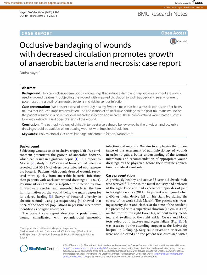
A nursing assistant at the Occupational Health Center cared for the patient’s abrasion; it was covered regularly during 4 weeks every other day with an occlusive band-age (Mepilex foam dressing, Mölnlycke Health Care, Sweden), a wound dressing material that is routinely used at health centers. The patient had no fever dur-ing this period and did not change the dressing himself. There is no document about him contacting the Depart-ment of Infectious Diseases or the health center during this period. He had noticed that the ulcer was producing odorous discharge, but the attending nurse did not expe-rience a situation that should be referred to specialist. The patient was referred to the hospital (13th April) due to visible muscle necrosis accompanied by yellow, odor-ous secretion at the bottom of an ulcer on the front of the right leg (Fig. 2). The patient had no fever, and his vital parameters were stable, although he did have diffuse red-ness and pitting edema on the right leg. The laboratory analysis revealed normal white blood cells, creatinine, electrolytes, and c-reactive protein 30 (<5 mg/l).
The local status motivated an ulcer revision and debridement of muscle tissue necrosis. However, there was no sign of acute inflammation in the ulcer area and with respect to the severe necrosis and significant growth of anaerobic bacteria together with gram-positive bac-teria in a biofilm [5, 6], there was a risk of developing a larger ulcer area with impaired healing. The clinical status
of patient was stable. Intravenous meropenem (3 × 1 g) was started immediately after cultures from blood and ulcer secretion were secured. A culture from the ulcer secretion revealed growth of Staphylococcus aureus, Streptococcus beta hemolytic group G, Clostridium innoc-uum, and Bacterioides thetaiotaomicron. The ulcer was treated conservatively with the local application of an antibiotic gel containing 250 mg vancomycin and hepat-ocyte growth factor (HGF in 100 IU antithrombin III Baxter) [7] plus sodium chloride for 2 days, followed by antithrombin III plus sodium chloride gel for 5 days. The wound dressing comprised sterile cotton compresses that were changed daily during the first week.
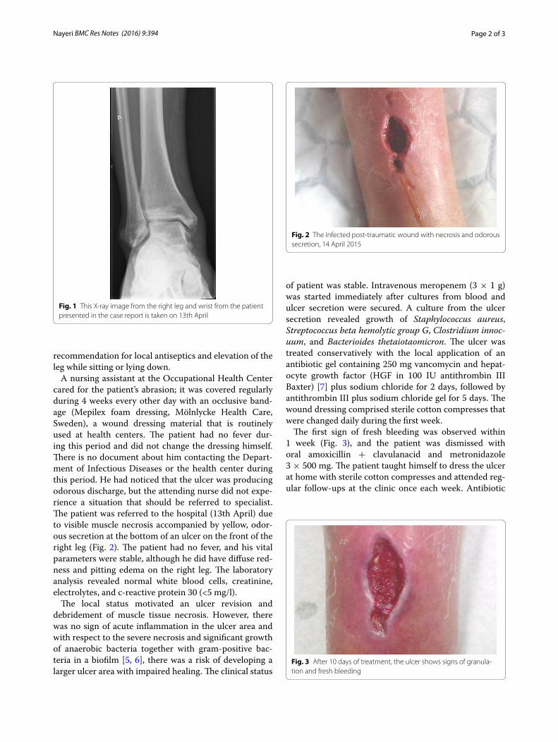
The first sign of fresh bleeding was observed within 1 week (Fig. 3), and the patient was dismissed with oral amoxicillin + clavulanacid and metronidazole 3 × 500 mg. The patient taught himself to dress the ulcer at home with sterile cotton compresses and attended reg-ular follow-ups at the clinic once each week. Antibiotic
Fig. 1 This X-ray image from the right leg and wrist from the patient presented in the case report is taken on 13th April
Fig. 2 The infected post-traumatic wound with necrosis and odorous secretion, 14 April 2015
Fig. 3 After 10 days of treatment, the ulcer shows signs of granula-tion and fresh bleeding
Page 3 of 3Nayeri BMC Res Notes (2016) 9:394
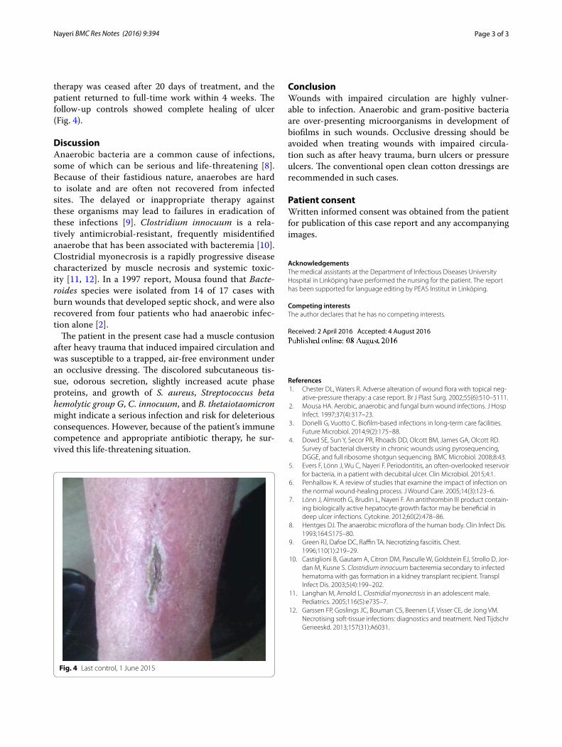
therapy was ceased after 20 days of treatment, and the patient returned to full-time work within 4 weeks. The follow-up controls showed complete healing of ulcer (Fig. 4).
DiscussionAnaerobic bacteria are a common cause of infections, some of which can be serious and life-threatening [8]. Because of their fastidious nature, anaerobes are hard to isolate and are often not recovered from infected sites. The delayed or inappropriate therapy against these organisms may lead to failures in eradication of these infections [9]. Clostridium innocuum is a rela-tively antimicrobial-resistant, frequently misidentified anaerobe that has been associated with bacteremia [10]. Clostridial myonecrosis is a rapidly progressive disease characterized by muscle necrosis and systemic toxic-ity [11, 12]. In a 1997 report, Mousa found that Bacte-roides species were isolated from 14 of 17 cases with burn wounds that developed septic shock, and were also recovered from four patients who had anaerobic infec-tion alone [2].
The patient in the present case had a muscle contusion after heavy trauma that induced impaired circulation and was susceptible to a trapped, air-free environment under an occlusive dressing. The discolored subcutaneous tis-sue, odorous secretion, slightly increased acute phase proteins, and growth of S. aureus, Streptococcus beta hemolytic group G, C. innocuum, and B. thetaiotaomicron might indicate a serious infection and risk for deleterious consequences. However, because of the patient’s immune competence and appropriate antibiotic therapy, he sur-vived this life-threatening situation.
ConclusionWounds with impaired circulation are highly vulner-able to infection. Anaerobic and gram-positive bacteria are over-presenting microorganisms in development of biofilms in such wounds. Occlusive dressing should be avoided when treating wounds with impaired circula-tion such as after heavy trauma, burn ulcers or pressure ulcers. The conventional open clean cotton dressings are recommended in such cases.
Patient consentWritten informed consent was obtained from the patient for publication of this case report and any accompanying images.
AcknowledgementsThe medical assistants at the Department of Infectious Diseases University Hospital in Linköping have performed the nursing for the patient. The report has been supported for language editing by PEAS Institut in Linköping.
Competing interestsThe author declares that he has no competing interests.
Received: 2 April 2016 Accepted: 4 August 2016
References 1. Chester DL, Waters R. Adverse alteration of wound flora with topical neg-
ative-pressure therapy: a case report. Br J Plast Surg. 2002;55(6):510–5111. 2. Mousa HA. Aerobic, anaerobic and fungal burn wound infections. J Hosp
Infect. 1997;37(4):317–23. 3. Donelli G, Vuotto C. Biofilm-based infections in long-term care facilities.
Future Microbiol. 2014;9(2):175–88. 4. Dowd SE, Sun Y, Secor PR, Rhoads DD, Olcott BM, James GA, Olcott RD.
Survey of bacterial diversity in chronic wounds using pyrosequencing, DGGE, and full ribosome shotgun sequencing. BMC Microbiol. 2008;8:43.
5. Evers F, Lönn J, Wu C, Nayeri F. Periodontitis, an often-overlooked reservoir for bacteria, in a patient with decubital ulcer. Clin Microbiol. 2015;4:1.
6. Penhallow K. A review of studies that examine the impact of infection on the normal wound-healing process. J Wound Care. 2005;14(3):123–6.
7. Lönn J, Almroth G, Brudin L, Nayeri F. An antithrombin III product contain-ing biologically active hepatocyte growth factor may be beneficial in deep ulcer infections. Cytokine. 2012;60(2):478–86.
8. Hentges DJ. The anaerobic microflora of the human body. Clin Infect Dis. 1993;164:S175–80.
9. Green RJ, Dafoe DC, Raffin TA. Necrotizing fasciitis. Chest. 1996;110(1):219–29.
10. Castiglioni B, Gautam A, Citron DM, Pasculle W, Goldstein EJ, Strollo D, Jor-dan M, Kusne S. Clostridium innocuum bacteremia secondary to infected hematoma with gas formation in a kidney transplant recipient. Transpl Infect Dis. 2003;5(4):199–202.
11. Langhan M, Arnold L. Clostridial myonecrosis in an adolescent male. Pediatrics. 2005;116(5):e735–7.
12. Garssen FP, Goslings JC, Bouman CS, Beenen LF, Visser CE, de Jong VM. Necrotising soft-tissue infections: diagnostics and treatment. Ned Tijdschr Geneeskd. 2013;157(31):A6031.
Fig. 4 Last control, 1 June 2015


