Occipital peripheral nerve stimulation in the management ...€¦ · Occipital peripheral nerve...
6
CASE REPORT Open Access Occipital peripheral nerve stimulation in the management of chronic intractable occipital neuralgia in a patient with neurofibromatosis type 1: a case report Ioannis Skaribas 1,2* , Octavio Calvillo 1,2 and Evangelia Delikanaki-Skaribas 1,2 Abstract Introduction: Occipital peripheral nerve stimulation is an interventional pain management therapy that provides beneficial results in the treatment of refractory chronic occipital neuralgia. Herein we present a first-of-its-kind case study of a patient with neurofibromatosis type 1 and bilateral occipital neuralgia treated with occipital peripheral nerve stimulation. Case presentation: A 42-year-old Caucasian woman presented with bilateral occipital neuralgia refractory to various conventional treatments, and she was referred for possible treatment with occipital peripheral nerve stimulation. She was found to be a suitable candidate for the procedure, and she underwent implantation of two octapolar stimulating leads and a rechargeable, programmable, implantable generator. The intensity, severity, and frequency of her symptoms resolved by more than 80%, but an infection developed at the implantation site two months after the procedure that required explantation and reimplantation of new stimulating leads three months later. To date she continues to experience symptom resolution of more than 60%. Conclusion: These results demonstrate the significance of peripheral nerve stimulation in the management of refractory occipital neuralgias in patients with neurofibromatosis type 1 and the possible role of neurofibromata in the development of occipital neuralgia in these patients. Introduction Chronic daily headache (CDH) syndromes represent a major health issue worldwide in terms of lost workdays and revenue [1-3]. Diagnoses include migraine, atypical migraine, cluster headaches, transformed migraines, cer- vicogenic headaches, occipital and facial hemicranias, or any combination of these diagnoses. Many of the patients who experience these syndromes become totally disabled after conservative and pharmacological treat- ments fail to relieve their symptoms [4,5]. Occipital neuralgia is described by the National Insti- tute of Neurological Diseases and Stroke as a “distinct type of headache characterized by piercing, throbbing, or electric-shock-like chronic pain in the upper neck, back of the head, and behind the ears, usually on one side” [6]. Typically, the pain of occipital neuralgia begins at the base of the head and spreads upward within the distribution of the greater and lesser occipital nerves. Characteristically, it is neuropathic, with paroxysmal epi- sodes of shooting electric shock-like symptoms. Although the symptom etiology includes trauma, infec- tion, and surgery, most patients with occipital neuralgia have idiopathic etiologies of their pain [7]. Neurofibromatosis type 1 (NF-1) is a possible but undocumented idiopathic etiology of occipital neuralgia. This human genetic disease, which is caused by muta- tions of the NF-1 tumor suppressor gene, has an inci- dence of about one in every 2500 live births and has a high rate of spontaneous mutations [8]. The characteris- tic feature of NF-1 is neurofibromata, which are com- plex lesions of the peripheral nervous system [8]. These lesions are composed of abnormal local cells, including * Correspondence: [email protected] 1 Greater Houston Pain Consultants, Greater Houston Anesthesiology, 2411 Fountain View Drive #200, Houston, TX 77057-4832, USA Full list of author information is available at the end of the article Skaribas et al. Journal of Medical Case Reports 2011, 5:174 http://www.jmedicalcasereports.com/content/5/1/174 JOURNAL OF MEDICAL CASE REPORTS © 2011 Skaribas et al; licensee BioMed Central Ltd. This is an Open Access article distributed under the terms of the Creative Commons Attribution License (http://creativecommons.org/licenses/by/2.0), which permits unrestricted use, distribution, and reproduction in any medium, provided the original work is properly cited.
Transcript of Occipital peripheral nerve stimulation in the management ...€¦ · Occipital peripheral nerve...
CASE REPORT Open Access
Occipital peripheral nerve stimulation in themanagement of chronic intractable occipitalneuralgia in a patient with neurofibromatosistype 1: a case reportIoannis Skaribas1,2*, Octavio Calvillo1,2 and Evangelia Delikanaki-Skaribas1,2
Abstract
Introduction: Occipital peripheral nerve stimulation is an interventional pain management therapy that providesbeneficial results in the treatment of refractory chronic occipital neuralgia. Herein we present a first-of-its-kind casestudy of a patient with neurofibromatosis type 1 and bilateral occipital neuralgia treated with occipital peripheralnerve stimulation.
Case presentation: A 42-year-old Caucasian woman presented with bilateral occipital neuralgia refractory tovarious conventional treatments, and she was referred for possible treatment with occipital peripheral nervestimulation. She was found to be a suitable candidate for the procedure, and she underwent implantation of twooctapolar stimulating leads and a rechargeable, programmable, implantable generator. The intensity, severity, andfrequency of her symptoms resolved by more than 80%, but an infection developed at the implantation site twomonths after the procedure that required explantation and reimplantation of new stimulating leads three monthslater. To date she continues to experience symptom resolution of more than 60%.
Conclusion: These results demonstrate the significance of peripheral nerve stimulation in the management ofrefractory occipital neuralgias in patients with neurofibromatosis type 1 and the possible role of neurofibromata inthe development of occipital neuralgia in these patients.
IntroductionChronic daily headache (CDH) syndromes represent amajor health issue worldwide in terms of lost workdaysand revenue [1-3]. Diagnoses include migraine, atypicalmigraine, cluster headaches, transformed migraines, cer-vicogenic headaches, occipital and facial hemicranias, orany combination of these diagnoses. Many of thepatients who experience these syndromes become totallydisabled after conservative and pharmacological treat-ments fail to relieve their symptoms [4,5].Occipital neuralgia is described by the National Insti-
tute of Neurological Diseases and Stroke as a “distincttype of headache characterized by piercing, throbbing,or electric-shock-like chronic pain in the upper neck,
back of the head, and behind the ears, usually on oneside” [6]. Typically, the pain of occipital neuralgia beginsat the base of the head and spreads upward within thedistribution of the greater and lesser occipital nerves.Characteristically, it is neuropathic, with paroxysmal epi-sodes of shooting electric shock-like symptoms.Although the symptom etiology includes trauma, infec-tion, and surgery, most patients with occipital neuralgiahave idiopathic etiologies of their pain [7].Neurofibromatosis type 1 (NF-1) is a possible but
undocumented idiopathic etiology of occipital neuralgia.This human genetic disease, which is caused by muta-tions of the NF-1 tumor suppressor gene, has an inci-dence of about one in every 2500 live births and has ahigh rate of spontaneous mutations [8]. The characteris-tic feature of NF-1 is neurofibromata, which are com-plex lesions of the peripheral nervous system [8]. Theselesions are composed of abnormal local cells, including
* Correspondence: [email protected] Houston Pain Consultants, Greater Houston Anesthesiology, 2411Fountain View Drive #200, Houston, TX 77057-4832, USAFull list of author information is available at the end of the article
Skaribas et al. Journal of Medical Case Reports 2011, 5:174http://www.jmedicalcasereports.com/content/5/1/174 JOURNAL OF MEDICAL
CASE REPORTS
© 2011 Skaribas et al; licensee BioMed Central Ltd. This is an Open Access article distributed under the terms of the Creative CommonsAttribution License (http://creativecommons.org/licenses/by/2.0), which permits unrestricted use, distribution, and reproduction inany medium, provided the original work is properly cited.
Schwann cells, endothelial cells, fibroblasts, and a largenumber of infiltrating inflammatory mast cells [8,9].They can cause a variety of symptoms when they invadesurrounding tissues. Other characteristics of NF-1 areflat, pigmented lesions of the skin (café au lait spots),axillary freckles, pigmented iris hamartomas (Lischnodules), and a variety of central nervous system mani-festations, such as optic nerve tumors, unidentifiedbright objects in the visual field, and neurofibromas ofthe spinal nerve roots (schwannomas) [10]. Althoughheadaches are very common in patients with NF-1, thespecific diagnosis of occipital neuralgia is not [11-13].The initial treatment for both CDH syndromes and
occipital neuralgia is pharmacologic and is focused onsymptom relief [14]. Patients whose symptoms do notrespond to this initial therapy are treated secondarilywith occipital nerve blockade [15], radiofrequency abla-tion [16], botulinum toxin A injections [17,18], surgicaldecompression [19], and occipital peripheral nerve sti-mulation (OPNS) [7,20-23]. OPNS involves the place-ment of trial peripheral nerve-stimulating electrodesover the occipital nerves. If the prerequisite dermatomalparesthesia is achieved, then pain relief as a result ofpermanent implantation has been reported to be as highas 80% [7,20-23]. In this report, we present the case of awoman with NF-1 and bilateral occipital neuralgia whoexperienced pain relief after OPNS.
Case presentationPatient historyA 42-year-old Caucasian woman was referred to ourhospital for pain management by a neurologist specializ-ing in the treatment of daily headaches. She had experi-enced daily intractable headaches since age 18 years.She also had chronic bilateral occipital neuralgia on thebasis of the diagnostic criteria outlined in the secondedition of The International Classification of HeadacheDisorders [24]. Her occipital neuralgia persisted formore than 15 days monthly and was distributedthroughout the greater occipital nerves, beginning in theoccipital region and radiating upward to the top of thehead. When the occipital neuralgia occurred, her occipi-tal area became very tender to palpation. Complete alle-viation of her pain had been achieved for a limited timewith diagnostic bilateral greater occipital nerve blocks.Her medical history included NF-1, which was first
diagnosed in childhood. Several neurofibromas had beenremoved from her sacrum 10 years previously, as well asmany from her upper extremities. She also had had pro-blems with depression, anxiety, alcohol consumption,and smoking. She has been a housewife throughout heradult life. With regard to her family medical history, hermother had died at 68 years of age as a result of heartdisease, and her father was alive at 72 years of age with
a history of cancer. An older sister has rheumatoidarthritis but not NF-1.Before her referral to our service, she had undergone
extensive medical management with biofeedback train-ing, physical therapy, massage, acupuncture, and phar-macological management with narcotic and non-narcotic pain medications. Her medications includedsustained-release morphine (30 mg every 12 hours),hydrocodone and acetaminophen (10 mg and 325 mg,respectively, every four to six hours), and pregabaline(75 mg every eight hours). More recently, she hadundergone three greater occipital nerve blocks thatresulted in complete pain resolution that lasted fromtwo to three days. Because she required an ever-increas-ing dose of morphine for pain relief, and because shehad responded to the occipital nerve blocks, she wasconsidered to be a good candidate for OPNS.
Trial procedureAt her baseline office visit, the patient underwent a dis-ability and quality-of-life assessment by completing aseries of questionnaires (see “Quality-of-life assessment”section below) and was found to be a suitable candidatefor a trial of OPNS. After the risks and benefits of theprocedure were discussed with the patient and herinformed consent was obtained, the trial of OPNS wascarried out in October 2008 by using two percutaneouseight-contact leads (Octrode; St Jude Medical Neuromo-dulation Division, Plano, TX, USA). After a week-longsuccessful trial with more than 80% symptom improve-ment, the patient was deemed a suitable candidate forpermanent implantation and she underwent implanta-tion of two permanent percutaneous eight-contact leads(Octrode) and a conventional implantable pulse genera-tor (IPG) (Genesis; St Jude Medical NeuromodulationDivision).
Permanent implantation procedureOn the day of the procedure, which was carried out inan operating room, a slow intravenous infusion of 2 g ofcefazolin was started, and the patient was placed in aprone position with pillows under her chest to augmentneck flexion. Monitored anesthesia was administered byusing intravenous fentanyl and midazolam at a level thatallowed the patient to be comfortable but able to inter-act with medical personnel throughout the procedure.The patient’s hair was shaved below a line connectingthe external occipital protuberance to the mastoid pro-cesses, and her skin was treated with chlorhexidine. Asterilely draped C-arm was introduced to obtain a trueanteroposterior image of the cervical spine at the C1-C2interspace, and the overlying skin was marked with asterile marker. Thereafter a portable ultrasound with asterile linear array transducer of 5 MHz to 13 MHz
Skaribas et al. Journal of Medical Case Reports 2011, 5:174http://www.jmedicalcasereports.com/content/5/1/174
Page 2 of 6
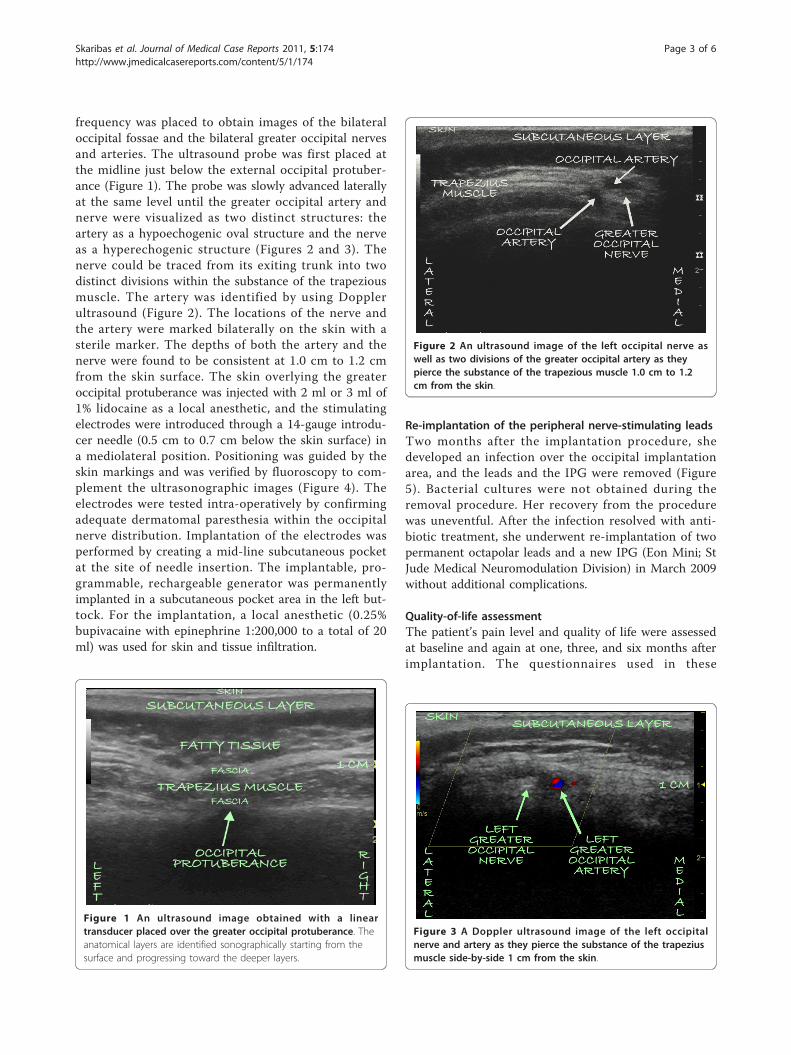
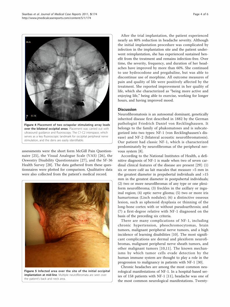
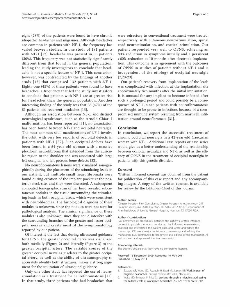
frequency was placed to obtain images of the bilateraloccipital fossae and the bilateral greater occipital nervesand arteries. The ultrasound probe was first placed atthe midline just below the external occipital protuber-ance (Figure 1). The probe was slowly advanced laterallyat the same level until the greater occipital artery andnerve were visualized as two distinct structures: theartery as a hypoechogenic oval structure and the nerveas a hyperechogenic structure (Figures 2 and 3). Thenerve could be traced from its exiting trunk into twodistinct divisions within the substance of the trapeziousmuscle. The artery was identified by using Dopplerultrasound (Figure 2). The locations of the nerve andthe artery were marked bilaterally on the skin with asterile marker. The depths of both the artery and thenerve were found to be consistent at 1.0 cm to 1.2 cmfrom the skin surface. The skin overlying the greateroccipital protuberance was injected with 2 ml or 3 ml of1% lidocaine as a local anesthetic, and the stimulatingelectrodes were introduced through a 14-gauge introdu-cer needle (0.5 cm to 0.7 cm below the skin surface) ina mediolateral position. Positioning was guided by theskin markings and was verified by fluoroscopy to com-plement the ultrasonographic images (Figure 4). Theelectrodes were tested intra-operatively by confirmingadequate dermatomal paresthesia within the occipitalnerve distribution. Implantation of the electrodes wasperformed by creating a mid-line subcutaneous pocketat the site of needle insertion. The implantable, pro-grammable, rechargeable generator was permanentlyimplanted in a subcutaneous pocket area in the left but-tock. For the implantation, a local anesthetic (0.25%bupivacaine with epinephrine 1:200,000 to a total of 20ml) was used for skin and tissue infiltration.
Re-implantation of the peripheral nerve-stimulating leadsTwo months after the implantation procedure, shedeveloped an infection over the occipital implantationarea, and the leads and the IPG were removed (Figure5). Bacterial cultures were not obtained during theremoval procedure. Her recovery from the procedurewas uneventful. After the infection resolved with anti-biotic treatment, she underwent re-implantation of twopermanent octapolar leads and a new IPG (Eon Mini; StJude Medical Neuromodulation Division) in March 2009without additional complications.
Quality-of-life assessmentThe patient’s pain level and quality of life were assessedat baseline and again at one, three, and six months afterimplantation. The questionnaires used in these
Figure 1 An ultrasound image obtained with a lineartransducer placed over the greater occipital protuberance. Theanatomical layers are identified sonographically starting from thesurface and progressing toward the deeper layers.
Figure 2 An ultrasound image of the left occipital nerve aswell as two divisions of the greater occipital artery as theypierce the substance of the trapezious muscle 1.0 cm to 1.2cm from the skin.
Figure 3 A Doppler ultrasound image of the left occipitalnerve and artery as they pierce the substance of the trapeziusmuscle side-by-side 1 cm from the skin.
Skaribas et al. Journal of Medical Case Reports 2011, 5:174http://www.jmedicalcasereports.com/content/5/1/174
Page 3 of 6
assessments were the short form McGill Pain Question-naire [25], the Visual Analogue Scale (VAS) [26], theOswestry Disability Questionnaire [27], and the SF-36Health Survey [28]. The data gathered from these ques-tionnaires were plotted for comparison. Qualitative datawere also collected from the patient’s medical record.
After the trial implantation, the patient experiencednearly an 80% reduction in headache severity. Althoughthe initial implantation procedure was complicated byinfection in the implantation site and the patient under-went reimplantation, she has experienced sustained ben-efit from the treatment and remains infection-free. Overtime, the severity, frequency, and duration of her head-aches have improved by more than 60%. She continuedto use hydrocodone and pregabaline, but was able todiscontinue use of morphine. All outcome measures ofpain and quality of life were positively affected by thetreatment. She reported improvement in her quality oflife, which she characterized as “being more active andenjoying life,” being able to exercise, working for longerhours, and having improved mood.
DiscussionNeurofibromatosis is an autosomal dominant, geneticallyinherited disease first described in 1882 by the Germanpathologist Friedrich Daniel von Recklinghausen. Itbelongs to the family of phakomatoses and is subcate-gorized into two types: NF-1 (von Recklinghausen’s dis-ease) and NF-2 (bilateral acoustic neurofibromatosis).Our patient had classic NF-1, which is characterizedpredominately by neurofibromas of the peripheral ner-vous system [8].According to the National Institutes of Health, a defi-
nitive diagnosis of NF-1 is made when two of seven car-dinal clinical features of the disease are present [29]: (1)six or more café au lait macules that measure >5 mm inthe greatest diameter in prepubertal individuals and >15mm in the greatest diameter in postpubertal individuals;(2) two or more neurofibromas of any type or one plexi-form neurofibroma; (3) freckles in the axillary or ingu-inal region; (4) optic nerve glioma; (5) two or more irishamartomas (Lisch nodules); (6) a distinctive osseouslesion, such as sphenoid dysplasia or thinning of thelong-bone cortex with or without pseudoarthrosis; and(7) a first-degree relative with NF-1 diagnosed on thebasis of the preceding six criteria.There are many complications of NF-1, including
chronic hypertension, pheochromocytomas, braintumors, malignant peripheral nerve tumors, and a highincidence of learning disabilities [10]. The most signifi-cant complications are dermal and plexiform neurofi-bromas, malignant peripheral nerve sheath tumors, andother malignant tumors [10,11]. The known mechan-isms by which tumor cells evade detection by thehuman immune system are thought to play a role in theprogression to malignancy in patients with NF-1 [30].Chronic headaches are among the most common neu-
rological manifestations of NF-1. In a hospital-based ser-ies of 158 patients with NF-1 [11], headache was one ofthe most common neurological manifestations. Twenty-
Figure 4 Placement of two octapolar stimulating array leadsover the bilateral occipital areas. Placement was carried out withultrasound guidance and fluoroscopy. The C1-C2 interspace, whichserves as a key fluoroscopic landmark for occipital peripheral nervestimulation, and the dens are easily identifiable.
Figure 5 Infected area over the site of the initial occipitalimplantation at mid-line. Multiple neurofibromata are seen overthe patient’s back and neck area.
Skaribas et al. Journal of Medical Case Reports 2011, 5:174http://www.jmedicalcasereports.com/content/5/1/174
Page 4 of 6
eight (28%) of the patients were found to have chronicidiopathic headaches and migraines. Although headachesare common in patients with NF-1, the frequency hasvaried between studies. In one study of 181 patientswith NF-1 [12], headache was present in 55 patients(30%). This frequency was not statistically significantlydifferent from that found in the general population,leading the study investigators to conclude that head-ache is not a specific feature of NF-1. This conclusion,however, was contradicted by the findings of anotherstudy [13] that comprised 132 patients with NF-1.Eighty-one (45%) of these patients were found to haveheadaches, a frequency that led the study investigatorsto conclude that patients with NF-1 are at greater riskfor headaches than the general population. Anotherinteresting finding of the study was that 38 (47%) of the81 patients had recurrent headaches [13].Although an association between NF-1 and distinct
neurological syndromes, such as the Arnold-Chiari Imalformation, has been reported [31], no associationhas been found between NF-1 and occipital neuralgia.The most common skull manifestations of NF-1 involvethe orbit, with very few reports of occipital defects inpatients with NF-1 [32]. Such occipital defects havebeen found in a 54-year-old woman with a massiveplexiform neurofibroma that extended from the auricu-lar region to the shoulder and was associated with largeleft occipital and left petrous bone defects [32].No neurofibromatous lesions were visualized sonogra-
phically during the placement of the stimulating leads inour patient, but multiple small neurofibromata werefound during creation of the implant pocket at the pos-terior neck site, and they were dissected. A subsequentcomputed tomographic scan of her head revealed subcu-taneous nodules in the tissue surrounding the stimulat-ing leads in both occipital areas, which were consistentwith neurofibromas. The histological diagnosis of thesenodules is unknown, since the nodules were not sent forpathological analysis. The clinical significance of thesenodules is also unknown, since they could interfere withthe surrounding branches of the greater and lesser occi-pital nerves and create most of the symptomatologyreported by our patient.Of interest is the fact that during ultrasound guidance
for OPNS, the greater occipital nerve was visualizedboth medially (Figure 2) and laterally (Figure 3) to thegreater occipital artery. The variable course of thegreater occipital nerve as it relates to the greater occipi-tal artery, as well as the ability of ultrasonography toaccurately identify both structures, makes a strong argu-ment for the utilization of ultrasound guidance.Only one other study has reported the use of neuro-
stimulation as a treatment for neurofibromatosis [11].In that study, three patients who had headaches that
were refractory to conventional treatment were treated,respectively, with cutaneous neurostimulation, spinalcord neurostimulation, and cortical stimulation. Ourpatient responded very well to OPNS, achieving an80% reduction in symptoms initially and a persistent>60% reduction at 10 months after electrode implanta-tion. This outcome is in agreement with the outcomesof OPNS in studies of patients without NF-1 and isindependent of the etiology of occipital neuralgia[7,20-23].Our patient’s recovery from implantation of the leads
was complicated with infection at the implantation siteapproximately two months after the initial implantation.It is unusual for any implant to become infected aftersuch a prolonged period and could possibly be a conse-quence of NF-1, since patients with neurofibromatosisare thought to be prone to infections because of a com-promised immune system resulting from mast cell infil-tration around neurofibromata [31].
ConclusionIn conclusion, we report the successful treatment ofchronic occipital neuralgia in a 42-year-old Caucasianwoman with NF-1. Additional case reports or case serieswould give us a better understanding of the relationshipbetween occipital neuralgia and NF-1 as well as the effi-cacy of OPNS in the treatment of occipital neuralgia inpatients with this genetic disorder.
ConsentWritten informed consent was obtained from the patientfor publication of this case report and any accompany-ing images. A copy of the written consent is availablefor review by the Editor-in-Chief of this journal.
Author details1Greater Houston Pain Consultants, Greater Houston Anesthesiology, 2411Fountain View Drive #200, Houston, TX 77057-4832, USA. 2Department ofAnesthesiology, University General Hospital, Houston, TX 77030, USA.
Authors’ contributionsIMS performed all procedures, obtained the patient’s written informedconsent to publish the report, conducted the follow-up examinations,analyzed and interpreted the patient data, and wrote and edited themanuscript. OC was a major contributor to reviewing and editing themanuscript. EDS contributed to the review and editing of the manuscript. Allauthors read and approved the final manuscript.
Competing interestsThe authors declare that they have no competing interests.
Received: 13 December 2009 Accepted: 10 May 2011Published: 10 May 2011
References1. Stewart WF, Wood GC, Razzaghi H, Reed ML, Lipton RB: Work impact of
migraine headaches. J Occup Environ Med 2008, 50:736-745.2. Weiss MD, Bernards P, Price SJ: Working through a migraine: addressing
the hidden costs of workplace headaches. AAOHN J 2008, 56:495-502.
Skaribas et al. Journal of Medical Case Reports 2011, 5:174http://www.jmedicalcasereports.com/content/5/1/174
Page 5 of 6

3. Stovner LJ, Hagen K, Jensen R, Katsarava Z, Lipton R, Scher A, Steiner T,Zwart JA: The global burden of headache: a documentation of headacheprevalence and disability worldwide. Cephalalgia 2007, 27:193-210.
4. Stewart WF, Lipton RB, Kolodner KB, Sawyer J, Lee C, Liberman JN: Validityof the Migraine Disability Assessment (MIDAS) score in comparison to adiary-based measure in a population sample of migraine sufferers. Pain2000, 88:41-52.
5. Silberstein S, Lipton R: Chronic daily headache including transformedmigraine, chronic tension type headache and medication overuse. InWolff’s Headache and Other Head Pain.. 7 edition. Edited by: Silberstein SD,Lipton RB, Dalessio DJ. New York: Oxford University Press; 2001:247-282.
6. National Institute of Neurological Disorders and Stroke: NINDS OccipitalNeuralgia Information Page.[http://www.ninds.nih.gov/disorders/occipitalneuralgia/occipitalneuralgia.htm].
7. Oh MY, Ortega J, Bellotte JB, Whiting DM, Aló K: Peripheral nervestimulation for the treatment of occipital neuralgia and transformedmigraine using a C1-2-3 subcutaneous paddle style electrode: atechnical report. Neuromodulation 2004, 7:103-112.
8. Riccardi VM: Neurofibromatosis: past, present, and future. N Engl J Med1991, 324:1283-1285.
9. Viskochil DH: It takes two to tango: mast cell and Schwann cellinteractions in neurofibromas. J Clin Invest 2003, 112:1791-1793.
10. Riccardi VM: Neurofibromatosis: Phenotype, Natural History, and Pathogenesis.2 edition. Baltimore: Johns Hopkins University Press; 1992.
11. Créange A, Zeller J, Rostaing-Rigattieri S, Brugières P, Degos JD, Revuz J,Wolkenstein P: Neurological complications of neurofibromatosis type 1 inadulthood. Brain 1999, 122:473-481.
12. Clement M, Battistella PA, Rizzi L, Boni S, Tenconi R: Headache in patientswith neurofibromatosis type 1. Headache 1996, 36:10.
13. DiMario FJ Jr, Langshur S: Headaches in patients with neurofibromatosis-1. J Child Neurol 2000, 15:235-238.
14. Evers S, Frese A: Recent advances in the treatment of headaches. CurrOpin Anaesthesiol 2005, 18:563-568.
15. Ashkenazi A, Levin M: Greater occipital nerve block for migraine andother headaches: is it useful? Curr Pain Headache Rep 2007, 11:231-235.
16. Navani A, Mahajan G, Kreis P, Fishman SM: A case of pulsedradiofrequency lesioning for occipital neuralgia. Pain Med 2006,7:453-456.
17. Taylor M, Silva S, Cottrell C: Botulinum toxin type-A (BOTOX) in thetreatment of occipital neuralgia: a pilot study. Headache 2008,48:1476-1481.
18. Kapural L, Stillman M, Kapural M, McIntyre P, Guirgius M, Mekhail N:Botulinum toxin occipital nerve block for the treatment of severeoccipital neuralgia: a case series. Pain Pract 2007, 7:337-340.
19. Pikus HJ, Phillips JM: Outcome of surgical decompression of the secondcervical root for cervicogenic headache. Neurosurgery 1996, 39:63-70.
20. Weiner RL, Reed KL: Peripheral neurostimulation for control of intractableoccipital neuralgia. Neuromodulation 1999, 2:217-221.
21. Popeney CA, Aló KM: Peripheral neurostimulation for the treatment ofchronic, disabling transformed migraine. Headache 2003, 43:369-375.
22. Slavin KV, Nersesyan H, Wess C: Peripheral neurostimulation for treatmentof intractable occipital neuralgia. Neurosurgery 2006, 58:112-119.
23. Skaribas I, Aló K: Ultrasound imaging and occipital nerve stimulation.Neuromodulation Technol Neural Interface 2010, 13:126-130.
24. Headache Classification Subcommittee of the International HeadacheSociety: The International Classification of Headache Disorders.Cephalalgia , 2 2004, 24(Suppl 1):9-160.
25. Melzack R: The short-form McGill Pain Questionnaire. Pain 1987,30:191-197.
26. Crichton N: Visual Analogue Scale (VAS). J Clin Nurs 2001, 10:706.27. Fairbank JC, Couper J, Davies JB, O’Brien JP: The Oswestry low back pain
questionnaire. Physiotherapy 1980, 66:271-273.28. Ware JE Jr: SF-36 health survey update. Spine (Phila Pa 1976) 2000,
25:3130-3139.29. National Institutes of Health Consensus Development Conference:
Neurofibromatosis: conference statement. Arch Neurol 1988, 45:575-578.30. Lee PR, Cohen JE, Fields RD: Immune system evasion by peripheral nerve
sheath tumor. Neurosci Lett 2006, 397:126-129.31. Herrero Valverde A, Moiron Simões R, Mera Campillo J, Palma T: [Headache
in patient with neurofibromatosis type 1] [in Spanish]. Neurologia 2007,22:911-914.
32. Renshaw A, Borsetti M, Nelson RJ, Orlando A: Massive plexiformneurofibroma with associated meningo-encephalocoele and occipitalbone defect presenting as a cervical mass. Br J Plast Surg 2003,56:514-517.
doi:10.1186/1752-1947-5-174Cite this article as: Skaribas et al.: Occipital peripheral nerve stimulationin the management of chronic intractable occipital neuralgia in apatient with neurofibromatosis type 1: a case report. Journal of MedicalCase Reports 2011 5:174.
Submit your next manuscript to BioMed Centraland take full advantage of:
• Convenient online submission
• Thorough peer review
• No space constraints or color figure charges
• Immediate publication on acceptance
• Inclusion in PubMed, CAS, Scopus and Google Scholar
• Research which is freely available for redistribution
Submit your manuscript at www.biomedcentral.com/submit
Skaribas et al. Journal of Medical Case Reports 2011, 5:174http://www.jmedicalcasereports.com/content/5/1/174
Page 6 of 6