Obstructive Jaundice Caused by Tuberculous Lymphadenopathies
5
232 http://jsms.sch.ac.kr Obstructive Jaundice Caused by Tuberculous Lymphadenopathies Ju Ri Kim, Ji Woong Jang, So Hee Chung, Min Kyu Kang, Jin Young Kim, Jun Hyung Park Department of Internal Medicine, Eulji University Hospital, Eulji University School of Medicine, Daejeon, Korea Although pulmonary tuberculosis is known to be the most common type in tuberculosis, it actually can affect any organ system. However, abdominal type is very rare among the extra-pulmonary types, and obstructive jaundice caused by lymphadenopathies due to tuberculosis is especially uncommon manifestation even in endemic areas. Tuberculous lymphadenopathies can mimic lymphadenopathies by other metastatic tumors or lymphoma, thus early correct diagnosis is very important for avoiding unneces- sary surgical interventions. Here, we reported two cases of obstructive jaundice caused by tuberculous lymphadenopathies. Both were treated with anti- tuberculosis medications and endoscopic retrograde biliary drainage without surgery. Keywords: Jaundice; Tuberculosis; Lymphatic diseases INTRODUCTION Abdominal tuberculosis (TB) is known to be 12-25% of all extra- pulmonary TB [1], and diagnosis of lymphadenopathies (LAPs) by abdominal TB is not simple, because those often mimic LAPs by malignant tumor or lymphoma. For this reason, surgical interven- tions have been carried out globally for differentiating benign le- sions from malignancies up to date [2]. However, if correct diagno- sis can be established without surgical interventions, it would be helpful for saving patients ’ time and cost as well as avoiding severe complications from invasive procedures. Here, we report two cases of obstructive jaundice due to pericholedochal LAPs by TB, which were treated with anti-TB medications and endoscopic retrograde biliary drainage (ERBD) without surgery. CASE REPORTS 1. Case 1 A 29-year-old man visited Eulji University Hospital for inter- mittent epigastric pain. Seven months ago, he was diagnosed with TB abscess on his right axilla and had taken anti-TB medications for six months. Physical examinations revealed jaundice and right upper ab- dominal tenderness, but there was no fever. Laboratory data in- cluded the followings: hemoglobin 14.9 g/dL (normal range, 12 to 16 g/dL); white blood cell 9,280/ μL (normal range, 3,800 to 10,500/ μL); C-reactive protein 1.28 mg/dL (normal range, 0 to 0.5 mg/dL); aspartate aminotransferase (AST) 188 IU/L (normal range, 1 to 34 IU/L); alanine aminotransferase (ALT) 151 IU/L (normal range, 1 to 34 IU/L); alkaline phosphatase (ALP) 178 IU/L (normal range, 25 to 100 IU/L); γ-glutamyl transpeptidase ( γ-GTP) 450 IU/L (normal range, 1 to 35 IU/L); and total bilirubin 2.9 mg/dL (normal range, 0.1 to 1.2 mg/dL). Acid-fast bacillus (AFB) stain, culture, and poly- merase chain reaction (PCR) for Mycobacterium tuberculosis of sputum were negative. Abdominal computed tomography (CT) revealed narrowed mid common bile duct (CBD) with dilated intrahepatic bile duct and proximal CBD, in addition to suspicious fistula tract between lymph node and mid CBD (Fig. 1A). Several central necrotic LAPs at portocarval and porta hepatis area were also observed. Endo- scopic retrograde cholangiopancreatography (ERCP) demonstrated narrowed mid CBD with dilated intrahepatic bile duct and proxi- mal CBD, but fistula tract was not observed on the cholangiogram. Bile juice was aspirated for culture and PCR for M. tuberculosis and Soonchunhyang Medical Science 21(2):232-236, December 2015 pISSN: 2233-4289 I eISSN: 2233-4297 CASE REPORT Correspondence to: Ji Woong Jang Department of Internal Medicine, Eulji University Hospital, Eulji University School of Medicine, 95 Dunsanseo-ro, Seo-gu, Daejeon 35233, Korea Tel: +82-42-611-3063, Fax: +82-42-259-1211, E-mail: [email protected] Received: Sep. 21, 2015 / Accepted after revision: Oct. 26, 2015 © 2015 Soonchunhyang Medical Research Institute This is an Open Access article distributed under the terms of the Creative Commons Attribution Non-Commercial License (http://creativecommons.org/licenses/by-nc/3.0/).
Transcript of Obstructive Jaundice Caused by Tuberculous Lymphadenopathies
232 http://jsms.sch.ac.kr
Obstructive Jaundice Caused by Tuberculous LymphadenopathiesJu Ri Kim, Ji Woong Jang, So Hee Chung, Min Kyu Kang, Jin Young Kim, Jun Hyung Park
Department of Internal Medicine, Eulji University Hospital, Eulji University School of Medicine, Daejeon, Korea
Although pulmonary tuberculosis is known to be the most common type in tuberculosis, it actually can affect any organ system. However, abdominal type is very rare among the extra-pulmonary types, and obstructive jaundice caused by lymphadenopathies due to tuberculosis is especially uncommon manifestation even in endemic areas. Tuberculous lymphadenopathies can mimic lymphadenopathies by other metastatic tumors or lymphoma, thus early correct diagnosis is very important for avoiding unneces-sary surgical interventions. Here, we reported two cases of obstructive jaundice caused by tuberculous lymphadenopathies. Both were treated with anti- tuberculosis medications and endoscopic retrograde biliary drainage without surgery.
Keywords: Jaundice; Tuberculosis; Lymphatic diseases
INTRODUCTION
Abdominal tuberculosis (TB) is known to be 12-25% of all extra-pulmonary TB [1], and diagnosis of lymphadenopathies (LAPs) by abdominal TB is not simple, because those often mimic LAPs by malignant tumor or lymphoma. For this reason, surgical interven-tions have been carried out globally for differentiating benign le-sions from malignancies up to date [2]. However, if correct diagno-sis can be established without surgical interventions, it would be helpful for saving patients’ time and cost as well as avoiding severe complications from invasive procedures. Here, we report two cases of obstructive jaundice due to pericholedochal LAPs by TB, which were treated with anti-TB medications and endoscopic retrograde biliary drainage (ERBD) without surgery.
CASE REPORTS
1. Case 1
A 29-year-old man visited Eulji University Hospital for inter-mittent epigastric pain. Seven months ago, he was diagnosed with TB abscess on his right axilla and had taken anti-TB medications for six months.
Physical examinations revealed jaundice and right upper ab-dominal tenderness, but there was no fever. Laboratory data in-cluded the followings: hemoglobin 14.9 g/dL (normal range, 12 to 16 g/dL); white blood cell 9,280/μL (normal range, 3,800 to 10,500/ μL); C-reactive protein 1.28 mg/dL (normal range, 0 to 0.5 mg/dL); aspartate aminotransferase (AST) 188 IU/L (normal range, 1 to 34 IU/L); alanine aminotransferase (ALT) 151 IU/L (normal range, 1 to 34 IU/L); alkaline phosphatase (ALP) 178 IU/L (normal range, 25 to 100 IU/L); γ-glutamyl transpeptidase (γ-GTP) 450 IU/L (normal range, 1 to 35 IU/L); and total bilirubin 2.9 mg/dL (normal range, 0.1 to 1.2 mg/dL). Acid-fast bacillus (AFB) stain, culture, and poly-merase chain reaction (PCR) for Mycobacterium tuberculosis of sputum were negative.
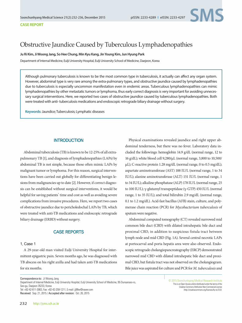
Abdominal computed tomography (CT) revealed narrowed mid common bile duct (CBD) with dilated intrahepatic bile duct and proximal CBD, in addition to suspicious fistula tract between lymph node and mid CBD (Fig. 1A). Several central necrotic LAPs at portocarval and porta hepatis area were also observed. Endo-scopic retrograde cholangiopancreatography (ERCP) demonstrated narrowed mid CBD with dilated intrahepatic bile duct and proxi-mal CBD, but fistula tract was not observed on the cholangiogram. Bile juice was aspirated for culture and PCR for M. tuberculosis and
Soonchunhyang Medical Science 21(2):232-236, December 2015 pISSN: 2233-4289 I eISSN: 2233-4297
CASE REPORT
Correspondence to: Ji Woong JangDepartment of Internal Medicine, Eulji University Hospital, Eulji University School of Medicine, 95 Dunsanseo-ro, Seo-gu, Daejeon 35233, KoreaTel: +82-42-611-3063, Fax: +82-42-259-1211, E-mail: [email protected]: Sep. 21, 2015 / Accepted after revision: Oct. 26, 2015
© 2015 Soonchunhyang Medical Research InstituteThis is an Open Access article distributed under the terms of the
Creative Commons Attribution Non-Commercial License (http://creativecommons.org/licenses/by-nc/3.0/).
Obstructive Jaundice Caused by Tuberculous Lymphadenopathies • Kim JR, et al.
Soonchunhyang Medical Science 21(2):232-236 http://jsms.sch.ac.kr 233
other mycobacteria (MTB-PCR), and finally, ERBD was inserted in the CBD. All bile examinations yielded negative results.
When considering his tuberculous history and CT findings, ob-structive jaundice due to pericholedochal LAPs by TB was strongly suspected. His abdominal pain and jaundice was improved gradu-ally with anti-TB medications and ERBD. Five weeks later, fol-lowed abdominal CT demonstrated regressed previous multiple enlarged lymph nodes and improvement of dilated CBD (Fig. 1B). Laboratory data was also within normal limit. ERBD was removed after three months from initial insertion.
2. Case 2
A 29-year-old man visited Eulji University Hospital with itching sensation and icteric sclera. He had no history of specific disease. At physical examinations, he had jaundice with right upper ab-dominal tenderness.
Laboratory data revealed the followings: hemoglobin 13.7 g/dL (normal range, 12 to 16 g/dL); white blood cell 8,270/μL (normal range, 3,800 to 10,500/μL); C-reactive protein 0.2 mg/dL (normal range, 0 to 0.5 mg/dL); AST 49 IU/L (normal range, 1 to 34 IU/L); ALT 83 IU/L (normal range, 1 to 34 IU/L); ALP 285 IU/L (normal range, 25 to 100 IU/L); γ-GTP 63 IU/L (normal range, 1 to 35 IU/L);
A B
Fig. 1. (A) Abdominal CT at admission revealed mild bile duct dilatation due to lymphadenopathies (arrowhead) and suspicious fistula tract between lymph node and CBD (arrow). (B) After five weeks with anti-tuberculosis medications and endoscopic retrograde biliary drainage, abdominal CT showed regressed previous enlarged lymph nodes and improvement of dilated CBD. CT, computed tomography; CBD, common bile duct.
A B
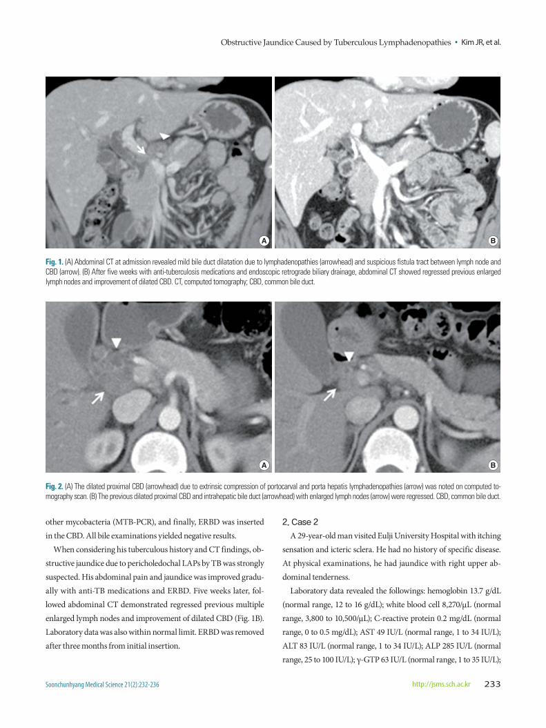
Fig. 2. (A) The dilated proximal CBD (arrowhead) due to extrinsic compression of portocarval and porta hepatis lymphadenopathies (arrow) was noted on computed to-mography scan. (B) The previous dilated proximal CBD and intrahepatic bile duct (arrowhead) with enlarged lymph nodes (arrow) were regressed. CBD, common bile duct.
Kim JR, et al. • Obstructive Jaundice Caused by Tuberculous Lymphadenopathies
Soonchunhyang Medical Science 21(2):232-236234 http://jsms.sch.ac.kr
and total bilirubin 6.82 mg/dL (normal range, 0.1 to 1.2 mg/dL). Tu-mor marker, carbohydrate antigen 19-9, was within normal limit.
Abdominal CT demonstrated compression of mid CBD with dilated proximal CBD and intrahepatic bile duct due to extrinsic compression of portocarval and porta hepatis LAPs (Fig. 2A). The
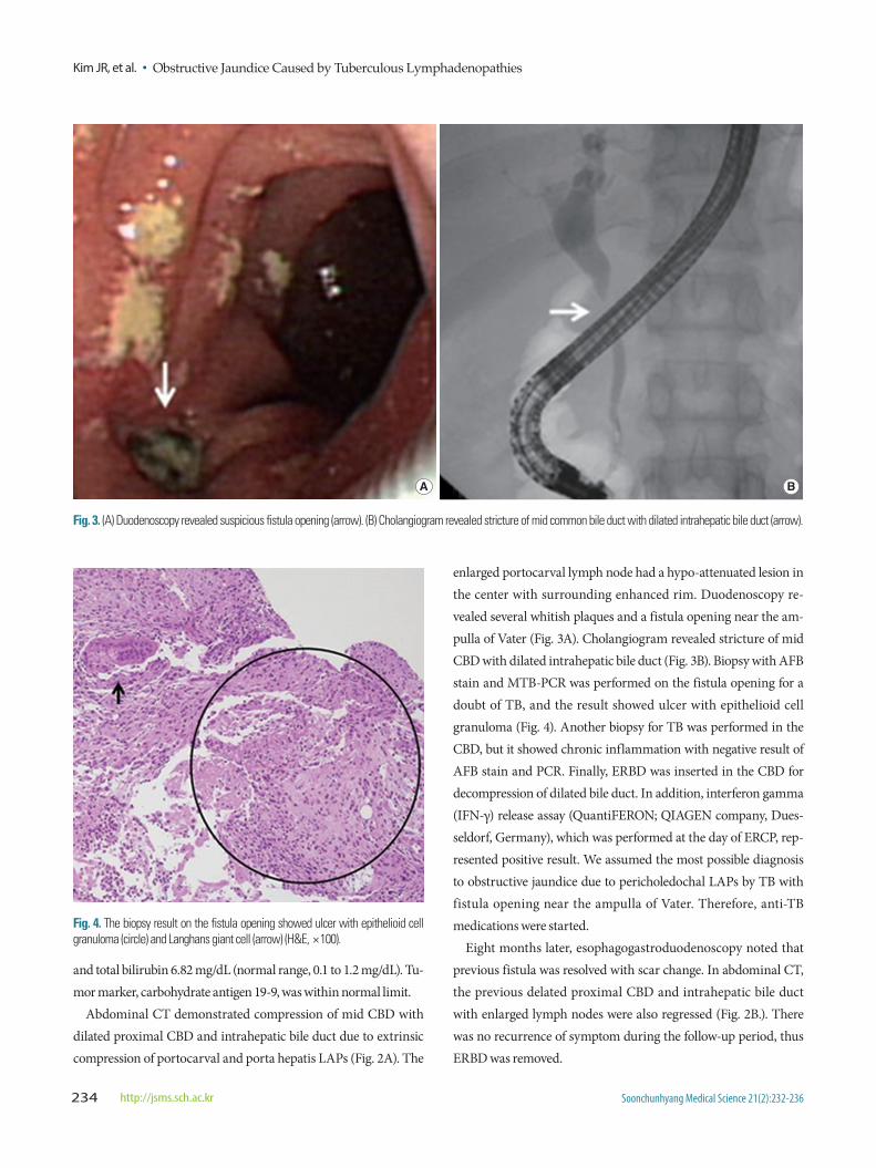
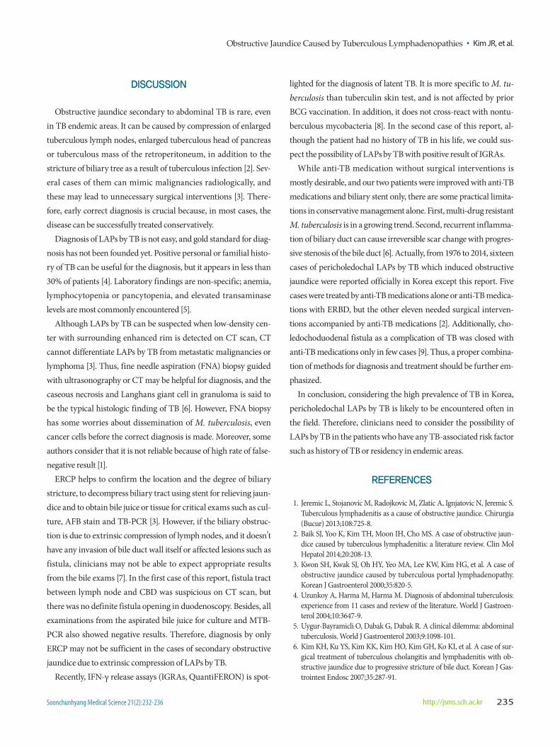
enlarged portocarval lymph node had a hypo-attenuated lesion in the center with surrounding enhanced rim. Duodenoscopy re-vealed several whitish plaques and a fistula opening near the am-pulla of Vater (Fig. 3A). Cholangiogram revealed stricture of mid CBD with dilated intrahepatic bile duct (Fig. 3B). Biopsy with AFB stain and MTB-PCR was performed on the fistula opening for a doubt of TB, and the result showed ulcer with epithelioid cell granuloma (Fig. 4). Another biopsy for TB was performed in the CBD, but it showed chronic inflammation with negative result of AFB stain and PCR. Finally, ERBD was inserted in the CBD for decompression of dilated bile duct. In addition, interferon gamma (IFN-γ) release assay (QuantiFERON; QIAGEN company, Dues-seldorf, Germany), which was performed at the day of ERCP, rep-resented positive result. We assumed the most possible diagnosis to obstructive jaundice due to pericholedochal LAPs by TB with fistula opening near the ampulla of Vater. Therefore, anti-TB medications were started.
Eight months later, esophagogastroduodenoscopy noted that previous fistula was resolved with scar change. In abdominal CT, the previous delated proximal CBD and intrahepatic bile duct with enlarged lymph nodes were also regressed (Fig. 2B.). There was no recurrence of symptom during the follow-up period, thus ERBD was removed.
A B
Fig. 3. (A) Duodenoscopy revealed suspicious fistula opening (arrow). (B) Cholangiogram revealed stricture of mid common bile duct with dilated intrahepatic bile duct (arrow).
Fig. 4. The biopsy result on the fistula opening showed ulcer with epithelioid cell granuloma (circle) and Langhans giant cell (arrow) (H&E, ×100).
Obstructive Jaundice Caused by Tuberculous Lymphadenopathies • Kim JR, et al.
Soonchunhyang Medical Science 21(2):232-236 http://jsms.sch.ac.kr 235
DISCUSSION
Obstructive jaundice secondary to abdominal TB is rare, even in TB endemic areas. It can be caused by compression of enlarged tuberculous lymph nodes, enlarged tuberculous head of pancreas or tuberculous mass of the retroperitoneum, in addition to the stricture of biliary tree as a result of tuberculous infection [2]. Sev-eral cases of them can mimic malignancies radiologically, and these may lead to unnecessary surgical interventions [3]. There-fore, early correct diagnosis is crucial because, in most cases, the disease can be successfully treated conservatively.
Diagnosis of LAPs by TB is not easy, and gold standard for diag-nosis has not been founded yet. Positive personal or familial histo-ry of TB can be useful for the diagnosis, but it appears in less than 30% of patients [4]. Laboratory findings are non-specific; anemia, lymphocytopenia or pancytopenia, and elevated transaminase levels are most commonly encountered [5].
Although LAPs by TB can be suspected when low-density cen-ter with surrounding enhanced rim is detected on CT scan, CT cannot differentiate LAPs by TB from metastatic malignancies or lymphoma [3]. Thus, fine needle aspiration (FNA) biopsy guided with ultrasonography or CT may be helpful for diagnosis, and the caseous necrosis and Langhans giant cell in granuloma is said to be the typical histologic finding of TB [6]. However, FNA biopsy has some worries about dissemination of M. tuberculosis, even cancer cells before the correct diagnosis is made. Moreover, some authors consider that it is not reliable because of high rate of false-negative result [1].
ERCP helps to confirm the location and the degree of biliary stricture, to decompress biliary tract using stent for relieving jaun-dice and to obtain bile juice or tissue for critical exams such as cul-ture, AFB stain and TB-PCR [3]. However, if the biliary obstruc-tion is due to extrinsic compression of lymph nodes, and it doesn’t have any invasion of bile duct wall itself or affected lesions such as fistula, clinicians may not be able to expect appropriate results from the bile exams [7]. In the first case of this report, fistula tract between lymph node and CBD was suspicious on CT scan, but there was no definite fistula opening in duodenoscopy. Besides, all examinations from the aspirated bile juice for culture and MTB-PCR also showed negative results. Therefore, diagnosis by only ERCP may not be sufficient in the cases of secondary obstructive jaundice due to extrinsic compression of LAPs by TB.
Recently, IFN-γ release assays (IGRAs, QuantiFERON) is spot-
lighted for the diagnosis of latent TB. It is more specific to M. tu-berculosis than tuberculin skin test, and is not affected by prior BCG vaccination. In addition, it does not cross-react with nontu-berculous mycobacteria [8]. In the second case of this report, al-though the patient had no history of TB in his life, we could sus-pect the possibility of LAPs by TB with positive result of IGRAs.
While anti-TB medication without surgical interventions is mostly desirable, and our two patients were improved with anti-TB medications and biliary stent only, there are some practical limita-tions in conservative management alone. First, multi-drug resistant M. tuberculosis is in a growing trend. Second, recurrent inflamma-tion of biliary duct can cause irreversible scar change with progres-sive stenosis of the bile duct [6]. Actually, from 1976 to 2014, sixteen cases of pericholedochal LAPs by TB which induced obstructive jaundice were reported officially in Korea except this report. Five cases were treated by anti-TB medications alone or anti-TB medica-tions with ERBD, but the other eleven needed surgical interven-tions accompanied by anti-TB medications [2]. Additionally, cho-ledochoduodenal fistula as a complication of TB was closed with anti-TB medications only in few cases [9]. Thus, a proper combina-tion of methods for diagnosis and treatment should be further em-phasized.
In conclusion, considering the high prevalence of TB in Korea, pericholedochal LAPs by TB is likely to be encountered often in the field. Therefore, clinicians need to consider the possibility of LAPs by TB in the patients who have any TB-associated risk factor such as history of TB or residency in endemic areas.
REFERENCES
1. Jeremic L, Stojanovic M, Radojkovic M, Zlatic A, Ignjatovic N, Jeremic S. Tuberculous lymphadenitis as a cause of obstructive jaundice. Chirurgia (Bucur) 2013;108:725-8.
2. Baik SJ, Yoo K, Kim TH, Moon IH, Cho MS. A case of obstructive jaun-dice caused by tuberculous lymphadenitis: a literature review. Clin Mol Hepatol 2014;20:208-13.
3. Kwon SH, Kwak SJ, Oh HY, Yeo MA, Lee KW, Kim HG, et al. A case of obstructive jaundice caused by tuberculous portal lymphadenopathy. Korean J Gastroenterol 2000;35:820-5.
4. Uzunkoy A, Harma M, Harma M. Diagnosis of abdominal tuberculosis: experience from 11 cases and review of the literature. World J Gastroen-terol 2004;10:3647-9.
5. Uygur-Bayramicli O, Dabak G, Dabak R. A clinical dilemma: abdominal tuberculosis. World J Gastroenterol 2003;9:1098-101.
6. Kim KH, Ku YS, Kim KK, Kim HO, Kim GH, Ko KI, et al. A case of sur-gical treatment of tuberculous cholangitis and lymphadenitis with ob-structive jaundice due to progressive stricture of bile duct. Korean J Gas-trointest Endosc 2007;35:287-91.
Kim JR, et al. • Obstructive Jaundice Caused by Tuberculous Lymphadenopathies
Soonchunhyang Medical Science 21(2):232-236236 http://jsms.sch.ac.kr
7. Probst A, Schmidbaur W, Jechart G, Hammond A, Zentner J, Niculescu E, et al. Obstructive jaundice in AIDS: diagnosis of biliary tuberculosis by ERCP. Gastrointest Endosc 2004;60:145-8.
8. Longo DL, Kasper DL, Jameson JL, Fauci AS, Hauser SL, Loscalzo J. Har-rison’s principles of internal medicine. 18th ed. New York (NY): McGraw-
Hill Medical; 2011. p. 1351.9. Yoon SJ, John BM, Jung SH, Kim A, Ko BS, Yang HW, et al. Choledocho-
duodenal fistula caused by tuberculosis. Korean J Gastrointest Endosc 2005;30:286-9.

![Follow Sipi cantpancreatitis · tuberculous]Tuberculous 38. 2010167550 lymphaderioPathy [lymph Fallow Up: 4 Korea Republ.. 09-Sep- node 11. tuberculosis]Tuberculous Pleural effusion](https://static.fdocuments.net/doc/165x107/5f7d6a51d573d133e30b0217/follow-sipi-tuberculoustuberculous-38-2010167550-lymphaderiopathy-lymph-fallow.jpg)