Obstetric operations: induced abortion, forceps delivery ...
58
Obstetric operations: induced abortion, forceps delivery, vacuum extraction, cesarean section Gyula Richárd Nagy MD, PhD
Transcript of Obstetric operations: induced abortion, forceps delivery ...

Obstetric operations:
induced abortion,
forceps delivery,
vacuum extraction,
cesarean section
Gyula Richárd Nagy MD, PhD

Obstetricoperations
Before the 24th week
Induced abortion
1st trimester
D & C
2nd trimester
Medical induction, oxytocin infusion,
curettage
After the 24th week
Delivery
At the 1st stage of labor
Cesarean section
At the 2nd stage of labor
Forceps delivery
Vacuum extraction(at term)

Induced abortion

Induced abortion
(abortus arteficialis, interruptio graviditatis):
An induced abortion is the medical or surgical
termination of pregnancy before 24 weeks of
gestation (before the time of fetal viability).

Indications
Non-medical indications
Until the 12th week
• „Social” indication
Until the 18th week
• Crime (rape)
• Partial or total legal incapacity
• Misdiagnosis of pregnancy
• Youthful pregnant (<18 years of age)

Medical indicationsMaternal indication (Termination of the pregnancy may be
permitted at any time when serious illness threatens the mother’s
life.)
Fetal indication (When the risk of serious genetic disease or
malformation in the fetus is between 50% and 100%, and there is
no possibility of treatment, termination may be permitted until the
20th week. When a late diagnosis is made because of laboratory
delay and not through any negligence on the mother’s part,
termination may be permitted up to the 24th week.)
Anomalies fatal in the postnatal period (Termination of the
pregnancy may be permitted at any time.)
Genetical, teratologic indication (When the risk of genetic
disorder or teratogenic demage to the fetus exceeds 10%, and the
disorder/demage is likely to be severe, termination up to the 12th
week of pregnancy is permitted.)

Abortion techniques
Induced abortion for early pregnancies
(up to the 6th week)
„Menstrual aspiration” („mini-suction”)
Medical abortion
• Mifepristone (RU-486) and adjuvant prostaglandine
• Methotrexate

Induced abortion in the 1st trimester
(up to the 12th week)
Cervical dilatation
• Metal/Mechanical dilators (metal Hegar-dilators)
• Hygroscopic dilators (Laminaria, Dilapan)
• Chemical (Rivanol, Prostaglandins)
Evacuating the pregnancy
• Vacuum aspiration
• Curettage

Induced abortion in the 2nd trimester
(up to the 24th week)
Medical induction
(extraamnial Rivanol, Laminaria, vaginal prosztaglandin pill)
Abortion due to oxytocin infusion
Curettage

Complications
Intraoperative complications:
perforatio uteri
hemorrhage
• injury
• uterine atony
• retention
• coagulopathy
Early postoperative complication: infection
Late postoperative complication: infertility (Asherman-syndrome: intrauterine synechiae due to injury of the basal layer of the endometrium)

Operative vaginal delivery

At the second-stage labor operative vaginal
delivery (forceps or vacuum extraction) is
indicated in any condition threatening the
mother or fetus that is likely to be relieved
by delivery, whereas at the first-stage labor
cesarean section is the choice (partus cum
operatione obstetrica).

Operative vaginal delivery
Forceps delivery

The forceps
Basically consist of two crossing branches;
components: handle, lock, shank, blades.
The cephalic curve conforms to the shape of
the fetal head, the pelvic curve corresponds
more or less to the axis of the birth canal.

Types
Pajot-Naegele-forceps. Outlet forceps, has cephalic and pelvic curve.
Kielland-forceps. Able to rotate the head. Can be used for all forceps deliveries as it has cephalic curve only.
Piper-forceps. Can be used in breech presentation to deliver the fetal head.
Shute-forceps. Has cephalic and pelvic curve. Used in preterm deliveries. Here the goal is not to pull out the fetal head, but to protect the fragile head of the preterm fetus by pushing the tissues of the birth canal away.
Prerequisites for forceps
application
Trained obstetrician in forceps deliveries
Completely dilated cervix
Ruptured membranes
The fetal head must be engaged (the greatest transverse
diameter in an occiput presentation passes through the
pelvic inlet)
The size and consistence of fetal head is suitable for a
forceps delivery
Living fetus
No suspicion of cephalopelvic disproportion

Indications for forceps delivery
ProphylacticMaternal diseases (CNS vascular malformations, aortaaneurysm, heart disease, etc.)
Previous operations on the uterus (CS, metroplasty)
Prolonged second-stage labor
Threatened intrauterine fetal asphyxia
VitalHeart failure, pulmonary edema
Eclampsia
Severe hemorrhage, DIC
Definite fetal asphyxia

Overall rules of forceps delivery
Preparation
Mother in lithotomy position, disinfect and isolate the area of the operation.
The bladder should be emptied
The exact position of the fetal head must be checked once again with inner examination
Episiotomy in regional analgesia
Steps
Check in front of the vaginal inlet how the forceps will stay in the birth canal.
Application of the blades
After positioning, the branches are articulated. Closure. Traction probe
Traction

Types of forceps delivery I.
Outlet forceps delivery
Scalp is visible at the introitus without
separating the labia
Fetal skull has reached pelvic floor
Sagittal suture is in anterioposterior diameter or
rotation does not exceed 45 degrees
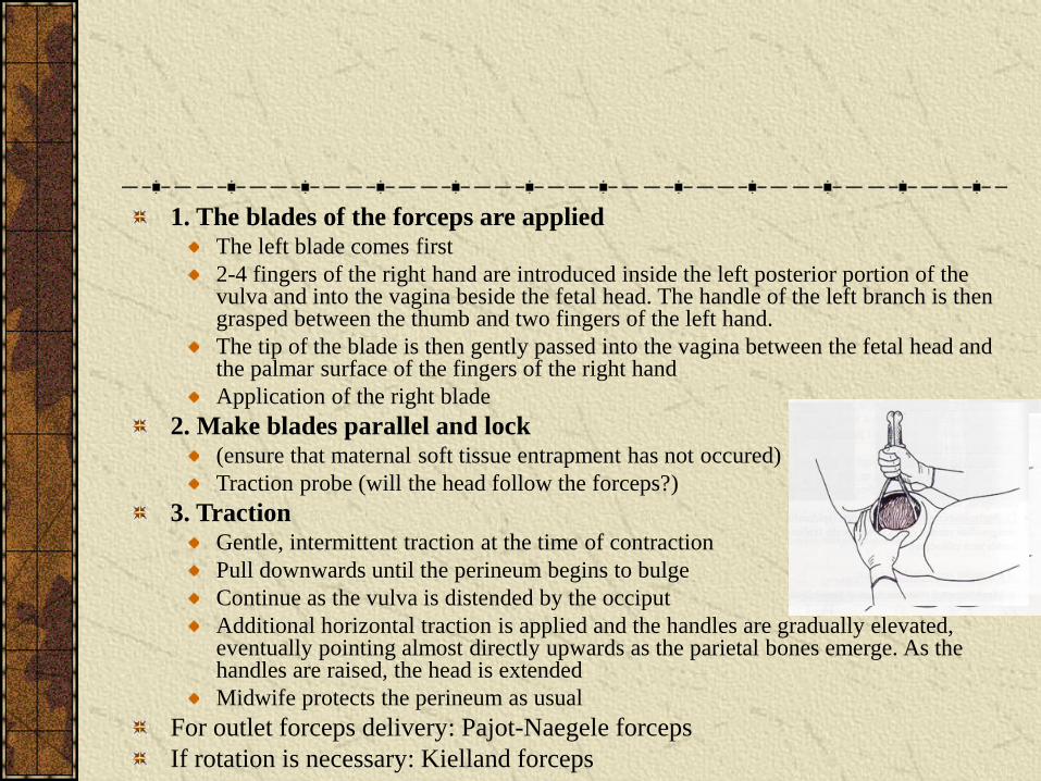
1. The blades of the forceps are appliedThe left blade comes first
2-4 fingers of the right hand are introduced inside the left posterior portion of the vulva and into the vagina beside the fetal head. The handle of the left branch is then grasped between the thumb and two fingers of the left hand.
The tip of the blade is then gently passed into the vagina between the fetal head and the palmar surface of the fingers of the right hand
Application of the right blade
2. Make blades parallel and lock (ensure that maternal soft tissue entrapment has not occured)
Traction probe (will the head follow the forceps?)
3. TractionGentle, intermittent traction at the time of contraction
Pull downwards until the perineum begins to bulge
Continue as the vulva is distended by the occiput
Additional horizontal traction is applied and the handles are gradually elevated, eventually pointing almost directly upwards as the parietal bones emerge. As the handles are raised, the head is extended
Midwife protects the perineum as usual
For outlet forceps delivery: Pajot-Naegele forceps
If rotation is necessary: Kielland forceps

Types of forceps delivery II.
Low- and midforceps operations
If the greatest transverse diameter of the fetal head
passes through the pelvic inlet
Leading point of fetal skull is at the interspinal plane.
This is point „0”.
Position of the head in the birth channel is the distance
in cm from this point: +1, +2, +3, +4, +5. (If the leading
point is above the interspinal plane: from -1 to -5.)

Low forceps operation
Leading point of fetal skull is at station ≥ +2 cm
Midforceps operation
Station is between 0 and +2 cm

1. the blades of the forceps are applied
2. make blades parallel and lock
check
traction probe
3. traction
Mimic the remaining rotation of the head. Withintermittent traction rotate the minor fontanella in front and pull downwards until the sagittal suture is in theanterioposterior diameter, and the subocciput is underthe symphysis

Types of forceps delivery III.
Forceps delivery in breech presentation
Piper forceps
Assistant holds the fetal legs and body
horizontal to avoid abnormal extension of the
neck
After episiotomy first the left then the right
blade is applied
Traction is applied upwards

Complications of forceps
operations
Mostly at midforceps operations; these should be performed by trained obstetricians
In the mother: vaginal, bladder, perineal rectal injuries
In the fetus: cephalhaematoma, facial paresis, skull fraction, intracranial hemorrhage
After forceps operation it is necessary to check the uterine cavity and the vagina for injuries

Operative vaginal delivery
Vacuum extraction

Developed by Malmström Swedish obstetrician (1954)
Vacuum extraction contains a suction unit, a flexible
tubing and a cup (metal or plastic).
Vacuum be created gradually until a negative pressure of
80 kPa (0.8 kg/cm2) is reached. Traction should be
intermittent and coordinated with maternal expulsive
efforts.
Negative pressure can be increased to 0.8 kg/cm2 in 1-2
minutes.

Contraindications include malposition of the fetal
head, such as face and brow presentation. In these
cases cesarean section is indicated.
Vacuumextraction is contraindicated in preterm
delivery.
Indications of vacuum extraction
Same as for forceps delivery.
Technique of vacuum extraction
Check if your vacuum delivery system is working.
Proper cup placement is the most important determinant of success. The center of the cup should be over the saggitalsuture and about 3 cm in front of the posterior fontanellatoward the face. (Anterior placement can result in cervicalspine extension. Asymmetrical placement may worsenasynclitism.)
The full circumference of the cup should be palpated bothbefore and after the vacuum has been created to ensure thatmaternal soft tissue entrapment has not occured.

Technique of vacuum extraction
Traction should be intermittent and coordinated withmaternal expulsive efforts. The fingers of one hand areplaced against the suction cup, while the other hand graspsthe handle of the instrument. No data are availableregarding the number of pulls required to effect delivery.
When the head is delivered, turn off the suction.

Complications of vacuum extraction
Scalp lacerations, subgaleal hematomas,
cephalohematomas, subconjunctival hemorrhage,
retinal hemorrhage, hyperbilirubinaemia.

Cesarean delivery
(sectio caesarea)

If the cervix is not fully dilated,
or if the cervix is fully dilated but the head
is not engaged,
to finish the delivery fast cesarean section is
the safest choice.

Prerequisites for cesarean section
No absolute prerequisite and
No absolute contraindication
This is due to precise operation techniques, antibiotics, blood transfusion possibilities and safe anaesthesia.
Only prerequisite is, that the fetal head is not engaged.
(If contracted outlet is diagnosed after fetal head engagement, cesarean section should be performed; during the operation the head should be pushed back via a vaginal examination, and pulled out from the pelvis by grabbing the shoulders.)

Indications of cesarean section
Absolute indication: no possibility for vaginal delivery
Relative indication: possibility for vaginal delivery, but higher risk of fetal and/or maternal mortality and morbidity without the cesarean section.
Vital indication: if done to prevent an immediate life threatening situation.
Prophylactic indication: if done to prevent complications.

Indications of cesarean section
Prophylactic
maternal
fetal
maternal/fetal
Vital
maternal
fetal
maternal/fetal

Prophylactic maternal indications
Maternal illnesses
Previous operations on the uterus (previous
cesarean section, metroplasty, etc.)
Contractions of the pelvis (diminished pelvic
capacity)
Late primiparity (≥30 years primipara)
Prophylactic fetal indications
Threatened fetal asphyxia (scalp pH 7,21-7,25)
Placental dysfunction, hypoxia
Fetal illnesses, and risk of it (fetopathy, Rh-
isoimmunisation, operable anomalies)
Pregnancy after infertility treatment

Prophylactic maternal/fetal indication
Damning gestational history
Dystocia, prolonged labor
Fetopelvic disproportion, malpresentation or
position of the fetus
Some cases of twin pregnancy

Vital maternal indications
Heart failure
Pulmonary edema
Severe hemorrhage (ie. vital maternal/fetal
indications)
DIC

Vital fetal indications
Fetal asphyxia (scalp pH <7,21)
Prolapse of the umbilical cord
Neglected transverse lie (after a while also is a
vital maternal indication)
Ascending infection, fetal pneumonia

Vital maternal/fetal indications
Eclampsia
Uterine rupture
Placenta praevia
Placental abruption

Technique for cesarean delivery
Intratracheal narcosis or regional (spinal orepidural) anaesthesia is performed
Abdominal incisionVertical incision (lower median) or
suprapubic transverse incision (Pfannenstiel-) is usedto open the abdominal cavity
• Pfannenstiel incision is made at the level of the pubic hairlineand it is extended somewhat beyond the lateral borders of therectus muscles.
• The skin and subcutaneous tissue are incised, dissection is continued to the level of the fascia.
• Fascia is incised.
• The rectus muscles are separated.
• The underlying peritoneum is opened.

Uterine incision„Classical” (corporal longitudinal) incision is a vertical incision above thelower uterine segment and reaches theuterine fundus.
(Seldom used today. Indications: transverse lie of a largefetus, very small fetus especially if breech and the loweruterine segment is not thinned out, some cases of placenta previa with anterior implantation, leiomyoma occupies thelower uterine segment,etc.)
Transperitoneal cervical transverse(„t.c.t.”) incision is easier to repair, islocated at a site least likely to ruptureduring a subsequent pregnancy, and doesnot promote adherence of bowel oromentum to the incisional line.

The loose vesicouterine serosa is grasped and inceased
transversely.
The lower flap of peritoneum is elevated and the bladder is
gently separated from the underlying myometrium.
The uterus is entered through the lower uterine segment (1
cm below the upper margin of the peritoneal reflection).
The incision should be made large enough to allow
delivery of the head and trunk of the fetus. If the placenta
is encountered in the line of incision, it must be either
detached or incised.
If membranes are still, we rupture them
(In a cephalic presentation) the head is elevated gently with the fingers and palm through the incision, aided by modest transabdomnial fundal pressure
Eliminate mucus from fetal mouth
After clamping and cutting the umbilical cord the fetus is given to the neonatologist
Intravenous and/or intramyometral oxytocin is given to contract the uterus satisfactory and help the detachment of the placenta
The placenta and membranes are then delivered (manual removal or spontaneous delivery), afterwards the uterine cavity is inspected (check manually for remnants, or abnormalities (injuries, myomas, etc.))

If the cervix is closed, we have to dilate it to let the lochia
to go through
(Hegar dilators, from the uterine cavity through the cervix into the
hand of the assistant doctor, who performs a vaginal examination and
protects soft tissue injury with his fingers. Easier to use Zangemeister-
dilator, a horn-shaped instrument to dilate the cervix. It is pulled out
by the assisitant who performs the vaginal examination.)
The uterine incision is then closed in one or two
layers (muscular and seromuscular)
Closure or nonclosure of visceral peritoneum
After the sponge and instrument counts are found
to be correct, the abdominal incision is closed in
layers. If the subcutaneous tissue is at least 2 cm
thick, it should be closed and drained.
Patient is observed in a postoperative ward.

Complications of cesarean section
Nowadays complications of cesarean section are rare. The risk is higher, if
The pregnant is obese, or having preeclampsia
The presenting fetal part is in a very low or very high position
The operation is after preterm membrane rupture
There is severe haemorrhage
The pregnancy is preterm
The operation is done „fast”, in a rush
The obstetrician is not well trained, or the professional surveillance is lacking

Bladder injury 0,3%
Transurethral injected methylene blue dye can help
visualisation, should be sutured in double-layer, Foley-
catheter should be used until microscopic hematuria is
present, antibiotic-prophylaxis is recommended
Ureter injury <0,1%
Consult with urologist
Bowel injury 0,2%
Should be closed in double layer, consult with surgeon

Hemorrhage
Infection
Pulmonary embolism
Fetal injuries: very rare. If the delivery of the fetus is complicated, bone- or nerve injuries can be expected.
Anaesthesiological complications are very rare after the introduction of regional anaesthesia

Postpartum hysterectomy
Hysterectomy performed at or following delivery
may be lifesaving if there is severe obstetrical
hemorrhage. It can be carried out in conjunction
with cesarean delivery or following vaginal
delivery. Postpartum hysterectomy (Porro-
operation) is done from a laparotomy.

Major complication is increased blood loss (pelvic vessels
are appreciably hypertrophied). The morbidity rate
associated with emergency hysterectomy is substantively
increased as compared to a planned operation
In most cases blood transfusion is necessary
In severe bleeding ligation of the hypogastric arteries can
be used

Postpartum hysterectomy should be
considered
Severe hemorrhage (uterine atony, uterine
rupture, DIC, coagulation problems)
Abnormal placentation (placenta accreta,
increta, percreta)
Severe infection, sepsis, septic shock
Gynecological malignancy

After cesarean section, the uterine incision should be closed before hysterectomy
Postpartum hysterectomy can be supracervical or total hysterectomy using standard operative techniques. The operation should be done by the most trained obstetrician
In case of invasive cervical cancer, radical hysterectomy (Wertheim-operation) should be performed

Obstetricoperations
Before the 24th week
Induced abortion
1st trimester
D & C
2nd trimester
Medical induction, oxytocin infusion,
curettage
After the 24th week
Delivery
At the 1st stage of labor
Cesarean section
At the 2nd stage of labor
Forceps delivery
Vacuum extraction(at term)

Thank you for your attention!