
Obstetric Anaesthesia - Resources
100
Volume 34 September 2019 Editors: Christina Lundgren and Victoria Howell Education for anaesthesia providers worldwide The Journal of the World Federation of Societies of Anaesthesiologists Available at: www.wfsahq.org/resources/update-in-anaesthesia Obstetric Anaesthesia • Obstetric anaesthesia in resource limited settings • Obstetric airway management • General anesthesia for elective cesarean section in resource-limited settings • Obstetric spinal anaesthesia • Management of total spinal block in obstetrics • Labour epidural the basics • Labour epidrual troubleshooting • Establishing an epidural service for labour analgesia in a variable resource environment • Emergency management of maternal collapse and arrest • Pre-eclampsia - prevention, diagnosis and management • Placental pathology: a review of placenta previa, placental abruption and placenta accreta • Anaesthetic implications of morbid obesity in pregnancy • Update in obstetric trauma management • Update of maternal sepsis • Oxytocics • Obstetric and foetal physiology • Newborn resuscitation • Anaesthesia for non obstetric surgery during pregnancy • Guide for contributors ISSN 1353-4882 SPECIAL EDITION OBSTETRIC ANAESTHESIA
Transcript of Obstetric Anaesthesia - Resources
Volume 34 September 2019
Editors: Christina Lundgren and Victoria Howell
Education for anaesthesia providers worldwide
The Journal of the World Federation of Societies of Anaesthesiologists
Available at: www.wfsahq.org/resources/update-in-anaesthesia
Obstetric Anaesthesia• Obstetric anaesthesia in resource limited settings
• Obstetric airway management
• General anesthesia for elective cesarean section in resource-limited settings
• Obstetric spinal anaesthesia
• Management of total spinal block in obstetrics
• Labour epidural the basics
• Labour epidrual troubleshooting
• Establishing an epidural service for labour analgesia in a variable resource environment
• Emergency management of maternal collapse and arrest
• Pre-eclampsia - prevention, diagnosis and management
• Placental pathology: a review of placenta previa, placental abruption and placenta accreta
• Anaesthetic implications of morbid obesity in pregnancy
• Update in obstetric trauma management
• Update of maternal sepsis
• Oxytocics
• Obstetric and foetal physiology
• Newborn resuscitation
• Anaesthesia for non obstetric surgery during pregnancy
• Guide for contributors
ISSN 1353-4882
SPECIAL
EDITION
OBSTETRIC ANAESTHESIA

NEW UPDATE TEAM
EditorsChristina Lundgren (SA)
Victoria Howell (UK)
Emeritus Editor-in-chiefBruce McCormick (UK)
Editorial Board of Update in AnaesthesiaDouglas Bacon (USA)
Gustavo Elena (Argentina)
Martin Chobli (Benin)
Dr Aboudoul-Fataou Ouro-Bang’na Maman (Togo)
Dr David Pescod (Australia)
Dr Jeanette Thirlwell (Australia)
Dr Isabeau Walker (UK)
SS Harsoor (India)
Kazuyoshi Hirota (Japan)
Zhanggang Xue (China)
Jing Zhao (China)
Typeset and printed by Sumographics Ltd
© World Federation of Societies of Anaesthesiologists 2019. This issue may be freely reproduced for the purposes of private research and study and extracts (or indeed, the full report) may be included in professional journals provided that suitable acknowledgement is made and the reproduction is not associated with any form of advertising. Applications for commercial reproduction should be addressed to: World Federation of Societies of Anaesthesiologists, Dean Bradley House, 52 Horseferry Rd, London SW1P 2AF, UK.
DisclaimerThe WFSA takes all reasonable care to ensure that the information contained in Update in Anaesthesia is accurate. We cannot be held responsible for any errors or omissions and take no responsibility for the consequences of error or for any loss or damage which may arise from reliance on information contained.
The WFSA would like to thank Baxter for their financial support of this edition of Update in Anaesthesia

Cont
ents
Contents
Obstetric AnaesthesiaObstetric anaesthesia in resource limited settings................................................................................... 5
Obstetric airway management ............................................................................................................................. 7
General anesthesia for elective cesarean section in resource-limited settings ...................... 14
Obstetric spinal anaesthesia ................................................................................................................................... 18
Management of total spinal block in obstetrics ........................................................................................ 22
Labour epidural the basics ...................................................................................................................................... 26
Labour epidrual troubleshooting ........................................................................................................................ 30
Establishing an epidural service for labour analgesia in a variable ...............................................
resource environment ................................................................................................................................................ 35
Emergency management of maternal collapse and arrest ................................................................ 41
Pre-eclampsia - prevention, diagnosis and management .................................................................. 46
Placental pathology: a review of placenta previa, placental
abruption and placenta accreta ........................................................................................................................... 51
Anaesthetic implications of morbid obesity in pregnancy ................................................................. 56
Update in obstetric trauma management .................................................................................................... 63
Update of maternal sepsis ....................................................................................................................................... 71
Oxytocics ............................................................................................................................................................................ 78
Obstetric and foetal physiology ........................................................................................................................... 81
Newborn resuscitation ............................................................................................................................................. 85
Anaesthesia for non obstetric surgery during pregnancy ................................................................... 91
Guide for contributors ................................................................................................................................................ 97
5
© World Federation of Societies of Anaesthesiologists 2019. This issue may be freely reproduced for the purposes of private research and study and extracts (or indeed, the full report) may be included in professional journals provided that suitable acknowledgement is made and the reproduction is not associated with any form of advertising. Applications for commercial reproduction should be addressed to: World Federation of Societies of Anaesthesiologists, Dean Bradley House, 52 Horseferry Rd, London SW1P 2AF, UK.
Editor’s Notes
Edit
oria
l
Welcome to Volume 34 of Update in Anaesthesia, and this special Obstetric themed edition of the journal. This edition contains a range of articles covering many important aspects of obstetric anaesthesia from obstetric physiology to the management of pre-eclampsia, and from obstetric airway management to neonatal resuscitation. I hope that you are able to find something that is of interest and use to you.
This obstetric edition was partly inspired by the SAFE Obstetrics courses that are run in collaboration with the WFSA, so thank you to Dr James Leedham co-author of the SAFE Obstetric Handbook. The Safer Anaesthesia From Education (SAFE) courses are aimed at anaesthesia providers working with limited resources, and are an opportunity to update skills and knowledge of obstetric anaesthesia. We hope this edition of Update in Anaesthesia also helps to refresh your obstetric knowledge, and we have included topics that we hope are relevant whatever setting you work in. Further information on the SAFE courses can be found on the WFSA website https://www.wfsahq.org/wfsa-safer-anaesthesia-from-education-safe.
My grateful thanks must also go to Dr Mauricio Vasco, Chair of the WFSA Obstetric Committee. He helped recruit authors for this edition in addition to writing an interesting editorial on Obstetric Anaesthesia in Resource Limited Settings. Many members of the Obstetric Committee contributed to this edition of the journal, whether through writing articles, recruiting authors, or reviewing manuscripts. Thanks must go to all the reviewers who have peer reviewed every single article in this edition. They are an invaluable part of the process but, as they are part of the blind peer review process, must remain nameless. They should know that they have my sincerest gratitude though, as we wouldn’t be able to publish this journal without them. If you would like to become a peer reviewer for Update in Anaesthesia, please email [email protected]. Thank you also to Dr Maytinee Lilaonitkul, Editor-in-Chief of Anaesthesia Tutorial of the Week, the online open access educational resource from the WFSA, for allowing us to reproduce 2 tutorials for inclusion in this journal.
Looking ahead, the World Congress of Anaesthesiologists will be held in Prague in the Czech Republic from 5th-9th September 2020. These quadrennial congresses organised by the WFSA are an amazing opportunity to meet fellow anaesthesiologists from around the world, as well as being of great scientific and educational value. Whether your interest is in Obstetric Anaesthesia, or one of the other 23 subspecialty areas, there really is something for everyone with many excellent speakers and interesting workshops. Abstract submission and registration for the Congress are now open and I look forward to seeing you there.
Since the last edition of Update in Anaesthesia, Dr Christina Lundgren and I have taken over as Editors-in-Chief. However, despite the change of leadership, the aims and objectives of Update in Anaesthesia remain the same. To produce high-quality, clinically relevant educational articles that can be used by anaesthesia practitioners the world over. We aim to produce one themed edition a year such as this obstetric edition, but in addition to this we are also keen to publish educational articles on other subjects as well as original research, audits or case reports. We welcome your contribution to the journal, and if you have any suggestions about the journal or manuscripts that you would like to be published, please do not hesitate to get in touch. You can submit manuscripts directly through our online submission system at https://www.editorialmanager.com/wfsa/default.aspx or email [email protected]
Victoria HowellCo-Editor-in-Chief
Update in AnaesthesiaConsultant Anaesthetist
The Queen Elizabeth Hospital King’s Lynn
UK
4 www.wfsahq.org/resources/update-in-anaesthesia
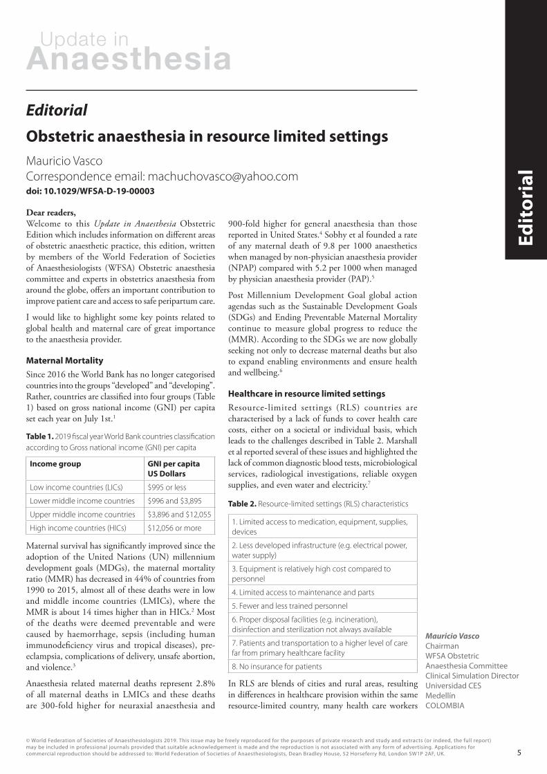
Editorial
Dear readers,Welcome to this Update in Anaesthesia Obstetric Edition which includes information on different areas of obstetric anaesthetic practice, this edition, written by members of the World Federation of Societies of Anaesthesiologists (WFSA) Obstetric anaesthesia committee and experts in obstetrics anaesthesia from around the globe, offers an important contribution to improve patient care and access to safe peripartum care.
I would like to highlight some key points related to global health and maternal care of great importance to the anaesthesia provider.
Maternal MortalitySince 2016 the World Bank has no longer categorised countries into the groups ‘‘developed” and ‘‘developing”. Rather, countries are classified into four groups (Table 1) based on gross national income (GNI) per capita set each year on July 1st.1
Table 1. 2019 fiscal year World Bank countries classification according to Gross national income (GNI) per capita
900-fold higher for general anaesthesia than those reported in United States.4 Sobhy et al founded a rate of any maternal death of 9.8 per 1000 anaesthetics when managed by non-physician anaesthesia provider (NPAP) compared with 5.2 per 1000 when managed by physician anaesthesia provider (PAP).5
Post Millennium Development Goal global action agendas such as the Sustainable Development Goals (SDGs) and Ending Preventable Maternal Mortality continue to measure global progress to reduce the (MMR). According to the SDGs we are now globally seeking not only to decrease maternal deaths but also to expand enabling environments and ensure health and wellbeing.6
Healthcare in resource limited settingsResource-limited settings (RLS) countries are characterised by a lack of funds to cover health care costs, either on a societal or individual basis, which leads to the challenges described in Table 2. Marshall et al reported several of these issues and highlighted the lack of common diagnostic blood tests, microbiological services, radiological investigations, reliable oxygen supplies, and even water and electricity.7
Table 2. Resource-limited settings (RLS) characteristics
Income group GNI per capita US Dollars
Low income countries (LICs) $995 or less
Lower middle income countries $996 and $3,895
Upper middle income countries $3,896 and $12,055
High income countries (HICs) $12,056 or more1. Limited access to medication, equipment, supplies, devices
2. Less developed infrastructure (e.g. electrical power, water supply)
3. Equipment is relatively high cost compared to personnel
4. Limited access to maintenance and parts
5. Fewer and less trained personnel
6. Proper disposal facilities (e.g. incineration), disinfection and sterilization not always available
7. Patients and transportation to a higher level of care far from primary healthcare facility
8. No insurance for patients
In RLS are blends of cities and rural areas, resulting in differences in healthcare provision within the same resource-limited country, many health care workers
Obstetric anaesthesia in resource limited settings
Edit
oria
lMaternal survival has significantly improved since the adoption of the United Nations (UN) millennium development goals (MDGs), the maternal mortality ratio (MMR) has decreased in 44% of countries from 1990 to 2015, almost all of these deaths were in low and middle income countries (LMICs), where the MMR is about 14 times higher than in HICs.2 Most of the deaths were deemed preventable and were caused by haemorrhage, sepsis (including human immunodeficiency virus and tropical diseases), pre-eclampsia, complications of delivery, unsafe abortion, and violence.3
Anaesthesia related maternal deaths represent 2.8% of all maternal deaths in LMICs and these deaths are 300-fold higher for neuraxial anaesthesia and
Mauricio Vasco Chairman WFSA Obstetric Anaesthesia Committee Clinical Simulation Director Universidad CESMedellínCOLOMBIA
Mauricio VascoCorrespondence email: [email protected]: 10.1029/WFSA-D-19-00003
5
© World Federation of Societies of Anaesthesiologists 2019. This issue may be freely reproduced for the purposes of private research and study and extracts (or indeed, the full report) may be included in professional journals provided that suitable acknowledgement is made and the reproduction is not associated with any form of advertising. Applications for commercial reproduction should be addressed to: World Federation of Societies of Anaesthesiologists, Dean Bradley House, 52 Horseferry Rd, London SW1P 2AF, UK.

have little or no access to basic, practical information. Indeed, many have come to rely on observation, on advice from colleagues, and on building experience empirically through their own treatment successes and failures. The disparity between theoretical and practical availability of information is due to several factors, including a failure to apply international development policies and guidelines, failure to engage with modern educational initiatives as massive open online courses and open access electronic journals/textbooks and tended to focus on approaches for higher-level health professionals, while ignoring other approaches that remain essential for the vast majority of primary health care workers.8
There is an interest by HICs academic centres in aiding LMICs in anaesthesia and surgical care; a large proportion of this aid is in the form of short-term medical missions, provision of equipment, and the training of personnel no only for clinical aspect but also for getting skills in research; the best way to achieve this in LMICs is building local capacity by training and mentoring healthcare workers, together with technology and skill transfer by HIC academic centres, rather than short-term aid programs.9,10
As in all fields of skill development, the risk to countries and institutions in LMICs is that heath care trainees can use newly acquired skills to move to the more desirable urban areas, another countries and/or higher paying posts; to avoid this it is important to build successful incentives local programs to retain these trainees.8
Strengthening emergency and essential surgical care and anaesthesia as a component of universal health coverage (UHC)World Health Assembly resolution 68.15 recognizes access to emergency and essential anaesthesia and surgical care as an integral part of UHC.11 There is also growing recognition that up to one third of the global burden of disease is surgically correctable, which is a greater burden than that of human immunodeficiency virus, tuberculosis and malaria combined. There is an urgent need to address deficiencies in access to safe anaesthesia care. An additional 1.27 million surgical, obstetric and anaesthesia providers will be required by 2030 to achieve Universal Health Coverage.12 The World Federation of Societies of Anaesthesiologists (WFSA) is committed to working with governments and non-governmental organisations to improve patient care and access to safe anaesthesia worldwide. Anaesthesiologist led development of anaesthesia services is vital if we are to achieve Universal Health Coverage by 2030.13
In RLS countries, anaesthesia is associated with unacceptably high mortality rates, training and ongoing maintenance of standards for a safe practice of anaesthesia and tools to assess surgical and anaesthesia capacity are essential for increasing the number of providers and improving the safety for patients worldwide.14,15,16
Training future anaesthesia provider in obstetric careAnaesthesia should be provided, led, or overseen by an anaesthesiologist, the anaesthesia provider is an essential member of the delivery unit team. Nearly 60% of women require anaesthetic intervention around the time of delivery. The number of patients who deliver by caesarean section is increasing in all the world and many more require anaesthetic care for operative/assisted deliveries, obstetric emergencies treatment and procedures during pregnancy or puerperium on the labour and delivery (L&D) suite, operating rooms and critical care facilities.
Additionally, obstetric practice carries a high risk of medical liability and anaesthesiologists are frequently named as part of the obstetrics team. Through increasing patient safety initiatives, practicing patient safety behaviours and prevention of fatigue and burnout, we can continue to improve clinical care and decrease medical error in patient care. As a result, the modern obstetric anaesthesia provider must have a role of peripartum/perioperative physician.6
Finally I hope that the readers of UIA find it a useful addition to their anaesthesia libraries; this edition will be available along with all the other WFSA education resources at www.wfsahq.org.
Mauricio Vasco RamirezChairman, WFSA Obstetric Anaesthesia Committee
Clinical Simulation Director, Universidad CES Medellín, Colombia
References1. World Bank Country and Lending Groups. https://datahelpdesk . worldbank.org/knowledgebase/articles/906519-worldbankcountry-and lending-groups. Accessed 10 December, 2018.
2. Alkema L, Chou D, Hogan D, et al. Global, regional, and national levels and trends in maternal mortality between 1990 and 2015, with scenario based projections to 2030: a systematic analysis by the UN Maternal Mortality Estimation Inter-Agency Group. Lancet. 2016; 387: 462-74.
3. Nair M, Nelson-Piercy C, Knight M. Indirect maternal deaths: UK and global perspectives. Obstet Med 2017; 10: 10–5.
4. Mhyre JM. The critical role of obstetric anaesthesia in low-income and middleincome countries. Lancet Glob Health. 2016; 4: e290-1.
5. Sobhy S, Zamora J, Dharmarajah K, et al. Anaesthesia-related maternal mortality in low-income and middle-income countries: a systematic review and metaanalysis. Lancet Glob Health 2016; 4: e320–e327.
6. Vasco M. Training future anesthesiologists in obstetric care. Curr Opin Anesthesiol 2017, 30: 313–318.
7. Marshall JC, Bosco L, Adhikari NK, et al. What is an intensive care unit? A report of the task force of the World Federation of Societies of Intensive and Critical Care Medicine. J Crit Care 2017; 37: 270–6.
8. Vasco M, Pandya S, Van Dyk D, et al. Maternal critical care in resource limited settings. Narrative review. Int J Obstet Anesth. 2019; 37: 86–95.
9. Chellam S, Ganbold L, Gadgil A, et al. Contributions of academic institutions in high income countries to anesthesia and surgical care in low- and middle-income countries: are they providing what is really needed? Can J Anaesth. 2019; 66: 255-262.
10. Bashford T, Vercueil A. Anaesthetic research in low- and middle-income countries. Anaesthesia. 2019; 74: 143-146
11. WHA Resolution 68.15. Strengthening emergency and essential surgical care and anaesthesia as a component of universal health coverage. World Health Assembly, Geneva, May 2015. Available at http://apps.who. int/medicinedocs/documents/s21904en/s21904en.pdf. Accessed 10 December 2018.
12. The WFSA Global Anesthesia Workforce Survey. Anesth Analg. 2017; 125: 981-990.
13. WFSA Releases Position Statement on Anaesthesiology and Universal Health Coverage (UHC). https://www.wfsahq.org/images/UHC_Position Statement_Final.pdf Accessed 10 December, 2018.
14. Morriss W, Ottaway A, Milenovic M, et al. A Global Anesthesia Training Framework. Anesth Analg. 2019 Feb; 128: 383-387
15. Gelb AW, Morriss WW, Johnson W, et al. World Health Organization World Federation of Societies of Anaesthesiologists ( WHO-WFSA) International Standards for a Safe Practice of Anesthesia. Anesth Analg. 2018; 126: 2047-2055.
16. Anaesthesia Facility Assessment Tool. https://www.wfsahq.org/images/ wfsaanaesthesia-facility-assessment-tool.pdf. Accessed 10 December, 2018.
6 www.wfsahq.org/resources/update-in-anaesthesia

Obstetric airway management
L Bordoni,1 K Parsons,2 and MWM Rucklidge3* *Correspondence email: [email protected]: 10.1029/WFSA-D-18-00019
1Registrar, 3Consultant Department of Anaesthesia Perioperative and Pain Medicine King Edward Memorial Hospital PerthWESTERN AUSTRALIA 2Consultant Anaesthetic Department, Chesterfield Royal Hospital NHS Foundation Trust UNITED KINGDOM
INTRODUCTIONObstetric patients are at increased risk of failed intubation due to a number of unique clinical, environmental and human factors. Despite widely publicised ‘failed intubation drills’ and advances in airway equipment and techniques, the incidence of failed obstetric tracheal intubation has not changed for more than 40 years, and remains higher than in the non-obstetric population.1 A recent literature review found an incidence of failed tracheal intubation of 2.6 per 1000 obstetric general anaesthetics (1 in 390) and associated maternal mortality of 2.3 per 100 000 general anaesthetics (one death for every ninety failed intubations).1 Given the difficulties in accurately predicting difficult intubation, and the unchanged rate of failed obstetric tracheal intubation, there has been a shift in focus away from efforts to primarily reduce rates of failed intubation towards a greater appreciation of measures to maintain oxygenation and to control associated human factors that may impact on delivery of safe airway management. These are described in recent UK obstetric-specific airway guidelines jointly published by the Obstetrics Anaesthetists’ Association (OAA) and Difficult Airway Society (DAS)2 and are explored in the following article.
Why is obstetric airway management more difficult?
Anatomical and physiological factorsMaternal anatomical and physiological changes of pregnancy may contribute to the increased failed tracheal intubation rate and airway-related adverse events. (Table 1) Obesity, increased maternal age and associated co-morbidities may further exacerbate the impact of these changes. A 2-year case-control study of failed obstetric intubation found age, body mass index and Mallampati score were significant independent predictors of failed obstetric tracheal intubation.3
Situational factorsThere is increasing awareness of the contribution of situational and human factors to complications encountered during airway management.4 Cognitive load may be increased in the obstetric setting by the unique emotional environment and dual demands of managing maternal and fetal wellbeing. The declining frequency of obstetric general anaesthesia (GA) in several parts of the world has led to many anaesthetists having little experience of the technique. Time constraints in the emergency setting may lead to inadequate airway assessment and patient positioning.
Key words: obstetric anaesthesia; airway management; pre-oxygenation; failed intubation
7
© World Federation of Societies of Anaesthesiologists 2019. This issue may be freely reproduced for the purposes of private research and study and extracts (or indeed, the full report) may be included in professional journals provided that suitable acknowledgement is made and the reproduction is not associated with any form of advertising. Applications for commercial reproduction should be addressed to: World Federation of Societies of Anaesthesiologists, Dean Bradley House, 52 Horseferry Rd, London SW1P 2AF, UK.
Abstract
Obstetric airway management has long been associated with an increased risk of failed tracheal intubation and
airway-related morbidity and mortality. However, there is little evidence that failed intubation rates have fallen
despite recent advances in airway equipment and techniques. Airway difficulties may be encountered due to
maternal physiological and anatomical changes associated with pregnancy and the unique situational factors
associated with emergency obstetric general anaesthesia in which the wellbeing of both mother and unborn child
may be at risk. Recent guidelines have highlighted the importance of maintaining oxygenation following failed
intubation and the decision making required to safely manage obstetric airway emergencies. This article reviews
the recent literature and describes recommendations for the management of the difficult obstetric airway.
Clin
ical
Art
icle
s

Patient positioningOptimal patient positioning is essential prior to induction of obstetric GA. A 20-30° head-up position should be considered for obstetric patients.2 A head-up position may facilitate insertion of the laryngoscope, improve the glottic view, increase functional residual capacity (FRC), and reduce the risk of gastric regurgitation. Aligning the external auditory meatus with the supra-sternal notch may be superior to the typical ‘sniffing’ position and is particularly helpful in the obese patient. This ‘ramped’ position can be achieved with the use of specific equipment, (e.g. Oxford HELP Pillow (Alma Medical, London, UK)) or with the use of pillows placed under the patient’s shoulders and head.
Pre-oxygenationEffective pre-oxygenation delays desaturation following induction of obstetric GA, especially in the parturient with an already decreased FRC (e.g. obesity). Lung denitrogenation is best indicated by end-tidal oxygen fraction (FETO2), and ensuring a FETO2 of ≥ 0.9 prior to induction is recommended.2 Fresh gas flows of over 10L.min-1 and a tight fitting facemask are required for effective pre-oxygenation. Eight deep breaths of 100% oxygen over one minute may be as effective as the more commonly adopted three minutes of normal tidal breathing.
Currently there is interest in alternative techniques to provide pre-oxygenation and/or apnoeic oxygenation during tracheal intubation in both non-obstetric and obstetric patients.2 Insufflation of oxygen at 5L.min-1 via nasal cannulae may maintain bulk flow of oxygen during intubation attempts and prolong the apnoeic time. Delivery of high flow humidified nasal oxygen (also referred to as ‘transnasal humidified rapid insufflation ventilatory exchange’ (THRIVE)) may provide an alternative method of pre-oxygenation and/or apnoeic oxygenation.8,9 While reports of the effective use of THRIVE in critical care and perioperative settings are increasing, there are few data in the obstetric population. Potential complications, including gastric insufflation and epistaxis, exist in this patient group and further investigation is required before widespread adoption of these techniques into obstetric GA practice.
It is worthwhile palpating and confirming the position of the cricothyroid membrane during pre-oxygenation in the event supraglottic attempts at airway management fail and front-of-neck access is required. More recently, US of the neck has been shown to accurately aid identification of the cricothyroid membrane and this is an emerging skill anaesthesia providers may wish to acquire.10
Knowledge of the increased risk of failed intubation in this patient group may further heighten anxiety, erode confidence and lead to the “self-fulfilling prophecy” of failure to secure the airway.5
MANAGEMENT OF THE OBSTETRIC AIRWAYPlanning and preparation for safe obstetric GASafe obstetric airway management goes hand-in-hand with many elements of the obstetric GA technique. Important components include: adequate and timely airway assessment, consideration of fasting status, pharmacological aspiration prophylaxis, optimal patient positioning, adequate pre-oxygenation and provision of a secure airway (typically with an endotracheal tube following rapid sequence induction of anaesthesia). Importantly, focus on airway management must continue until the patient has recovered from GA and is able to maintain her own airway. (Figure 1)
Airway assessmentBedside predictive tests of difficult intubation are notoriously unreliable. However, every woman undergoing obstetric surgery should have an airway assessment, and this should be clearly documented and communicated when necessary.2 Assessment should not only consider potential difficulties with tracheal intubation but also difficulties with facemask and supraglottic airway device (SAD) ventilation, and front-of-neck access. Several factors have been identified that may predict airway difficulties and these are shown in Table 2.
Pulmonary aspiration risk reductionMeasures to avoid or reduce the harm of pulmonary aspiration of gastric contents are a key component of obstetric GA. Gastric emptying in the non-labouring pregnant women is similar to the non-pregnant patient but is delayed by labour and opioid analgesia.6 The combination of H2-receptor antagonist (e.g. ranitidine) and an antacid (e.g. sodium citrate) has been shown to increase gastric acid pH; thereby reducing the potential harm should pulmonary aspiration occur.
More recently, point-of-care ultrasound (US) assessment of gastric content has been described to individualise the risk of regurgitation and tracheal aspiration in non-obstetric and obstetric patients.7 Further investigation is required to determine its utility in the emergency obstetric patient.
8 www.wfsahq.org/resources/update-in-anaesthesia
Table 1: Pregnancy related maternal anatomical and physiological factors that may contribute to airway difficulties and adverse airway-related events
Anatomical and physiological changes Clinical consequences
Airway • Increased breast size
• Weight gain in pregnancy
• Increased vascularity and oedema of the airway mucosa
• Difficulty with laryngoscope insertion
• Difficulty with positioning and increased oxygen desaturation
• Increased risk of airway bleeding and potential difficulty with tracheal intubation
Respiratory • Reduced functional residual capacity • Increased oxygen desaturation
Gastrointestinal • Decreased lower oesophageal sphincter tone
• Delayed gastric emptying
• Increased risk of gastric regurgitation and pulmonary aspiration

undergoing elective caesarean section. While significant airway-related complications were not found in these studies, higher risk women, including those with obesity, were generally excluded.
Direct and videolaryngoscopyDirect laryngoscopy using a standard laryngoscope (e.g. Macintosh laryngoscope) is commonly performed. A short-handled laryngoscope should be available for pregnant women because enlarged breasts may impede insertion of a laryngoscope with a standard-length handle.
Over recent years there has been widespread adoption of videolaryngoscopy into many areas of anaesthesia and critical care. Videolaryngoscopy has been shown to improve the glottic view in the non-obstetric population when compared with direct laryngoscopy and a combined videolaryngoscope bougie technique has recently been found to provide a high success rate for tracheal intubation in the emergency out-of-hospital setting.11 Such is the potential benefit of these devices that there is an argument for their first-line use for all tracheal intubations. Consequently, current guidelines recommend a videolaryngoscope should be immediately available for all obstetric GAs.2 Videolaryngoscopy has the additional advantage of enabling the view to be observed by the anaesthetic assistant, improving teamwork and communication and allowing cricoid pressure to be modified if required. However, tracheal intubation may still be difficult despite an adequate glottic view, especially when using a videolaryngoscope with a hyper-angulated blade. Because several
Cricoid pressureThe use of cricoid pressure is controversial. The OAA/DAS airway guidelines recommend application of cricoid pressure during rapid sequence induction of obstetric GA.2 However, due to limited evidence for its effectiveness in decreasing aspiration risk and potential for making airway management more difficult if incorrectly applied, its use has been questioned. Consequently, many guidelines recommend a low threshold to reduce or release cricoid pressure if it impairs the laryngoscopic view and/or insertion of the endotracheal tube, or impedes mask or SAD ventilation.2 Should a SAD be required after tracheal intubation fails, cricoid pressure should be temporarily released during its insertion.
Facemask ventilation prior to tracheal intubationFacemask ventilation following induction of obstetric GA has traditionally been avoided because of the fear of gastric insufflation and risk of regurgitation. However, gentle facemask ventilation (maximal inflation pressure <20cmH2O along with application of cricoid pressure) has been recommended in recent guidelines because it may reduce the risk of oxygen desaturation and provide an indication of ease (or otherwise) of ventilation in the event tracheal intubation fails.2
Elective use of SADs for Caesarean SectionTracheal intubation following rapid sequence induction of GA is generally recommended in the obstetric patient. However, there are a number of reports of the elective use of SADs in fasted women
9
© World Federation of Societies of Anaesthesiologists 2019. This issue may be freely reproduced for the purposes of private research and study and extracts (or indeed, the full report) may be included in professional journals provided that suitable acknowledgement is made and the reproduction is not associated with any form of advertising. Applications for commercial reproduction should be addressed to: World Federation of Societies of Anaesthesiologists, Dean Bradley House, 52 Horseferry Rd, London SW1P 2AF, UK.
Figure 1: OAA/DAS Algorithm 1 – safe obstetric general anaesthesia. WHO, World Health Organization; FET O2, end-tidal fraction of oxygen; Pmax, maximal inflation pressure. This algorithm is reproduced with permission from the OAA and DAS and is available online in pdf and PowerPoint formats.2

tube of smaller diameter. In order to decrease the risk of airway trauma, the most experienced anaesthetist present should carry out the second intubation attempt. Should the second intubation attempt fail, a ‘failed intubation’ should be clearly communicated and further help sought.
Failed intubationFollowing declaration of a failed intubation, oxygenation via a facemask or a SAD should be prioritized with simultaneous consideration of measures to avoid awareness and aspiration, both of which are increased in this patient group. (Figure 2) Effectiveness of facemask ventilation may be improved by insertion of an oropharyngeal airway and using two hands to hold the facemask with a second person squeezing the bag. If facemask ventilation is inadequate, and/or a decision is made to proceed with surgery, then insertion of a SAD is recommended. A second generation SAD is recommended because these devices enable drainage of gastric contents and provide higher inflation pressures.2 Cricoid pressure should be temporarily released during SAD insertion, which may be facilitated by a laryngoscope. A maximum of two attempts at SAD insertion are recommended to avoid oropharyngeal trauma that may impair subsequent oxygenation of the patient.
Can’t Intubate, Can’t OxygenateIf adequate ventilation and oxygenation cannot be achieved via facemask or SAD, then a ‘can’t intubate, can’t oxygenate’ situation should be clearly communicated. (Figure 3) If transitioning to front-of-neck airway access, specialist help should be sought (e.g. laryngologist surgeon and/or intensivists) but their availability should not delay attempts at re-establishing oxygenation. If front-of-neck access is unsuccessful then maternal advanced life support should be instigated and a peri-mortem caesarean section considered if over twenty weeks gestation.
different videolaryngoscopes are currently available, experience with one type does not equate to skill with all, and the optimal device is currently unknown.12
MANAGEMENT OF THE ANTICIPATED DIFFICULT AIRWAYShould potential airway difficulties be identified in the antenatal period, the woman should be referred early for formulation of an airway management plan. The optimal plan will depend on the specific airway-related issue. In some cases, involvement of the obstetric team will be required since plans will be influenced by intended mode of delivery. If a plan is made for a specific airway intervention performed under controlled conditions (e.g. awake fibreoptic intubation (AFOI) prior to elective caesarean section under GA), contingency plans should be considered in the event the woman presents in the after-hours period requiring an emergency intervention.
A detailed discussion of techniques of AFOI in the pregnant woman is beyond the scope of this article, but similar techniques for the non-pregnant woman can generally be used. The oral route for tracheal intubation is recommended in this patient group to reduce the risk of bleeding from trauma to the nasopharynx. Since AFOI is best performed in a controlled environment with a cooperative patient, performing this technique in the often chaotic and stressful setting of an emergency caesarean section is challenging and in such circumstances alternative airway approaches may be safer.13
MANAGEMENT OF THE UNANTICIPATED DIFFICULT AIRWAYMeasures to improve the glottic view should be performed if a poor view of the larynx is obtained during the first intubation attempt. These include changing the position of the patient’s head and neck, and decreasing, readjusting the direction of, or releasing cricoid pressure. If passage of the endotracheal tube is the issue then use of a bougie or stylet should be considered as well as using an endotracheal
10 www.wfsahq.org/resources/update-in-anaesthesia
Tracheal intubation Facemask ventilation SAD insertion Front-of-neck access
Body mass index >35 kg/m2
X X X X
Neck circumference >50cm
X X X X
Thyromental distance <6cm
X X X
Cricoid pressure X X X
Mallampati grade 3-4 X X
Fixed cervical spine flexion deformity
X X
Dentition problems (poor dentition, buck teeth)
X X
Misc. (obstructive sleep apnoea, reduced lower jaw protrusion, airway oedema)
X X
Mouth opening <4cm X
Table 2: Factors that predict problems with tracheal intubation, mask ventilation, insertion of a supraglottic airway device and front-of-neck airway access. This table is reproduced with permission from the OAA and DAS and is available online in pdf and PowerPoint formats.2

11
© World Federation of Societies of Anaesthesiologists 2019. This issue may be freely reproduced for the purposes of private research and study and extracts (or indeed, the full report) may be included in professional journals provided that suitable acknowledgement is made and the reproduction is not associated with any form of advertising. Applications for commercial reproduction should be addressed to: World Federation of Societies of Anaesthesiologists, Dean Bradley House, 52 Horseferry Rd, London SW1P 2AF, UK.
Figure 2: OAA/DAS Algorithm 2 – obstetric failed tracheal intubation. The diamonds represent decision-making steps. This algorithm is reproduced with permission from the OAA and DAS and is available online in pdf and Powerpoint formats.2
Algorithm 2 - obstetric failed tracheal intubation
Declare failed intubationTheatre team to call for help
Priority is to maintain oxygenation
Supraglottic airway device(2nd generation preferable)Remove cricoid pressure during insertion (maximum 2 attempts)
Facemask +/- oropharyngeal airwayConsider• 2-person facemask technique • Reducing/removing cricoid pressure
Follow Algorithm 3Can’t intubate
can’t oxygenate
Is adequate oxygenation possible?
1See Table 1, §See Table 2
Is it essential/safeto proceed with surgery
immediately?
Wake* Proceed with surgery§
No
No
Yes
Yes
Figure 3: OAA/DAS Algorithm 3 – ‘ can’t intubate, can’t oxygenate’. The diamonds represent decision-making steps. ENT, ear, nose and throat. This algorithm is reproduced with permission from the OAA and DAS and is available online in pdf and PowerPoint formats.2
Algorithm 2 - can’t intubate, can’t oxygenate
Declare emergency to theatre teamCall additional specialist help (ENT surgeon, intensivist
Give 100% oxygenExclude laryngospasm - ensure neuromuscular blockade
Perform front of neck procedure
Maternal advanced life support
Perimortem caesarean section
Is oxygenation restored?
*See Table 1, §See Table 2
Is it essential/safeto proceed with surgery
immediately?
Wake* Proceed with surgery§
No
No
Yes
Yes

Front-of-neck access techniquesEmergency front-of-neck access can be achieved via small-bore cannula placement or use of a scalpel but the optimal technique is unclear. The OAA/DAS obstetric airway guidelines recommend following DAS non-obstetric guidance and perform a scalpel cricothyroidotomy because the authors consider this technique to be faster and more reliable in the emergency setting.2,14 However, other guidelines support the initial use of the cannula cricothyroidotomy technique followed by a scalpel technique should the former fail.15 While definitive evidence supporting one technique over another is lacking, performing this procedure in the context of an evolving life-threatening emergency is undoubtedly challenging. Suitable equipment should be immediately available and all anaesthetists should be trained in this emergency procedure.
To wake or proceed with surgeryWaking the obstetric patient following failed intubation may not always be the optimal course of action if maternal and/or fetal life is at risk should the operation be abandoned. However, in the elective setting with no risk to either mother or unborn child, waking and subsequently proceeding with an alternative anaesthetic technique may be the correct decision. There are a number of factors that influence this decision and these are shown in Figure 4. Ultimately, the decision will depend on the clinical judgment of the anaesthetist and the evolving situation.16
If the decision is made to wake the patient, oxygenation should be prioritised with simultaneous measures taken to decrease aspiration risk (cricoid pressure) and potential awareness (small boluses of IV
anaesthetic agent) if there is persisting neuromuscular blockade. Neuromuscular function should be monitored and sugammadex (if available) administered if rocuronium was used at induction. Waking a pregnant woman following failed intubation may not be straightforward and in transitioning from the anaesthetised, paralysed state, there is a risk of airway complications on emergence including laryngospasm and pulmonary aspiration. Whether the patient should be left in the supine position or turned to the lateral head-down position during emergence from GA will depend on several factors including patient weight, ease of maintaining oxygenation and risk of regurgitation. Waking the patient in the supine head-up position may be favourable if the anaesthetist is most familiar with this position and oxygenation has been difficult. Since caesarean section was abandoned, lateral uterine displacement should be maintained throughout.
If the patient is woken, subsequent anaesthetic management will depend on several factors including the urgency of surgery and patient suitability for other anaesthetic techniques including neuraxial block and GA after AFOI. If the decision is made to proceed with AFOI, this will require cooperation from the woman and may need to be delayed until she has recovered from her earlier GA.
If the decision is made to continue with the caesarean section, surgery should be performed by the most senior member of the obstetric team, fundal pressure minimized at delivery to reduce the risk of gastric emptying and/or impairment of ventilation and the neonatal team informed that a failed intubation has occurred. There are several
Figure 4: Wake or proceed with surgery? Criteria to be used in the decision to wake or proceed following failed tracheal intubation (Table 1 from the OAA/DAS Guidelines). This algorithm is reproduced with permission from the OAA and DAS and is available online in pdf and PowerPoint formats.2
12 www.wfsahq.org/resources/update-in-anaesthesia

factors to consider if proceeding with surgery in the unintubated patient including: whether to use positive pressure or spontaneous ventilation, whether to maintain neuromuscular blockade, whether to maintain cricoid pressure throughout the procedure, whether to continue with the current airway device (facemask or SAD) or attempt to intubate the trachea using the SAD as a conduit, and the ideal agent to maintain anaesthesia. The clinical situation and the individual preferences and skills of the anaesthetist will likely dictate many of these decisions. Airway management may become easier once the woman has been delivered because of the decrease in maternal oxygen consumption and reduced intra-abdominal pressure and subsequent improvement in chest compliance. It is prudent to use a non-irritant volatile agent (e.g. sevoflurane). Whether to attempt tracheal intubation with a fibreoptic scope using the SAD as a conduit, demands careful consideration. The anaesthetist should consider their own skills, availability of suitable equipment and weigh up the benefits of securing the airway with an endotracheal tube with the risk of failure to intubate and potentially worsening the clinical situation.
EXTUBATION AND POSTOPERATIVE CAREAirway complications occur at extubation and recovery and the anaesthetist should remain vigilant until the patient is awake and able to maintain her own airway. Obstetric patients should be extubated awake in the left lateral or head-up position once neuromuscular blockade has been reversed.
TrainingGiven the reduced exposure of trainee anaesthetists to obstetric GA in many parts of the world, simulation-based training may aid acquisition and maintenance of skills for difficult obstetric airway management and other high-stakes clinical situations and its adoption into training programs has been advocated. Other novel approaches and visual aids that help clinical teams perform in life-threatening situations have been described. The ‘Vortex Approach’, designed for use in a developing, time-critical airway emergency, aims to provide a simple and consistent mental model and implementation tool for the real-time management of an airway emergency and may be valuable if faced with an evolving obstetric airway emergency.17
CONCLUSIONObstetric GA is often uneventful but is associated with a higher rate of failed intubation and associated adverse events. Greater focus on oxygenation via alternative airway devices and techniques is recommended along with an appreciation of the situational and human factors that commonly accompany an obstetric airway emergency. Widespread adoption of videolaryngoscopy is likely to reduce rates of failed intubation in this patient group.
REFERENCES1. Kinsella SM, Winton AL, Mushambi MC, et al. Failed tracheal intubation during obstetric general anaesthesia: a literature review. Int J Obstet Anesth. 2015; 24: 356-74.
2. Mushambi MC, Kinsella SM, Popat M, et al. Obstetric Anaesthetists’ Association and Difficult Airway Society guidelines for the management of difficult and failed tracheal intubation in obstetrics. Anaesthesia. 2015; 70: 1286-306.
3. Quinn AC, Milne D, Columb M, Gorton H, Knight M. Failed tracheal intubation in obsteric anaesthesia: 2 yr national case-control study in the UK. Br J Anaesth 2012: 110: 74-80.
4. Flin R, Fioratou E, Frerk C, Trotter C, Cook TM. Human factors in the development of complications of airway management: preliminary evaluation of an interview tool. Anaesthesia. 2013; 68: 817-25
5. Russell R. Failed intubation in obstetrics: a self-fulfilling prophecy? Int J Obstet Anesth. 2007; 16: 1-3.
6. O’Sullivan G. Gastric emptying during pregnancy and the puerperium. Int J Obstet Anesth 1993; 2: 216-24.
7. Van de Putte P, Perlas A. Ultrasound assessment of gastric content and volume. Br J Anaesth 2014; 113: 12-22.
8. Mir F, Patel A, Iqbal R, Cecconi M, Nouraei SAR. A randomised controlled trial comparing transnasal humidified rapid insufflation ventilatory exchange (THRIVE) pre-oxygenation with facemask pre-oxygenation in patients undergoing rapid sequence induction of anaesthesia. Anaesthesia 2017; 72: 439-443.
9. Tan PCF, Dennis AT. High flow humidified nasal oxygen in pregnant women. Anaesth Intensive Care 2018; 46: 36-41.
10. Kristensen MS, Teoh WH, Rudolph SS. Ultrasonographic identification of the cricothyroid membrane: best evidence, techniques, and clinical impact. Br J Anaesth 2016; 117 (S1): i39-i46.
11. Angerman S, Kirves H, Nurmi J. A before-and-after observational study of a protocol for use of the C-MAC videolaryngoscope with a Frova introducer in pre-hospital rapid sequence intubation. Anaesthesia. 2018; 73: 348-55.
12. Kelly FE, Cook TM. Seeing is believing: getting the best out of videolaryngoscopy. Br J Anaesth 2016 suppl 1; 117: i9-i13.
13. Girard T, Palanisamy A. The obstetric airway: if we can’t predict it can we prevent it? Anaesthesia 2017; 72: 143-47.
14. Frerk C, Mitchell VS, McNarry AF, Mendonca C, Bhagrath R, Patel A, et al. Difficult Airway Society 2015 guidelines for management of unanticipated difficult intubation in adults. Br J Anaesth 2015; 115: 827-48.
15. Heard AM, Green RJ, Eakins P. The formulation and introduction of a ‘can’t intubate, can’t ventilate’ algorithm into clinical practice. Anaesthesia 2009; 64: 601-8.
16. Rucklidge MW, Yentis SM. Obstetric difficult airway guidelines - decision making in critical situations. Anaesthesia. 2015; 70: 1221-5.
17. Chrimes N. The Vortex: a universal ‘high-acuity implementation tool’ for emergency airway management. Br J Anaesth. 2016; 117 suppl 1: i20-i27.
13
© World Federation of Societies of Anaesthesiologists 2019. This issue may be freely reproduced for the purposes of private research and study and extracts (or indeed, the full report) may be included in professional journals provided that suitable acknowledgement is made and the reproduction is not associated with any form of advertising. Applications for commercial reproduction should be addressed to: World Federation of Societies of Anaesthesiologists, Dean Bradley House, 52 Horseferry Rd, London SW1P 2AF, UK.

General anaesthesia for elective cesarean section in resource-limited settings
Hiroyuki Sumikura* and Eichi Inada*Correspondence: [email protected]: 10.1029/WFSA-D-18-00032
Hiroyuki Sumikura Eichi Inada
Juntendo University Faculty of Medicine
Department of Anesthesiology and Pain
Medicine3-1-3 Hongo Bunkyo-ku
Tokyo JAPAN
Clin
ical
Art
icle
s
INTRODUCTIONGeneral anaesthesia for caesarean section entails the risk of life-threatening complications such as difficult airway management and aspiration pneumonia, and it is therefore recommended that it be avoided whenever possible in favour of neuraxial anaesthesia.1 High-income countries (HICs) where this policy is widely followed have seen a rapid decrease in maternal mortality associated with general anaesthesia for caesarean section2, but in low- and middle-income countries (LMICs), general anaesthesia still remains a risk factor for caesarean-related maternal mortality.3 In this article, we explain safe methods of general anaesthesia for elective caesarean section for use in LMICs.
Indication of general anaesthesia for elective caesarean The most common indication for a general anaesthesia for caesarean section in the HICs is for a category 1 section, and it is thought that general anaesthesia is relatively contraindicated for elective caesarean section. In LMICs, however, general anaesthesia may be chosen even for elective caesarean section for the following three reasons.
a) If the anaesthesia provider is technically unable to provide neuraxial anaesthesia: Recently, the World Health Organization (WHO) and World Federation of Societies of Anaesthesiologists (WFSA) have published guidelines stating that a fully trained anaesthetist should be responsible for all anaesthetic procedures4, but in LMICs the anaesthesia provider may not necessarily be a trained anaesthetist. Furthermore, training in general anaesthesia is prioritized in the training of anaesthesia providers in LMICs.5 However, the managers of facilities where caesarean sections may be performed should actively train the anaesthesia providers employed in these facilities to enable them to provide neuraxial anaesthesia in addition to general anaesthesia.
b) If the risks of general anaesthesia are not fully understood by anaesthesia providers: Even when an anaesthesia provider is capable of providing both general and neuraxial anaesthesia, if they do not fully understand the risks of general anaesthesia for caesarean section, they may choose general anaesthesia without due consideration, putting the patient at risk as a result. To avoid such situations, the managers of institutions where caesarean sections may be performed should offer anaesthesia providers opportunities for training with a special focus on obstetric anaesthesia. Examples are the Safer Anaesthesia From Education (SAFE) obstetrics project in the UK and the No Pain Labour & Delivery Global Health Initiative in China.6,7 If the managers of an institution where caesarean sections may be performed are unable to offer such educational opportunities, neuraxial anaesthesia should be specified to be the standard method for caesarean sections in institutional protocols. (This article is mainly written for anaesthetic practitioners. However, it is sometime difficult for them to change their daily practice without supportive understanding of their manager. Hence, it is recommended to persuade the managers using this article.)
c) If neuraxial anaesthesia may be associated with risk: For patients in whom neuraxial anaesthesia may entail a medical risk, general anaesthesia can be considered, but it should be remembered that in most cases general anaesthesia is also highly risky for these patients. For example, in patients with clotting function problems (such as haemolysis, elevated liver enzymes, and low platelets (HELLP) syndrome), neuraxial anaesthesia entails the risk of neuraxial haematoma, but general anaesthesia carries the more serious risk of intracranial haemorrhage. In obese patients, neuraxial anaesthesia entails the risk of puncture difficulty, but general anaesthesia carries the more serious risk of difficult
Key words: general anaesthesia; cesarean section; difficult airway
14 www.wfsahq.org/resources/update-in-anaesthesia
airway management. The choice of anaesthesia method in at-risk pregnant women requires a high level of judgment, and managers of institutions dealing with high risk pregnant women should enable those responsible for anaesthesia to undergo further higher level training.
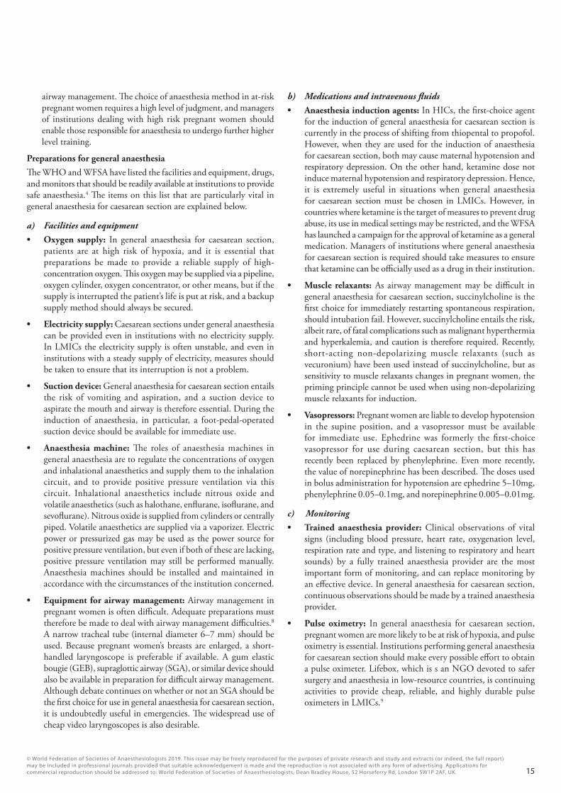
Preparations for general anaesthesiaThe WHO and WFSA have listed the facilities and equipment, drugs, and monitors that should be readily available at institutions to provide safe anaesthesia.4 The items on this list that are particularly vital in general anaesthesia for caesarean section are explained below.
a) Facilities and equipment• Oxygen supply: In general anaesthesia for caesarean section,
patients are at high risk of hypoxia, and it is essential that preparations be made to provide a reliable supply of high-concentration oxygen. This oxygen may be supplied via a pipeline, oxygen cylinder, oxygen concentrator, or other means, but if the supply is interrupted the patient’s life is put at risk, and a backup supply method should always be secured.
• Electricity supply: Caesarean sections under general anaesthesia can be provided even in institutions with no electricity supply. In LMICs the electricity supply is often unstable, and even in institutions with a steady supply of electricity, measures should be taken to ensure that its interruption is not a problem.
• Suction device: General anaesthesia for caesarean section entails the risk of vomiting and aspiration, and a suction device to aspirate the mouth and airway is therefore essential. During the induction of anaesthesia, in particular, a foot-pedal-operated suction device should be available for immediate use.
• Anaesthesia machine: The roles of anaesthesia machines in general anaesthesia are to regulate the concentrations of oxygen and inhalational anaesthetics and supply them to the inhalation circuit, and to provide positive pressure ventilation via this circuit. Inhalational anaesthetics include nitrous oxide and volatile anaesthetics (such as halothane, enflurane, isoflurane, and sevoflurane). Nitrous oxide is supplied from cylinders or centrally piped. Volatile anaesthetics are supplied via a vaporizer. Electric power or pressurized gas may be used as the power source for positive pressure ventilation, but even if both of these are lacking, positive pressure ventilation may still be performed manually. Anaesthesia machines should be installed and maintained in accordance with the circumstances of the institution concerned.
• Equipment for airway management: Airway management in pregnant women is often difficult. Adequate preparations must therefore be made to deal with airway management difficulties.8 A narrow tracheal tube (internal diameter 6–7 mm) should be used. Because pregnant women’s breasts are enlarged, a short-handled laryngoscope is preferable if available. A gum elastic bougie (GEB), supraglottic airway (SGA), or similar device should also be available in preparation for difficult airway management. Although debate continues on whether or not an SGA should be the first choice for use in general anaesthesia for caesarean section, it is undoubtedly useful in emergencies. The widespread use of cheap video laryngoscopes is also desirable.
b) Medications and intravenous fluids• Anaesthesia induction agents: In HICs, the first-choice agent
for the induction of general anaesthesia for caesarean section is currently in the process of shifting from thiopental to propofol. However, when they are used for the induction of anaesthesia for caesarean section, both may cause maternal hypotension and respiratory depression. On the other hand, ketamine dose not induce maternal hypotension and respiratory depression. Hence, it is extremely useful in situations when general anaesthesia for caesarean section must be chosen in LMICs. However, in countries where ketamine is the target of measures to prevent drug abuse, its use in medical settings may be restricted, and the WFSA has launched a campaign for the approval of ketamine as a general medication. Managers of institutions where general anaesthesia for caesarean section is required should take measures to ensure that ketamine can be officially used as a drug in their institution.
• Muscle relaxants: As airway management may be difficult in general anaesthesia for caesarean section, succinylcholine is the first choice for immediately restarting spontaneous respiration, should intubation fail. However, succinylcholine entails the risk, albeit rare, of fatal complications such as malignant hyperthermia and hyperkalemia, and caution is therefore required. Recently, short-acting non-depolarizing muscle relaxants (such as vecuronium) have been used instead of succinylcholine, but as sensitivity to muscle relaxants changes in pregnant women, the priming principle cannot be used when using non-depolarizing muscle relaxants for induction.
• Vasopressors: Pregnant women are liable to develop hypotension in the supine position, and a vasopressor must be available for immediate use. Ephedrine was formerly the first-choice vasopressor for use during caesarean section, but this has recently been replaced by phenylephrine. Even more recently, the value of norepinephrine has been described. The doses used in bolus administration for hypotension are ephedrine 5–10mg, phenylephrine 0.05–0.1mg, and norepinephrine 0.005–0.01mg.
c) Monitoring• Trained anaesthesia provider: Clinical observations of vital
signs (including blood pressure, heart rate, oxygenation level, respiration rate and type, and listening to respiratory and heart sounds) by a fully trained anaesthesia provider are the most important form of monitoring, and can replace monitoring by an effective device. In general anaesthesia for caesarean section, continuous observations should be made by a trained anaesthesia provider.
• Pulse oximetry: In general anaesthesia for caesarean section, pregnant women are more likely to be at risk of hypoxia, and pulse oximetry is essential. Institutions performing general anaesthesia for caesarean section should make every possible effort to obtain a pulse oximeter. Lifebox, which is s an NGO devoted to safer surgery and anaesthesia in low-resource countries, is continuing activities to provide cheap, reliable, and highly durable pulse oximeters in LMICs.9
15
© World Federation of Societies of Anaesthesiologists 2019. This issue may be freely reproduced for the purposes of private research and study and extracts (or indeed, the full report) may be included in professional journals provided that suitable acknowledgement is made and the reproduction is not associated with any form of advertising. Applications for commercial reproduction should be addressed to: World Federation of Societies of Anaesthesiologists, Dean Bradley House, 52 Horseferry Rd, London SW1P 2AF, UK.

• Non-invasive arterial blood pressure (NIBP): Because maternal hypotension reduces the oxygen supply to the fetus, blood pressure is measured once a minute from the induction of anaesthesia until delivery. As haemorrhage may also cause hypotension after the infant has been delivered, blood pressure is also measured at least once every five minutes after delivery. An automated sphygmomanometer is useful for this purpose.
• Electrocardiography (ECG): In HICs, ECG is a standard form of monitoring during anaesthesia, but if the pulse rate can be confirmed by pulse oximetry then an ECG monitor may not be necessarily required. Of course, if an ECG monitor is available it is useful for detecting problems such as arrhythmias and electrolyte disturbances.
• End-tidal carbon dioxide detector: As airway management in pregnant women is often difficult, an end-tidal carbon dioxide detector is useful for definite confirmation that tracheal intubation has been successful. Capnography is even more useful.
• Audible signals and alarms at all times: Alarms to signal abnormal monitor results should be activated at all times.
Specific methods of general anaesthesiaIn HICs, the safety of general anaesthesia for caesarean section has dramatically improved in recent years thanks to advances in monitoring, drugs, and medical devices.10,11 However, in LMICs where these advances are not so widely available, general anaesthesia for caesarean section still remains a procedure that may place patients’ lives at risk. The following points must be followed to provide general anaesthesia for caesarean section safely in LMICs.
a) Preoperative assessment: A preoperative assessment is performed based on an understanding of the physiological changes associated with pregnancy, and a careful judgment made on the method of anaesthesia. Airway assessment is particularly important.
b) Fasting from drink and food: Because gastric emptying time is prolonged during pregnancy, fasting from food and drink must be strictly imposed before a planned caesarean section, and all patients should be treated as having a full stomach even with a sufficient fasting period.
c) Premedication: When general anaesthesia is chosen, an antiemetic and a non-powder antacid are administered.
d) Sign-in: Time-outs are implemented following the WHO checklist.
e) Monitor attachment: Because pregnancy-associated physiological changes mean pregnant women are liable to develop hypoxia, pulse oximetry is essential. Blood pressure should ideally be measured frequently with an automated sphygmomanometer. Measurements are made once a minute until the infant is delivered, and once every five minutes after delivery.
f ) Preoxygenation: Pure oxygen is inhaled for five minutes before anaesthesia is induced. For an emergency section, the patient can be asked to take four deep breaths for denitrification.
g) Induction of anaesthesia: If thiopental is used, 4mg/kg is administered intravenously, and a muscle relaxant (succinylcholine) is administered immediately after the patient has fallen asleep. After the airway has been secured, the inhalational anaesthetic is started.
h) Airway management: After muscle relaxant administration, the lower jaw is raised, and positive pressure ventilation is not performed while spontaneous respiration is present. Even after spontaneous respiration has ceased, the use of positive pressure ventilation is kept to a minimum as long as blood oxygen saturation (SpO2) is maintained, and intubation is performed as soon as muscle relaxation has been confirmed. Cricoid pressure is recommended to prevent aspiration during tracheal intubation. Cricoid pressure is released if it interferes with tracheal intubation.
i) Start of surgery: Surgery is started after confirmation that the airway has been properly secured. The inhalational anaesthetic is administered at 1-1.5 minimum alveolar concentration (MAC) until the infant is delivered. If nitrous oxide can be used, it is administered at a concentration of around 50%.
j) Post-delivery management: Because inhalational anaesthetics relax the uterus, after delivery of the infant their concentration should be reduced to around 0.5 MAC. The incidence of awareness is increased among the patients undergoing general anaesthesia for caesarean section, and caution is therefore required. Because pregnant women are more sensitive to muscle relaxants, additional administration is not necessarily required.
k) Oxytocin administration: After the delivery of infant, intravenous administration of 5 U of the uterotonic oxytocin has been recommended. It should be noted, however, that excessive oxytocin may cause maternal hypotension.
l) Waking: Complications such as aspiration and hypoxia on extubation are greater risks in general anaesthesia for caesarean section, and the same level of caution is required as during induction. If a non-depolarizing muscle relaxant has been used, an antagonist is required. A muscle relaxation monitor should ideally be used if possible. Neostigmine, the non-depolarizing muscle relaxant antagonist, entails the risk of arrhythmia, and atropine is therefore administered prophylactically.
m) Postoperative analgesia: Early mobility is recommended to prevent pulmonary thrombosis following caesarean section. Efforts should be made to provide adequate postoperative pain relief with local anaesthetic infiltration, acetaminophen, NSAIDs, and other analgesics to enable early mobility.
CONCLUSIONSCircumstances under which general anaesthesia must be chosen because neuraxial anaesthesia cannot be provided should be urgently improved. The WHO and WFSA have published statements that anaesthesia is clearly a medical procedure and that it should be provided by a trained anaesthetist to assure its safety.
16 www.wfsahq.org/resources/update-in-anaesthesia

Whether neuraxial or general anaesthesia is chosen for caesarean section, both entail greater risks than normal anaesthesia. If general anaesthesia must be chosen, efforts should be made to manage the anaesthesia in full consideration of the special nature of pregnant women. Make use of the SAFE-OB educational project and other available information.
In HICs, while every effort is made to avoid general anaesthesia for caesarean section and choose neuraxial anaesthesia whenever possible, efforts to improve the safety of general anaesthesia for caesarean section are also ongoing, and mortality associated with general anaesthesia for caesarean section has dropped dramatically. Safe methods of general anaesthesia for caesarean section that are appropriate to the circumstances of LMICs must be established. The use of ketamine is particularly important.
REFERENCES1. Hawkins JL, Koonin LM, Palmer SK, Gibbs CP. Anesthesia-related deaths during obstetric delivery in the United States, 1979-90. Anesthesiology. 1997; 86: 277-84.
2. Hawkins JL, Chang J, Palmer SK, Gibbs CP, Callaghan WM. Anesthesia-related maternal mortality in the United States: 1979-2002. Obstet Gynecol. 2011;117: 69 74.
3. Sobhy S, Zamora J, Dharmarajah K, Arroyo-Manzano D, Wilson M, Navaratnarajah R, Coomarasamy A, Khan KS, Thangaratinam S. Anaesthesia-related maternal mortality in low-income and middle-income countries: a systematic review and meta-analysis. Lancet Glob Health. 2016; 4: e320-7.
4. Gelb AW, Morriss WW, Johnson W, Merry AF; International Standards for a Safe Practice of Anaesthesia Workgroup. World Health Organization-World Federation of Societies of Anaesthesiologists (WHO-WFSA) International Standards for a Safe Practice of Anaesthesia. Can J Anaesth. 2018. doi: 10.1007/s12630-018-1111-
5. Dohlman LE. Providing anaesthesia in resource-limited settings. Curr Opin Anaesthesiol. 2017; 30: 496-500.
6. Enright A, Grady K, Evans F. A new approach to teaching obstetric anaesthesia in low-resource areas. J Obstet Gynaecol Can. 2015; 37: 880–4.
7. Hu LQ, Flood P, Li Y, et al. No Pain Labour & Delivery: a global health initiative’s impact on clinical outcomes in China. Anesth Analg. 2016; 122: 1931–8.
8. Mushambi MC, Kinsella SM, Popat M, Swales H, Ramaswamy KK5, Winton AL, Quinn AC; Obstetric Anaesthetists’ Association; Difficult Airway Society. Obstetric Anaesthetists’ Association and Difficult Airway Society guidelines for the management of difficult and failed tracheal intubation in obstetrics. Anaesthesia. 2015; 70: 1286-306.
9. Enright A, Merry A, Walker I, Wilson I. Lifebox: A Global Patient Safety Initiative. A A Case Rep. 2016; 6: 366-9.
10. Devroe S, Van de Velde M, Rex S. General anaesthesia for caesarean section. Curr Opin Anaesthesiol. 2015; 28: 240-6.
11. Sumikura H, Niwa H, Sato M, Nakamoto T, Asai T, Hagihira S. Rethinking general anaesthesia for caesarean section. J Anesth. 2016; 30: 268-73.
17
© World Federation of Societies of Anaesthesiologists 2019. This issue may be freely reproduced for the purposes of private research and study and extracts (or indeed, the full report) may be included in professional journals provided that suitable acknowledgement is made and the reproduction is not associated with any form of advertising. Applications for commercial reproduction should be addressed to: World Federation of Societies of Anaesthesiologists, Dean Bradley House, 52 Horseferry Rd, London SW1P 2AF, UK.

INTRODUCTIONCaesarean section is the most frequently performed obstetric surgical procedure and may be performed under spinal (intrathecal), epidural or general anaesthesia. This article will focus on spinal anaesthesia, discussing the preoperative evaluation and preparation of patients, the indications and contraindications for spinal anaesthesia, and potential complications of the procedure.
Caesarean delivery rates vary significantly throughout the world, with around 140 million Caesarean sections performed globally during 2015.1 Rates vary from 4% in West and Central Africa to around 23% in the U.K, almost 32% in the USA and over 44% of all deliveries in Latin America and the Caribbean.1-3 The World Health Organisation (WHO) suggests that at a population level, Caesarean delivery rates of up to 10-15% are associated with decreases in maternal and neonatal mortality, but rates above this are not associated with reduced mortality.4
The risks of mortality in those women who undergo Caesarean section also vary widely. A recent systematic review and meta-analysis has shown a mortality rate of 7.6 per 1000 women who undergo Caesarean sections in low- and middle-income countries (LMICs), with the highest mortality rate of 10.9 per 1000 women in
Summary
Caesarean section is the most frequently performed obstetric surgical procedure, and spinal anaesthesia is a
common anaesthetic technique used across the world. It produces rapid, dense, predictable block, is relatively
easy to perform with a definite end point and has a very high success rate.
However, there are contraindications to its use, and complications associated with spinal anaesthesia, which
all patients should be counselled about. Spinal anaesthesia is inevitably associated with hypotension and it is
important to manage this to avoid adverse outcomes in the foetus.
Obstetric spinal anaesthesia
S S Harsoor* and S Bala Bhaskara*Correspondence email: [email protected]: 10.1029/WFSA-D-18-00017
SS Harsoor md
Gadag Institute of Medical Sciences
Gadag Karnataka
INDIA
S Bala Bhaskara da, dnb
Professor of AnaesthesiologyVijayanagar Institute of Medical
SciencesBellaryINDIA
sub-Saharan Africa.5 To compare, the risk of mortality following Caesarean section in the UK is around 8 per 100,000 women, showing an approximate 100-fold increase in risk of death following Caesarean section for those living in LMICs.
Indications for Caesarean SectionCaesarean section may be undertaken for the benefit of the mother, the baby or both. The most common indications are that of failure to progress in labour
Clin
ical
Art
icle
s
Indications for Caesarean Section
Previous Caesarean section
Obstructed labour or failure to progress
Pre-eclampsia or eclampsia
Placenta praevia or abruption
Foetal compromise
Malpositions of the foetus e.g. breech or transverse lie
Multiple pregnancy
Cord prolapse
Worsening of pre-existing maternal condition e.g. cardiac
Maternal choice
Table 1. Indications for Caesarean section
Category Classification of Urgency of Caesarean section
1 Immediate threat to life of woman or foetus
2 Maternal or foetal compromise which is not immediately life-threatening
3 Requires early delivery but no maternal or foetal compromise
4 At a time to suit the woman and maternity team
Table 2. Classification of Caesarean section
18 www.wfsahq.org/resources/update-in-anaesthesia

and prior Caesarean section, and further indications can be found in Table 1 overleaf.
Urgency of Caesarean sectionKnowledge of the indication for the Caesarean section is important as it often determines the urgency of delivery of the baby. Caesarean delivery can be classified as elective – usually performed around 39 weeks gestation at atime to suit the mother and maternity team, or emergency – performed at an unplanned time. As ‘emergency’ is a very broad term, a further classification of Caesarean section has been made by Royal College of Obstetrician and Gynaecologists, in order to help guide management of patients and resources, and is shown in Table 2.
There is much debate as to the maximum time to delivery for each of the suggested classifications, and there is little evidence base for this. However, delivery within 30 minutes of the decision to operate is usual for Category 1 Caesarean sections, where prolonged periods of intrauterine hypoxia may be associated with adverse foetal outcomes.
Spinal anaesthesiaSpinal anaesthesia is the commonest type of anaesthesia used for lower segment caesarean section (LSCS). Compared with epidural technique, spinal anesthesia is quicker and easier to perform, with a definite end point, and a high success rate. It produces rapid, dense and predictable block especially with hyperbaric solutions. There is minimal risk of regurgitation and aspiration of gastric contents. There is minimal transfer of drug across placenta to the foetus and even when transferred, there is minimal risk of foetal toxicity. The mother is awake and is able to enjoy the encounter with her baby.
Pre-operative evaluationAll patients undergoing Caesarean section should be assessed by the anaesthesia team. A thorough preanaesthetic evaluation is performed to elicit co-existing diseases, anaesthetic and obstetric history, contraindications to spinal anaesthesia such as those listed in Table 3, as well as a thorough examination of the patient including back and airway assessment.
Despite planning for spinal anaesthesia, the availability of equipment and medication to safely provide general anesthesia for an unanticipated emergency situation and difficult airway must always be considered.
In patients with pre-eclampsia and HELLP syndrome, both the platelet number and functionality may be poor. Although there is
no strong evidence to specify the exact platelet count for safe spinal anaesthesia to avoid a spinal hematoma, in the absence of other additional coagulation risk factors, a platelet count of 50,000/µl is considered safe.6,7 In addition to the actual number, the quality of platelet function should influence the decision to administer spinal anaesthesia. It must be noted that in pre-eclamptic patients, hyperactivity of the angiotensin II receptors causes hypertension and vasoconstriction and since spinal anesthesia does not influence the angiotensin system, it will cause lesser degree of hypotension in pre-eclamptic patients than in healthy patients.8
Care must be exercised in conditions like placenta praevia where spinal anaesthesia may have advantages of better uterine contractility as compared to general anaesthesia if the volume status is satisfactory.
Complications of spinal anaesthesiaThe mother, who could be in labour and not able to clearly understand the implications of anaesthesia, should still have an explanation of the procedure and consent should be obtained. The potential complications are shown in Table 4, and these risks along with the possibility of failure of spinal anaesthesia and the need to convert to general anaesthesia should be clearly explained to the mother.
Preparation of the patientFasting during labour is a tradition that continues without any strong evidences of improved outcomes either for mother or newborn.9 Hence it is suggested that, during an uncomplicated elective caesarean section, mother should undergo a fasting period for solids of 6 hours, but may have clear liquids up to 2 hours before anaesthesia. During emergency cases, all patients are assumed to be having full stomach. It is generally recommended that before any caesarean section an H2-blocker and a nonparticulate antacid be given with or without metoclopramide.10
At term pregnancy the compression of the inferior vena cava by the gravid uterus in the supine position results in supine hypotension syndrome, and the resulting severe hypotension is not easily managed by treatment with vasopressors. The aortic compression is not too significant to be of consequence as previously thought. Twin and singleton pregnancies cause a similar degree of compression. Measures to avoid aortocaval compression should be continued on
Table 3. Contraindications to spinal anaesthesia. HELLP – haemolysis, elevated liver enzymes, low platelets.
Contraindications to spinal anaesthesia
Coagulation disorders (e.g. due to pre-eclampsia or HELLP Syndrome)
Thrombocytopaenia
Hypovolaemia from active bleeding
Systemic sepsis
Localised sepsis at needle insertion point
Patient refusal
Potential complications of spinal anaesthesia
Hypotension (sympathetic blockade)
Urinary retention
Nausea and vomiting
Shivering
Respiratory depression or sedation (if intrathecal opioids are used)
High block or total spinal
Systemic local anaesthetic toxicity
Post dural puncture headache (PDPH)
Neuropathy – may be temporary or permanent
Epidural or spinal abscess or haematoma
Meningitis or arachnoiditis
Table 4. Potential complications of spinal anaesthesia
19
© World Federation of Societies of Anaesthesiologists 2019. This issue may be freely reproduced for the purposes of private research and study and extracts (or indeed, the full report) may be included in professional journals provided that suitable acknowledgement is made and the reproduction is not associated with any form of advertising. Applications for commercial reproduction should be addressed to: World Federation of Societies of Anaesthesiologists, Dean Bradley House, 52 Horseferry Rd, London SW1P 2AF, UK.

the operating table and after spinal anaesthesia by providing either leftward tilt of table, or wedge under right buttock, or even by obstetrician manually displacing the uterus to left.
Intravenous fluidsPreloading or co-loading with crystalloids before or during spinal anaesthesia is widely practiced in an attempt to reduce the incidence of spinal induced hypotension.11 Ringers Lactate (alternatively called Hartmann’s solution or Compound Sodium Lactate) is the most preferred crystalloid, both for preloading and for maintenance, since it is isoosmolar with plasma. It is also useful as a carrier for oxytocin. Intravenous Dextrose (5%)in water is not an ideal solution as a carrier, since it exhibits hypotonic properties in vivo and the use with oxytocin in dextrose can potentially lead to water retention. In addition, there is risk of foetal hyperglycaemia, acidosis and neonatal hypoglycaemia. However, dextrose can be used when there are clear indications such as in diabetic state.12,13
It is vital to avoid maternal hypotension following spinal anaesthesia, as placental blood flow is entirely dependent on maternal blood pressure. Other interventions to reduce the incidence of hypotension include the use of ephedrine, phenylephrine and lower limb compression.14
Administration of Spinal AnaesthesiaThe intrathecal injection is performed either in sitting or lateral position. The sitting position is preferred when it is difficult to identify the landmarks, as in obese patients, or when combined spinal and epidural technique is attempted. The aim is to attain a sensory block up to T4-T6 segmental level. Sensory blockade beyond T4 segmental level can cause a sense of dyspnoea, as the feel of chest expansion and voluntary sigh are lost due to intercostal muscle paralysis. Quiet reassurance and encouragement to breathe deeply till extraction of foetus will be adequate and no sedative should be administered. Such sensation disappears once the baby is delivered, with improved respiratory movements as the uterus is empty and contracted. Pain associated with traction on peritoneum and exteriorization of uterus can be reduced by administration of analgesics such as fentanyl or alfentanil.
The choice of space for intrathecal injection is at the L3-L4 level to ensure that the needle is inserted well below the termination of the spinal cord. Thin gauge (sizes 25F to 27F), pencil-point needles (Sprotte or Whitacre type) are preferred, as they reduce the incidence of post dural puncture headache(PDPH) as compared to Quincke cutting tip needles. If pencil point needles are not available, the thinnest gauge Quincke needles may be used. Once the free flow of cerebrospinal fluid (CSF) is seen, the chosen local anaesthetic drug dose is injected.
The commonly used drugs and doses for spinal anaesthesia are shown in the Table 5. Inj. Lignocaine (lidocaine) 5% produces faster onset and moderate duration of action of about 45 to 75 minutes, but concerns of transient neurological symptoms(TNS) reported with hyperbaric lignocaine have limited its use. Inj. Bupivacaine is a popular agent with rapid onset, longer duration of action and satisfactory muscle relaxation. The optimum dose of intrathecal heavy bupivacaine 0.5% for the parturients is 10-12mg. Inj. Levobupivacaine, which is a pure S(-) enantiomer of racemic bupivacaine, when used in a dose of 4-12 mg, has the efficacy equivalent to that of heavy bupivacaine 0.5%. The opioids can be added to the neuraxial local anaesthetic, to provide postoperative analgesia after LSCS. Preservative-free morphine 0.10 to 0.25 mg may be added to intrathecal local anaesthetics to prolong postoperative analgesia for 18 to 24 hours. Recent reports indicate that 5mcg dexmedetomidine added to hyperbaric bupivacaine potentiates and prologs spinal anaesthesia without any untoward effects on neonate and hence can be used when it is appropriate15.
Following injection of the spinal anaesthetic, the patient should be turned supine with a wedge under the right buttock, or a tilt on the operating table, to avoid supine hypotension.
MonitoringMandatory monitoring should consist of pulse oximetry, non-invasive blood pressure monitoring and electrocardiogram. The blood pressure should be checked every 2-3 minutes initially as rapid falls are anticipated, necessitating immediate intervention. The hypotension due to sympathetic block may be accentuated by aorto-caval compression caused by enlarged uterus in the supine position. Vasopressors such as ephedrine, phenylephrine, mephentermine or metaraminol should be drawn up in a syringe and kept ready before administering spinal anaesthesia. The infusions of phenylephrine (100mcg/min) are more effective in preventing hypotension. All patients should be monitored for incidences of tachycardia or bradycardia. Tachycardia associated with labour pain may continue for some time or it may occur due to hypotension. Intra-operatively bradycardia may occur because of higher levels of spinal blockade, or due to vagal stimulation caused by traction on peritoneum. Monitoring is especially important during the time when the uterine sinuses are open, until the suturing is complete, as there is risk of amniotic fluid embolism or venous air embolism. The risk of air embolism may be greater with exteriorization of uterus undertaken by some obstetric practitioners.
Blood lossIn low risk patients undergoing elective LSCS, the technique of spinal anaesthesia is associated with a lower risk of operative blood
20 www.wfsahq.org/resources/update-in-anaesthesia
Drug Dosage range (mg)
Duration (min)
Lignocaine (5%) Heavy 60–75 45–75
Bupivacaine (0.5%) 7.5–15.0 60–120
Ropivacaine (0.75%) 10-15 60-90
Levobupivacaine 8-12 60-120
Tetracaine 7.0–10.0 120–180
Procaine 100–150 30–60
Adjuvant drugs
Epinephrine 0.1–0.2 —
Morphine 0.1–0.25 360–1080
Fentanyl 0.010–0.025 180–240
Table 5. Dosage ranges for different local anaesthetic agents and the additives

loss when compared to general anaesthesia.16 Inj. Oxytocin 5-10 IU should be administered by infusion after delivery of the baby, but may be associated with maternal hypertension and tachycardia. In cases of uterine atony, administration of intramuscular (IM) carboprost may be required, but the incidence of nausea, vomiting may increase and rarely, bronchospasm may be precipitated with its use. IM Methyl ergometrine (‘methergine’) has also traditionally been used as uterotonic , however it is associated with increased adverse effects such as hypertension, and nausea and vomiting.
In addition to uterotonic medications, the use of tranexamic acid (TxA) decreases postpartum blood loss and may reduce the incidence of post-partum haemorrhage (PPH) and blood transfusions following LSCS in women at low risk of PPH.17 The World Maternal Antifibrinolytic (WOMAN) trial, has also demonstrated a significant reduction in deaths due to bleeding in patients who received intravenous TxA.18
Nausea and vomiting can be seen in some patients, which may be caused by hypotension or vagal reflexes due to visceral handling. The uterotonic drugs like misoprostol, carboprost or methergine have strong emitogenic potential. Intravenous use of antiemetics such as ondansetron, dexamethasone, droperidol, metoclopramide or combinations are tried with varying degrees of success.
Postoperative CarePostoperative care should continue until the effects of spinal anaesthesia have completely receded. Further monitoring for PDPH should be continued for 48 hours. PDPH should be considered for any headache following spinal anaesthesia for LSCS. The cerebrospinal fluid (CSF) pressure is increased in pregnancy and any breach in dura mater is associated with greater loss of CSF and consequent fall of CSF pressure. The patient manifests with severe headache, neck stiffness, nausea, tinnitus and photophobia. The PDPH is usually self-limiting and may respond to conservative management, which includes regular analgesics, bed rest and oral or IV fluids. An epidural blood patch is considered the gold standard for managing PDPH,19 when supportive measures fail. However, the procedure of epidural blood patch itself can lead to another inadvertent dural puncture and other adverse events can occur during a blood patch, such as meningitis or neurological deficits. The minimally invasive, simple procedure of bilateral greater occipital nerve block has been used for treating chronic headaches in patients with PDPH, or in patients who have failed conservative management.20, 21 Transnasal sphenopalatine ganglion block (SPGB) has also been proposed for the management of postdural puncture headache.22
REFERENCES1 Boerma T, Ronsmans C, Melesse DY et al. Global epidemiology of use of and disparities in caesarean sections. Lancet 2018; 392: 1341-1348
2 Hamilton BE, Martin JA, Osterman MJ, Curtin SC, Matthews TJ. Births: final data for 2014. Natl Vital Stat Rep. 2015; 64: 1–64.
3 Wagner M. Choosing caesarean section. Lancet. 2000; 356(9242): 1677–1680.
4 World Health Organization. WHO Statement on Caesarean Section Rates. WHO/RHR/15.02. 2015: Geneva, WHO.
5 Sobhy S, Arroyo-Manzano D, Murugesu N et al. Maternal and perinatal mortality and complications associated with caesarean section in low-income
and middle-income countries: a systematic review and meta-analysis. Lancet 2019; 393: 1973-82
6 van Veen JJ, Nokes TJ, Makris M The risk of spinal haematoma following neuraxial anaesthesia or lumbar puncture in thrombocytopenic individuals. Br J Haematol. 2010 Jan; 148(1): 15-25. doi: 10.1111/j.1365-2141.2009.07899.
7 nglbrecht JS, Pogatzki-Zahn EM, Zahn P. Spinal and epidural anesthesia in atients with hemorrhagic diathesis : decisions on the brink of minimum evidence? Anaesthesist. 2011 Dec; 60(12): 1126-34. doi: 10.1007/s00101-011 1930-z. [Article in German]
8 Atanas Sivevski, Emilija Ivanov, Dafina Karadjova, Maja Slaninka-Miceska, Igor Kikerkov. Spinal - Induced Hypotension in Preeclamptic and Healthy Parturients Undergoing Cesarean Section. Open Access Maced J Med Sci. 2019 Mar 30; 7(6): 996–1000.
9 Sleutel M, Golden SS. Fasting in labor: relic or requirement. J Obstet Gynecol Neonatal Nurs. 1999 Sep-Oct; 28(5): 507-12.
10 Practice Guidelines for Obstetric Anesthesia: An Updated Report by the American Society of Anesthesiologists Task Force on Obstetric Anesthesia and the Society for Obstetric Anesthesia and Perinatology – Anesthesiology. 2. 2016, Vol.12: 270-300.
11 Park GE, Hauch MA, Curlin F, Datta S, Bader AM. The effects of varying volumes of crystalloid administration before cesarean delivery on maternal hemodynamics and colloid osmotic pressure. Anesth Analg. 1996; 83(2): 299 303
12 Kenepp NB, Kumar S, Shelley WC, Stanley CA, Gabbe SG, Gutsche BB. Fetal and neonatal hazards of maternal hydration with 5% dextrose before caesarean section. Lancet. 1982; 1(8282): 1150–1152
13 Philipson EH, Kalhan SC, Riha MM, Pimentel R. Effects of maternal glucose infusion on fetal acid–base status in human pregnancy. Am J Obstet Gynecol. 1987; 157(4 Pt 1): 866–873
14 Chooi C, Cox JJ, Lumb RS, Middleton P, Chemali M, Emmett RS, Simmons SW, Cyna AM. Techniques for preventing hypotension during spinal anaesthesia for caesarean section. Cochrane Database of Systematic Reviews 2017, Issue 8. Art. No.: CD002251. DOI: 10.1002/14651858.CD002251.pub3
15 Xia F, Chang X, Zhang Y, Wang L, Xiao F. The effect of intrathecal dexmedetomidine on the dose requirement of hyperbaric bupivacaine in spinal anaesthesia for caesarean section: a prospective, double-blinded, randomized study. BMC Anesthesiol. 2018 Jun 23; 18(1): 74. doi: 10.1186/ s12871-018-0528-2.
16 Aksoy H, Aksoy Ü, Yücel B, Özyurt SS, Açmaz G, Babayiğit MA, Gökahmetoğlu G, Aydın T. Blood loss in elective cesarean section: is there a difference related to the type of anesthesia? A randomized prospective study. J Turk Ger Gynecol Assoc. 2015 Jul 14;16(3):158-63. doi: 10.5152/jtgga. 2015.15034.
17 Novikova N, Hofmeyr GJ, Cluver C. Tranexamic acid for preventing postpartum haemorrhage. Cochrane Database Syst Rev. 2015 Jun 16;(6):CD007872. doi: 10.1002/14651858.CD007872.pub3.
18 WOMAN Trial Collaborators. Effect of early tranexamic acid administration on mortality, hysterectomy, and other morbidities in women with post-partum haemorrhage (WOMAN): an international, randomised, double-blind, placebo controlled trial. Lancet 2017;389:2105-2116
19 Sachs A, Smiley R. Post-dural puncture headache: the worst common complication in obstetric anesthesia. Semin Perinatol. 2014 Oct; 38(6): 386-94. doi: 10.1053 / j.semperi.2014.07.007.
20 Nair AS, Kodisharapu PK, Anne P, Saifuddin MS, Asiel C, Rayani BK. Efficacy of bilateral greater occipital nerve block in postdural puncture headache: a narrative review. Korean J Pain. 2018 Apr; 31(2): 80-86. doi: 10.3344 kjp.2018.31.2.80.
21 Niraj G, Kelkar A, Girotra V. Greater occipital nerve block for postdural puncture headache (PDPH): a prospective audit of a modified guideline for the management of PDPH and review of the literature. J Clin Anesth. 2014 Nov; 26(7): 539-44. doi: 10.1016/j.jclinane.2014.03.006.
22 Kent S, Mehaffey G. Transnasal sphenopalatine ganglion block for the treatment of postdural puncture headache in obstetric patients. J Clin Anesth. 2016 Nov; 34: 194-6. doi: 10.1016/j.jclinane. 2016.04.009.
21
© World Federation of Societies of Anaesthesiologists 2019. This issue may be freely reproduced for the purposes of private research and study and extracts (or indeed, the full report) may be included in professional journals provided that suitable acknowledgement is made and the reproduction is not associated with any form of advertising. Applications for commercial reproduction should be addressed to: World Federation of Societies of Anaesthesiologists, Dean Bradley House, 52 Horseferry Rd, London SW1P 2AF, UK.

Management of total spinal block in obstetrics
Shiny Sivanandan* and Anoop Surendran*Correspondence email: [email protected]: 10.1029/WFSA-D-18-00034
INTRODUCTION Total spinal or a high neuraxial blockade is a recognised complication of central neuraxial techniques that include spinal and epidural anaesthesia. A high number of incidents of a high neuraxial block are being reported in obstetrics following the increased use of neuraxial anaesthesia. The leading cause of maternal death in the UK during or up to six weeks after the end of pregnancy is due to thrombosis and thromboembolism.1 But high neuraxial block is now identified as the leading cause of antepartum cardiac collapse in the UK.2
DEFINITIONA high neuraxial block is a sensorimotor block that has reached a spinal segmental level higher than that required to achieve surgical anaesthesia. The terms high, total or complete block are used interchangeably. A sensory level of T3 or above can be associated with significant cardiovascular and respiratory compromise and can hence be considered a high block. Involvement of the cranial nerves signifies intracranial spread of local anaesthetic which can culminate in complete loss of consciousness and cardiorespiratory arrest3.
EPIDEMIOLOGYThe incidence of high neuraxial block associated with
SummaryTotal spinal or a high neuraxial blockade is a recognised complication of central neuraxial techniques. A high
number of incidents of a high neuraxial block are being reported in obstetrics following the increased use of
neuraxial anaesthesia.
Unrecognised subdural or intrathecal placement of an epidural catheter and an intended spinal technique after
a failed epidural analgesia are two main identified causes of high spinal block in obstetrics. The anaesthetist
performing these procedures must be aware of this serious complication and must remain vigilant throughout.
One should have a high level of suspicion of a high spinal while giving a test dose or topping up an epidural in
the delivery suite or in the theatre especially if there is a rapid development of sensory and motor block within
minutes.
Shiny Sivanandan Specialty Registrar
Norfolk and Norwich University Hospital
NorwichUNITED KINGDOM
Anoop SurendranConsultant Anaesthetics
Queen Elizabeth Hospital King’s Lynn
PE30 4ETUNITED KINGDOM
Clin
ical
Art
icle
s
obstetric anaesthesia is not known. Estimates vary between 1:2,9714 and 1:16,2005 anaesthetics.
AETIOLOGYDosage - An accurate estimation of the required dosage of local anaesthetic (LA) to achieve an appropriate level of intrathecal anaesthesia is difficult and we rely on factors such as nature of surgery, patient anatomy and technique of injection. The baricity, volume/dose and injection technique can have an effect on the cephalad spread of intrathecal LA.
A dose of 10mg hyperbaric bupivacaine plus 10 microgram fentanyl is usually appropriate for caesarean section. If no fentanyl is available, administer hyperbaric bupivacaine 10mg alone.6
In general, the evidence suggests that faster injections produce greater spread with plain solutions, but that the effect is less marked with hyperbaric solutions.7 Short stature can be a risk factor for a high spinal as height influences lumbosacral CSF volume and hence intrathecal spread of drug.7 It is assumed that in obesity and pregnancy, the area of central neuraxial compartment could be reduced due to venous engorgement from raised intraabdominal pressure, and excessive adipose tissue.
Key words: total spinal block; high neuraxial blockade; unrecognised subdural catheter; intrathecal catheter; maternal cardiac arrest
22 www.wfsahq.org/resources/update-in-anaesthesia

of the intradural solution into the subdural or subarachnoid spaces.
PREVENTIONUnrecognised subdural or intrathecal placement of an epidural catheter and an intended spinal technique after a failed epidural analgesia are two main identified causes of high spinal block. The anaesthetist performing these procedures must be aware of this serious complication and must remain vigilant throughout with continual assessment of progression of the block in a well monitored environment.
Any abnormally functioning epidural must be followed up closely. Development of a dense motor block of lower limbs during epidural analgesia using a low dose epidural infusion is abnormal and a high-level suspicion of intrathecal placement must be maintained. This suspicion must be shared with the entire team looking after the patient and emphasised during handover care. Physical barriers in the form of prominent labels must be placed over the epidural catheter to prevent an accidental top up with large volume of LA.
Preparation prior to performing any neuraxial blockEnsure airway and resuscitation equipment, vasopressors and drugs essential for an emergency general anaesthesia are within immediate reach. In obstetrics, the team must be aware of the location of a perimortem caesarean section pack. A designated emergency team and a cardiac arrest team must be identifiable. The clinician must be familiar with the locally agreed protocols to activate the emergency call out system.9
Evidence does not confirm that barbotage, by the repeated aspiration and re-injection of CSF and local anaesthetic, increases spread.7
Positioning of patient - Position of patient during and immediately after injection of local anaesthetic may determine the cephalad spread.
Pre-existing epidural block8 The interaction between epidural and spinal injections is not always easy to predict. Unexpected high block can happen when a spinal is administered after the epidural space has presumably been expanded – and the subarachnoid space compressed – by recent epidural top-ups.9a
Unrecognised dural puncture and intrathecal injection of local anaesthetic following epidural top up.
Accidental subdural block. Subdural needle or catheter placement may account not only for delayed onset, profound and extensive conduction blockade, but also for ‘unexplained’ post epidural headaches, false negative aspiration tests and test doses, accidental total spinal, both early and late, some unilateral block and ‘unexplained’ neurological sequelae of spinal and epidural blockade.10
Accidental intradural space11 Local anaesthetic can be injected into the substance of the dura, to create an intradural space, to form a localised and swelling collection within the layers of the dura. Repeated doses of local anaesthetic may escape retrogradely from the intradural space to the epidural space around the outside of the epidural catheter, eventually producing a clinically acceptable block. But there is a slight risk of an extensive block developing some time later, following rupture of the remaining layers of dura and sometimes the arachnoid as well, leading to diversion
Symptoms and signs Root levels System affected Management
Bradycardia
Hypotension+/-
Nausea
T 1-4 Cardiac sympathetic fibres blocked
• Vagolytics like Atropine 0.6mg
• Sympathomimetics such as Ephedrine 6 mg boluses
• Left lateral tilt /wedge
• Phenylephrine 50-100mcg boluses
• Metaraminol 0.5mg boluses
• Mephentermine 3-5 mg boluses
• IV Fluids
Tingling of hand with progressive weakness of hand grip
C 6-8 Arms and hands
Accessory muscles of respiration
• Reassure patient
• Reverse Trendelenburg position in spinal block
• Stop local anaesthetic injection in epidural top up
Difficulty in breathing
Difficulty in speaking
Desaturation
C3-5 Shoulder weakness
Diaphragmatic innervation involved
• Assess airway
• Oxygen supplementation
• May require intubation and ventilation
Slurring of speech
Sedation
Loss of consciousness
Intracranial spread
• Call for help
• Airway, Breathing, Circulation (ABC)approach
• RSI with intubation and ventilation
• Circulatory support with sympathomimetics or vasopressors
• Epinephrine boluses of 50-100mcg may be required if persistent hypotension.
• Foetal monitoring
Table 1: Symptoms, signs and management of the different levels of spinal block
23
© World Federation of Societies of Anaesthesiologists 2019. This issue may be freely reproduced for the purposes of private research and study and extracts (or indeed, the full report) may be included in professional journals provided that suitable acknowledgement is made and the reproduction is not associated with any form of advertising. Applications for commercial reproduction should be addressed to: World Federation of Societies of Anaesthesiologists, Dean Bradley House, 52 Horseferry Rd, London SW1P 2AF, UK.

ProcedureAfter a sub-arachnoid injection or epidural top up, close monitoring of heart rate, blood pressure, oxygen saturations, respiratory rate and level of neuraxial block is necessary. Monitoring should follow clear written protocols.12 The frequency of observations should be determined by normal clinical considerations.12 In hospitals in UK, clinical guidelines suggest that after each epidural bolus or top up for labour analgesia, blood pressure should be recorded for 5 min for the first 20 minutes and thereafter every 30 minutes.
Sensory levels can be tested with ice cubes, ice packs, ethyl chloride sprays, alcohol wipes or with pin-prick
During a spinal block, focus on:
• Dose of local anaesthetic required,
• Baricity of drug,
• Position of patient after spinal
During epidural test dose/top up:
• Always aspirate with a 2ml syringe for blood/CSF before any top up.
• For labour analgesia, test dose with a weaker solution, i.e., 10ml of 0.1% Levobupivacaine is enough to rule out sub arachnoid block. But go as per hospital protocol.
• Always check the level of neuraxial block before any epidural top up.
• If possible, top up epidural for procedural anaesthesia in theatre only for ease of managing emergencies.
• Always give local anaesthetic solution in increments.
Post procedureWritten documentation of any difficulty in neuraxial block is of utmost importance.
Staffing levels sufficient to provide the necessary standard of care are essential in areas providing care for patients with neuraxial blocks, but the individuals need to be trained to the requisite standard as well, and they must know when (and how) to obtain anaesthetic advice9.
HOW TO RECOGNISE HIGH/TOTAL SPINAL BLOCKOne should have a high level of suspicion of a high spinal while giving a test dose or topping up an epidural in the delivery suite or in the theatre especially if there is a rapid development of sensory and motor block within minutes.
After any neuraxial block, there should be constant monitoring of heart rate, blood pressure, respiratory rate and level of neuraxial block.
Constant communication with the mother is very important as it will help to detect any early changes in the voice, effort of breathing or the conscious levels.
Symptoms and signs of a high spinal correlate with the ascending level of neuraxial block.
Early detection and management can prevent deleterious effects to both mother and foetus.
MANAGEMENT OF HIGH SPINAL BLOCK1. Recognition of high spinal and call for help
2. If only circulatory compromiseCorrection of bradycardia and hypotension.
Lateral displacement of uterus manually, with a wedge under the patient or by tilting the theatre table.
Vagolytics like Atropine 0.6mg can be useful for severe bradycardia.
For hypotension, Phenylephrine13 boluses of 50-100mcg can be given. It can also be given as an infusion 20-40ml/hr (in a concentration of 100mcg/ml or as per hospital protocol).
Ephedrine13 in 6mg boluses can also be given if there is hypotension and bradycardia.
Metaraminol boluses of 0.5mg or as an infusion in a concentration of 0.5mg/ml.
Mephentermine14 has been used as a 3-5mg intravenous bolus or intravenous infusion of 2-5mg/min, or 25-50mg intramuscularly. Limited information is available regarding placental transfer and foetal metabolic effects, although it is a popular agent in a number of low and middle-income countries.6
IV fluids -500ml to 1 litre to be given rapidly. To be cautious in cardiac patients and in those with pre-eclampsia.
Reassure the patient as she might be nauseous and will feel faint. Keeping a conversation also will help to assess if the neuraxial block is ascending.
3. If circulatory and respiratory compromise +/- neurological deteriorationIf neuraxial block is ascending with breathing difficulties and desaturation, then reassure the patient, assess the airway, and give supplemental oxygen.
If the patient loses her airway, becomes sedated or unconscious, then secure the airway which includes intubation with Rapid Sequence Intubation (RSI).
If high doses of vasopressors are required, consider epinephrine boluses of 50-100mcg (epinephrine dilution of 100mcg/ml) or infusion.
Maintain anaesthesia as there is possibility for awareness in an apparently unconscious patient.
Patient will have to be sedated and ventilated until neuraxial block has worn off, so intensive care will need to be involved.
In the event of a cardiac arrest, immediate cardiopulmonary resuscitation (CPR) as per Advanced Life support and to start perimortem caesarean section within 4 minutes of arrest.
4. Foetal monitoringAssess the foetal wellbeing. If compromised, then the obstetric team to consider emergency delivery of foetus.
24 www.wfsahq.org/resources/update-in-anaesthesia

5. To rule out other causes of cardiovascular deteriorationThese causes may include local anaesthetic toxicity if intravascular injection, thromboembolism, major haemorrhage, amniotic fluid embolism, profound vasovagal effect
6. Written documentation of the events is of utmost importance for continual care of patient, for future reference and for medico legal purposes.
7. After patient has been resuscitated and woken up (if applicable), update the patient and family on course of events and offer follow up if needed.
REFERENCES1. Knight M, Bunch K, Tuffnell D, Jayakody H, Shakespeare J, Kotnis R, Kenyon S, Kurinczuk JJ (Eds.) on behalf of MBRRACE-UK. Saving Lives, Improving Mothers’ Care - Lessons learned to inform maternity care from the UK and Ireland Confidential Enquiries into Maternal Deaths and Morbidity 2014-16. Oxford: National Perinatal Epidemiology Unit, University of Oxford 2018.
2. Beckett VA, Knight M, Sharpe P. The CAPS Study: incidence, management and outcomes of cardiac arrest in pregnancy in the UK: a prospective, descriptive study. BJOG 2017; 124:1374–1381.
3. UK Obstetric Surveillance System(UKOSS) https://www.npeu.ox.ac.uk/ukoss
4. Scott DB, Tunstall ME. Serious complications associated with epidural/spinal blockade in obstetrics: A two-year prospective study. Int J Obstet Anesth 1995; 4: 133–9.
5. Jenkins JG. Some immediate serious complications of obstetric epidural analgesia and anaesthesia: A prospective study of 145,550 epidurals. Int J Obstet Anesth 2005; 14: 37-42.
6. S. M. Kinsella, B. Carvalho, R. A. Dyer, R. Fernando, N. McDonnell, F. J. Mercier, A. Palanisamy, A. T. H. Sia, M. Van de Velde, A. Vercueil and the Consensus Statement. International consensus statement on the management of hypotension with vasopressors during caesarean section under spinal anaesthesia. Anaesthesia 2018, 73, 71–92.
7. G. Hocking, J.A.W.Wildsmith.Intrathecal drug spread BJA: British Journal of Anaesthesia, Volume 93, Issue 4, October 2004, Pages 568–578.
8. D’Angelo R, Smiley RM, Riley ET, Segal S. Serious complications related to obstetric anesthesia: the serious complication repository project of the Society for Obstetric Anesthesia and Perinatology. Anesthesiology. 2014; 120:1505-5.
9. NAP 3 Report and findings of the 3rd National Audit Project of the Royal College of Anaesthetists, Pages 8-9, 108-110, 115.
9a. NAP 3 Major complications of central neuraxial block in the UK - Chapter 16 - Complications after Obstetric CNB, Page 122.
10. F. Reynolds and H. M. Speedy. The subdural space: the third place to go astray. Anaesthesia,1990. Volume 45, pages 120-123.
11. C.B. Collier. The intradural space: the fourth place to go astray during epidural block. Int J Obstet Anesth. 2010 Apr; 19(2): 133-41.
12. Best practice in the management of epidural analgesia in the hospital setting (page 7) - AAGBI guidelines.
13. D.W. Cooper, S. Sharma, P. Orakkan, S. Gurung. Retrospective study of association between choice of vasopressor given during spinal anaesthesia for high-risk caesarean delivery and fetal pH. IJOA January 2010 Volume 19, Issue 1, Pages 44–49.
14. Deb Sanjay Nag, Devi Prasad Samaddar, Abhishek Chatterjee, Himanshu Kumar, and Ankur Dembla. Vasopressors in obstetric anaesthesia: A current perspective. World J Clin Cases. 2015 Jan 16; 3(1): 58–64.
15. Obstetric Anaesthetists’ Association Guideline Initiative – High Regional Block Guideline Examples - Southampton University Hospitals/Stockport NHS Foundation Trust/University Hospitals Coventry and Warwickshire. Available at http://www.oaa-anaes.ac.uk/ content.
25
© World Federation of Societies of Anaesthesiologists 2019. This issue may be freely reproduced for the purposes of private research and study and extracts (or indeed, the full report) may be included in professional journals provided that suitable acknowledgement is made and the reproduction is not associated with any form of advertising. Applications for commercial reproduction should be addressed to: World Federation of Societies of Anaesthesiologists, Dean Bradley House, 52 Horseferry Rd, London SW1P 2AF, UK.

The labour epidural: the basics
Charlotte Kingsley* and Alan McGlennan,*Correspondence email: [email protected]:10.1029/WFSA-D-18-00002
INTRODUCTIONLabour epidurals are popular and safe; they provide effective analgesia for labouring parturients. Lower dose epidural regimes limit motor block, do not affect progress of labour, and have minimal side effects to mother and fetus. Labour epidurals can also be used to provide anaesthesia for assisted vaginal delivery or caesarean section.
Specific circumstances when labour epidurals may be beneficial1
• Pre-eclampsia (without severe thrombocytopenia or coagulopathy)
• High body mass index (BMI)
• Anticipated difficult airway or other risk factors for general anaesthetic
Charlotte Kingsley Anaesthetic Registrar
Royal Free London NHS Foundation Trust
LondonUNITED KINGDOM
Alan McGlennanConsultant AnaesthetistRoyal Free London NHS
Foundation TrustLondon
UNITED KINGDOM
• High risk for assisted vaginal delivery eg. breech or multiple gestation
• Trial of labour after previous caesarean section
• Maternal cardiovascular, cerebrovascular or respiratory disease1
• Spinal disorders when ‘urgent’ neuraxial anaesthesia placement may be difficult, for example with scoliosis.
Consent and risks for labour epidurals2
• 1 in 10 need further attention to help function (e.g. pull catheter back)
• 1 in 20 need catheter re-siting
• 1 in 100 accidental dural puncture
Clin
ical
Art
icle
s
ABSOLUTE RELATIVE
Patient refusal Fixed cardiac output state
Coagulopathy Anatomical abnormalities of the vertebral column e.g. previous spinal surgery, spina bifida and severe spinal deformity
Severe thrombocytopenia Pre-existing central and peripheral neurological disease
Hypovolaemia or uncontrolled haemorrhage Uncooperative patient
Local infection or systemic sepsis
Local anaesthetic allergy
Raised intracranial pressure
Table 1: Contraindications to labour epidurals1
26 www.wfsahq.org/resources/update-in-anaesthesia
Originally published as Anaesthesia Tutorial of the Week 365
Summary
• Labour epidurals provide safe, effective analgesia with minimal side effects to mother and fetus
• Knowledge of anatomy and pain pathways are key to providing epidural analgesia to labouring parturients
• Consent can be challenging during active labour
• Labour epidurals are beneficial in certain circumstances: high probability of emergency operative delivery;
patients with predictors of a difficult airway; and medical conditions benefiting from reducing the stress
response of labour, e.g. pre-eclampsia
• Risk of permanent nerve damage in obstetric epidurals may be as high as 1 in 80,000
• Combined spinal-epidurals (CSE), single-shot spinals (SSS), and dural puncture epidurals (DPE) are alternative
neuraxial techniques to epidurals for labour analgesia.

• 1 in 24,000 temporary nerve damage, such as temporary motor weakness or paraesthesia of a limb lasting less than 6 months
• 1 in 80,000 permanent nerve damage, such as permanent motor weakness or paraesthesia of a limb
• Bleeding, including epidural haematoma
• Infection, including epidural abscess
• Pruritus
• Hypotension
• Increased risk of assisted vaginal delivery.
Boundaries of the epidural space are as follows:
• Superior fusion of the spinal and periosteal layers of dura mater at the foramen magnum
• Inferior sacro-coccygeal membrane
• Anterior posterior longitudinal ligament, vertebral bodies and intervertebral discs
• Lateral pedicles and intervertebral foramina
• Posterior ligamentum flavum and vertebral laminae
The epidural space contains fat, spinal nerve roots, spinal arteries, extra-dural venous plexuses, connective tissue, lymphatics and the dural sac.4
In adults the spinal cord most commonly ends at L1-L2 (L3 in 10% of adults), the dural sac ends at S2, continuing below this is the filum terminale, which attaches to the coccygeal ligament.5
Surface anatomyKnowledge of surface anatomy is essential in identifying the correct vertebral level for epidural insertion. An imaginary line is drawn between the top of the iliac crests, which corresponds to the level of the L4 spinous process or the L4-L5 interspace, and is known as “Tuffier’s line”. In parturients, tuffier’s line crosses the spine at a higher level (L3-4) due to the forward rotation of the pelvis.6 As a result anaesthetists are often at a higher level than anticipated. This is especially pertinent when a CSE technique is being planned.7 Pre-puncture neuraxial ultrasound can help confirm the correct vertebral level, midline and depth of the epidural space.8
Pain pathways in labourDuring the first stage of labour, afferent nerve impulses from the lower uterine segment and cervix cause visceral pain, which is poorly localised and diffuse in nature. These nerve cell bodies are located in the dorsal root ganglia of T10 to L1. During the second stage of labour, afferent nerves innervating the vagina and perineum cause somatic pain, which is better localised. These somatic impulses travel primarily via the pudendal nerve to dorsal root ganglia of S2 to S4.9
The ideal labour epidural block should cover sensory loss from T10 – S5 dermatomes (with minimal motor block) to provide analgesia for the first and second stages of labour.
Patient positioning for neuraxial blockadeInsertion of labour epidurals is commonly performed in either the sitting, or the flexed lateral position. Positioning is governed by maternal comfort and compliance, as well as anaesthetist preference. Epidural placement in the sitting position has a higher success rate of first-pass insertion and the procedure can be performed faster compared with the lateral position.10
Equipment and Insertion Technique
The basic equipment required for epidural Insertion is:
• Scrub Pack: Hibiscrub, surgical hat, mask, gown, gloves
• Sterile Pack with swabs and drape
Figure 1. Anatomy relevant to epidural anaesthesia
Informed consent for epidural insertion can be challenging in active labour. The process is improved if information can be given antenatally, e.g. with information leaflets.1 Retention of details of the consent discussion may be improved by requesting the parturient sign a written consent form prior to being in active labour.
ANATOMY OF THE LUMBAR SPINE AND THE EPIDURAL SPACEKnowledge of lumbar spine anatomy is the cornerstone of providing safe labour epidural analgesia.
The vertebral columnThe vertebral column provides support and protection for the spinal cord. There are five lumbar vertebrae, which have large vertebral bodies for weight-bearing, increasing in size from L1 to L5.3
Intervertebral discs separate each vertebral body. The spinal canal encloses the epidural and subarachnoid spaces. The vertebral bodies are connected anteriorly by the anterior longitudinal ligament and posteriorly (on the anterior wall of the vertebral canal) by the posterior longitudinal ligament; both extend from the occiput to the sacrum. Three ligaments are pierced during epidural insertion: supraspinous, interspinous, and ligamentum flavum (Figure 1).
The epidural spaceThe epidural space is a ‘potential space’ that surrounds the dura mater and extends from the foramen magnum to the sacral hiatus at the level of S2/3.
27
© World Federation of Societies of Anaesthesiologists 2019. This issue may be freely reproduced for the purposes of private research and study and extracts (or indeed, the full report) may be included in professional journals provided that suitable acknowledgement is made and the reproduction is not associated with any form of advertising. Applications for commercial reproduction should be addressed to: World Federation of Societies of Anaesthesiologists, Dean Bradley House, 52 Horseferry Rd, London SW1P 2AF, UK.

experienced; potentially allowing the parturient to be ambulatory.13
There a several different regimes for administering labour epidural analgesia. Current practices are: intermittent physician or nurse bolus, Patient Controlled Epidural Analgesia (PCEA), Programmed Intermittent Epidural Boluses (PIEB) or continuous infusions.
Labour epidurals provide safe continuous analgesia throughout labour and can be converted with higher concentration local anaesthetic top-up to anaesthesia for operative delivery.
Combined spinal-epidural (CSE)A CSE combines rapid onset of analgesia from the spinal component, with the benefit of continuing labour analgesia with the epidural catheter. A CSE can be performed as an individual single-shot spinal followed by placement of an epidural catheter as a separate technique (see below), or with a needle-through-needle technique. For dosing of the CSE’s spinal component, please see single-shot spinal section below.
When comparing CSEs with labour epidurals, there is no difference in: unintentional dural puncture; incidence of PDPH; rescue analgesia requirements, maternal satisfaction scores; and mode of delivery. There is an increased risk of transient hypotension and fetal bradycardia requiring intervention with CSE compared with labour epidural.14
CSE is a slightly more complicated technique and there is a theoretical risk of having an untested epidural catheter for labour analgesia and surgery if close to the time of CSE placement.
The 3rd National Audit Project2 in the UK showed there was an increased overall risk with the use of CSEs compared to epidurals. Both the optimistic and pessimistic interpretations of the incidence
• Cleaning solution e.g. Chlorhexidine 0.5%
• Local anaesthetic for skin e.g. 1% lignocaine
• 0.9% saline
• Tuohy needle (18 or 16G)
• Loss of resistance syringe
• Epidural catheter
• Epidural filter
• Epidural lock/device for securing epidural to skin
There is no consensus over the use of loss of resistance to air (LORA) or saline (LORS) for epidural insertion. However, there are case reports of pneumocephalus with LORA and increased incidence of dural punctures. As a result, LORS is now commonly practiced.11
NEURAXIAL TECHNIQUES ON LABOUR WARD: WHAT ARE THE OPTIONS?There are alternative neuraxial techniques to the traditional labour epidural. The first choice technique may differ with anaesthetist experience, institutional preference and the clinical situation.
Labour epiduralLabour epidurals do not increase caesarean section rates, but marginally prolong the second stage of labour and increase assisted vaginal delivery rates.12 Labour epidurals improve maternal pain and satisfaction scores in comparison to systemic analgesics and are the most effective analgesic option for labour.
Modern labour epidural dosing regimens (e.g. 0.0625 to 0.1% bupivacaine with 2-4mcg/ml fentanyl or 0.4mcg/ml sufentanil) reduce the total local anaesthetic dose required and motor block
Table 1: Advantages and disadvantages of neuraxial techniques for labour analgesia
Advantages DisadvantagesEpidural • Continuous analgesia
• Ability to convert from analgesia to anaesthesia for operative delivery
• Longer time to insert compared with spinal
• 10-15 minutes to establish analgesia
• Higher failure rate
CSE • Rapid analgesia
• Benefits of spinal and epidural
• Continuous analgesia
• Ability to convert from analgesia to anaesthesia for operative delivery
• Increased likelihood of functional epidural catheter due to confirmation of midline on placement
• Initially untested epidural catheter
• Potentially longer insertion time than an epidural or spinal
• Risk of fetal bradycardia/ hypotension with spinal component
• Unfamiliarity of labour ward staff with management of spinal component
Spinal • Rapid analgesia
• Fast insertion time
• Less risk of epidural haematoma than epidural
• Analgesia duration limited, lasting 60-120 minutes
• Greater risk of hypotension/ fetal bradycardia
• Potential unfamiliarity on labour ward with management
DPE • Reduced haemodynamic instability compared with spinal/ CSE
• Increased likelihood of functional epidural catheter due to confirmation of midline on placement
• Rarely practised – relatively new technique
28 www.wfsahq.org/resources/update-in-anaesthesia

The current trend is towards using low dose local anaesthetic without adrenaline as a ‘test dose’. This helps reduce motor block, thereby allowing better chance of ambulation. There is a large variation in drugs/ doses currently used for test doses, with ranges of 3-20mg bupivacaine and 15-90mg lidocaine.18 Every dose administered via an epidural catheter, whether to initiate a block, or treat breakthrough pain should be treated as a ‘test dose’ as catheters can migrate intrathecally and intravascularly, despite initially being placed correctly in the epidural space.
REFERENCES AND FURTHER READING1. Obstetrics Anaesthetists Association. Available from: http://www.oaa-anaes. ac.uk/assets/_managed/editor/File/Guidelines/epidural%20for%20labour/ Painrelief_for_labour_Swales_Southampton.pdf (accessed 14 January 2016)
2. The 3rd National Audit Project of the The Royal College of Anaesthetists. Available from: http://www.rcoa.ac.uk/system/files/CSQ-NAP3-Full_1.pdf (accessed 14 January 2016)
3. Lumbar Spine Anatomy. Available from: http://emedicine.medscape.com article/1899031-overview (accessed 14 January 2016)
4. Richardson J, Groen G. Applied Epidural Anatomy. Contin Educ Anaesth Crit Care Pain 2005; 5(3): 98-100
5. Barash PG, Cullen BF, Stoelting RK et al. Clinical anesthesia. 6th Edition. 1989. p565.
6. Bucklin B, Gambling D, Wlody D. A Practical Approach to Obstetric Anaesthesia. 1st Edition. 2009. P145
7. Margarido CB, Mikhael R, Arzola C et al.The intercristal line determined by palpation is not a reliable anatomical landmark for neuraxial anesthesia. Can J Anaesth 2011; 58(3): 262-6
8. Amin WA, Osama Abou Seada M, Bedair EMA et al. . Comparative study between ultrasound determination and clinical assessment of the lumbar interspinous level for spinal anaesthesia. M E J Anesth 2014; 22(4): 407-12
9. Shorthouse JR. SAQs for the Final FRCA Examination (Oxford Specialty Training Revision Texts) Paperback 28 July 2011
10. Nishi M, Usukaura A, Kidani Y et al. Which is a better position for insertion of a high thoracic epidural catheter: sitting or lateral decubitus? J Cardiothorac Vasc Anesth. 2006; 20(5): 656-8
11. Nistal-Nuño B, Gómez-Ríos MÁ. Case Report: Pneumocephalus after labor epidural anesthesia. F1000Research. 2014; 3: 166.
12. Gaiser RR. Labor epidurals and outcome. Best Pract Res Clin Anaesthesiol. 2005; 19(1): 1-16
13. Sunil T. Labour analgesia: Recent advances. Ind J Anaesth 2010; 54(5): 400–408
14. McGrady E, Litchfield K. Epidural analgesia in labour. Contin Educ Anaesth Crit Care Pain 2004; 4(4): 114-17
15. Viitanen H, Viitanen M, Heikkila M. Single-shot spinal block for labour analgesia in multiparous parturients. Acta Anaesthesiol Scand 2005; 49(7): 1023-9
16. Cappiello E, O’Rourke N, Segal S et al.A randomized trial of dural puncture epidural technique compared with the standard epidural technique for labor analgesia. Anesth Analg 2008; 107(5): 1646-51
17. Hermanides J, Hollmann MW, Stevens MF et al. Failed epidural: causes and management. Br J Anaesth 2012; 109(2): 144-54
18. Gardner IC, Kinsella SM. Obstetric epidural test doses: a survey of UK practice. Int J Obstet Anesth. 2005 Apr; 14(2): 96-103.
A B
of permanent harm, and paraplegia or death per 100,000 was greater for CSEs than epidurals when used perioperatively in the general patient population. This was not shown in obstetric patients.
Single-shot spinalSingle-shot spinal block for labour analgesia can provide pain relief for immediate delivery.15 Multiparous parturients are probably the most suitable candidates for this technique due to rapid labour progression. A dose of 2.5mg bupivacaine and 25mcg fentanyl has been shown to last up to two hours in duration.15
There is greater incidence of transient hypotension and fetal bradycardia with a single-shot spinal compared with an epidural.2 The anaesthetist should anticipate this and have phenylephrine, ephedrine, and/or glyceryl trinitrate (GTN) immediately available. Transient hypotension may contribute to the fetal bradycardia but it is most likely to be caused by increased uterine tone secondary to the rapid reduction in circulating catecholamines (especially adrenaline). Administering GTN (intravenously or sublingually) provides rapid tocolysis improving the fetal bradycardia.
Single-shot spinal followed by epiduralA single-shot spinal can be immediately followed with an epidural. This is a useful technique in a distressed parturient to facilitate fast pain relief and better positioning.
Dural puncture epidural (DPE)An alternative for labouring parturients is the DPE technique. This technique is similar to a CSE, performing an intentional dural puncture with a spinal needle but without administering intrathecal drugs. DPE avoids the potential haemodynamic instability caused by intrathecal local anaesthetics and enhances labour analgesia when compared with standard epidural techniques.16 DPE improves analgesia compared with epidurals alone by “epidural rent” of the intrathecal space; when there is a puncture in the dura the anaesthetic can flow from the epidural space into the intrathecal space. This technique, along with the CSE technique allows partial confirmation of epidural catheter placement, e.g. cerebrospinal fluid (CSF) is seen in the spinal needle placed through the epidural needle, and therefore the epidural catheter itself is more likely to be midline.
The DPE technique is not currently widely practised.
Test doseAn epidural test dose can identify inadvertent intrathecal or intravascular catheter placement. Unidentified intrathecal or intravascular epidural catheter placement can lead to a high or total spinal, or local anaesthetic systemic toxicity (LAST).
Historically, 3ml of 1.5% lidocaine with 1 in 200,000 adrenaline (epinephrine) was used as a ‘test dose’: intrathecal lidocaine would rapidly produce evidence of a spinal block; intravenous adrenaline would produce transient tachycardia. However, using adrenaline is unreliable (low sensitivity) because of confusion with transient tachycardia seen with contraction pains.16,17
29
© World Federation of Societies of Anaesthesiologists 2019. This issue may be freely reproduced for the purposes of private research and study and extracts (or indeed, the full report) may be included in professional journals provided that suitable acknowledgement is made and the reproduction is not associated with any form of advertising. Applications for commercial reproduction should be addressed to: World Federation of Societies of Anaesthesiologists, Dean Bradley House, 52 Horseferry Rd, London SW1P 2AF, UK.

The labour epidural: troubleshooting
Charlotte Kingsley* and Alan McGlennan*Correspondence email: [email protected]
INTRODUCTIONThe incidence of labour epidural failure is approximately 9-12%.1 Epidurals fail for many reasons, however knowledge of the potential causes can allow anaesthetists to troubleshoot and potentially salvage inadequately functioning epidural catheters.
WHY LABOUR EPIDURALS FAIL Inadequate epidural activation or maintenance doseAdequate volumes and concentrations of local anaesthetic must be administered to establish an adequate epidural block for labour. Once an epidural block is established an adequate maintenance dose is required. The maintenance dose maybe administered via physician or midwife led boluses, patient
Clin
ical
Art
icle
s
QUESTIONSBefore continuing, try to choose an appropriate neuraxial technique with justification for each case. Discussion can be found at the end of the article.
Case 1A 30-year-old primigravid patient, with prolonged first stage, now fully dilated. Requesting an epidural. She is in severe distress with pain, although it appears that she will tolerate sitting still for the procedure.
Case 2A 38-year-old multiparous patient, G3P2, had previous uneventful vaginal deliveries with epidurals. She went into labour at home 2 hours ago and is now fully dilated. She is requesting an epidural but reports she cannot sit down due to sacral pressure or stay still due to pain.
Case 3A 24-year-old primigravid patient is 4cm dilated. She has received multiple intramuscular doses of pethidine (meperidine). You are requested urgently for an epidural. On your arrival, the parturient is on her knees crying in pain. She is very distressed, requesting an epidural. You manage to get her onto the bed, but despite this she is moving around the bed constantly. She is intermittently drowsy with medical nitrous oxide and oxygen gas mixture (50:50), which she will not stop using.
Case 4A 32-year-old multiparous patient, G2P1, is 3cm dilated. She has a BMI of 35 and gestational hypertension. Previous prolonged second stage requiring assisted vaginal delivery.
30 www.wfsahq.org/resources/update-in-anaesthesia
Originally published as Anaesthesia Tutorial of the Week 366
Charlotte Kingsley Anaesthetic Registrar
Royal Free London NHS Foundation Trust
LondonUNITED KINGDOM
Alan McGlennanConsultant AnaesthetistRoyal Free London NHS
Foundation TrustLondon
UNITED KINGDOM
Summary
• Labour epidural failure rate is 9-12% and can be multifactorial
• Poor insertion technique (multiple needle passes), needle mal-placement (off midline), inadequate loading
dose and/or infusion rate, catheter migration and rapid labour progression are the commonest causes of
labour epidural failure
• Anatomical factors such as previous spinal surgery can affect placement and subsequent epidural failure
• Immediate identification of intrathecal and intravascular catheters are vital
• Continuous active management of epidurals is required, with a low threshold to replace inadequately
functioning catheters.

controlled epidural analgesia (PCEA) or programmed intermittent boluses (PIB). A range of local anaesthetic concentrations from 0.0625-0.1% with additional opiate (discussed later in the article) are used in larger volumes for establishing the epidural for labour. The method of providing the epidural maintenance doses and the institutional guidelines will determine the concentration, volume and time interval for administration of maintenance local anaesthetic.
Incorrect needle and/or catheter placement
There are several sites an epidural can be erroneously placed, some more easily identified than others: subcutaneous, subdural, intrathecal (IT) and intravenous.
Subcutaneous placement of epidural catheters will result in complete block failure.2 This is the most common cause of complete block failure, that frequently occurs with a false loss of resistance (loss of resistance prior to entering the epidural space), most commonly in parturients with a raised BMI.
The subdural space is a potential space between the dura and pia-arachnoid mater. Insertion into the subdural space would imply advancement beyond the epidural space, piercing the dura mater but not entering the subarachnoid space. The effects of subdural placement can range from complete block failure, to a patchy asymmetric block, with delayed or minimal motor and autonomic blockade.6
Intrathecal catheter placement can occur after unintentional dural puncture with the needle or catheter. It is identified by cerebrospinal fluid (CSF) flowing through the Tuohy needle, or during catheter aspiration, or it may not be identified until the patient displays symptoms or signs.2 Intrathecal placement can occur at the time of catheter insertion or due to migration from the epidural space. Rapid onset of motor blockade following a test dose should alert the anaesthetist of IT catheter placement. In the case of unintentional dural puncture there are several management options. The epidural catheter can be threaded intrathecally and used to provide low-dose
continuous spinal analgesia7, or the catheter can be removed and re-sited in the epidural space.
A unilateral block can occur when the catheter is located in the lateral aspect of the epidural space. This can sometimes be improved by withdrawing the catheter 1-2cm, with the aim of positioning the catheter midline in the posterior epidural space.
Intravascular placement of an epidural catheter is a potentially life threatening complication and can occur in up to 3-7% of placements with nylon catheters.5 This can be identified if blood is aspirated via the epidural catheter, however there is a high false-negative rate.8 The intravascular catheter can either be withdrawn (1cm at a time then flushed with saline and aspirated) until no more blood is be aspirated, or it can be totally removed and replaced. The gravid uterus compresses the vena cava causing epidural vein distension increasing venous cannulation risk.2 Lateral positioning for placement, not advancing the needle or catheter during a contraction, limiting the depth of catheter insertion to ≤5cm, and using a soft tipped flexible epidural catheter may reduce the risk of intravascular placement.9, 10
Intravenous injection of local anaesthetic can lead to Local Anaesthetic Systemic Toxicity (LAST), which can cause cardiac
Location of Catheter Placement
Incidence 2,3,4,5
Speed of Onset Motor Block Complications
Subcutaneously Unknown Never No block No analgesia
Local tissue swelling
Subdural 1-13% 5-15minutes Minimal motor block with sympathetic sparing
High sensory block (including cervical with Horner’s syndrome)
Patchy block
Intrathecal 1.5% 2-5 minutes Dose dependent Haemodynamic instability
Respiratory compromise
High/total spinal
Fetal bradycardia
Post dural puncture headache (PDPH)
Infection
Intravenous 3-7% (with rigid catheters) Immediate signs and symptoms
No Block LAST
Haemodynamic collapse
Table 2: Potential sites for incorrect epidural catheter placement and their associated complications
PDPH = post dural puncture headache; LAST = local anaesthetic systemic toxicity
31
© World Federation of Societies of Anaesthesiologists 2019. This issue may be freely reproduced for the purposes of private research and study and extracts (or indeed, the full report) may be included in professional journals provided that suitable acknowledgement is made and the reproduction is not associated with any form of advertising. Applications for commercial reproduction should be addressed to: World Federation of Societies of Anaesthesiologists, Dean Bradley House, 52 Horseferry Rd, London SW1P 2AF, UK.
Table 1: Reasons for epidural failure
Reasons for epidural failure
Inadequate activation dose (volume and/or concentration) and maintenance dose
Incorrect needle and/or catheter placement
Migration of epidural catheter
Altered anatomy (e.g. previous spinal surgery)
Sacral sparing
Precipitous labour
Unrealistic patient expectations and low pain thresholds

arrest. Key treatment of any symptoms or signs of LAST is to immediately stop administering the epidural bolus and/or infusion and commence treatment with intravenous lipid emulsion (Intralipid®) in conjunction with advanced life support, if required.
Migration of the catheterMigration of epidural catheters can occur despite correct placement initially.
Migration of the catheter through the intervertebral foramina laterally may produce a unilateral block.11 The catheter can also migrate into the IT and subdural spaces.
Epidural catheters can migrate posteriorly into subcutaneous tissue, more commonly in parturients with high BMI, or in patients where the catheter has not been secured in place effectively.
The incidence of epidural catheter migration can be minimized by leaving 5cm or less in the epidural space and attention to fixation only after repositioning the patient from a flexed position to an extended position (sitting upright, or lying with hips extended). Epidural catheter fixation devices and flexible catheters may help prevent migration. Subcutaneous migration of the catheter is more likely to be seen if less than 3cm is left in the epidural space, and unilateral analgesia is more likely if greater than 5cm of the catheter is left in the epidural space.12
Previous spinal surgery Spinal surgery does not preclude a parturient from receiving neuraxial anaesthesia. However, patients with previous spinal surgery are at increased risk of difficult epidural insertion, inadequate analgesia and increased unintentional dural puncture.2 Post-operative scarring and obliteration of the epidural space distorts normal anatomy and may interfere with normal loss of resistance and spread of local anaesthetic solution.
Sacral sparingDuring labour, afferents innervating the vagina and perineum cause somatic pain (S2-4 nerve roots). The S2-4 nerve roots are covered with thick dura mater, have a large diameter and are further away from the tip of the epidural catheter than the roots transmitting pain in the first stage of labour (T10-L1). These factors in addition to the normal propensity for local anaesthetic solution to travel cephalad can reduce diffusion to sacral nerve roots leading to sacral sparing and failure of labour analgesia in the second stage.2
A subarachnoid block (spinal) in conjunction with epidural (combined spinal and epidural - CSE) reduces sacral sparing.2
Precipitous labourLabour epidurals alone may fail with rapid progress of labour and a precipitous delivery due to insufficient time to establish effective analgesia; a CSE is an alternative technique that can be used in these situations.
Patient expectations and pain thresholdsManaging individual patient expectations are vital. Current labour epidural infusions consist of a low concentration of local anaesthetic plus low dose narcotic and it is expected that parturients may feel
contractions as a pressure sensation. Pain thresholds and response to local anaesthesia can vary between individuals and also between different labour processes and fetal positions.
WHAT TO DO IF THE EPIDURAL CATHETER FAILS? An incomplete block can range from a missed segment, a patchy block, a unilateral block, sacral sparing, not dense enough block, or complete failure. There are a number of techniques that can be employed to rescue an inadequate block.
A thorough assessment should be carried out prior to any intervention of an inadequate block, which includes:
1. History from the patient – pain score, location of pain (abdominal vs perineal), type of pain (pressure?), has a bolus dose helped?
2. Examination – check the position of the catheter (compare with what was documented at the time of placement), check sensory/ motor block.
3. Optimise the patient’s position.
4. Assess effectiveness of a bolus dose (if not already done) prior to catheter manipulation and/ or further boluses with increased local anaesthetic concentration ± supplemental epidural narcotic (fentanyl).
Physician epidural-top-upInadequately functioning epidural catheters can be topped-up by a physician-bolus of local anaesthetic. A large volume of low concentration (e.g. 10ml 0.125% bupivacaine), or a small volume of high concentration (e.g. 5ml 0.25% bupivacaine) local anaesthetic can be administered depending on whether spread or density is required. A physician-administered bolus is effective in approximately 70% of cases.13
Catheter manipulationEpidural catheters can pass cranially, caudally, laterally or into the anterior epidural space.11 Withdrawing catheters 1-2cm (in a sterile fashion) and giving an additional dose of local anesthetic can improve the block in 77% of patients.13
Epidural opioidsMorphine, diamorphine, sufentanil and fentanyl can all be administered epidurally. Fentanyl is the commonest opioid used for labour analgesia.14 Epidural fentanyl is commonly used in the UK, whereas sufentanil is often used in the US.
Fentanyl is a lipophilic drug with an epidural onset time of 10 minutes. Epidural fentanyl has been shown to be three times more effective than IV fentanyl in labouring women, suggesting a spinal mechanism of action.15 A common concentration of epidural infusion solution contains 2mcg/ml fentanyl combined with local anaesthetic.
Epidural fentanyl can be administered as an initial loading dose and/or for breakthrough pain (50-100mcg) to improve the quality of analgesia.14 It can also be administered to manage sacral sparing or as a supplement when converting labour analgesia to surgical anaesthesia for operative delivery.
32 www.wfsahq.org/resources/update-in-anaesthesia

Sufentanil is more potent than fentanyl. An appropriate epidural loading dose is 10mcg and 0.4mcg/ml for an infusion combined with local anaesthetic.
Positioning the patientAnother potential rescue measure for asymmetrical blocks is to alternate the position of the parturient between the right and left lateral positions prior to local anaesthetic top-ups.
WHEN TO RE-SITE THE LABOUR EPIDURAL?Inadequate labour analgesia despite the above interventions (and if more than two physician boluses are required) should prompt the anaesthetist to consider replacement of the epidural catheter.16
Can the epidural be topped up for caesarean section?Active management of epidurals throughout labour is vital to ensure adequate pain relief, maternal satisfaction and the ability to convert labour analgesia to surgical anaesthesia if required for caesarean section.
The following criteria should be satisfied before topping-up a labour epidural for surgery:
1. There is no suspicion of an intrathecal or intravascular catheter
2. Parturient has had adequate labour epidural analgesia, and had resolution of pain from any intervention(s) performed, i.e. catheter withdrawal and/ or physician bolus
Topping up an inadequately functioning epidural catheter risks failure and conversion to general anaesthetic during caesarean section. Options for managing a non-functioning epidural prior to caesarean section include: replacing the epidural and topping it up, performing a single shot spinal (SSS) or a CSE. Depending on intrathecal dosing, care needs to be taken to avoid high spinal blockade or conversely a low surgical sensory block. Although a more involved procedure, a CSE in this setting is a more titratable technique avoiding the risk of a low or high block.
33
© World Federation of Societies of Anaesthesiologists 2019. This issue may be freely reproduced for the purposes of private research and study and extracts (or indeed, the full report) may be included in professional journals provided that suitable acknowledgement is made and the reproduction is not associated with any form of advertising. Applications for commercial reproduction should be addressed to: World Federation of Societies of Anaesthesiologists, Dean Bradley House, 52 Horseferry Rd, London SW1P 2AF, UK.
REFERENCES 1. Pan PH, Bogard TD, Owen MD. Incidence and characteristics of failures in obstetric neuraxial analgesia and anesthesia: a retrospective analysis of 19,259 deliveries. Int J Obstet Anesth. 2004; 13(4): 227-33
2. Arendt K, Segal S. Why epidurals do not always work. Rev Obstet Gynecol. 2008; 1(2): 49-55
3. Chestnut DH, Wong C, Tsen LC, Ngan Kee WD, Beilin Y, Mhyre J. Chestnut’s Obstetric Anaesthesia: Principles and Practice. 5th Edition. 253-254.
4. Agarwal D, Mohta M, Tyagi A, Sethi AK. Subdural Block and the Anaesthetist. Anaesthesia and Intensive Care, 2010, Vol 38, No. 1
5. Paech MJ, Godkin R, Webster S. Complications of obstetric epidural analgesia and anaesthesia: a prospective analysis of 10,995 cases. Int J Obstet Anesth. 1998 Jan; 7(1): 5-11.
6. Collier CB. Accidental subdural injection during attempted lumbar epidural block may present as a failed or inadequate block: radiographic evidence. Reg Anesth Pain Med. 2004; 29(1): 45-51
7. Ng A, Shah J, Smith G. Is continuous spinal analgesia via an epidural catheter appropriate after accidental subarachnoid administration of 15 mL of bupivacaine 0.1% containing fentanyl 2 micrograms/mL? Int J Obstet Anesth. 2004; 13(2): 107-9
8. Bell DN, Leslie K. Detection of intravascular epidural catheter placement: a review. Anaesth Intens Care. 2007; 35(3): 335-41
9. Lubenow T, Keh-Wong E, Kristof K, Ivankovich O, Ivankovich AD. Inadvertent subdural injection: a complication of an epidural block. Anesth Analg. 1988; 67(2): 175-9
10. Mhyre JM, Greenfield ML, Tsen LC, Polley LS. A systematic review of randomized controlled trials that evaluate strategies to avoid epidural vein cannulation during obstetric epidural catheter placement. Anesth Analg. 2009; 108(4): 1232 42
11. Hogan Q. Epidural catheter tip position and distribution of injectate evaluated by computed tomography. Anesthesiology. 1999; 90: 964–70
12. Afshan G, Chohan U, Khan FA, Chaudhry N, Khan ZE, Khan AA. Appropriate length of epidural catheter in the epidural space for postoperative analgesia: evaluation by epidurography. Anaesthesia. 2011; 66(10): 913-8
13. Beilin Y, Zahn J, Bernstein HH, Zucker-Pinchoff B, Zenzen WJ, Andres LA. Treatment of incomplete analgesia after placement of an epidural catheter and administration of local anesthetic for women in labor. Anesthesiology. 1998; 88(6): 1502-6
14. Khangure N. Adjuvant agents in neuraxial blockade. Anaesthesia Tutorial of The Week 2011;230
15. Ginosar Y, Columb MO, Cohen SE, Mirikatani E, Tingle MS, Ratner EF, Angst MS, Riley ET. The site of action of epidural fentanyl infusions in the presence of local anesthetics: a minimum local analgesic concentration infusion study in nulliparous labor. Anesth Analg. 2003; 97(5): 1439-45
16. Riley ET and Papasin J. Epidural catheter function during labor predicts anesthetic efficacy for subsequent cesarean delivery. IJOA 2002; 11; 81-84

34 www.wfsahq.org/resources/update-in-anaesthesia
ANSWERS TO CASES
Case 1
A 30-year-old primigravid patient, with prolonged first stage, now fully dilated. Requesting an epidural. She is in severe distress with pain, although she will tolerate sitting still for the procedure.
COMBINED SPINAL EPIDURAL (CSE) This parturient is likely to be suffering perineal and pelvic pain as she is fully dilated. The spinal component will provide immediate analgesia with increased likelihood of blocking the S2-4 somatic afferents.
She is a primip with a prolonged first stage. A rapid second stage is unlikely and she is at risk of an instrumental delivery. The epidural component of the CSE will allow continued analgesia throughout labour and provide the option of anaesthesia for an assisted or operative delivery if required.
An epidural alone takes time to establish adequate analgesia and there is risk of sacral sparing.
A single-shot spinal may not provide an adequate duration of analgesia.
Case 2
A 38-year-old multiparous patient, G3P2, had previous uneventful precipitous vaginal deliveries with epidurals, is fully dilated. She went into labour at home 2 hours ago. She is requesting an epidural but reports she cannot sit down due to the pelvic pressure or stay still due to the pain.
SINGLE SHOT SPINAL (SSS) IN THE LATERAL POSITIONThis parturient is a multip and requires fast-acting analgesia. She is unable to sit due to sacral pressure, likely from a low-lying fetal head. Performing the neuraxial procedure in the lateral position has a number of benefits: improved patient comfort and increased probability of the patient remaining still during the procedure.
Primarily, blockade of the S2-4 somatic afferents is required for analgesia, which will take time to achieve with an epidural a. A SSS will provide immediate effective analgesia. A CSE could be performed but it is unlikely she will need the epidural component, exposing her to unnecessary procedural risk. Intraveous opiates can be considered if there is no time for a SSS. The paediatricians must be informed of potential fetal respiratory depression.
Case 3
A 24-year-old primigravid patient is 4cm dilation. She has received multiple doses of pethidine (meperidine). You are requested urgently for an epidural. On your arrival, the parturient is face down on the floor of the room, on her knees crying in pain. She is very distressed, asking you
to take her pain away. You manage to get her onto the bed, but despite this she is moving around the bed constantly. She is intermittently drowsy with the use of medical nitrous oxide and oxygen gas mixture (50:50), which she will not stop using.
CSE or SSS IN THE LATERAL POSITION FOLLOWED BY SITTING EPIDURALThis scenario poses a challenge to even an experienced obstetric anaesthetist. This parturient is unlikely to tolerate labour without any form of neuraxial analgesia, however it can only be provided for her in an environment that is safe. The risk of complications in a poorly compliant, moving and distressed parturient are high. In addition, the anaesthetist needs to consider that a number of factors may impact the patient’s capacity to consent: understanding of the procedure, ability to understand risks and benefits. Junior anaesthetists with concerns that it is unsafe to proceed with the procedure should call for senior help.
Focusing on breathing control improves the effectiveness of Nitrous oxide/oxygen and a calm explanation from the anaesthetist may help. The patient may be more comfortable in the lateral position (especially if they are drowsy), but this needs to be balanced with the familiarity of the anaesthetist with this position. A rapid SSS in the lateral position may provide enough analgesia to calm the patient and allow a longer term epidural to be established in a calm, safer environment.
A patient may require IV narcotics to help pain control and the ability to stay still during neuraxial analgesia placement.
Case 4
A 32-year-old multiparous patient, G2P1, is 3 cm dilation. She has a BMI of 35 and gestational hypertension. Previous prolonged second stage, requiring assisted vaginal delivery.
EPIDURAL Epidural-alone is an appropriate choice of neuraxial technique in this case. In view of gestational hypertension the platelet count should be checked prior to epidural insertion and removal. Early insertion of epidural catheters in patients with a high BMI is recommended. Placement can be challenging in obese patients, therefore it is easier to place it in a controlled situation rather than in an emergency situation, such as in an urgent caesarean section for example. Intubation can also potentially be more difficult in obese patients, therefore having a catheter in-situ and time to ensure it is functioning well, may avoid the need to perform general anesthesia in an emergency situation. Epidurals also reduce the stress response from labour, which can improve blood pressure control.

Establishing an epidural service for labour analgesia in a variable resource environment
Andrew Kintu, Hilary MacCormick and Ronald B. George**Correspondence email: [email protected]: 10.1029/WFSA-D-18-00022
INTRODUCTIONThe health of women correlates strongly with the social and economic well-being of a community or society. Acute pain during childbirth is undertreated in variable resource environments (VRE).1 Women are dying in childbirth from treatable conditions because they don’t have access to the most basic surgeries and perioperative care, including analgesia. Pain associated with labour and delivery is the most important anesthetic-related concern for expectant mothers in high income countries.2 Epidurals are widely regarded as the gold standard for labour analgesia with their analgesic efficacy confirmed many times.3-5 Titration of low-dose, low-concentration local anesthetics produces safe, reliable analgesia during labour and delivery. Epidurals provide safe and efficient provision of anaesthesia for unplanned or emergent caesarean delivery. A properly monitored labour epidural has a low incidence of side effects or serious complications to the parturient or fetus.6
In the year 1900, subarachnoid injection of cocaine was described with the result of total lower body anaesthesia in six parturients in labour.7 Years of innovation, including needle design, catheter development, and medication discovery, have led to modern day epidural analgesia. In the past 40 years new developments in epidural delivery pumps have shaped current practice in high resource environments.8,9 Current estimated rate of neuraxial labour analgesia use ranges from 66-82% in the United States.10 Estimates of neuraxial labour analgesia usage in
VRE are challenging. However, one study estimated that only 2.2% of parturients at a South African public hospital received neuraxial labour analgesia.11 Barriers to neuraxial analgesia include lack of skilled anaesthesia personnel, knowledgeable support staff, and equipment.
This article will focus primarily on establishing an epidural service to provide labour analgesia and anaesthesia for non-scheduled caesarean delivery. Considerations for two other neuraxial techniques, such as single-shot spinal analgesia and combined spinal epidural (CSE), will be covered briefly. An overview of neuraxial analgesia techniques will be followed by discussion of various elements critical to successful establishment of an epidural service, safety considerations, and highlighting challenges within VRE. Every epidural provides analgesia; a managed epidural service provides analgesia, anaesthesia, and conveys a level of safety to women within the service. The development of a labour epidural service should encompass patients’ safety as its key tenant. Articles covering the broader topic of establishing an obstetrical anaesthesia service exist for the interested reader.4,12
CURRENT EPIDURAL TECHNIQUE FOR LABOUR ANALGESIANeuraxial labour analgesia has been provided via single shot epidural or intrathecal injections, continuous epidural infusions, and, much less frequently, through
Ronald B. George md, frcpc
Department of Women’s & Obstetric AnaesthesiaIWK Health Centre 5980 University AvenueHalifaxNova Scotia CANADA
Andrew Kintu, mbchb, mmed AnaesthetistDepartment of AnaesthesiaMulago HospitalMakerere University College of Health Sciences KampalaUGANDA
Hilary MacCormick, md, frcpc
Department of Anaesthesia Pain Management and Perioperative MedicineDalhousie UniversityHalifaxNova ScotiaCANADA
35
© World Federation of Societies of Anaesthesiologists 2019. This issue may be freely reproduced for the purposes of private research and study and extracts (or indeed, the full report) may be included in professional journals provided that suitable acknowledgement is made and the reproduction is not associated with any form of advertising. Applications for commercial reproduction should be addressed to: World Federation of Societies of Anaesthesiologists, Dean Bradley House, 52 Horseferry Rd, London SW1P 2AF, UK.
Summary
The health of women correlates strongly with the social and economic well-being of a community. However, pain
during childbirth is undertreated in variable resource environments Epidurals provide safe and efficient provision
of anesthesia for unplanned or emergent cesarean delivery. This article will focus primarily on establishing an
epidural service to provide labour analgesia and anesthesia for nonscheduled cesarean delivery. The development
of a labour epidural service should encompass patients’ safety as its key tenant. Developing a high quality epidural
service requires a well-trained, cooperative multidisciplinary team, an adequately equipped unit, and dedicated
leadership whose role is to ensure the service maintains high standards. Ongoing monitoring and evaluation
of the service will create the best environment for continued improvements and longevity of a means to strive
towards provision of excellent care for parturients and their babies.
Clin
ical
Art
icle
s

They suggest that patient preference is all that is necessary for an indication to provide labour analgesia; “Labour causes severe pain for many women. There is no other circumstance where it is considered acceptable for an individual to experience untreated severe pain, amenable to safe intervention, while under a physician’s care. In the absence of a medical contraindication, maternal request is a sufficient medical indication for pain relief during labour. Pain management should be provided whenever medically indicated”.16
There are certain maternal comorbidities, such as mitral stenosis and other cardiac conditions, that would benefit haemodynamically from labour analgesia.17 Patients with stenotic valvulopathies do not tolerate the tachycardia that may accompany labour pain. By providing analgesia via epidural, the increase in heart rate secondary to pain can be mitigated, which allows for improved haemodynamic stability. However, one must be cautious to avoid excessive sympathectomy in these preload-dependent lesions.18 A more common example of maternal disease that may benefit from labour analgesia would be preeclampsia, along with other hypertensive disorders of pregnancy.12 Adequate analgesia can play an important
continuous intrathecal infusions. Epidural analgesia is a technical procedure, performed under aseptic technique, where medication is injected into the epidural space with the intention of providing analgesia to a specific region of sensory dermatomes.13 Alternatively, spinal analgesia is obtained via injection of medication into the subarachnoid space.7 For applications in labour analgesia, single-shot spinal typically consists of small doses of local anaesthetic, lipophilic opioid (e.g. fentanyl), or a combination of both.14 These techniques can be combined and analgesia performed with a CSE, which combines aspects of both spinal and epidural techniques.13 Table 1 highlights several key differences between these neuraxial procedures. All three of these neuraxial techniques share the benefit of avoiding maternal sedation, which may occur with labour analgesia provided via parenteral opioids or inhaled nitrous oxide.15
INDICATIONSA joint statement by the American Society of Anaesthesiologists (ASA) and the American College of Obstetricians and Gynaecologists (ACOG) highlights the patient centered aspect of labour analgesia.16
Epidural Single-shot Spinal CSE
Site of medication injection
Epidural space Subarachnoid space Both epidural and subarachnoid spaces
Duration of effect Can provide analgesia as long as epidural catheter remains in situ
Limited duration, depending on medication choice, dose and volume injected15
Subarachnoid injection is of limited duration, but can continue to provide analgesia as long as epidural catheter remains in situ
Advantages • In situ catheter allows for titration of medication throughout labour, can control the spread and duration of analgesia and anesthesia
• Can extend use to provide anesthesia for instrumental or cesarean delivery15
• Predictable spread of sensory blockade
• Technically easier in obese parturients (versus spinal)
• Does not require dural puncture
• Rapid onset
• Does not require ongoing availability of anesthesia providers
• Technically simple procedure
• See advantages of epidural
• Lower failure rate (versus epidural)13
• Allows for more rapid onset of analgesia and anesthesia (versus epidural alone)15
Disadvantages • Requires knowledgable healthcare providers to monitor for appropriate use and potential complications
• In situ catheter has potential for misuse (e.g. being mistaken for an intravenous catheter) if not clearly labelled
• Greater quantities of medications required (versus spinal)
• Increased risk of maternal local anesthetic toxicity (versus spinal) if catheter positioned intravascularly15
• Limited duration
• May result in greater degree of motor blockade
• Inadequate analgesia for second stage of labour
• May result in greater sympathectomy, resulting in maternal hypotension
• See disadvantages of epidural
• Delayed ability to detect non-functioning epidural catheter15
• Increased maternal pruritus if intrathecal opioids administered (versus epidural alone)15
• Requires dural puncture, although does not appear to impact incidence of PDPH15
• Potentially increased rate of fetal bradycardia, but not associated with increased cesarean delivery rate13
Table 1: Highlighted comparisons between epidural, single-shot spinal, and CSE techniques for labour analgesia. Adapted from Report of Best practice in the management of epidural analgesia in the hospital setting.45
36 www.wfsahq.org/resources/update-in-anaesthesia
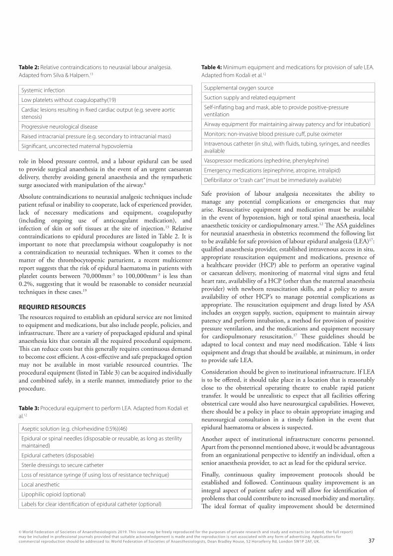
role in blood pressure control, and a labour epidural can be used to provide surgical anaesthesia in the event of an urgent caesarean delivery, thereby avoiding general anaesthesia and the sympathetic surge associated with manipulation of the airway.6
Absolute contraindications to neuraxial analgesic techniques include patient refusal or inability to cooperate, lack of experienced provider, lack of necessary medications and equipment, coagulopathy (including ongoing use of anticoagulant medication), and infection of skin or soft tissues at the site of injection.13 Relative contraindications to epidural procedures are listed in Table 2. It is important to note that preeclampsia without coagulopathy is not a contraindication to neuraxial techniques. When it comes to the matter of the thrombocytopenic parturient, a recent multicenter report suggests that the risk of epidural haematoma in patients with platelet counts between 70,000mm-3 to 100,000mm-3 is less than 0.2%, suggesting that it would be reasonable to consider neuraxial techniques in these cases.19
REQUIRED RESOURCESThe resources required to establish an epidural service are not limited to equipment and medications, but also include people, policies, and infrastructure. There are a variety of prepackaged epidural and spinal anaesthesia kits that contain all the required procedural equipment. This can reduce costs but this generally requires continuous demand to become cost efficient. A cost-effective and safe prepackaged option may not be available in most variable resourced countries. The procedural equipment (listed in Table 3) can be acquired individually and combined safely, in a sterile manner, immediately prior to the procedure.
Table 2: Relative contraindications to neuraxial labour analgesia. Adapted from Silva & Halpern.13
Table 3: Procedural equipment to perform LEA. Adapted from Kodali et al.12
Table 4: Minimum equipment and medications for provision of safe LEA. Adapted from Kodali et al.12
Systemic infection
Low platelets without coagulopathy(19)
Cardiac lesions resulting in fixed cardiac output (e.g. severe aortic stenosis)
Progressive neurological disease
Raised intracranial pressure (e.g. secondary to intracranial mass)
Significant, uncorrected maternal hypovolemia
Aseptic solution (e.g. chlorhexidine 0.5%)(46)
Epidural or spinal needles (disposable or reusable, as long as sterility maintained)
Epidural catheters (disposable)
Sterile dressings to secure catheter
Loss of resistance syringe (if using loss of resistance technique)
Local anesthetic
Lipophilic opioid (optional)
Labels for clear identification of epidural catheter (optional)
Supplemental oxygen source
Suction supply and related equipment
Self-inflating bag and mask, able to provide positive-pressure ventilation
Airway equipment (for maintaining airway patency and for intubation)
Monitors: non-invasive blood pressure cuff, pulse oximeter
Intravenous catheter (in situ), with fluids, tubing, syringes, and needles available
Vasopressor medications (ephedrine, phenylephrine)
Emergency medications (epinephrine, atropine, intralipid)
Defibrillator or “crash cart” (must be immediately available)
Safe provision of labour analgesia necessitates the ability to manage any potential complications or emergencies that may arise. Resuscitative equipment and medication must be available in the event of hypotension, high or total spinal anaesthesia, local anaesthetic toxicity or cardiopulmonary arrest.12 The ASA guidelines for neuraxial anaesthesia in obstetrics recommend the following list to be available for safe provision of labour epidural analgesia (LEA)17: qualified anaesthesia provider, established intravenous access in situ, appropriate resuscitation equipment and medications, presence of a healthcare provider (HCP) able to perform an operative vaginal or caesarean delivery, monitoring of maternal vital signs and fetal heart rate, availability of a HCP (other than the maternal anaesthesia provider) with newborn resuscitation skills, and a policy to assure availability of other HCP’s to manage potential complications as appropriate. The resuscitation equipment and drugs listed by ASA includes an oxygen supply, suction, equipment to maintain airway patency and perform intubation, a method for provision of positive pressure ventilation, and the medications and equipment necessary for cardiopulmonary resuscitation.17 These guidelines should be adapted to local context and may need modification. Table 4 lists equipment and drugs that should be available, at minimum, in order to provide safe LEA.
Consideration should be given to institutional infrastructure. If LEA is to be offered, it should take place in a location that is reasonably close to the obstetrical operating theatre to enable rapid patient transfer. It would be unrealistic to expect that all facilities offering obstetrical care would also have neurosurgical capabilities. However, there should be a policy in place to obtain appropriate imaging and neurosurgical consultation in a timely fashion in the event that epidural haematoma or abscess is suspected.
Another aspect of institutional infrastructure concerns personnel. Apart from the personnel mentioned above, it would be advantageous from an organizational perspective to identify an individual, often a senior anaesthesia provider, to act as lead for the epidural service.
Finally, continuous quality improvement protocols should be established and followed. Continuous quality improvement is an integral aspect of patient safety and will allow for identification of problems that could contribute to increased morbidity and mortality. The ideal format of quality improvement should be determined
37
© World Federation of Societies of Anaesthesiologists 2019. This issue may be freely reproduced for the purposes of private research and study and extracts (or indeed, the full report) may be included in professional journals provided that suitable acknowledgement is made and the reproduction is not associated with any form of advertising. Applications for commercial reproduction should be addressed to: World Federation of Societies of Anaesthesiologists, Dean Bradley House, 52 Horseferry Rd, London SW1P 2AF, UK.

on an institutional or departmental basis, but indicators of safety of neuraxial analgesia need to be determined, measured and acted upon. A system of regular evaluation and response should be put in place, which includes efficacy, patient satisfaction and feedback, incidence of complications, and adherence to institutional protocols. All relevant department members should have the opportunity to be involved in the process. In the instance of a poor outcome or near miss, emphasis should be placed on determining systemic issues to prevent future similar errors, rather than any penalizing action against a specific individual. This places emphasis on a systems approach to medical error, rather than a person approach, and is much better suited to the healthcare setting.20
et al.28 compared preferences between American-born parturients and parturients immigrated from Sudan and Somalia and found that only 12.5% of Sudanese women would prefer epidural analgesia during labour, compared to 66.7% and 64% of American and Somali women, respectively. The study did not further explore attitudes regarding epidural analgesia, so it is unclear why there was such a significant difference in preference amongst Sudanese women, but it seems reasonable to suspect that lack of knowledge surrounding the benefits and risks of LEA may have contributed.
Even in countries where LEA is widely accepted, many patients still have misconceptions about the procedure and its risks.29,30 Many parturients turn to the internet to find out information related to various aspects of pregnancy, including LEA.31,32 A recent study by Espitalier et al.33 found that the quality of information regarding LEA on both English and French language websites was poor overall.
Ogboli-Nwasor et al.34 surveyed HCPs in Zaria, Nigeria, and found that although the vast majority (94.8%) agreed that pain relief should be provided during labour, only half of the respondents actually provided labour analgesia during the 8 weeks preceding the study’s survey. This highlights a gap between the attitudes and practices of HCPs in that setting. Many of the reasons cited for non-provision of labour analgesia, such as lack of resources and skills, are challenges encountered in many other VRE. In many VRE, midwives or non-traditional healers are often primary care providers for parturients. Special attention should be paid to involving and engaging our midwifery colleagues in discussions around labour analgesia.
Educating non-anaesthesia HCPs regarding LEA extends beyond explaining the procedure’s risks and benefits. There must also be education surrounding how to monitor for post-procedural complications. Epidural haematoma and abscess are emergencies that require timely diagnosis and treatment to minimize mortality and long-term morbidity.
Although extremely rare, there is potential for delayed respiratory depression following administration of intrathecal morphine.35 Lipophilic opioids, such as fentanyl, may be safer alternatives when using neuraxial opioids;35 but low dose intrathecal morphine has been shown to prolong duration of labour analgesia when combined with bupivacaine and fentanyl.15 It should also be noted that there is only limited research on this complication within the obstetrical population.
Post-dural puncture headache (PDPH) typically does not present immediately after performance of a neuraxial procedure, and can create significant short-term morbidity for patients.13,36 This particular complication requires that non-anaesthesia HCPs are able to recognize potential symptoms in order to alert the anaesthesia providers, and there should be an established process for follow-up of PDPH patients to ensure resolution of symptoms. These examples illustrate that the window for potential complications following LEA extends beyond the time that the epidural catheter is discontinued.
Finding an effective method of knowledge translation is heavily dependent on local context.37,38 It is critical for anaesthesia providers to access the most appropriate methods to increase knowledge of patients and other healthcare providers. Ongoing communication
Table 5: Complications of neuraxial analgesia and anesthesia in obstetrical population. Adapted from D’Angelo et al (2014)21
Post-dural puncture headache 1:114
Epidural abscess/meningitis 1:62, 866
Epidural hematoma 1:251, 463
Serious neurologic injury 1:35, 923
High neuraxial block 1:4, 336
Unrecognized intrathecal catheter (intended to be epidural)
1:15, 435
POTENTIAL COMPLICATIONSMaternal hypotension, pruritus, fetal bradycardia, maternal fever, urinary retention, and shivering have all been reported as adverse effects related to neuraxial analgesia.6,15 Estimates of incidence for these adverse effects are related to the type and dose of medications used to provide analgesia.15 A combination of low-dose local anaesthetic (for example, bupivacaine) and lipid-soluble opioid results in good analgesia with minimal adverse effects.21 There is a dearth of large obstetrical anaesthesia databases, which makes it very challenging to estimate the incidences of serious complications. The Society for Obstetrical Anaesthesia and Perinatology has begun a repository project to better document serious complications related to obstetrical anaesthesia and we have summarized selected complications in Table 5.21 It is important to note that these incidences cannot be generalized to all high income countries, and that incidences in VRE, where LEA may not yet be commonplace, are not widely available at this time. Ensuring appropriate continuous quality improvement measures are in place will help to determine what the incidence of each complication and guide efforts to reduce them.
AWARENESS & EDUCATIONIn countries where labour analgesia is not widely utilized the first step in establishing an LEA service is to educate both patients and HCPs involved in the care of parturients. If HCPs are not educated about benefits, risks, and options for labour analgesia, they cannot be expected to pass along information to their patients. If patients are not aware of their options for labour analgesia, they cannot be expected to request it or provide informed consent.
Several studies have demonstrated low levels of awareness amongst parturients regarding epidural analgesia in Saudi Arabia22, India23, Uganda24, Nigeria25,26, and Hong Kong.27 One study by Ogunleye
38 www.wfsahq.org/resources/update-in-anaesthesia

between anaesthesia providers, other HCPs, patients, and their families, will undoubtedly enhance knowledge translation efforts. Social media may facilitate health education and knowledge translation in VRE, however currently misinformation and lack of credible sites may contribute to negative outcomes.39
SAFETY CONSIDERATIONS Patient safety should be the top priority as we plan to provide pain relief. Availability of resuscitation equipment, development of institutional protocols, setting of minimum required levels of training, fostering communication among members of the multidisciplinary team, and initiating quality control measures all play key roles in the safe provision of epidural services.
Development of institutional protocols Protocols are used to standardize clinical practice; their use has been associated with improvement in patient outcomes.40 The development of epidural protocols should be multidisciplinary, involving nurses, anaesthesia providers, obstetricians, patient advocates, and hospital administrators. Usually these protocols are adapted from local national guidelines, or speciality governing bodies like the Society for Obstetrical Anaesthesia and Perinatology and the Obstetric Anaesthetists’ Association (OAA). Each hospital must develop their own protocols, adapted to the local setting and context. Table 6 shows an example of what information should be addressed in a protocol. Documentation of the epidural procedure should include the date and time when the procedure was performed, consent (including alternatives, risks, and benefits discussed with the patient), the name of the person performing the procedure, relevant patient history, vital signs relevant to the pharmacodynamics of the specific medication, medications used,
a description of the epidural insertion itself, the time of catheter removal, and any complications or side effects of the procedure.
Standardized order forms help to improve quality of patient care by having preset safety checks available, hence improving on clinical decisions and health outcomes.41 However, this may be a challenge in variable resource settings where printing of order forms may not be possible.
Patient educationAs emphasized in the section regarding awareness, patient education is critical for successful implementation of a LEA service. Information regarding labour analgesia options should be available to patients as early as possible, ideally during antenatal care. This information can be in the form of pre-printed epidural cards, from HCPs during antenatal visits, word of mouth from friends, and internet sources from official institutional web sites. In order to control the quality of information given to the patients, there should be means of educating all HCPs that play a role in the service through departmental Continuing Medical Education opportunities. The information given to the patient should include: an explanation of the procedure, possible benefits, risks, and alternatives (such as opting for no LEA). The OAA website has an example of patient information about labour epidurals and is also available in a printable format.42
CONCLUSIONSetting up a high quality epidural service requires a well-trained, cooperative multidisciplinary team (anaesthesia providers, obstetricians, midwives, nursing, and paediatricians), an adequately equipped unit, and dedicated leadership whose role is to ensure the service maintains high standards. Several studies in East Africa have identified lack of monitoring equipment, lack of local protocols, and low staffing as major challenges in providing obstetrical anaesthesia services.43, 44 Despite those undeniable challenges, the authors believe that the provision of labour analgesia by working towards establishing an epidural service should remain an important goal for anaesthesia departments worldwide. Ongoing monitoring and evaluation of the service will create the best environment for continued improvements and longevity of a means to strive towards provision of excellent care for parturients and their babies.
REFERENCES1. Imarengiaye CO, Ande AB. Demand and utilisation of labour analgesia service by Nigerian women. J Obstet Gynaecol. 2006; 26(2): 130-2.
2. Carvalho B, Cohen SE, Lipman SS, Fuller A, Mathusamy AD, Macario A. Patient preferences for anaesthesia outcomes associated with caesarean delivery Anesth Analg. 2005; 101(4): 1182-7.
3. Halpern SH, Leighton BL, Ohlsson A, Barrett JF, Rice A. Effect of epidural vs parenteral opioid analgesia on the progress of labour: a meta-analysis. JAMA. 1998; 280(24): 2105-10.
4. Sng BL, Sia ATH. Maintenance of epidural labour analgesia: The old, the new and the future. Best Pract Res Clin Anaesthesiol. 2017; 31(1): 15-22.
5. Jones L, Othman M, Dowswell T, Alfirevic Z, Gates S, Newburn M, et al. Pain management for women in labour: an overview of systematic reviews. Cochrane Database Syst Rev. 2012; 14(3).
Table 6: Recommended information to be addressed within epidural protocol. Adapted from Report of Best practice in the management of epidural analgesia in the hospital setting45
Overall management of patients with epidural infusions
Instructions for the use of the pump/ boluses
Description of the drug concentrations used in the hospital
Description of infusion rates and how to adjust them
Instructions for changing epidural solution bags or syringes
Frequency of observations
Maintenance of intravenous access throughout the infusion period
Identification and management of early and late complications
Management of inadequate analgesia
Management of accidental catheter disconnection
Instructions for removal of the epidural catheter and monitoring for complications
Insertion and removal of epidural catheters in patients receiving anticoagulants
Pain management after cessation of the epidural infusion
Management of opioid and local anaesthetic toxicity
Mobilization after epidural removal (e.g. during enhanced recovery programs)
39
© World Federation of Societies of Anaesthesiologists 2019. This issue may be freely reproduced for the purposes of private research and study and extracts (or indeed, the full report) may be included in professional journals provided that suitable acknowledgement is made and the reproduction is not associated with any form of advertising. Applications for commercial reproduction should be addressed to: World Federation of Societies of Anaesthesiologists, Dean Bradley House, 52 Horseferry Rd, London SW1P 2AF, UK.

6. Leighton BL, Halpern SH. The effects of epidural analgesia on labour, maternal, and neonatal outcomes: a systematic review. American Journal of Obstetrics and Gynecology. 2002; 186(5): 69-77.
7. Doughty A. Walter Stoeckel (1871-1961)A pioneer of regional analgesia in obstetrics. Anaesthesia. 1990; 45: 468-71.
8. Capogna G, Camorcia M, Stirparo S, Farcomeni A. Programmed intermittent epidural bolus versus continuous epidural infusion for labour analgesia: the effects on maternal motor function and labour outcome. A randomized double-blind study in nulliparous women Anesth Analg. 2011; 113: 826-31.
9. Wong CA, Ratliff JT, Sullivan JT, Scavone BM, Toledo P, McCarthy RJ. A randomized comparison of programmed intermittent epidural bolus with continuous epidural infusion for labour analgesia Anesth Analg. 2006; 102(3): 904-9.
10. Traynor AJ, Aragon M, Ghosh D, Choi RS, Dingmann C, Vu Tran Z, et al. Obstetric Anaesthesia Workforce Survey: A 30-Year Update. Anesth Analg. 2016; 122(6): 1939-46.
11. Jacobs-Martin GG, Burke JL, Levin AI, Coetzee AR. Labour epidural analgesia audit in a teaching hospital in a developing country. Southern African Journal of Anaesthesia and Analgesia. 2014; 20(4): 174-8.
12. Kodali BS, Jagannathan DK, Owen MD. Establishing an obstetric neuraxial service in low-resource areas. Int J Obstet Anesth. 2014; 23(3): 267-73.
13. Silva M, Halpern SH. Epidural analgesia for labour: Current techniques. Local Reg Anesth. 2010; 3: 143-53.
14. Anabah T, Olufolabi A, Boyd J, George R. Low-dose spinal anaesthesia provides effective labour analgesia and does not limit ambulation. Southern African Journal of Anaesthesia and Analgesia. 2015; 21(1) :19-22.
15. Wong CA. Advances in labour analgesia. International Journal of Women’s Health. 2009; 1: 139-54.
16. ACOG A. Statement on Pain Relief During Labour. 2010.
17. Force ASoAT. Practice Guidelines for Obstetric Anaesthesia: An Updated Report by the American Society of Anaesthesiologists Task Force on Obstetric Anaesthesia and the Society for Obstetric Anaesthesia and Perinatology. 2016 Feb. Report No.: 1528-1175 (Electronic) 0003-3022 (Linking) Contract No.: 2.
18. Regitz-Zagrosek V, Blomstrom Lundqvist C, Borghi C, Cifkova R, Ferreira R, Foidart JM, et al. ESC Guidelines on the management of cardiovascular diseases during pregnancy: the Task Force on the Management of Cardiovascular Diseases during Pregnancy of the European Society of Cardiology (ESC). Eur Heart J. 2011; 32(24): 3147-97.
19. Lee L, Bateman B, Kheterpal S, Klumpner T, Housey M, Aziz M, et al. Risk of epidural haematoma after neuraxial techniques in thrombocytopenic parturients: A report from the multicenter perioperative outcomes group. Anesthesiology. 2017; 126(6): 1053-64.
20. Reason J. Human error: models and management British Medical Journal. 2000; 320: 768-70.
21. D’Angelo R, Smiley RM, Riley ET, Segal S. Serious complications related to obstetric anaesthesia: the serious complication repository project of the Society for Obstetric Anaesthesia and Perinatology Anesthesiology 2014; 120(6): 1505-12.
22. Gari A, Aziz A, N AL, Hamour Y, Abdelal H, Ahmed RS. Awareness of epidural analgesia among pregnant women in Jeddah, Saudi Arabia. Electron Physician. 2017; 9(5): 4274-80.
23. Hussain SST, Maheswari P. Barriers for labour analgesia in South India- knowledge and attitude of relevant stakeholders: A hospital-based cross sectional study Indian Journal of Anaesthesia 2017; 61(2): 170-3.
24. Nabukenya MT, Kintu A, Wabule A, Muyingo MT, Kwizera A. Knowledge, attitudes and use of labour analgesia among women at a low-income country antenatal clinic. BMC Anesthesiol. 2015; 15: 98.
25. Ezeonu PO, Anozie OB, Onu FA, Esike CU, Mamah JE, Lawani LO, et al. Perceptions and practice of epidural analgesia among women attending antenatal clinic in FETHA. Int J Womens Health. 2017; 9: 905-11.
26. Oladokun A, Eyelade O, Morhason-Bello I, Fadare O, Akinyemi J, Adedokun B. Awareness and desirability of labour epidural analgesia: a survey of Nigerian women. Int J Obstet Anesth. 2009; 18(1): 38-42.
27. To WWK. A questionnaire survey on patients’ attitudes towards epidural analgesia in labour. Hong Kong Med J. 2007; 13(3): 208-15.
28. Ogunleye O, Shelton JA, Ireland A, Glick M, Yeh J. Preferences for Labour and Delivery Practices between Pregnant Immigrants and US Born Patients: A Comparative Prospective Antenatal Survey Study. J Natl Med Assoc. 2010; 102(6): 481-4.
29. Echevarria GC, Grant GJ, Chung Y, Lax J. Survey of nulliparous parturients’ attitudes regarding timing of epidural analgesia initiation. J Clin Anesth. 2017; 41: 106-11.
30. Ituk U, Wong CA. Epidural labour analgesia: Whence come our patients’ misconceptions? J Clin Anesth. 2017; 42: 84-5.
31. Lagan BM, Sinclair M, Kernohan G. Internet Use in Pregnancy Informs Women’s Decision Making: A Web-Based Survey. Birth. 2010; 37(2): 106-15.
32. Sutton CD, Carvalho B. What’s trending now? An analysis of trends in internet searches for labour epidurals. Int J Obstet Anesth. 2017; 30: 52-7.
33. Espitalier F, De Lamer S, Barbaz M, Laffon M, Remerand F. Evaluation and comparison of epidural analgesia in labour related information on French and English-speaking websites. Anaesth Crit Care Pain Med. 2018.
34. Ogboli-Nwasor E, Adaji S, Bature S, Shittu O. Pain relief in labour: a survey of awareness, attitude, and practice of health care providers in Zaria, Nigeria. J Pain Res. 2011; 4: 227-32.
35. Force ASoAT. Practice Guidelines for the Prevention, Detection, and Management of Respiratory Depression Associated with Neuraxial Opioid Administration: An Updated Report by the American Society of Anaesthesiologists Task Force on Neuraxial Opioids and the American Society of Regional Anaesthesia and Pain Medicine. Anesthesiology. 2016; 124(3): 535 52.
36. Turnbull DK, Shepherd DB. Post-dural puncture headache: pathogenesis, prevention and treatment. British Journal of Anaesthesia. 2003;91(5):718-29.
37. Orem JN, Mafigiri DK, Nabudere H, Criel B. Improving knowledge translation in Uganda: more needs to be done. Pan Afr Med J. 2014; 17 Suppl 1:14.
38. Santesso N, Tugwell P. Knowledge translation in developing countries. J Contin Educ Health Prof. 2006;26(1):87-96.
39. Hagg E, Dahinten SV, Currie LM. The emerging use of social media for health-related purposes in low and middle-income countries: A scoping review International Journal of Medical Informatics. 2018; 115: 92-105.
40. Improvement CoPSaQ. Clinical Guidelines and Standardization of Practice to Improve Outcomes. The American College of Obstetricians and Gynaecologists 2015; 629: 1-3.
41. Document AP. Guidelines for quality assurance in anaesthesia. ANZCA Professional Document. 2005.
42. Midwives TRCo. LabourPains.com Reliable information from doctors, midwives & mothers on pain relief & anaesthesia choices for your baby’s birth [Available from: http://www.labourpains.com/home.
43. Epiu I, Wabule A, Kambugu A, Mayanja-Kizza H, Tindimwebwa JVB, Dubowitz G. Key bottlenecks to the provision of safe obstetric anaesthesia in low- income countries; a cross-sectional survey of 64 hospitals in Uganda. BMC Pregnancy Childbirth. 2017; 17(1): 387.
44. Epiu I, Tindimwebwa JV, Mijumbi C, Chokwe TM, Lugazia E, Ndarugirire F, et al. Challenges of Anaesthesia in Low- and Middle-Income Countries: A Cross-Sectional Survey of Access to Safe Obstetric Anaesthesia in East Africa. Anesth Analg. 2017; 124(1): 290-9.
45. Anaesthetists FoPMotRCo. Best practice in the management of epidural analgesia in the hospital setting. 2010 Accessed; May 15, 2018.
46. Campbell J, Plaat F, Checketts M, Bogod D, Tighe S, Moriarty A, et al. Safety guideline: Skin antisepsis for central neuraxial blockade. Anaesthesia 2014; 69: 1279-86.
40 www.wfsahq.org/resources/update-in-anaesthesia

Emergency management of maternal collapse and arrest
Elizabeth Yates and Richard Kaye**Correspondence email: [email protected]: 10.1029/WFSA-D-18-00022
INTRODUCTIONMaternal collapse is a spectrum of clinical presentations from an uncomplicated faint to sudden unexpected cardiac arrest in a term mother.
Around two-thirds of pregnancy-related deaths occur during childbirth or in the immediate postpartum period.1 The commonest causes of world-wide maternal mortality are shown in Table 1, although it should be noted that there is widespread regional variation.
Indirect causes may be due to cardiac disease, non-pregnancy related sepsis (e.g. influenza), neurological conditions, psychiatric illness and malignancy.2 Less common causes include pulmonary or amniotic fluid embolism, cardiovascular disease, trauma and complications from anaesthesia.
Fundamentally, the reason for the collapse may not initially be obvious. Therefore, a generic approach to resuscitation may be helpful, and can be augmented by specific treatments as the diagnosis become apparent.
Resuscitation during pregnancyPrior to 22-24 weeks gestation, resuscitation of a collapsed pregnant woman follows the European Resuscitation Council Basic and Advanced Life Support algorithms (BLS and ALS). After this point, resuscitation is complicated by the progressively significant maternal anatomical and physiological changes discussed in this article.
Whilst the algorithms and the ABC (airway, breathing, circulation) approach remain the basis of cardiopulmonary resuscitation, modifications
are required in this group. Sample obstetric arrest algorithms are shown in Figure 2 and 3. Senior multidisciplinary help should be summoned immediately by defined and rehearsed emergency pathways.
Factors affecting airway management
Difficult intubation
• High reported difficult intubation rate 1:2503
• Worsened by obesity and oedema (including larynx)
• Increased aspiration risk
• Increased intragastric pressure, reduced oesophageal tone and gastric motility delayed4
Prompt and effective airway management is critical to successful resuscitation. Efforts are directed at early intubation of the trachea, as it protects from aspiration of stomach contents and facilitates effective ventilation of the mother. It should be considered early in resuscitation, although attempts must not be at the expense of oxygen delivery. In the face of respiratory arrest, simple airway manoeuvres and positive pressure mask ventilation with or without cricoid pressure may be necessary until such time as intubation can be achieved. Repetitive attempts at intubation may lead to trauma and hypoxia, worsening an already disastrous situation.
The increased rate of difficult or failed intubation in obstetric patients is multi-factorial. Proposed factors
Elizabeth ThompsonSpecialty Anaesthetic Registrar Oxford DeaneryUNITED KINGDOM
Richard KayeConsultant Anaesthetist Stoke Mandeville Hospital Buckinghamshire UNITED KINGDOM
41
© World Federation of Societies of Anaesthesiologists 2019. This issue may be freely reproduced for the purposes of private research and study and extracts (or indeed, the full report) may be included in professional journals provided that suitable acknowledgement is made and the reproduction is not associated with any form of advertising. Applications for commercial reproduction should be addressed to: World Federation of Societies of Anaesthesiologists, Dean Bradley House, 52 Horseferry Rd, London SW1P 2AF, UK.
Summary
• The cause of maternal collapse and cardiorespiratory arrest is not always immediately apparent
• A generic approach based on Basic and Advanced Adult Life Support is recommended
• Key modifications to these algorithms are required in pregnancy. These include early intubation and the use
of lateral tilt or uterine displacement
• If circulation is not restored during cardiopulmonary resuscitation, delivery of the baby by perimortem
caesarean section should be accomplished within 5 minutes of arrest
• Senior multidisciplinary help should be summoned immediately by defined emergency pathways.
Clin
ical
Art
icle
s

include a reduction in training and expertise due to the increasing use of regional techniques, situational stress, and is worsened in the presence of large breasts, distortion of anatomy by cricoid pressure, obesity and oedema of the soft tissues and airway.
Difficult airway equipment in a well-organised trolley should be available in clinical areas, and staff practised in using it. Gum elastic bougies, alternative laryngoscopes may improve success, but should not delay ventilation by other means.
Since 2009, when the first edition of this article was published, conceptual advances have been made in respect to emergency obstetric airway management. In part, this reflects the increasingly cheaper availability of newer equipment such as optical laryngoscopes (see Figure 1), but importantly represents a much greater focus on oxygenation rather than achieving a definitive airway. Recent obstetric guidelines from the UK Difficult Airway Society (DAS) suggest only two attempts at intubation (or three if an experienced colleague is available) before moving onto a supraglottic airway device or facemask ventilation.
In the emergency ‘cannot intubate, cannot oxygenate’ situation, emergency cricothyroidotomy should be considered early.
The large NAP4 audit project suggested a surgical technique may be more successful than a cannula cricothyroidotomy6.
Although this is a less comfortable technique for many anaesthetists unused to wielding a scalpel, it should be practised as part of the emergency drill and the appropriate equipment should be standardised on the difficult airway trolley as part of ‘plan D’.
In short, the Resuscitation Council ALS guidance to ‘intubate early’ in maternal collapse must not take primacy over establishment of oxygenation by any means. Supraglottic airways (and particularly second-generation laryngeal mask airways, if available) represent a reasonable compromise, and careful consideration of continued
resuscitation and maintenance of anaesthesia with such devices should be made. This point is elucidated well in the DAS ‘Proceed with Surgery?’ risk-balance criteria.7 Ultimately, the final decision regarding airway management will depend on a practitioner’s clinical judgement.
Factors affecting breathing (ventilatory) managementHigher ventilatory requirements
• Decreased functional residual capacity (FRC) by 10-15%
• Increase in basal oxygen requirements by 20-30%
• Decreased chest compliance due to abdominal pressure
A combination of increased oxygen requirements and reduced ventilatory capacity results in rapid hypoxia once normal breathing ceases. The diaphragm is displaced upwards by the gravid uterus and exacerbates the difficulties in achieving effective positive pressure ventilation. Whilst an endotracheal tube allows high positive pressures to be employed, this may have a further deleterious effect on the cardiac output from chest compressions. This is improved following perimortem caesarean section (see section below).
Ventilation should follow usual Adult Life Support guidelines, with 100% oxygen if available. Initially two ventilations after every 30 chest compressions then asynchronous (uninterrupted) following intubation, at a rate of about 10 breaths min-1.
Factors affecting circulatory management
Anatomical
• Mediastinum displaced upwards in chest• Aortocaval compression by gravid uterus when supine
Physiological
• Increased cardiac output at rest (around 40- 50%)• Increased blood volume (up to 60%)
Both blood volume and basal cardiac output increase dramatically from the first trimester, with around 25% of cardiac output supplying the utero-placental circulation at term. During cardiac arrest in non-pregnant subjects, closed chest compressions provide up to 30% of normal cardiac output.9 In pregnancy, the effect of aorto-caval compression by the bulky uterus in the supine position is likely to worsen this considerably. For this reason, it is imperative to mechanically displace the uterus leftwards from the midline to reduce this effect. The ideal full left lateral position is not compatible with cardiopulmonary resuscitation, and so a compromise must be reached. A left lateral tilt of 15-30° can be achieved using a hard board but soft pillows or wedges should be avoided as chest compressions are unlikely effective9. Alternatively, manual leftwards displacement of the uterus using external pressure can be employed.Figure 1: AirTraq battery-powered optical laryngoscope
42 www.wfsahq.org/resources/update-in-anaesthesia
Abortion Embolism Haemorrhage Hypertension Sepsis Other Indirect
Worldwide 7.9% 3.2% 2.7% 14% 10.7% 9.6% 27.5%
Developed 7.5% 13.8% 16.3% 12.9% 4.7% 20% 24.7%
Developing 7.9% 3.1% 27.1% 14% 10.7% 9.6% 27.5%
Table 1: Global causes of maternal death 2003-20121

Circulatory life support should generally follow standard guidelines, with large-bore IV access, use of epinephrine, amiodarone and defibrillation as appropriate, and identification and treatment of the underlying cause. Chest compressions should be performed at a rate of 100-120min-1 to a depth of 5-6cm, with 30 chest compressions followed by a pause for 2 ventilations.
Once an airway is inserted chest compressions should become continuous. Exclusion of the 4 ‘H’s and 4 ‘T’s in the ALS algorithm to determine the cause of arrest may help. Briefly these are: hypoxia, hypothermia, hypovolaemia (include haemorrhage) and hyperkalaemia (including other biochemical abnormalities including hypokalaemia, hypocalcaemia, acidaemia and other metabolic disorders) followed by thrombosis (consider acute coronary syndrome or pulmonary embolus), toxins, tamponade (cardiac tamponade usually only seen after penetrating chest trauma), and tension pneumothorax.
It has become clear that cardio-pulmonary resuscitation remains significantly impaired by the gravid uterus after around 22-24 weeks gestation, despite the above management. Accordingly, surgical evacuation of the uterus has preceded many successful resuscitation attempts. Therefore if cardiopulmonary resuscitation is unsuccessful, delivery of the baby by perimortem section (‘resuscitative hysterotomy’) should be accomplished within 5 minutes. The indications for this are shown in Table 2. The logistics of this are challenging, and the need to avoid delay is likely to preclude transfer of the mother to an operating theatre.
A simple kit of gloves, scalpel and swabs is potentially life saving and should form part of a readily accessible emergency obstetric trolley.
If unknown, estimation of gestational age should be made clinically by observation and palpation. Intervention should not be delayed for formal uterine or foetal assessment.
Whilst primarily a life saving procedure for the mother, infants appear to have the best chance of survival when delivered within 5 minutes of maternal arrest (although some reports show survival up to 30 minutes10) and peri-mortem caesarean section should be considered at the earliest stage. The recommendation for performing peri-mortem caesarean section within 4 minutes of arrest was made by the American Heart Association in 1986. Following this, a review of cases11 over the next two decades suggested that early delivery of the infant in cardiac arrest was associated with much improved outcomes for both mother and child (including neurologically), and certainly did not worsen the situation. The intervention should be completed within 5 minutes. A suitably specialised doctor may not always be
readily available, so the skill set and experience of the operator needs to be balanced with the dire circumstances. A midline laparotomy may suit non-obstetricians as it is more familiar and avoids a possibly full bladder. A midline laparotomy will also allow better exposure to intra-abdominal organs, which may be relevant in the context of cardiovascular collapse without obvious cause.
Multidisciplinary team involvementEffective management of obstetric emergencies relies heavily on the skills and support of several individuals and services (Table 3).
Adequate planning, preparation and rehearsal of emergency drills are crucial to this process. Many hospitals will have protocols and activation pathways to ensure that these services are rapidly engaged in the event of an emergency. Daily tasks involve checking of equipment, drugs and communication systems. Longer term tasks involve training, audit, service development, case review and risk management.
If unsuccessful, the decision to terminate CPR should be agreed by the entire resuscitation team. It is unlikely to be appropriate to stop if the patient is in VT or VF, however prolonged asystole without the identification of a reversible cause should prompt discussions about stopping resuscitation attempts.
Thorough records should be kept throughout and following the resuscitation, noting times of drugs, decisions, interventions and transfers.
Post resuscitation careFollowing successful resuscitation, meticulous attention must be paid to on-going support and treatment of the mother, ideally in a high dependency or intensive care environment. Less immediate complications of obstetric emergencies, such as myocardial damage from post-partum haemorrhage12, renal failure and pulmonary thrombo-embolic disease13, may be underestimated contributors to mortality and morbidity.
It is good practice that senior staff members take responsibility for informing the family of key progress and outcomes throughout. Additionally, a team debrief should be carried out whether the resuscitation is successful or not.
REFERENCES AND FURTHER READING1. Global causes of maternal death: a WHO systematic analysis: Say, Lale et al. The Lancet Global Health, Volume 2, Issue 6, e323 - e333 2014
2. MBRACE-UK – Saving Lives, Improving Mother’s care 2017
Indications for perimortem caesarean section
• Estimated gestational age > 20 weeks
• Person able to perform procedure*
• Resources to allow post-operative care of mother (and ideally child, although of secondary importance)
• Peri-mortem caesarean section should be considered at the earliest stage unless there is return of spontaneous circulation
*This does not need to be an obstetrician
Services involved in effective obstetric emergency plan
• Obstetricians
• Midwives
• Anaesthetist
• Critical Care
• Haematology
• Ancillary (theatre staff, porters etc.)
Table 2 Table 3
43
© World Federation of Societies of Anaesthesiologists 2019. This issue may be freely reproduced for the purposes of private research and study and extracts (or indeed, the full report) may be included in professional journals provided that suitable acknowledgement is made and the reproduction is not associated with any form of advertising. Applications for commercial reproduction should be addressed to: World Federation of Societies of Anaesthesiologists, Dean Bradley House, 52 Horseferry Rd, London SW1P 2AF, UK.

3. Barnardo P, Jenkins J. Failed tracheal intubation in obstetrics: a 6-year review in a UK region. Anaesthesia. 2000; 55: 685 - 94
4. The Merck Manual for healthcare professionals. Accessed 20 Feb 2010. Available at www.merck.com/mmpe
5. Mushambi et al. | Guidelines for failed intubation in obstetrics Anaesthesia 2015; 70: 1286–1306
6. 4th National Audit Project (NAP4): Major complications of airway management in the United Kingdom Report and Findings – March 2011 https://www nationalauditprojects.org.uk/NAP4-Report.
7. 2015 OAA/DAS obstetric airway guideline algorithms. Available at http:// www.oaa-anaes.ac.uk/assets/_managed/cms/files/01-15%20DASalgorithms web-PRINT20092015(1).pdf
8. Sanders A, Meislin H, Ewy G. The physiology of cardiopulmonary resuscitation. JAMA. 1984; 252(23): 3283-6
9. Rees , Willis B. Resuscitation in late pregnancy. Anaesthesia. 1988; 43: 347-9
10. Capobianco G, Balata A, Mannazzu M, et al. Perimortem cesarean delivery 30 minutes after a laboring patient jumped from a fourth-floor window: baby survives and is normal at age 4 years. Am J Obstet Gynecol. 2008; 198(1): e15-6
11. Katz V, Balderston M, DeFreest M. Perimortem cesarian delivery: Were our assumptions correct? American Journal of Obstetrics and Gynecology. 2005; 192: 1916-21
12. Karpati P, Rossignol M, Pirot M, Cholley B, et al. High incidence of myocardial ischaemia during postpartum haemorrhage. Anesthesiology. 2004; 100: 30-6
13. Kuklina E, Meikle S, Jamieson D, et al. Severe obstetric morbidity in the United States: 1998-2005. Obstet Gynecol. 2009; 113(2 Pt 1): 293-9
Figure 2: Emergency management of maternal collapse and arrest (BLS). Modified Basic Life Support algorithm for in-hospital obstetric emergencies
44 www.wfsahq.org/resources/update-in-anaesthesia
Collapsed/unwell pregnant women
Shout for help and assess
patient
Signs of life?
NO YES
Call obstetric Emergency
Team
Commence CPR 30:2 and attach
defib
Shock if appropriate
Advance Life Support when
team arrives
Obtain expert help including obstetrician, anaesthetist and neonatologist
Hand position may have to be higher on sternum in advanced pregnancy
After 20 weeks gestation add left lateral tilt 15-30o, if feasible, on a hard surface. Otherwise maintain left uterine displacement.
Avoid soft pillows and wedges.
Seek expert help
Left lateral tilt or manually
displace uterus
ABCDE approach
High flow O2

Figure 3: Emergency management of maternal collapse and arrest (ALS). Modified Advanced Life Support algorithm for in-hospital obstetric emergencies
45
© World Federation of Societies of Anaesthesiologists 2019. This issue may be freely reproduced for the purposes of private research and study and extracts (or indeed, the full report) may be included in professional journals provided that suitable acknowledgement is made and the reproduction is not associated with any form of advertising. Applications for commercial reproduction should be addressed to: World Federation of Societies of Anaesthesiologists, Dean Bradley House, 52 Horseferry Rd, London SW1P 2AF, UK.
Collapsed/unwell pregnant women
Shout for help and assess
patient
Signs of life?
NO YES
Assess rhythmn
Non-shockable
PEA/Asystole
Commence CPR 30:2 and attach
defib
Shockable
VT/VF
1 shock
Resume CPR for 2 mins
Immediately resume CPR for
2 minutes
Obtain expert help including obstetrician, anaesthetist and neonatologist
Hand position may have to be higher on sternum in advanced pregnancy
After 20 weeks gestation add left lateral tilt 15-30o, if feasible, on a hard surface. Otherwise maintain left uterine displacement.
Avoid soft pillows and wedges
Due to the increased risk of aspiration of gastric contents aim to intubate early then give uninterrupted chest compressions.
Seek expert help
Left lateral tilt or manually
displace uterus
ABCDE approach
High flow O2
Start preparation for perimortem section early in
women >20 weeks pregnant
Best infant survival rates occur when infants are delivered
within 5 mins of CPR

Philip Hassell and Anoop Surendran
Queen Elizabeth Hospital King’s Lynn NHS
Foundation TrustKings Lynn
UNITED KINGDOM
Pre-eclampsia: prevention, diagnosis and management
Philip Hassell* and Anoop Surendran *Correspondence email: [email protected]:10.1029/WFSA-D-19-00004
DEFINITION AND DIAGNOSISPre-eclampsia or pre-eclamptic toxaemia (PET) is defined as a hypertensive syndrome that occurs in pregnant women after 20 weeks’ gestation, consisting of new-onset, persistent hypertension (defined as a BP ≥140mmHg systolic and/or ≥90mmHg diastolic, based on at least 2 measurements taken at least 4 hours apart) with one or more of the following:
1) proteinuria (defined as urinary excretion of ≥0.3g/24 hours of protein, or ≥1+ (on 2 random urine samples, collected at least 4 hours apart ))
2) evidence of systemic involvement, such as renal insufficiency (elevated creatinine - >1.1mgdL or >97.2µmol/L), liver involvement (elevated transaminases and/or right upper quadrant pain), neurological complications, haematological complications
3) foetal growth restriction.
Pre-eclampsia usually resolves within 48 hours of the post-partum period, however it can be diagnosed up to six weeks post-partum without any signs during the pregnancy.
AETIOLOGY AND RISK FACTORSPET is associated with hyper-placentation disorders such as diabetes, hydatidiform mole, and multiple pregnancy. There are numerous risk factors that increase the probability and severity of pre-eclampsia, including primiparity, previous maternal history or family history, BMI >30, maternal age >35 years, multiple pregnancy, pre-gestational
diabetes, autoimmune disease, renal disease, chronic hypertension, hypertension at booking, and an interval of 10 years or more since a previous pregnancy. However, these risk factors do not account for all cases and complications such as eclampsia, HELLP syndrome (a subtype of severe pre-eclampsia characterised by haemolysis [H], elevated liver enzymes [EL], and low platelets [LP]), and foetal growth restriction are not present in all patients.
PATHOGENESISThere is much, on-going, research regarding the pathophysiology of PET, currently thought to be multifactorial in origin with genetics, immunology, and endothelial dysfunction each playing a role. However, it is generally accepted that there is an association with a failure of the normal invasion of trophoblast cells leading to maladaptation of maternal spiral arterioles. The maladaptation of these vessels can interfere with normal villous development; leading to placental insufficiency and subsequently hypoxia leading to foetal growth restriction, and the release of both vasoconstrictors and vasodilators leading to increased perfusion of vascular beds with endothelial damage and hypertension.
The cardiac pathophysiology subsequently manifests in other organ dysfunctions, such as:• cerebral vascular dysregulation leading to cerebral oedema• liver vascular dysregulation and associated oedema• pulmonary oedema.
Clin
ical
Art
icle
s
46 www.wfsahq.org/resources/update-in-anaesthesia
Summary
Pre-eclampsia causes significant morbidity and mortality to both mother and foetus worldwide, the major causes
being delayed diagnosis and poor management. Identifying the parturient at risk of developing pre-eclampsia is
paramount and optimising this patient cohort is key. Early management of pre-eclampsia favours a better outcome
with magnesium sulphate and anti-hypertensive medications being the mainstay of treatment. A multi-disciplinary
approach involving Anaesthesia is most likely to confer more favourable outcomes to both mother and foetus. This
review article will describe the diagnosis, pathogenesis, prevention and management of pre-eclampsia; in particular
its treatment options with reference to anaesthetic practice.

SEVERITY OF PRE-ECLAMPSIA The National Institute for Health and Care Excellence (UK), (NICE) have classified the severity of pre-eclampsia based on blood pressure measurement:• Mild – Systolic BP 140-149mmHg and/or diastolic BP 90 99mmHg• Moderate - Systolic BP 150-159mmHg and/or diastolic BP 100 109mmHg• Severe – Systolic BP ≥160mmHg and/or diastolic BP ≥110mmHg
In addition, severe pre-eclampsia can be associated with the following features: • severe headache• visual disturbance eg. flashing lights or blurred vision• vomiting• papilloedema• clonus (≥3 beats)• liver tenderness and subcostal pain• thrombocytopaenia (<100x109 litre-1)
• abnormal liver enzymes (AST or ALT >70iu litre-1)• HELLP syndrome (haemolysis, elevated liver enzymes and low platelets)
PREVENTIONThe significant morbidity associated with pre-eclampsia has led to considerable interest in preventative measures. Although it is difficult to assess what measures will show a positive outcome due to an incomplete understanding of the pathogenesis, a number of preventative measures are currently recommended.
AspirinNICE have recommended a daily dose of 75mg aspirin from 12 weeks until 36-37 weeks’ gestation for any woman with on high, or two or more moderate risk factors2 (Table 1). This was based on a sub-group comparison of a large Cochrane meta-analysis demonstrating a 50% relative risk reduction for the development of PET. The mechanism of action is thought to be as a result of reduced platelet production of thromboxane relative to prostacyclin and hence reduced vasoconstriction.
CalciumA Cochrane review found evidence from 13 studies that calcium supplementation in high doses (≥1 gram daily) during pregnancy may be a safe way of reducing the risk of pre-eclampsia, especially
in women from communities with low dietary calcium and those at increased risk of pre-eclampsia.
One drawback of routine calcium supplementation is the development of HELLP syndrome, found by a Cochrane review. The absolute numbers were very small (high-quality evidence) and further research is needed into the ideal dosage of supplementation.
Folic acidThere was thought to be a clear dose-response relationship between increasing folic acid supplementation and decreasing risk of pre-eclampsia in women with additional identified risk factors. However, the recently published Folic Acid Clinical Trial (FACT) suggests that high dose folic acid supplementation in later pregnancy has no benefit for preventing pre-eclampsia. Folic acid supplementation remains indicated in pre-conception and early pregnancy but there is a need to define when to discontinue supplementation as current clinical practice guidelines do not provide clear guidance beyond the first trimester.
Other preventative management Women with the diagnoses of hypertension and renal disease prior to pregnancy should be optimised prior to conception. Women with hypertension, including those with an isolated elevated diastolic blood pressure at booking, should be followed up in an increased-frequency surveillance programme.
A controlled weight loss program reduces the incidence of pre-eclampsia and as such, evidence suggests that exercise in pregnancy should be encouraged, albeit in the absence of complications, such as risk factors for bleeding, premature delivery, and maternal co-morbidities.
MANAGEMENT
Blood PressureThe main aim of blood pressure management in pre-eclampsia is to prevent intra-cerebral haemorrhage. It is recommended to aim for systolic and diastolic blood pressures of <150 and 80–100mmHg, respectively, although rapid reductions in blood pressure may result in complications to both mother and foetus. Oral labetalol is often first choice, but alternatives include nifedipine and methyldopa.
Caution must be taken when administering calcium channel blockers with magnesium as they may precipitate profound hypotension. If the blood pressure remains elevated, intravenous agents may be required to lower the blood pressure – hydralazine or labetolol can be titrated to effect and will more reliably reduce blood pressure as they did not rely on absorption from the gastrointestinal tract.
High risk factors Moderate risk factors
Hypertensive disease in previous pregnancy
Chronic kidney disease
Autoimmune disease (eg. antiphospholipid syndrome)
Type 1 or 2 diabetes mellitus
Chronic hypertension
First pregnancy
Age ≥ 40 years
Pregnancy interval ≥ 10years
Family history of PET
Multiple pregnancy
Table 1: Risk factors for PET
47
© World Federation of Societies of Anaesthesiologists 2019. This issue may be freely reproduced for the purposes of private research and study and extracts (or indeed, the full report) may be included in professional journals provided that suitable acknowledgement is made and the reproduction is not associated with any form of advertising. Applications for commercial reproduction should be addressed to: World Federation of Societies of Anaesthesiologists, Dean Bradley House, 52 Horseferry Rd, London SW1P 2AF, UK.

If intravenous agents are required to reduce the blood pressure, fluid restriction, hourly urine output monitoring and six-hourly blood tests to monitor platelet count, renal and liver function must be carried out as well as continuous monitoring of the foetus using CTG or foetal scalp electrode (FSE).
SeizuresEclamptic seizures may develop as a consequence of not recognising or inappropriate management of PET. This can lead to significant morbidity and mortality due to intracerebral haemorrhage and cardiac arrest. First-line therapy for eclampsia and its prevention is with magnesium sulphate. The Collaborative Eclampsia Trial recommended a loading dose of 4–5g over 5 min followed by an infusion of 1g.h−1 for 24 hours through an infusion device. Any further seizures are treated with a further 2g bolus of magnesium whilst observing the patient for signs of magnesium toxicity (see Table 3).
Where infusion devices are not widely available, similar plasma concentrations can be achieved by a slow manual push of 4g, administered over 20 minutes, followed by deep intramuscular (IM) loading of 10g (10ml in each buttock) followed by one 5g deep IM injection every 4 hours .
Whilst magnesium alone is not recommended as the sole antihypertensive agent in PET, repeated boluses may cause cardiovascular instability and invasive blood pressure monitoring is advised, especially if the urine output is diminished and magnesium clearance is reduced.
Magnesium also has the added benefit of neuroprotection for the foetus if preterm delivery is anticipated; reducing the risk of cerebral palsy.
The treatment for magnesium toxicity is calcium gluconate (10ml of 10% solution over 10min).
PULMONARY OEDEMA
Approximately 3% of pre-eclamptic patients will develop acute-onset pulmonary oedema following delivery due to excess fluid administration in the antenatal period. Fluid restriction to 80ml.hour-1 is recommended including intravenous fluids, medications and oral intake, provided there are no on-going fluid losses. Clinical examination, chest x-ray and echocardiogram can all be useful to assess left ventricular dysfunction.
Treatment includes sitting the patient upright, supplementary oxygen, fluid restriction and intravenous furosemide boluses (20-
Medication Dosing regime Cautions
Ora
l
Labetolol 100mg bds, increasing in 100mg increments to a maximum of 2.4g daily; >800mg daily qds regime
Diabetes (may mask signs of hypoglycaemia); first-degree AV block; history of obstructive airways disease (introduce cautiously) and asthma; symptoms of thyrotoxicosis may be masked
Methyldopa 250mg bds/tds, increasing every 3 days to a maximum of 3g/day
Acute porphyria, depression, phaeochromocytoma
Nifedipine MR 10mg bds increasing to a maximum of 40mg bds Poor cardiac reserve, significantly impaired left ventricular function, *modified release must be used as large blood pressure variations occur with short-acting preparations
Intr
aveo
us
Labetolol 50mg over 1 minute for rapid control, then 20mg/hour increased by 20mg/hour every 30 minutes to a maximum of 160mg/hour
Same as oral labetolol
Hydralazine 5-10mg by slow IV injection for rapid control, or;
200-300mcg/min for rapid control then 50-150mcg/min for maintenance control
Acute porphyria, cor pulmonale, idiopathic systemic lupus erythematosus; severe tachycardia, *patient may require fluid bolus 250-500ml prior to administration
Annotation: bds (latin: bis die sumendum) twice daily; tds (latin: ter die sumendum) three time daily; qds (latin: quarter die sumendum) four times daily
Table2: Pharmacological agents to reduce blood pressure3
Magnesium concentration (mmol/L) Effects
0.8-1.0 Normal plasma levels
1.7-3.5 Suggested therapeutic range
2.5-5.0 ECG changes, increased PR interval, widened QRS
4.0-5.0 Reduced deep tendon reflexes, sedation, slurred speech, nausea, diplopia
>5.0 Loss of deep tendon reflexes
>7.5 Respiratory paralysis, CNS depression, SA and AV node blockade
>12 Complete heart block, cardiac arrest
Table 3: Magnesium toxicity
48 www.wfsahq.org/resources/update-in-anaesthesia

Postpartum analgesia can be provided by paracetamol, in combination with intrathecal, epidural or intravenous opioids, abdominal wall nerve blocks or local infiltration depending on the mode of delivery.
Non-steroidal anti-inflammatory drugs (NSAIDs) must be avoided in PET as they can precipitate renal and platelet dysfunction.
ANAESTHESIARegional techniques are the anaesthetic of choice when performing a caesarean section. They provide a profound depth of block and obtund the surgical stress response during both the intra-operative and the immediate post-operative period. They also serve to manage blood pressure control, however caution must be given to inducing maternal hypotension leading to poor utero-placental perfusion. A phenylephrine infusion can be used to antagonise the peripheral vasodilatory actions of the central neuraxial techniques by using its actions as an alpha-agonist. Due consideration must be given to the highly responsive nature of the maternal vasculature system, therefore lower infusion rates must be used compared to normal. Invasive blood pressure monitoring can be invaluable to titrate the blood pressure carefully, avoiding periods of profound hypo- and hypertension but also large variations between both.
Caution must be used when administering uterotonic agents; syntocinon causes profound hypotension and must be administered slowly, ergometrine may cause hypertension and myocardial infarction and must therefore be avoided in PET.
Where the use of regional anaesthetic techniques are precluded, general anaesthetic technique must be adapted.
Pre-eclamptic patients are prone to desaturation; standard pre-oxygenation may still lead to profound hypoxaemia during induction of anaesthesia. The use of the Head Elevating Laryngoscopy Position (HELP) pillow and Transnasal Humidified Rapid-Insufflation Ventilatory Exchange (THRIVE) can aid in reducing the rate at which the parturient desaturates. In the absence of this equipment, using the ramped position (inducing anaesthesia in the semi-recumbent position) and the use of high-flow oxygen through standard nasal cannulae may be advantageous .
When performing intubation, the airway may be oedematous requiring a smaller endotracheal tube than usual and the frequency of difficult intubations are increased. This should be anticipated and the Obstetric Anaesthetists Association (OAA) and Difficult Airway Society (DAS) guidelines should be followed. Use of video laryngoscopes can aid intubation and are more commonly being used as the first line equipment in Obstetric Anaesthesia.
Blood pressure control must be carefully titrated, with the expectation of hypertension commonly associated with laryngoscopy, surgical stimulation and emergence. Where the blood pressure is not adequately controlled, significant morbidity and mortality can occur from intra-cerebral haemorrhage and is demonstrated in the EMBRACE reports. Appropriate medications can include opioid analgesics (alfentanil, remifentanil), beta-blockers (labetolol, esmolol), local anaesthetic agents (intravenous lidocaine) and magnesium sulphate or a combination of the aforementioned. These medications are not without their side effects however; maternal
Blood test Result
Haemolysis LDH >600iu litre-1, total bilirubin >20mmol litre-1, blood film
Elevated liver enzymes AST, ALT or GGT >70iu litre-1
Low platelets <100x10-9 litre-1
Table 4: HELLP syndrome diagnosis
60mg) titrated to effect. Oxygen can be delivered in a number of different ways, however non-invasive ventilation may be required to achieve oxygen saturations >95%. Opioid analgesics are not routinely used; although they dilate the pulmonary vasculature, they have the negative effect of sedation and reducing ventilatory drive.
HAEMOLYSIS, ELEVATED LIVER ENZYMES AND LOW PLATELETS (HELLP) SYNDROMEHELLP and ELLP (without the presence of haemolysis) syndrome are very serious manifestations of pre-eclampsia.
Both syndromes are associated with a high maternal and foetal morbidity. The commonest complaint is due to engorgement of the liver leading to right upper-quadrant abdominal pain however patients can often present with disseminated intravascular coagulation (DIC), placental abruption or hepatic rupture or infarction. The severity of the maternal disease state or in the presence of a non-reassuring CTG, urgent delivery of the foetus (and placenta) is indicated.
Consideration must be given to the timing of such deliveries, for example if the administration of corticosteroids may benefit foetal lung maturation in the preterm foetus (<37 weeks gestation). The World Health Organisation (WHO) recommend four doses of intramuscular (IM) dexamethasone 6mg twelve hours apart or two doses of IM betamethasone 12mg twenty-four hours apart. It is important to consider dosing frequency and timing of preterm birth to ensure the woman receives preferably the total dose, or at least a substantial amount, prior to delivery .
HELLP syndrome can progress very rapidly, however, and a full blood count within the preceding 2 hours of performing central neuraxial blockade is vital as platelet count can fall extremely quickly. In the presence of thrombocytopaenia or a rapidly falling platelet count the use of regional techniques are precluded and the use of general anaesthesia is required for operative delivery. In these circumstances, eclamptic seizures are common, so magnesium sulphate should be strongly considered.
ANALGESIANeuraxial analgesia can be used to obtund the hypertensive response to pain and block the sympathetically-mediated hypertension that women with PET encounter. Care must be taken to ensure it is safe to perform such blocks; a platelet count >75-100×10−9 litre−1 is required within 6 hours of such techniques but shorter if there is suspicion of a rapidly decreasing platelet count. Careful titration of low concentration epidural mixtures does not normally require administration of intravenous fluid boluses nor vasopressors; but care must be taken not to induce hypotension as this is poorly tolerated by the foetus; there is no utero-placental autoregulation.
49
© World Federation of Societies of Anaesthesiologists 2019. This issue may be freely reproduced for the purposes of private research and study and extracts (or indeed, the full report) may be included in professional journals provided that suitable acknowledgement is made and the reproduction is not associated with any form of advertising. Applications for commercial reproduction should be addressed to: World Federation of Societies of Anaesthesiologists, Dean Bradley House, 52 Horseferry Rd, London SW1P 2AF, UK.

beta-blockade may induce foetal bradycardia or hypoglycaemia, opioids can precipitate respiratory depression in the neonate and magnesium inhibits platelet aggregation and can prolong blockade with non-depolarising neuromuscular blockers.
Care must be taken during emergence from anaesthesia; to ensure adequate respiratory effort, ensure that airway oedema will not obstruct spontaneous ventilation and that blood pressure is controlled. Frequently, patients are being nursed in a high dependency environment with invasive blood pressure and central venous pressure monitoring to ensure their care standard is optimised.
POST-PARTUM HAEMORRHAGEPre-eclampsia is a recognized risk factor for postpartum haemorrhage (PPH) possibly due to the associated disease characteristics mentioned previously along with the limited use of uterotonic agents. As such, a second large-bore peripheral intravenous cannula should be considered and good communication with the haematology laboratory ensuring group specific blood cross-matched, where available. Tranexamic acid can be used as an antifibrinolytic agent to help reduce blood loss.Table 6 shows the pharmacological uterotonic agents that can normally be used to enhance uterine tone. Ergometrine is not recommended in PET as it precipitates hypertension.
Alternatively, surgical techniques such as the Bakri balloon or B-Lynch suture (if intra-operative) can be used to optimise uterine tone. In the case of a significant PPH associated with large fluid shifts, a central venous cannula may be useful in determining right heart filling pressures and the patient managed accordingly.
REFERENCES1. American College of Obstetricians and Gynecologists; Task Force on Hypertension in Pregnancy. Hypertension in pregnancy: report of the American College of Obstetricians and Gynecologists’ Task Force on Hypertension in Pregnancy. Obstet Gynecol. 2013 Nov; 122(5): 1122-31.
2. National Institute for Health and Clinical Excellence. Hypertension in Pregnancy: the management of hypertensive disorders during pregnancy CG107. 2010. Available from http://guidance.nice.org.uk/cg107 (Accessed February 2019)
3. British National Formulary. Hypertension in Pregnancy. https://www.bnf.org products/bnf-online (Accessed February 2019)
4. Magnesium sulphate for prevention of eclampsia: are intramuscular and intravenous regimens equivalent? A population pharmacokinetic study. Salinger DH, Mundle S, Regi A, Bracken H, Winikoff B, Vicini P, Easterling T. BJOG. 2013 Jun; 120(7): 894-900.
5. WHO recommendation on use of either dexamethasone or betamethasone as the antenatal corticosteroid of choice. 17 November 2015. https://extranet who.int/rhl/topics/preconception-pregnancy-childbirth-and-postpartum care/pregnancy-complications/preterm-birth/who-recommendation-use either-dexamethasone-or-betamethasone-antenatal-corticosteroid-choice (Accessed February 2019)
6. Preoxygenation and prevention of desaturation during emergency airway management. Weingart SD, Levitan RM. Ann Emerg Med. 2012; 59(3): 165-175
7. https://www.oaa-anaes.ac.uk/ui/content/content.aspx?ID=70 Accessed April 2019
8. Royal College of Obstetricians and Gynaecologists. Prevention and Management of Postpartum Haemorrhage. Green-top Guideline No. 52. December 2016. https://www.rcog.org.uk/en/guidelines-research-services guidelines/gtg52/ (Accessed February 2019)
Absolute contraindication Relative contraindication
Patient refusal Platelet count (<100 epidural, <70 spinal) (x109/L)
Local anaesthetic allergy INR and APTTr ≥ 1.5
Active infection at site of insertion Continuing anticoagulation
Previous lumbar back surgery - epidural Severe stenotic cardiac valve lesions
Thrombophilia
Raised intra-cranial pressure
Previous lumbar back surgery - spinal
Table 5: Contraindications to neuraxial techniques
Medication Contraindications and Side Effects
Syntocinon
- bolus
- infusion
5 units, repeated once iv/im
10 units.hour-1 iv
Tachycardia, hypotension, nausea and vomitting
Ergometrine 500mcg im Hypertension, contra-indicated in PET
Carboprost 250mcg im every 15 minutes up to 8 doses
Reactive airways disease, bronchospasm
Misoprostol 800mcg rectally or sublingually Ischaemic heart disease, arrhythmia
Table 6: Pharmacologic uterotonic agents
50 www.wfsahq.org/resources/update-in-anaesthesia

Placental pathology: A review of placenta previa, placental abruption and placenta accreta
Kristin Brennan MDCorrespondence email: [email protected]: 10.1029/WFSA-D-18-00010
INTRODUCTIONAlthough placental pathology is relatively rare, escalation in associated risk factors – including history of caesarean delivery (CD) - portend an increased incidence in placenta-related morbidity and mortality. Between 1990 and 2014, the global CD rate increased an average of 4.4% per year from 12.4% to 19.1% overall1. Regional increases in CD rates are variable. Eastern and Western sub-regions of Africa report CD rates less than 4%, while China’s CD rate exceeds 54%.1,2 Many middle- and high-socioeconomic status countries have reported an almost 10-fold increase in the incidence of placental pathology.3 This trend carries implications for increased risk of emergent delivery, preterm birth and peripartum haemorrhage.
DEFINITIONSPlacental pathology comprises conditions related to abnormal implantation and separation. Placenta previa is abnormal placental tissue development, overlying or in close proximity to the internal cervical os, in advance of the presenting fetus. Placenta previa can be “total” (completely covering the os of the cervix) or “partial”; the condition is termed “marginal” if the placenta lies within 2 centimetres of but does not cover the os. Placental abruption refers to complete or partial separation of the placenta from the decidua basalis layer of the uterine endometrium, occurring progressively or suddenly before delivery and leading to uteroplacental insufficiency. Diagnoses on the abnormally invasive placenta (AIP) or placenta
accreta spectrum (PAS) include placenta accreta vera (direct placental adherence to uterine myometrium), placenta increta, (invasion of the placental chorionic villi into the myometrium), and placenta percreta (invasion through the myometrium into the serosa and possibly adjacent organs, usually the bladder). Placenta accreta is the least severe and most common presentation (78%) of PAS, while placenta percreta (also termed “morbidly adherent placenta” MAP), though rare (5%), is most severe.4
INCIDENCE The worldwide prevalence of placenta previa is between 4 to 5 per 1000 pregnancies.5,6 The highest prevalence was detected in Asia (12.2 per 1000 pregnancies), and a lower prevalence was reported in Europe (3.6 per 1000 pregnancies), North America (2.9 per 1000 pregnancies) and Sub-Saharan Africa (2.7 per 1000 pregnancies).6
Seven to twelve of every 1000 pregnancies in North America end in placental abruption, with the highest trends reported in the United States (US), especially among African Americans. European countries have demonstrated a temporal decline in placental abruption with a prevalence of 3 to 6 per 1000 pregnancies.7
Complicated by variation in definition of diagnosis and clinical confirmation, estimates for the incidence of placenta accreta range from 1.7 to 900 per 100,000
Kristin Brennan MDPenn State Health Milton S. Hershey Medical Center500 University DrPO Box 850 MC H187Hershey PA 17033UNITED STATES
SummaryOver the last four decades, primary and emergency cesarean delivery rates have risen worldwide. An almost
simultaneous increase in the incidence of placental abnormalities - including placenta previa, placental abruption,
and placenta accreta - has prompted growing interest into the management of their potentially life-threatening
obstetric outcomes. The anaesthesiologist’s expertise in treatment of the parturient and massive haemorrhage
resuscitation is a valuable component of the multidisciplinary care team’s capacity to reduce maternal and
perinatal morbidity and mortality. This article will review recent updates in the diagnosis and management of
placental pathology and will present challenges facing today’s obstetric anesthesiologist.
Key words: placenta praevia; placental abruption; placenta accreta; placenta increta, placenta percreta; anaesthesia; obstetric
51
© World Federation of Societies of Anaesthesiologists 2019. This issue may be freely reproduced for the purposes of private research and study and extracts (or indeed, the full report) may be included in professional journals provided that suitable acknowledgement is made and the reproduction is not associated with any form of advertising. Applications for commercial reproduction should be addressed to: World Federation of Societies of Anaesthesiologists, Dean Bradley House, 52 Horseferry Rd, London SW1P 2AF, UK.
Clin
ical
Art
icle
s

deliveries, with the highest incidence reported in Israel.8 The reported rate of placenta accreta in the US is 40 per 100,0009, in the United Kingdom is 17 per 100,00010, and the reported average incidence worldwide is 189 per 100,000 deliveries.11 Variation in birth rate and inter-pregnancy intervals will potentially delay the increased incidence of PAS disorders corresponding to rising CD rates. The International Federation of Gynaecology and Obstetrics estimates that for the US alone, if the CD rate continues its current trend, the 2020 CD rate will be greater than 50%, resulting in over 4500 annual cases of PAS disorders and 130 deaths due to resulting obstetric complications.12
RISK FACTORSSeveral studies have investigated possible risk factors for placental pathology. Advanced maternal age is associated with increased risk of placenta previa.5 Compared to women <20 years old, primiparous women >39 years old are at an almost 10-fold higher risk of placenta previa.13,14 Other risk factors include previous placenta previa14, multiparity5, multifetal gestation15, smoking and cocaine use5, male fetal gender13, prior uterine surgical history or myometrial scarring4, prior pregnancy terminations4, polyhydramnios14, and birth intervals <1 year16 or >4 years.14 The direct relationship existing between the risk of placenta previa and prior history of CD is most significant.16 Gurol-Urganci et al14 found that risk of placenta previa increased by 60% after a women’s first CD.
History of CD has also been studied as a risk factor for placental abruption. Getahun et al16 found that following their first birth by CD, women were more likely to have a placental abruption in their second pregnancy compared to women who had a vaginal first birth. Two consecutive CDs were associated with a 30% increased risk of placental abruption in the third pregnancy. A population-based, retrospective cohort comparison of risk factors between placenta previa and placental abruption among primiparous and multiparous singleton pregnancies in the US found the effects of advanced maternal age, parity and previous CD to be stronger for risk of placenta previa. The same study also found cigarette smoking, two or more alcoholic beverages per week during pregnancy and prenatal care to have stronger effects on risk of placental abruption. The authors concluded that risk of placenta previa is more likely affected by conditions prior to pregnancy whereas risk of placental abruption is more likely affected by conditions occurring during pregnancy.13 Other conditions associated with placental abruption include preeclampsia and hypertensive disease, premature rupture of membranes, chorioamnionitis, cocaine abuse and trauma.17 Historically, women with placenta previa were found to be 13 to 14 times more likely to have a placental abruption than women without, possibly due to inadequate placental development at the abnormal implantation site.18 A growing number of studies suggest that environmental conditions, including variation in temperature, can affect the risk of placental abruption.19
The most important risk factor found in approximately half of all cases of PAS disorder is placenta previa.20 Existence of placenta previa combined with history of CD synergistically increases the risk of placenta accreta. Women with placenta previa face a 3% risk of an
abnormally invasive placenta if without prior uterine surgery, versus an 11% risk having a history of one previous CD, a 40% risk having two previous CDs and greater than 60% risk having a history ≥ 3 CDs.21 Interestingly, the incidence of placenta accreta was found to be <1% in the absence of placenta previa, unless the woman had undergone >5 previous CDs. A more recent, large population-based pregnancy cohort study in Scandinavian countries confirmed similar elevation in risk of abnormally implanted placenta after one prior CD versus after ≥ 3 CDs. This study also newly identified history of postpartum haemorrhage as a risk factor for placenta accreta.20
A study comparing the rate of subsequent placenta accreta in women who had undergone a primary CD without labor to the rate in women who had undergone primary emergency CD following labor onset. After a primary elective CD, women were found to be three times more likely to develop placenta accreta in a subsequent pregnancy complicated by placenta previa.22 Dowes et al23 reported a greater than two-fold increased risk of placenta previa in women undergoing previous pre-labor CD compared to women after previous vaginal delivery that was not associated with women undergoing previous intrapartum CD.
PRESENTATIONAlthough the obstetric anaesthesiologist is seldom the primary diagnostician of placental pathology, recognition of characteristic signs and symptoms are critical in expeditious detection and timely management. The classical presentation of placenta previa is painless vaginal bleeding during the second or third trimester. The first bleeding episode commonly occurs preterm, is unrelated to any inciting event, and typically resolves spontaneously without maternal or fetal compromise. Unlike placenta previa, placental abruption often (however not always) presents with uterine tenderness and/or increased uterine activity. Some cases of placental abruption may present with painless vaginal bleeding commonly occurring in late pregnancy, or as idiopathic preterm labor with a broad variety of non-reassuring fetal heart rate patterns. (A helpful overview of cardiotocography interpretation has been previously published).24
DIAGNOSISThe gold standard for placental pathology diagnosis is transvaginal ultrasound. While inexpensive and efficient to perform, ultrasound is subject to operator-dependence and limited availability. The expense and expertise required for magnetic resonance imaging (MRI) is not practical in most low- and middle-income countries—and in most obstetric emergencies—yet its described superior sensitivity and specificity compared to that of ultrasound25,26 may serve as a promising diagnostic tool in the future. Variable accuracy of antenatal imaging may explain why two-thirds of PAS disorders remain undiagnosed until delivery.20
Placental abruption is primarily diagnosed by history and clinical presentation and is often a diagnosis of exclusion in the parturient presenting with vaginal bleeding and no other identified etioloy.27 Normal findings do no exclude the diagnosis.
MATERNAL COMPLICATIONS Abnormal placentation increases the likelihood of serious
52 www.wfsahq.org/resources/update-in-anaesthesia

complications, such as maternal haemorrhage, peripartum hysterectomy and death.28 Parturients with placenta accreta are more likely to undergo CD and to require hysterectomy.29 One third of peripartum coagulopathies are attributed to placental abruption. The increased antepartum bleeding risk of patients with placenta accreta is primarily related to coexisting placenta previa and is greatest at the time of delivery.27
Placental pathologies can also be associated with increased risk of peripartum and chronic cardiovascular disease. Placenta previa is associated with higher rates of preeclampsia.18 Women with a history of placental abruption experience a 3 to 4-fold increased risk of longterm cardiovascular morbidity and mortality.30
FETAL AND NEONATAL COMPLICATIONSImpaired uteroplacental perfusion caused by abnormal placentation threatens fetus wellbeing. In the US, parturients with placental abruption are five times more likely to deliver preterm and face a perinatal mortality rate of 12%.31 Babies born to women with placenta accreta are more likely to be born preterm and to require neonatal resuscitation and intensive care4. Prematurity remains the most common cause of neonatal morbidity and mortality; however, abnormal placental implantation can also increase the risk of fetal growth restriction.
Placenta previa may be associated with an increased risk of major congenital malformations.32
OBSTETRIC MANAGEMENT The obstetric management of placental pathology is based upon symptom severity and the maturity and condition of the fetus. Consensus encourages individualization of care that delays delivery of stable patients with placenta previa, reducing sequelae of premature birth.4 Positive outcomes have resulted from reservation of outpatient management for stable patients without bleeding for at least 48 hours and with easily available access to a tertiary obstetric and neonatal care centre. American Maternal-Fetal Medicine (MFM) recommendations promote planned delivery of parturients with stable placenta previa and without obstetric complications at 36 to 37 6/7 weeks.27 Expectant inpatient management of the first bleeding episode has been shown to prolong pregnancy by an average of four weeks. Corticosteroid administration at 24 to 34 weeks is advised to accelerate fetal lung maturity; however delivery should not be delayed for corticosteroid administration in women with active haemorrhage in the late preterm period27. Indicators for delivery of placenta previa include active labor, persistent bleeding, and 36 weeks gestational age. Predictors for emergent delivery include history of CD, antepartum bleeding and blood transfusion requirement4. A direct relationship has been found between number of bleeding episodes and risk for emergent delivery.33
Definitive obstetric management of placental abruption consists of delivery of the infant and placenta. Delivery may be delayed to permit fetal maturation in a preterm parturient with minimal abruption and without evidence of maternal or fetal distress. Vaginal delivery may be appropriate if the fetus is at near term and the maternal and fetal conditions are reassuring. Vaginal delivery is also preferred for stable patients with placental abruption and intrauterine fetal demise.34 A
gestational age of 34 to 37 weeks has been identified as the most appropriate planned CD time for parturients with PAS disorder due to increased risk for severe maternal morbidity associated with emergent bleeding.27 An urgent CD is indicated for any signs of maternal or neonatal instability.
Multidisciplinary care - specifically experienced personnel plus resources for complex obstetric/surgical intervention and massive haemorrhage resuscitation - reduces maternal morbidity.35 International guidelines recommend the adoption of formal protocols for prevention and treatment of PPH, including the application of simulation training for dedicated care teams.36 Obstetric management ranges from conservative techniques aiming to avoid peripartum hysterectomy to scheduled caesarean-hysterectomy with placenta in situ.37 Current recommendations include reserving prophylactic intra-arterial balloon catheters for “well-counseled women with a strong desire for fertility preservation, those who decline blood products, and those with unresectable placenta percreta”.27 Partial myometrial resection of the accreta area and immediate uterine reconstruction and bladder reinforcement is specifically recommended for low- and middle-income countries where interventional radiology is unavailable.38 Successful management of MAP with uterine conservation has been reported to retain desired fertility and reduce surgery-related morbidity. Hysterectomy is considered the definitive treatment for ongoing, life-threatening PPH of any etiology, however should be weighed against the costs of permanent sterility and potential surgical complications.39 Patients treated at institutions with in-hospital obstetric and anaesthesia physicians, immediately available gynaecology/oncology specialists, a blood bank and interventional radiology services encounter less morbidity.40 Early and active involvement of the anaesthesiologist (including prenatal evaluation, admissions planning “huddles”, call-team hand-offs and postpartum rounds) reduces patient risk and improves quality of clinical care.
ANAESTHETIC MANAGEMENTAll patients with antepartum vaginal bleeding should undergo anaesthesia evaluation, including • physical examination of the patient and airway • assessment of intravascular volume status• establishment of large-gauge, intravenous cannulas for rapid volume resuscitation, and• collection of a complete medical, surgical and obstetric history.
Patients with placental pathology face increased risk of peripartum blood loss, possibly indicating a preoperative measurement of haemoglobin concentration and blood-type and screen or crossmatch. If the parturient requires transfusion before blood is crossmatched, type-specific or type-O, Rh-negative blood should be administered. International guidelines recommend estimation of blood loss by clinical signs and symptoms (i.e., tachycardia, tachypnea or hypotension) instead of visual approximation. A readily-available, regularly-serviced receptacle containing PPH emergency equipment is also recommended.36
Anaesthetic technique will depend on several factors, notably, the indication and urgency for delivery. Expert consensus supports
53
© World Federation of Societies of Anaesthesiologists 2019. This issue may be freely reproduced for the purposes of private research and study and extracts (or indeed, the full report) may be included in professional journals provided that suitable acknowledgement is made and the reproduction is not associated with any form of advertising. Applications for commercial reproduction should be addressed to: World Federation of Societies of Anaesthesiologists, Dean Bradley House, 52 Horseferry Rd, London SW1P 2AF, UK.

CONCLUSION Although relatively rare, placental pathology presents significant risk of maternal and perinatal morbidity and mortality. Awareness of increasing trends in risk factors associated with abnormal placental implantation and separation is critical to the anaesthesiologist’s management of these conditions in order to improve obstetric outcomes worldwide.
REFERENCES1. Betrán AP, Ye J, Moller AB et al. The increasing trend in caesarean section rates: global, regional and national estimates: 1990 - 2014. PLoS One 2016; 11(2): e0148343
2. Wang W, Hellerstein S, Hou L et al. Caesarean deliveries in China. BMC Pregnancy Childbirth 2014; 14: 410
3. Solheim KN, Esakoff TF, Little SE et al. The effect of caesarean delivery rates on the future incidence of placenta previa, placenta accreta, and maternal mortality J Matern Fetal Neonatal Med 2011; 24: 1341 - 6
4. Vahanian SA, Lavery JA, Ananth CV, Vintzileos AM. Placental implantation abnormalities and risk of preterm delivery: a systematic review and meta analysis. Am J Obstet Gynecol 2015; 213 (Suppl 4): S78-S90
5. Faiz AS and Ananth CV. Etiology and risk factors for placenta previa: an overview and meta-analysis of observational studies. J Matern Fetal Neonatal Med 2003; 13(3): 175 – 190
6. Cresswell JA, Ronsmans C, Calvert C, Filippi V. Prevalence of placenta praevia by world region: a systemic review and meta-analysis. Trop Med Int Health 2013; 18(6): 712 – 24
7. Ananth CV, Keyes KM, Hamilton A et al. An international contrast of rates of placental abruption: an age-period-cohort analysis. PLoS One 2015; 10(5): e0125246
8. Gielchinsky Y, Rojansky N, Fasouliotis SJ et al. Placenta accreta—summary of 10 years: a survey of 310 cases. Placenta 2002; 23: 210-4
9. Miller DA, CHollet JA, Goodwin TM. Clinical risk factors for placenta previa placenta accreta. Am J Obstet Gynecol 1997; 177: 210-4
10. Fitzpatrick KE, Sellers S, Spark P et al. The management and outcomes of placenta accreta, increta, and percreta in the UK: a population-based descriptive study. BJOG 2014; 121(1): 62-71
11. Balayla J, Bondarenko HD. Placenta accreta and the risk of adverse maternal and neonatal outcomes. J Perinat Med 2013; 41: 141-9
12. Jauniaux E, Bhide A, Kennedy A et al. FIGO consensus guidelines on placenta accreta spectrum disorders: prenatal diagnosis and screening. Int J Gynecol Obstet 2018; 140: 274 - 280
13. Yang Q, Wen SW, Phillips K et al. Comparison of maternal risk factors between placental abruption and placenta previa. Am J Perinatol 2009; 26(4): 279 – 86
14. Gurol-Urganci I, Cromwell DA, Edozien LC et al. Risk of placenta previa in second birth after first birth caesarean section: a population-based study and meta-analysis. BMC Pregnancy Childbirth 2011; 11: 95
15. Silver RM. Abnormal placentation: placenta previa, vasa previa, and placenta accreta. Obstet Gynecol 2015; 126: 654 - 668
16. Getahun D, Oyelese Y, Silihu HM, Ananth CV, Previous caesarean delivery and risks of placenta previa and placental abruption. Obstet Gynecol 2006; 107(4): 771 – 8
17. Scavone BM. Antepartum and postpartum haemorrhage. In: Chestnut’s Obstetric Anaesthesia (5th edition) Elsevier Inc, 2014: 881 – 907.
18. Baumfeld Y, Herskovitz R, Niv ZB et al. Placenta associated pregnancy complications in pregnancies complicated with placenta previa. Taiwanese J Obstet Gynecol 2017; 56: 331 - 335
19. He S, Kosatsky, Smargiassi A et al. Heat and pregnancy-related emergencies: risk of placental abruption during hot weather. Environmental International 2018; 111: 295 - 300
neuraxial technique for scheduled CD of placenta previa. Parturients with severe placental pathology have also been reported to tolerate complex surgery of prolonged duration under neuraxial technique. The sympathetic block induced by neuraxial anaesthesia (and consequent low arterial pressures) may beneficially decrease blood loss and blood transfusion requirement. Hong et al41 found lumbar epidural anaesthesia to be associated with more stable blood pressures after delivery, lower transfusion rates and lower transfusion volumes without significant differences in haematocrit concentrations, operative times, intraoperative blood loss or neonatal Apgar scores compared to those for patients under GA. Combined spinal-epidural technique to accommodate prolonged surgical duration may be acceptable for parturients without active bleeding or coagulopathy; however, the possibility of rapid fluid or blood resuscitation should be discussed preoperatively to prepare the awake patient. When neuraxial anaesthesia is planned, precautions should be taken to address obstacles in conversion to general anaesthesia (GA) (e.g., difficult airway, undiagnosed placenta accreta). International literature reports a conversion to GA risk of 8 - 45%; the higher rates occur in situations without prior suspicion of placental pathology and in low-income countries.42 Reasons for intraoperative conversion of neuraxial to GA after delivery include rapid volume resuscitation, patient pain and improved surgical exposure.43 Conversion of neuraxial to GA during caesarean-hysterectomy is associated with a history of > 2CDs, longer surgical durations, and > 3 units of PRBCs transfused.43 Reported times from the obstetrician decision to the caesarean delivery of the newborn are shortest for induction of general versus epidural or spinal anaesthesia44,45; however, neonatal respiratory complications appear to improve using neuraxial technique, likely by avoiding placental transfer of volatile agents.42
For haemodynamically unstable, actively bleeding and/or coagulopathic patients, rapid sequence induction of GA with endotracheal tube has been the traditionally preferred technique. Ketamine – or etomidate, where available—is an appropriate alternative to low dose propofol for patients with uncorrected hypovolemia.46 Placental abruption presents increased risk for persistent haemorrhage due to uterine atony or coagulopathy, requiring aggressive monitoring and repletion of coagulation factors, especially fibrinogen.47 Oxytocin is the first choice uterotonic agent for prevention of PPH. Dosing regimens vary broadly from slow intravenous (IV) bolus up to 10 IU to infusion of up to 40 IU over 4 hours48, or 10 IU intramuscular (IM) injection. Side-effects of oxytocin include hypotension, tachycardia and myocardial ischemia, and receptor desensitization during prolonged exposure is associated with increased risk of PPH and transfusion. Carbetocin – the long-acting, heat-stable, oxytocin analogue -given 100mcg IM was found to be noninferior to oxytocin in preventing PPH.49 Randomized controlled trials comparing different dose regimens for each agent are needed. Administration of antifibrinolytic agent tranexamic acid (TXA) within 3 hours of recognizing postpartum haemorrhage (e.g., 1 gram IV at a rate of 100mg/min), has been found to significantly reduce maternal mortality due to bleeding of any etiology.50,51 Although supported by little evidence52, prophylactic administration of TXA where blood product availability is limited is an area of recent focus to reduce global maternal mortality.53
54 www.wfsahq.org/resources/update-in-anaesthesia

20. Thurn L, Lindqvist PG, Jakobsson M et al. Abnormally invasive placenta prevalence, risk factors and antenatal suspicion: results from a large, population based pregnancy cohort study in the Nordic countries. BJOG 2016; 123: 1348 – 55
21. Silver RM, Landon MB, Rouse DJ et al. Maternal morbidity associated with multiple repeat caesarean deliveries. Obstet Gynecol 2006; 107: 1226-32
22. Shi XM, Wang Y, Zhang Y et al. Effect of primary elective caesarean delivery on placenta accreta: a case-control study. Chin Med J 2018; 131: 672 - 6
23. Dowes KL, Hinkle SN, Sjaarda LA, et al. Previous prelabor or intrapartum caesarean delivery and risk of placenta previa. Am J Obstet Gynecol 2015; 212: 669; e1-e6
24. Todd C, Rucklidge M, Kay T. Fetal heart rate monitoring – principles and interpretation of cardiotocography. Update in Anaesthesia 2013: https://www wfsahq.org
25. Masselli G, Brunelli R, Parasassi T et al. Magnetic resonance imaging of clinically stable late pregnancy bleeding: beyond ultrasound. Eur Radiol 2011; 21: 1841 - 9
26. Masselli G, Brunelli R, DiTola M et al. MR imaging in the evaluation of placental abruption: correlation with sonographic findings. Radiology 2011; 259: 222 - 30
27. Society for Maternal-Fetal Medicine. Consult series #44: management of bleeding in the late preterm period. Am J Obstet Gynecol 2018; 218(1): B2 - B8
28. Bateman BT, Mhyre JM, Callaghan WM, Kuklina EV. Peripartum hysterectomy in the United States: nationwide 14 year experience. Am J Obstet Gynecol 2012; 206: 63. e1-8
29. Nguyen-Lu N, Carvalho JC, Kingdom J et al. Mode of anaesthesia and clinical outcomes of patients undergoing caesarean delivery for invasive placentation: a retrospective cohort study of 50 consecutive cases. Can J Anaesth 2016; 63: 1233 - 44
30. Pariente G, Shoham-Vardi I, Kessous R et al. Placental abruption as a significant risk factor for long-term cardiovascular mortality in a follow-up period of more than a decade. Paediatr Perinat Epidemiol 2014; 28: 32 – 38
31. Ananth & Wilcox. Placental abruption and perinatal mortality in the United States. Am J Epidemiol 2001; 153: 332-7
32. Kancherla V, Räisänen S, Gissler M, et al. Placenta previa and risk of major congenital malformations among singleton births in Finland. Birth Defects Res A Clin Mol Teratol 2015; 103: 527 – 535
33. Ruiter L, Eschbach SJ, Burgers M, et al. Predictors for emergency caesarean delivery in women with placenta previa. Am J Perinatol 2016; 33(14): 1407 – 14
34. Oyelese and Ananth. Placental abruption. Obstet Gynecol 2006; 108: 1005-16
35. Farquhar CM, Zhuoyang L, Lensen S et al. Incidence, risk factors and perinatal outcomes for placenta accrete in Australia and New Zealand: a case control study. BMJ Open 2017; 7:e017713. DOI: 10.1136/bmjopen-2017-017713
36. Tunçalp O, Souza JP, Gülmezoglu M. New WHO recommendations on prevention and treatment of postpartum haemorrhage. Int J Gynecol Obstet 2013; 123: 254 – 6
37. American College of Obstetricians and Gynecologists. Placenta accreta. ACOG Committee Opinion No. 529. Obstet Gynecol 2012; 120: 207-11
38. Sentilhes L, Kayem G, Chandraharan et al. FIGO consensus guidelines on placenta accreta spectrum disorders: conservative management. Int J Gynecol Obstet 2018; 140: 291 – 8
39. American College of Obstetricians and Gynecologists. Postpartum haemorrhage. ACOG Practice Bulletin No 183. Obstet Gynecol 2017; 130: e168 – 186
40. Eller AG, Bennett MA, Sharshiner M et al. Maternal morbidity in cases of placenta accreta managed by a multidisciplinary care team compared with standard obstetric care. Obstet Gynecol 2011; 117: 331-7
41. Hong et al. Comparison of general and epidural anaesthesia in elective caesarean section for placenta previa totalis: maternal hemodynamics, blood loss and neonatal outcome. Int J Obstet Anesth 2003
42. Allen L, Jauniaux E, Hobson S et al. FIGO consensus guidelines on placenta accreta spectrum disorders: nonconservative surgical management. Int J Gynecol Obstet 2018; 140: 281 – 290
43. Markley et al. Neuraxial anaesthesia during caesarean delivery for placenta previa with suspected morbidly adherent placenta: a retrospective analysis. Anes Analg 2018; 127: 930 – 938
44. Popham P, Buettner A, Mendola M. Anaesthesia for emergency caesarean section, 2000 – 2004, at the Royal Women’s Hospital, Melbourne. Anaesth Intensive Care 2007; 35: 74 – 9
45. Ismail S, Huda A. An observational study of anaesthesia and surgical time in elective caesarean section: spinal compared with general anaesthesia. Int J Obstet Anesth 2009; 18(4): 352 – 5
46. Jabre et al. Etomidate versus ketamine for rapid-sequence intubation in acutely ill patients: a multicentre randomised controlled trial. Lancet 2009; 374: 293 - 300
47. Mercier & van der Velde: Major obstetric haemorrhage. Anestheiol Clin 2008; 26: 53-66
48. Roach MK, Abramovici A, Tita ALN. Dose and duration of oxytocin to prevent postpartum haemorrhage: a review. Amer J Perinatol 2013; 30: 523 – 528
49. Widmer et al. Heat-stable carbetocin versus oxytocin to prevent haemorrhage after vaginal birth. N Engl J Med 2018; 379: 743 – 52
50. WOMAN Trial Collaborators. Effect of early tranexamic acid administration on mortality, hysterectomy, and other morbidities in women with post-partum haemorrhage (WOMAN): an international, randomised, double-blind, placebo controlled trial. Lancet 2017; 389 (10084): 2105 - 21116
51. Vogel et al. Updated WHO recommendation on intravenous tranexamic acid for the treatment of postpartum haemorrhage. Lancet Glob Health 2018; 6: e18 – 19
52. Saccone et al. Prophylactic use of tranexamic acid after vaginal delivery reduces the risk of primary postpartum haemorrhage. J Matern Fetal Neonatal Med 2019; 31: 1 – 9
53. Bishop D et al. Maternal and neonatal outcomes after caesarean delivery in the African surgical outcomes study: a 7-day prospective observational cohort study. Lancet 2019; 7: PE513 – 22
SUGGESTED READING (from references) 1. Silver RM. Abnormal placentation: placenta previa, vasa previa, and placenta accreta. Obstet Gynecol 2015; 126: 654 - 668
2. Vahanian SA, Vintzileos AM. Placental implantation abnormalities: a modern approach. Curr Opin Obstet Gynecol 2016, 28: 477-484
3. Sentilhes L, Kayem G, Chandraharan et al. FIGO consensus guidelines on placenta accreta spectrum disorders: conservative management. Int J Gynecol Obstet 2018; 140: 291 – 298
4. Ananth CV, Keyes KM, Hamilton A et al. An international contrast of rates of placental abruption: an age-period-cohort analysis. PLoS One 2015; 10(5): e0125246
55
© World Federation of Societies of Anaesthesiologists 2019. This issue may be freely reproduced for the purposes of private research and study and extracts (or indeed, the full report) may be included in professional journals provided that suitable acknowledgement is made and the reproduction is not associated with any form of advertising. Applications for commercial reproduction should be addressed to: World Federation of Societies of Anaesthesiologists, Dean Bradley House, 52 Horseferry Rd, London SW1P 2AF, UK.
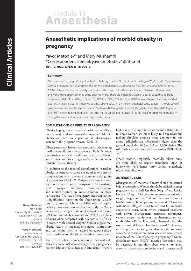
Anaesthetic implications of morbid obesity in pregnancy
Clin
ical
Art
icle
s
Yavor Metodiev* and Mary Mushambi*Correspondence email: [email protected]: 10.1029/WFSA-D-18-00013
COMPLICATIONS OF OBESITY IN PREGNANCYObesity in pregnancy is associated with adverse effects on maternal, fetal and neonatal outcomes.4,5 Morbid obesity can have an impact on all physiological systems in the pregnant woman (Table 1).
Obese parturients have an increased risk of developing medical complications in pregnancy (Table 2). Some pre-existing medical conditions, such as diabetes and asthma, are prone to get worse or become more resistant to usual therapy.
In addition to the medical complications related to obesity in pregnancy, there are number of obstetric complications which are more common in this group of parturients (Table 2). Peripartum complications, such as perineal trauma, postpartum haemorrhage, cord prolapse, infection, thromboembolism, and uterine rupture are more common in obese parturients.7 The rate of emergency caesarean section is significantly higher in the obese group, usually due to protracted labour or failed trial of vaginal delivery post caesarean section.4 A study by Wolfe demonstrated a higher failure of induction of labour (29% for morbid obese women and 23% for all obese women) when compared with a failure rate of 13% in women with normal weight.6 Studies suggest that obesity results in impaired myometrial contractility and that leptin, which is released by adipose tissue, may contribute to inhibition of uterine contractions.8
The fetus of obese women is also at increased risk. There is a higher risk of miscarriage in early pregnancy, preterm labour or fetal demise in later dates.4 There is
higher rate of congenital abnormalities. Babies born to obese women are more likely to be macrosomic making shoulder dystocia more common in this group. Stillbirths are substantially higher than the general population (8.6 vs. 3.9 per 1,000 births). The still birth rate increases with increasing BMI (Table 3).3
Obese women, especially morbidly obese ones, are more likely to require anaesthetic input or intervention and hence carry further anaesthetic related complications.
ANTENATAL CAREManagement of maternal obesity should be started before conception. Women should be advised to enter pregnancy with a BMI less than 30kg.m-2 and ideally less than 25 kg.m-2. On the first antenatal consultation weight, height and BMI should be recorded and a baseline arterial blood pressure measured. All women with BMI >40kg.m-2 must be referred for antenatal anaesthetic consultation where potential problems with airway management, neuraxial techniques, venous access, equipment requirements, or co-morbidities will be identified, discussed with the woman and delivery plan documented.9 However, it is important to recognise that despite antenatal anaesthetic consultation, many obese women, remain unaware of the risks of obesity in pregnancy.10 Multi-disciplinary team (MDT) meeting discussion may be necessary in morbidly obese women to allow obstetric, anaesthetic, midwifery and theatres staff
Yavor Metodiev md phdfrca
University Hospitals of Leicester NHS Trust
LeicesterUNITED KINGDOM
Mary Mushambi mbchb, frca, llm
University Hospitals of Leicester NHS Trust
LeicesterUNITED KINGDOM
56 www.wfsahq.org/resources/update-in-anaesthesia
Summary
Obesity is one of the greatest public health challenges of the 21st century. According to World Health Organisation
(WHO), the proportion of people in the general population classed as obese has risen by about 10 times since
1980s.1 Likewise, maternal obesity has mirrored this trend but with some variations between different parts of
the world, developed countries being affected more. There are different classes of obesity according to body
mass index (BMI): 30 – 34.9kg.m-2 (class 1); BMI 35 – 39.9kg.m-2 (class 2); and BMI above 40kg.m-2 (class 3 or morbid
obesity).1 Maternal obesity is defined as a BMI above 30kg.m-2 on the first antenatal consultation. In the UK, 20% of
pregnant women are classified as obese2, 5% have a BMI of greater than 35, 2% greater than 40 and 0.2% greater
than 50.3 Obesity during pregnancy puts the mother, fetus and neonate at higher risk of morbidity and mortality
during the antenatal, intrapartum and postnatal periods.

to plan for the care of such women and address requirements such as special equipment and manual handling issues. Attempts should be made to seek signs and symptoms of obstructive sleep apnoea (OSA) and, if necessary, appropriate investigations such as sleep studies requested and treatment with CPAP initiated. Consent issues should be discussed with the woman, particularly, with regards to the management of emergency caesarean section for fetal distress which may not be achievable in the time scale which is applicable for women with normal weight.
Parturients with BMI >35kg.m-2 are not suitable for home birth and most of them would not be under midwife-led care.9 All women with high BMI should be advised about healthy diet and exercise before and during pregnancy. Professional dietary advice should be sought especially with regards to folic acid and vitamin D supplements.
Thromboembolic risk must be assessed and women at high risk should be prescribed pharmacological thrombo-prophylaxis according to local and national guidelines.11 Continuous screening for hypertensive disorders and gestational diabetes is recommended.
INTRAPARTUM CAREMorbidly obese women should deliver in a consultant-led obstetric unit with continuous midwifery care. Obesity on its own is not an indication for induction of labour but the presence of co-morbidities may be.9 Vaginal delivery is associated with fewer complication rates in this group of parturients, but each case should be taken individually, and a delivery plan should be in place.
ANAESTHETIC MANAGEMENT OF VAGINAL DELIVERYThe anaesthetic team must be involved early in the intrapartum care of morbidly obese women. When a woman presents to labour ward, the duty anaesthetist and theatre team should be made aware. Any antenatal consultations and plans should be reviewed and possibly re-discussed with the team and the woman at the time.
Early venous cannulation is advised as these can prove difficult and are almost always needed for obstetric or anaesthetic reasons. If venous cannulation proves difficult, ultrasound can be used to aid identification of deep veins. In very rare cases, central venous cannulation is required but this is also associated with difficult insertion and increased risk of complications.
System Changes related to morbid obesity
Cardiovascular • Increased blood volume
• Increased cardiac output
• Hypertension and left ventricular hypertrophy
• Increased risk of myocardial ischaemia
• Systolic and diastolic dysfunction
• Supine hypotension due to higher risk of aorto-caval compression
• Pulmonary hypertension and cor pulmonale secondary to obstructive sleep apnoea (OSA)
Respiratory • Reduced FRC
• Increased closing capacity
• Restrictive respiratory changes due to weight of thoracic adipose tissue
• Decreased chest wall and lung compliance
• Obstructive sleep apnoea (OSA)
• Increased risk of difficult airway
• Obesity-related hypoventilation syndrome (Pickwickian syndrome)
• V/Q mismatching
Gastrointestinal • Increased risk of regurgitation and pulmonary aspiration (Hiatus hernia and increased residual gastric contents)
• Fatty liver infiltration
Renal • Increased angiotensin-converting enzyme and renin levels
• Glomerular filtration rate may be reduced
• Sodium retention
Endocrine • Peripheral insulin resistance
• Increased lipid levels
• Dysregulation of sex hormones
Haematological • Pro-coagulable state
• Increased risk of thromboembolic diseases
• Increased haematocrit due to hypoxia
• Reduced immune defence
Musculoskeletal • Poor mobility
Table 1. Maternal physiological changes associated with morbid obesity
57
© World Federation of Societies of Anaesthesiologists 2019. This issue may be freely reproduced for the purposes of private research and study and extracts (or indeed, the full report) may be included in professional journals provided that suitable acknowledgement is made and the reproduction is not associated with any form of advertising. Applications for commercial reproduction should be addressed to: World Federation of Societies of Anaesthesiologists, Dean Bradley House, 52 Horseferry Rd, London SW1P 2AF, UK.

Labour analgesia should be discussed early and epidural for labour advised. There is higher epidural failure rate in morbidly obese patients.12 As BMI increases, landmarks are more difficult to palpate, and the midline is hard to establish. Positioning the woman for epidural insertion is also challenging due to inability to flex the back. The use of ultrasound to identify the midline, determine the depth to the epidural space and level of lumbar spines is recommended as it has been demonstrated to reduce the number of attempts and increase the success rate.13 Longer needles may be needed but are not always available. Once the epidural catheter is sited, it is important to carefully sit the woman prior to fixation of the epidural catheter to prevent withdrawal of the catheter from the epidural space. Increased mobility of the skin and fat can cause displacement of the epidural catheter during any movement in labour. It is therefore essential to leave increased length of epidural catheter in the epidural space and to monitor the epidural closely during labour.
Combined spinal-epidural (CSE) is used for labour analgesia in some units but it may not always be a feasible option due to lack of long enough pencil-point needles. Continuous spinal analgesia can be offered, especially when accidental dural puncture occurs.14 The use of an intrathecal catheter in labour requires close monitoring by an anaesthetist. The intrathecal catheter can be used to top up for operative delivery or other procedures.
Patient controlled intravenous analgesia (PCA) with opioids (remifentanil, fentanyl, morphine) is not advisable in morbidly obese women because of the risks of respiratory depression and sedation. Many women with BMI >40kg.m-2 have OSA and/or obesity related hypoventilation. Opioid administration will exacerbate hypoventilation and increase the incidence of apnoea, leading to
prolonged hypoxic episodes. This is detrimental to the mother, as well as to the fetus. In addition to the physiological side effects of opioids, pharmacokinetic and pharmacodynamic considerations exist. The dosing regimen should be based on lean body weight. Most PCA pumps have limitations of their pharmacokinetic models when high weights are used, making dosing of drugs unreliable, unsafe, or inadequate.
Active management of third stage of labour is recommended because of the increased likelihood of postpartum haemorrhage. Cross-matched blood should be available and administered early if required. Uterotonic agents, such as syntocinon, should be used as a bolus and an infusion. Care with ergometrine should be taken, as there is higher chance that obese women are hypertensive or pre-eclamptic.
ANAESTHESIA FOR CASEAREAN SECTIONUnless there are contraindications, neuraxial anaesthesia remains the preferred modality of anaesthesia for caesarean section in morbidly obese parturients because general anaesthesia is associated with increased maternal mortality and morbidity.15
REGIONAL ANAESTHESIA
Epidural top upIn cases where an indwelling epidural catheter has been working well for labour analgesia, epidural top up is the safest and perhaps quickest anaesthetic technique Epidural top ups in the obese parturients should be carefully titrated and thorough check of the sensory block undertaken. Maximum doses of local anaesthetics should be calculated according to Ideal Body Weight (IBW). Volumes required may be lower than expected due to increased pressure in the epidural space and higher content of adipose tissue. Epidural opioids should be used to augment the block and to prolong analgesia in the postoperative period. The same precautions with epidural opioids should be considered as with intravenous ones. Long-acting opioids, such as diamorphine or morphine, may necessitate HDU care for close monitoring and possibly non-invasive ventilation (in some case of severe OSA). The advantages of a good working epidural catheter are that it can be topped up if required should surgery be prolonged, it can be used for postoperative analgesia or it can be topped up again should further operative procedure be required in the postoperative period.
Early mobilisation should be encouraged to reduce the risk of thromboembolic events. If mobilisation is delayed, intermittent compression devices should be used until the woman is mobilising.
Single-shot spinal anaesthesia Single-shot spinal anaesthesia (SSSA) is a procedure that most anaesthetists are familiar with and it provides reliable block with a rapid onset. It, therefore, makes it a reasonable option when faced
Maternal
Higher risk of maternal death
Thromboembolism
Gestational and type 2 diabetes
Hypertension
Pre-eclampsia
Obstructive sleep apnoea
Post –partum haemorrhage
Infections – Wound, urinary tract, genital tract
Increases Caesarean section rate
Increased rate of induction of labour
Higher rate of failed induction of labour
Fetal and neonatal risks
Miscarriage
Preterm labour
Stillbirth
Fetal abnormality such as neural defects and cardiac defects
Fetal distress
Macrosomia
Low Apgar scores
Table 2. Maternal, fetal and neonatal risks associated with maternal obesity BMI (kg/m2) Still births (per 1000 births)
35 – 39.99 7.9
40 – 49.99 8.8
>50 15.8
Table 3. Rates of still births in relation to BMI.3
58 www.wfsahq.org/resources/update-in-anaesthesia

with an urgent caesarean section for a morbidly obese parturient. However, there are some drawbacks to using a SSSA in morbidly obese women. The need for a longer needle may make the introducer needle inadequate for the long spinal needle. This can lead to more failed attempts causing delays and distress. Some authors suggest using a Tuohy needle as an introducer in the morbidly obese patients as it leads to fewer attempts in establishing anaesthesia.16 A second drawback is the inability to top up should surgery be prolonged. In this case, conversion to general anaesthesia (GA) remains the only rescue option. Conversion to general anaesthesia intraoperatively in morbidly obese woman can be challenging and can present the anaesthetist with a dilemma should failed intubation occur. The third drawback is the inability to titrate the spinal dose. There is no evidence that reduction in doses of intrathecal local anaesthetics is required in morbidly obese women.17 However, because of the increased risk of aorto-caval compression, morbidly obese patient may develop severe catastrophic post spinal hypotension.18
Combined spinal-epidural anaesthesiaCombined spinal-epidural anaesthesia (CSE) is the preferred neuraxial technique for morbidly obese parturients. It provides reliable anaesthesia with a rapid onset (spinal component) and the ability to extend the duration and the level of the block as required (epidural component). Other advantages of a CSE are the ability to give a lower spinal dose and to extend the block using the epidural catheter. This technique is also preferred in cases where opioids are considered higher risk such as patients with severe OSA because anaesthesia can be extended by epidural top ups with local anaesthetics. Disadvantages of CSEs include: possible failure to pass the epidural catheter, a slightly higher failure rate of the spinal component and the usage of an untested epidural catheter.
Continuous spinal anaesthesiaThis technique is rarely used as a primary anaesthetic plan, but there are suggestions that it should be considered more. It is usually employed in cases where an intrathecal catheter has been used for labour analgesia or an attempt at an epidural or CSE has produced a dural puncture.14 Intrathecal catheters should be managed very carefully, and local anaesthetic injected very slowly and with limited volumes. The risk of high spinal anaesthesia or total spinal anaesthesia is high in these cases. High spinal block is highly undesirable in the obese population as hypoventilation is more profound and leads to more pronounced hypoxaemia.
The use of continuous spinal anaesthesia is also limited by the higher risk of post-dural puncture headache (PDPH), despite the protective function of an indwelling catheter. There are suggestions that obese patient is at a lower risk of developing PDPH, but this is controversial.19, 20
Double catheter techniqueIn certain cases of parturients with extremely high BMI a midline laparotomy is performed instead of horizontal (Pfannenstiel) incision. Anaesthesia and analgesia provided by a lumbar epidural would not be sufficient in these patients. There are several cases reports of simultaneously using two epidural catheters – lumbar and low thoracic levels.21, 22 This is an option which needs appropriate
planning and is probably applicable to only few super morbidly obese parturients.
GENERAL ANAESTHESIAGeneral anaesthesia in morbidly obese parturients is usually reserved for the emergency situations and instances where neuraxial techniques are contraindicated or have failed. It is also sometimes the only option when faced with non-obstetric surgery.
Antacid PremedicationMorbid obesity is associated with increased risk of regurgitation and aspiration. It is therefore important that antacid prophylaxis using H2-blockers and sodium citrate are used appropriately in these patients.
Positioning Positioning on the operating table of a morbidly obese pregnant woman can prove to be a challenge. A compromise between left lateral tilt, appropriate position for airway management, and patient’s comfort, needs to be achieved. Elevating the woman’s torso in the head up ramped position improves ventilation and laryngoscopy views and reduces reflux symptoms. Aligning the tragus and the sternal notch improves airway management. Ramping position can be achieved by using head elevating laryngoscopy aids such as the Oxford Head Elevating Laryngoscopy Pillow HELP® or by using multiple pillows under the patient’s torso until an elevated desirable position is achieved.
Operating tables have limits on the weight they can sustain safely. This needs to be checked prior to positioning. Extra theatre staff is necessary to help with manual handling, especially when hoists are not available.
Monitoring Monitoring requirements for the morbidly obese patient should comply with the AAGBI monitoring standards.23 However, additional monitoring may be necessary in women with severe co-morbidities or women were non-invasive monitoring is inadequate. It is important to use appropriately sized blood pressure cuff in order to have accurate measurements. In some instances, invasive blood pressure monitoring may be the only option to have reliable measurements. Transabdominal fetal monitoring may prove difficult or impossible due to the large size of abdominal fat.
Anaesthetic drugs Administration of anaesthetic drugs in morbidly obese patients requires careful understanding of the altered pharmacodynamics and pharmacokinetics. Incorrect calculations can lead to overdosing or under dosing. Total body weight (TBW) should be used for suxamethonium, lean body weight (LBW) should be used for drugs such as thiopentone, propofol and opioids and ideal body weight (IBW) - for drugs such as non-depolarising muscle relaxants, paracetamol and non-steroidal anti-inflammatory drugs. Historically, the induction agent of choice in obstetrics in the UK has been thiopentone but there has been a recent change to using propofol following the publication of the DAS/OAA obstetric difficult airway guidelines and the 5th National Audit Project (NAP5)24 results.
59
© World Federation of Societies of Anaesthesiologists 2019. This issue may be freely reproduced for the purposes of private research and study and extracts (or indeed, the full report) may be included in professional journals provided that suitable acknowledgement is made and the reproduction is not associated with any form of advertising. Applications for commercial reproduction should be addressed to: World Federation of Societies of Anaesthesiologists, Dean Bradley House, 52 Horseferry Rd, London SW1P 2AF, UK.

Thiopentone was identified as a risk factor for accidental awareness under general anaesthesia as a result of inappropriate dosing (especially in the obese), short duration of action and infrequent use in non-obstetric cases.24 Propofol has many advantages such as familiarity, less confusion with other drugs, does not need mixing and drawing up in advance and produces better intubating conditions.
Rapid sequence induction (RSI) is almost universally employed in obstetric anaesthesia. Short acting muscle relaxant producing deep paralysis rapidly is ideal in these situations. In the morbidly obese parturients difficult airway is more likely and paralysis needs to be short or reversible to make intubation attempts safe. Succinylcholine is the classic muscle relaxant used in a dose of 2 – 2.5mg.kg-1 total body weight (TBW). Obesity and pregnancy reduce pseudocholinesterase levels and this makes the duration of action of succinylcholine unpredictable in some cases. After the introduction of sugammadex, there has been a slow but gradual change to using rocuronium for RSI in obstetrics in a dose of 1 – 1.2mg.kg-1 IBW. It produces adequate intubating conditions within 30 seconds and is comparable to succinylcholine. Unfortunately, sugammadex is still an expensive drug and is not always readily available; hence the slow change in practice. Immediate reversal of high-dose rocuronium paralysis is achieved by 16mg.kg-1 IBW of sugammadex. However, in this situation, it is easy to develop a false sense of security as reversal of muscle relaxant does not necessarily produce a clear airway.
Maintenance of anaesthesia in morbidly obese patients is largely achieved by inhalational agents, such sevoflurane or desflurane which have shorter recovery times compared with isoflurane.
Airway managementGeneral anaesthesia (GA) in morbidly obese patients is associated with significant morbidity and mortality. Three out of four obstetric difficult airway cases in the Fourth National Audit Project (NAP4) were obese.25 The physiological changes of pregnancy exacerbate the difficulties encountered when managing the airway in the obese patient. Therefore, general anaesthesia in morbidly obese parturient requires detailed airway assessment, careful planning and preparation, and communication with the team prior to induction of GA. Airway assessment should encompass prediction of: difficult tracheal intubation, mask ventilation, supraglottic device insertion and front-of-neck access. Neck circumference is a useful predictor of difficult intubation, mask ventilation and front-of-neck access. Ultrasound of the neck can be used to identify the cricothyroid membrane prior induction of GA in case failed tracheal intubation occurs and front-of-neck access is required. In cases of super morbidly obese, an awake intubation using either a fibreoptic scope or rigid laryngoscope may be chosen as the safest airway management prior to induction of general anaesthetic.14
In 2015, the Difficult Airway Society (DAS) produced algorithms for airway management specific to obstetrics (see Figure 1 and Figure 2).26 The main emphasis in these guidelines are planning and preparation, as well as communication with the whole team. Prior to induction of GA, the team should decide whether the patient should be woken up or surgery proceed, should failed intubation occur. This can be done using Table 1 of the DAS/OAA guidelines (Figure 2).
Figure 1. Safe Obstetric general anaesthetic algorithm – reproduced with permission form the Difficult Airway Society and Obstetric Anaesthetic Association.26
60 www.wfsahq.org/resources/update-in-anaesthesia

Figure 2. Decision whether to proceed or wake following failed tracheal intubation- reproduced with permission form the Difficult Airway Society and Obstetric Anaesthetic Association.26
Morbid obesity favours the waking of the patient because proceeding with surgery with an unsecured airway carries a high risk of maternal hypoxia. Pre-oxygenation is recommended prior to induction of GA in all patients, but in particular during RSI. However, despite good pre-oxygenation with face mask, pregnant women desaturate rapidly, and obese pregnant women desaturate even faster. The new guidelines recommend the use of nasal oxygenation to administer oxygen during apnoea in order to increase the safe apnoea time. Nasal cannula at 5 – 15L/min have been shown to be effective in reducing hypoxia during instrumentation of the airway.27 High flow humidified nasal oxygen using systems, such as Optiflow, have been used recently and shown to increase the safe apnoea time in obstetrics.28 Videolaryngoscopes increase successful rate of intubation and should therefore be available and be used in obese women.
A significant proportion of airway related complications arise during emergence of anaesthesia. Morbidly obese patients are prone to slower emergence and extubation should be carefully planned and performed according to the DAS extubation guidelines.29 The morbidly obese woman falls into the high risk extubation group and should have tracheal extubation performed with the woman fully awake and in a ramped position.
Postoperative care and analgesiaPostoperative analgesia following general anaesthesia is a challenge in the morbidly obese parturient. Multimodal pain management is advised – regional techniques are favoured in order to reduce the need for systemic opioids. Local infiltration with local anaesthetics or transverse abdominal plane (TAP) blocks performed by either the
anaesthetist under ultrasound guidance or the obstetrician provide significant pain relief and lead to less opioid usage. Paracetamol and non-steroidal anti-inflammatory drugs, where not contraindicated, reduce opioid consumption significantly. Certain analgesics, such as codeine, should be avoided in women who are breastfeeding.
Close post-operative monitoring of morbid obese women in an HDU setting is important in order to prevent avoidable complications such respiratory depression. Precautions against DVT should take place and appropriate doses of thrombo-prophylaxis administered.
CONCLUSIONObesity in the obstetric population continues to grow and present challenges to all healthcare workers. Morbid obesity increases morbidity and mortality in pregnant women and their babies. Careful planning and preparation in the antenatal, intrapartum and postnatal periods is necessary in order to ensure safe outcomes for the mother and baby. Regional analgesia and anaesthesia are recommended in order to avoid the risks of general anaesthesia. Should general anaesthesia be required, careful team planning, preparation and performance by experienced staff is essential.
REFERENCES1. www.who.int/topics/obesity/en/. Accessed 19th May 2018.
2. Heslehurst N, Lang R, Rankin J, Wilkinson JR, Summerbell CD. Obesity in pregnancy: a study of the impact of maternal obesity on NHS maternity services. BJOG 2007; 114(3): 334 – 42.
3. Centre for Maternal and Child Enquires (CMACE). Maternal obesity in the UK: Findings from a national project. London: CMACE 2010.
61
© World Federation of Societies of Anaesthesiologists 2019. This issue may be freely reproduced for the purposes of private research and study and extracts (or indeed, the full report) may be included in professional journals provided that suitable acknowledgement is made and the reproduction is not associated with any form of advertising. Applications for commercial reproduction should be addressed to: World Federation of Societies of Anaesthesiologists, Dean Bradley House, 52 Horseferry Rd, London SW1P 2AF, UK.

4. Leddy MA, Power ML, Schulkin J. The impact of maternal obesity on maternal and fetal health. Rev Obstet Gynecol 2008; 1(4): 170 – 8.
5. Tonidandel A, Booth J, D’Angelo R, Harris, L, Tonidandel S. Anesthetic and obstetric outcomes in morbidly obese parturients: a 20-year follow-up retrospective cohort study. Int J Obstet Anesth. 2014; 23(4): 357 – 64.
6. Wolfe KB, Rossi RA, Warshak CR. The effect of maternal obesity on the rate of failed induction of labour. Am J Obstet Gynecol 2011; 205(2): 128.e 1 – 7.
7. Conner SN, Verticchio JC, Tuuli MG, Odibo AO, Macones GA, Cahill AG. Maternal obesity and risk of postcesarean wound complications. Am J Perinatol 2014; 31(4): 299 – 304.
8. Moynihan AT, Hehir MP, Glavey SV, Smith TJ, Morrison JJ. Inhibitory effect of leptin on human uterine contractility in vitro. Am J Obstet Gynecol 2006; 195(2): 504 – 9.
9. Management of women with obesity in pregnancy. CMACE/RCOG Joint Guideline. March 2010.
10. Eley VA, Donovan K, Walters E, Brijball R, Eley DS. The effect of antenatal anaesthetic consultation on maternal decision-making, anxiety level and risk perception in obese pregnant women. Int J Obstet Anesth 2014; 23(2): 118 – 24.
11. Green-top Guideline No. 37a. Reducing the risk of venous thromboembolism during pregnancy and the puerperium. Royal College of Obstetricians and Gynaecologists (RCOG). April 2015.
12. Whitty RJ, Maxwell CV, Carvalho JC. Complications of neuraxial anesthesia in an extreme morbidly obese patient for Cesarean section. Int J Obstet Anesth 2007; 16(2): 139 – 44.
13. Balki M, Lee Y, Halpern S, Carvalho JC. Ultrasound imaging of the lumbar spine in the transverse plane: the correlation between estimated and actual depth to the epidural space in obese parturients. Anesth Analg 2009; 108(6): 1876 – 81.
14. Hood DD, Dewan DM. Anesthetic and obstetric outcome in morbidly obese parturients. Anesthesiology 1993; 79: 1210 - 18.
15. Lamon AM, Habib AS. Managing anesthesia for caesarean section in obese patients: current perspectives. Local Reg Anesth 2016; 16(9): 45 – 57.
16. Ross VH, Dean LS, Thomas JA, Harris LC, Pan PH. A randomized controlled comparison between combined spinal-epidural and single-shot spinal techniques in morbidly obese parturients undergoing caesarean delivery: time for initiation of anesthesia. Anesth Analg 2014; 118(1): 168 – 72.
17. Carvalho B, Collins J, Drover DR, Atkinson Ralls L, Riley ET. ED(50) and ED(95) of intrathecal bupivacaine in morbidly obese patients undergoing cesarean delivery. Anesthesiology 2011; 114(3): 529 – 35.
18. Tsueda K, Deband M, Zeok SS, Wright BD, Griffin WO. Obesity supine death syndrome: reports of two morbidly obese patients. Anesth Analg 1979; 58(4): 345 – 7.
19. Faure E, Moreno R, Thisfed R. Incidence of postdural puncture headache in morbidly obese parturients. Reg Anesth 1994; 19(5): 361 – 3.
20. Miu M, Paech MJ, Nathan E. The relationship between body mass and post dural puncture headache in obstetric patients. Int J Obstet Anesth 2014; 23(4): 371 – 5.
21. Polin CM, Hale B, Mauritz AA, et al. Anesthetic management of super-morbidly obese parturients for cesarean delivery with a double neuraxial catheter technique: a case series. Int J Obstet Anesth 2015; 24(3): 276 – 80.
22. McDonnell NJ, Paech MJ. The management of a super morbidly obese parturient delivering twins by cesarean section. Anesth Intensive Care 2007; 35(6): 979 – 83.
23. Association of Anaesthetists of Great Britain and Ireland. Recommendations for standards of monitoring during anaesthesia and recovery 2015. Anaesthesia 2016; 71: 85 – 93.
24. Pandit JJ, Cook TM, the NAP5 Steering Panel. NAP5. Accidental Awareness During General Anaesthesia. London: The Royal College of Anaesthetists and Association of Anaesthetists of Great Britain and Ireland 2014 ISBN 978-1 900936-11-8.
25. Cook TM, Woodall N, Frerk C; Fourth National Audit Project. Major complications of airway management in the UK: results of the Fourth National Audit Project of the Royal College of Anaesthetists and the Difficult Airway Society. Part 1: anaesthesia. Br J Anaesth 2011; 106: 617 – 31.
26. Mushambi MC, Kinsella SM, Popat M, Swales H, Ramaswamy KK, Winton AL, Quinn AC. Obstetric Anaesthetists’ Association and Difficult Airway Society guidelines for the management of difficult and failed tracheal intubation in obstetrics. Anaesthesia 2015; 70: 1286 – 306.
27. Baraka AS, Taha SK, Siddik-Sayyid SM, et al. Supplementation of pre-oxygenation in morbidly obese patients using nasopharyngeal oxygen insufflation. Anaesthesia 2007; 62: 769 – 73.
28. Mir F, Patel A, Iqbal R, Cecconi M, Nouraei SA. A randomised controlled trial comparing transnasal humidified rapid insufflation ventilatory exchange (THRIVE) pre-oxygenation with face-mask pre-oxygenation in patients undergoing rapid sequence induction of anaesthesia. Anaesthesia 2017; 72(4): 439 – 43.
29. Popat M, Mitchell V, Dravid R, Patel A, Swampillai C, Higgs A. Difficult Airway Society Guidelines for the management of tracheal extubation. Anaesthesia 2012; 67: 318 – 40.
62 www.wfsahq.org/resources/update-in-anaesthesia

Nesrine RefaiCairo UniversityCairoZamalek EGYPT
Reham Abdel Rahman AliLecturerCairo UniversityEGYPT
Hala GomaaProfessor of AnaesthesiaCairo UniversityEGYPT
Update in obstetric trauma management
Nesrine Refa*, Reham Abdel Rahman Ali and Hala Gomaa*Correspondence email: [email protected]: 10.1029/WFSA-D-18-00018
DEFINITION AND DIAGNOSISTrauma during pregnancy is the leading non-obstetrical cause of maternal death and remains the most common cause of foetal demise. It complicates 6% to 8% of pregnancies, 0.4% require hospital admission and 0.1% suffer major trauma (Injury Severity Score (ISS) >15).1, 2
The initial evaluation and management of the traumatized pregnant patient requires a multidisciplinary, team to provide the optimal care for both mother and fetus. Although the resuscitation of the injured pregnant patient follows the same algorithmic protocol of all trauma patients, but it is essential to understand all of the anatomical and physiological changes that occur during pregnancy. Foetal outcomes are directly correlated with early and aggressive maternal resuscitation.3
Motor vehicle accidents and domestic violence account for most cases of maternal major trauma, while falls, burns, homicide, suicide, penetrating trauma, and toxic exposure account for the majority of the remainder.4
A 2013 systematic review of studies on trauma in pregnancy4 reported that the incidence rate of motor vehicle crash (MVC) during pregnancy has been estimated at around 207/100,000 pregnancies. While the incidence of domestic violence was 8307/100,000 live births. Blunt trauma accounts for 69% of trauma among pregnant women, whereas penetrating trauma accounts for only 1.5% of trauma during pregnancy.
Epidemiology of obstetric Trauma
Physical ViolenceInterpersonal violence during pregnancy carries a high risk to both the mother and fetus. A global systematic review described a prevalence during pregnancy of 1-20%.4 Intimate partner violence constitutes the majority of cases and increases with poverty as seen in India (21-28%)5, and some Asian countries (4-48%).6
It is also related to the community culture, religion, poverty and large families. Das et al., in 2013 have reported that one in seven women suffered intimate partner violence during or shortly after pregnancy in Mumbai slums.5 A similar study in Vietnam suggested that exposure to intimate partner violence during pregnancy increases the risk of preterm birth and low birth weight.4
Ibrahim et al., studied violence during pregnancy in 1,857 Egyptian women aged 18 - 43 years and reported prevalence of violence among 44.1% of the studied group.7
Motor Vehicle AccidentMotor vehicle crashes involving a pregnant driver were associated with elevated rates of adverse pregnancy outcomes, including preterm birth, stillbirth, placental abruption, and premature rupture of membranes (PROM).
In 2013, Cathrine et al., suggested that among pregnant drivers who were involved in one or more
63
© World Federation of Societies of Anaesthesiologists 2019. This issue may be freely reproduced for the purposes of private research and study and extracts (or indeed, the full report) may be included in professional journals provided that suitable acknowledgement is made and the reproduction is not associated with any form of advertising. Applications for commercial reproduction should be addressed to: World Federation of Societies of Anaesthesiologists, Dean Bradley House, 52 Horseferry Rd, London SW1P 2AF, UK.
SummaryTrauma during pregnancy is the leading non-obstetrical cause of maternal death and remains the most common
cause of foetal demise. It complicates 6% to 8% of pregnancies, 0.4% require hospital admission and 0.1% suffer
major trauma (Injury Severity Score (ISS) >15).
Motor vehicle accidents and domestic violence account for most cases of maternal major trauma, while falls, burns,
homicide, suicide, penetrating trauma, and toxic exposure account for the remainder .
Trauma management needs multidisciplinary team of emergency physician, obstetrician, anesthesiologist &
surgeons. Major trauma causes obstetric & fetal complications & may end by maternal death. Perimortem cesarean
section is important to save both mother & fetus. Pregnant trauma victims experience nearly 2-fold higher
mortality compared to non-pregnant.
Clin
ical
Art
icle
s

crashes, 2% were reportedly unbelted and 18% were driving in vehicles without airbags.8
The strong association between motor vehicle accidents and trauma in pregnancy raises the importance of the proper use of seatbelts. The correct way to wear the seat belt during pregnancy is keeping the lap belt low, on the pelvic bones, and placing the shoulder strap between the breasts (Figure1).8, 9
Homicide & SuicideThe governmental health authorities in many countries have adopted a surveillance system, looking at the mortalities among pregnant women. They aimed to identify the most common non obstetric causes of death including violence, homicide, suicide and intoxication.
In 2016, a study done by Wallace et al., reviewed the deaths in US states with enhanced pregnancy mortality surveillance from 2005–2010. They compared mortality among four groups of women: pregnant, early postpartum, late postpartum and non-pregnant/non-postpartum. They estimated pregnancy-associated mortality ratios and compared to non-pregnant/non-postpartum mortality ratio. They observed that homicide was more common in pregnant victims who were young, Black, and undereducated while pregnancy-associated suicide occurred most frequently among older White women. They also concluded that risk of homicide among pregnant/postpartum women was 1.84 times that of non-pregnant/non-postpartum women, while risk of suicide was decreased.10
APPROACH TO ASSESSMENT AND MANAGEMENT OF OBSTETRIC TRAUMATrauma in pregnancy can be classified into three different types: blunt abdominal trauma, pelvic fractures, and penetrating trauma. Obstetric trauma has been associated with serious complications including maternal injury, shock, internal hemorrhage, intrauterine fetal demise, direct fetal injury, abruptio placentae, and uterine rupture. Hence, a rapid efficient evaluation is critical to ensure the well-being of the mother and fetus.11
Assessment and ManagementA multidisciplinary team is warranted to optimize outcome for both the mother and fetus. This team involves trauma surgeons, emergency medicine physicians, obstetricians, neonatologists, nursing staff, and technicians. Whenever the patient’s condition necessitates surgery, an expert obstetric anesthetist should be involved.12 The team should consider the anatomical and physiological changes in pregnancy13, which could affect the type of injury and the mother’s response to trauma.14
According to Obstetric Trauma Guidelines 201414, the following sequence of actions should take place upon initial notification:
1. Gather vital information from the notifier using the MIST mnemonic:
M Mechanism of injury I Injuries found or suspected S Signs: respiratory rate, pulse rate, blood pressure, SpO2, GCS or AVPU T Treatment given
Figure 2: Manual tilt of the uterus
Primary SurveyEvery female of reproductive age with significant injuries should be considered pregnant until proven otherwise .Immediately from the time of starting a primary survey, pregnant patients beyond 20 weeks gestation should be nursed in a left lateral tilt of 15–30 degrees to relieve the aorto-caval compression.
A 30-degree tilt can be achieved by placing a wedge under the patient’s right hip, using pillows, rolled towels or bags of fluid, or by manually displacing the uterus to the left. The latter is preferred as it allows effective chest compression during cardiopulmonary resuscitation (Figure 2).14, 15
Gestational age can be identified from the fundal level. At 24 weeks gestation, the fundal level will be at the level of umbilicus.
64 www.wfsahq.org/resources/update-in-anaesthesia
Figure1: The correct way to wear the seat belt

A systematic approach based on the ABCDE survey should be applied to assess and treat an acutely injured patient.
ABCDE approach: A for Airway, B: Breathing, C: Circulation, D: Disability and E for Exposure.
The goal is to manage any life-threatening conditions and identify any emergent concerns, especially in a pregnant patient who may present with polytrauma complications.
Airway with cervical spine protection
Assess for airway stability:Airway assessment includes signs of airway obstruction (use of accessory muscles, paradoxical chest movements and see-saw respirations). Intubation is considered early if there are any signs of decreased level of consciousness, or unprotected airway .Intubation is considered even earlier in pregnant patients compared with non-pregnant patients because pregnant women desaturate more rapidly and are more susceptible to irreversible hypoxic injury. Maternal hypoxia is associated with poor fetal outcomes.
However, the likelihood of failed intubation is higher in a pregnant patient. Hence, intubation of a pregnant patient should be attempted by the most senior and experienced airway-skilled practitioner.15 Direct laryngoscopy using a standard laryngoscope (Macintosh blade) is commonly performed. A short-handled laryngoscope should be available for pregnant women because enlarged breasts may impede insertion of a laryngoscope with a standard-length handle.16
Preparations for a difficult intubation should start early, with all available equipment for difficult airway management including rigid laryngoscopy with multiple blades, Mccoy laryngoscope (with movable tip), different sizes of nasal and oral airway, small size of endotracheal tubes, supraglottic devices, stylets , illuminating stylet, (Gum-elastic) bougie, and video assisted laryngoscopy if available.
Availability of the fibre-optic would be helpful for intubation of patients with a known difficult airway or facial or cervical fractures. In limited resources countries, the anesthetist may assign another colleague to maintain manual in-line stabilization or keep the collar in place during intubation.15
Cricoid pressure can be considered during intubation to decrease the risk of regurgitation of gastric contents into the pharynx. However, cricoid pressure maybe adjusted or removed if it comprises the view.
BreathingIt’s advised to give high flow oxygen (15L) to keep oxygen saturation of the traumatized pregnant about 100%14 or at least above 95%.15 This recommendation aims to avoid the well- known poor fetal outcomes from maternal hypoxia. Chest auscultation is mandatory to detect wheezes or decreased air entry caused by underlying chest injury. A nasogastric tube should be inserted in a semiconscious or unconscious injured pregnant woman to prevent aspiration of acidic gastric content. Suspected fracture base of skull should be excluded before insertion of the nasogastric tube.14
Blunt chest trauma may be complicated by life-threatening injuries; tension pneumothorax, hemothorax or aortic rupture. These injuries should be diagnosed by clinical assessment, chest x-ray, computed tomography or bedside ultrasonography. Management includes needle thoracostomy or chest tube.17, 18 In traumatized pregnant patient, chest tube should be inserted one or two rib spaces higher (in the third or fourth intercostal space) due to the elevation of diaphragm in pregnancy.14
CirculationThe mother’s heart rate, blood pressure and neck veins should be checked, while signs of hemorrhage are inspected. The potential for significant internal bleeding is suspected in relation to the mechanism of injury. However, pregnant patients may not display signs of haemorrhage until there is a 30% reduction in blood volume. Tachycardia with normotension may be considered an early sign of potentially significant blood loss.14
Insertion of 2 large bore intravenous cannulae (14–16 gauge) is recommended for all seriously injured trauma patients to facilitate initial rapid crystalloid infusion and possible further blood transfusion if required. Administration of fluids and blood products during resuscitation should proceed according to standard trauma protocols. However, some modifications should be made in the pregnant trauma victim.15
Fluid resuscitation should be initiated if hypovolemia is suspected to maintain both maternal and fetoplacental perfusion. In the resource-limited environment, the use of cheaper crystalloid solutions is still recommended as crystalloid is effective in improving neonatal oxygenation if there is evidence of maternal hypo tension.19, 20 Commence resuscitation with up to 1–2L of crystalloid solution. Blood or blood product transfusion should be considered in subsequent fluid administration. If uncross matched blood is indicated, group O Rh-negative blood should be used to prevent antibody development. The goal is to transfuse blood and crystalloid to maintain hematocrit at 25-30% and urine output greater than 30cc/hr.15
Vasopressors are known to have adverse effect on uteroplacental perfusion. Hence, vasopressors in pregnant women should be used only for intractable hypotension that is unresponsive to fluid resuscitation. In cases of maternal hypotension unresponsive to intravascular volume expansion, the vasopressor of choice is ephedrine. Ephedrine has both β-2 and α-1 agonist properties, thus increasing uterine blood flow and maternal blood pressure.3
The focused assessment with sonography for trauma (FAST)The focused assessment with sonography for trauma (FAST) examination is considered as the best practice at this time. A FAST evaluates four areas where fluid or blood may accumulate in the abdomen including the subxiphoid pericardial window, hepatorenal recess, perisplenic and suprapubic view.12
A large retrospective review suggested that FAST examination has a reasonable specificity and accuracy of > 90% to detect free fluid in pregnant trauma patients (positive FAST scan).13
65
© World Federation of Societies of Anaesthesiologists 2019. This issue may be freely reproduced for the purposes of private research and study and extracts (or indeed, the full report) may be included in professional journals provided that suitable acknowledgement is made and the reproduction is not associated with any form of advertising. Applications for commercial reproduction should be addressed to: World Federation of Societies of Anaesthesiologists, Dean Bradley House, 52 Horseferry Rd, London SW1P 2AF, UK.

Some authors believe that in the stable pregnant trauma patient, computed tomography (CT) scanning is indicated only if the FAST examination is positive. While a recent retrospective review study found that CT can accurately diagnose placental abruption, in the presence of a negative FAST examination.15 The obstetrical ultrasound assessment includes fetal heart rate, gestational age, presentation, placenta, abruption, evidence of fetal injury, and evidence of intraabdominal/pelvic fluid.
As cost of obstetric ultrasound becomes more affordable in Low and middle-income countries LMICs, it was found to improve patient management.21 The LMICs have published articles documenting the importance of the use of FAST in trauma care. Articles from South Africa, Liberia, Rwanda and Peru concluded that ultrasound is a reliable and affordable alternative to computer tomography (CT) in context of trauma.22
Disability: neurological statusAssessment of neurological status includes assessment of consciousness level using Glasgow Coma Scale (GCS) or AVPU assessment (Alert, responds to Voice, responds to Pain, Unresponsive).Pupillary response and blood sugar level should also be assessed.
ExposurePatient’s clothing and jewelry are removed in order to assess all areas of the body. It is mandatory to avoid hypothermia in traumatized pregnant patient, as it leads to metabolic acidosis and bleeding. Hence, it is advised to monitor the patient temperature aiming to keep the patient temperature above 36.5°C. The patient is kept warm by keeping her in warm environment and warming the intravenous fluids.14
Secondary surveySecondary survey should only be started once the patient is stable with no any life-threatening injuries.
A detailed Top to Toe examination is performed, followed by fetal assessment.
Fetal assessment:Electronic monitoring of the fetus is indicated where there is ascertained fetal viability (greater than 24 weeks) and the appropriate equipment is available (cardiotocography/CTG).
CTG allows monitoring of the fetal heart rate and uterine contractions. Normal fetal heart rate ranges from 120 to 160 bpm.17
Types of trauma
Blunt trauma:Damage control laparotomy (DCL) is considered as life-saving procedure with the potential to diagnose the destructive clinical outcomes that can be expected under the circumstances of blunt abdominal trauma with exsanguination.23
In a systematic review of the current literature from January 2006 to July 2016, including a total of 95,949 patients, motor vehicle crash was the most frequent cause of blunt trauma, followed by falls, assault, both domestic and interpersonal violence.24
Direct fetal injury is uncommon with blunt trauma owing to the absorption of forces by the uterus, placenta and amniotic fluid. However, foetal injury and death is an indirect result of maternal shock and death.24
During the first trimester, the uterus is protected by the bony pelvis.24 As the uterus enlarges, it displaces the bowel cephalad, rendering the fetus more vulnerable to injury. Thinning of the uterine wall with growth and the relative decrease in amniotic fluid volume also contribute to fetal vulnerability. Bladder and spleen injury and retroperitoneal bleeds are the most common injuries resulting from blunt trauma.25
Pelvic fractures are commonly associated with blunt trauma and are associated with significant retroperitoneal hemorrhage as a result of engorged pelvic vessels.
Obstetric complications of blunt abdominal trauma include preterm labor, preterm delivery, preterm premature rupture of membranes, abruption, fetomaternal hemorrhage, and rarely, uterine rupture.
Penetrating trauma:Penetrating trauma is due to knife wounds or gunshot injuries and requires immediate surgical exploration. The gravid uterus grows cephalic pushing in all the viscera and acts as a protective barrier in penetrating trauma. Therefore, uterine injury is common in these cases, thus causing direct fetal injury and
increasing both maternal and fetal mortalities.
Management of penetrating trauma does not differ in the pregnant patient than non-pregnant.12 Exploratory laparotomy is not an indication for caesarean delivery, but the gravid uterus makes abdominal surgery difficult; hence caesarean section may be required.15
Burns:The incidence of burn injuries during pregnancy ranges from 7% to 15%,26 with the majority of cases in developing countries. Burn
66 www.wfsahq.org/resources/update-in-anaesthesia
History is obtained using AMPLE acronym to assist with gathering important information:
A Allergies
M Medication
P Past medical history
L Last meal
E Events leading to injury
Head-to-toe examination: Abdominal examination in pregnancy is challenging and complicated by the enlarged uterus.
Particular attention is given to signs of injury around the pelvis; tenderness over the uterus, uterine contractions, vaginal bleeding or ruptured membranes.15

occurs from direct thermal injury, inhalation injury to the airway, carbon monoxide, cyanide, or dissipation of electrical current. Maternal and fetal mortality risk depend on the amount of total body surface area burned (TBSAB) which can be calculated by the rule of 9’s.27 The rule of nines is meant to be used for: second-degree burns (partial-thickness burns), third-degree burns (full-thickness burns). For adults, the rule of 9’s is: Arm (including the hand): 9% each, Anterior trunk (front of the body): 18% Posterior trunk (back of the body): 18% Genitalia: 1% Head and neck: 9%As severe burn in pregnant women is not common, thus lacking data to define an adequate therapy.
Fluid resuscitation is of paramount important in burn during pregnancy, being a hyperdynamic state with total body plasma volume is expanded. Thus, the risk of a hypovolemic shock is increased and can lead to placental insufficiency. However the resuscitation management is not different from that in the non-pregnant woman. Intravenous fluid replacement can be initiated using the Parkland formula (percentage of TBSA burned X 4mL per kg of body weight).28
There are many resuscitation formulas adopted by different burn units as Ruijin’s formula which is a modification of Evans formula. Ruijin’s formula sets the ratio of crystalloid solution versus colloid solution at 1:1.29 Appropriate modification of the volume of solution for fluid resuscitation based on vital signs and urine output of the patient is advised.
Much literature has indicated that septicemia and sepsis are the main causes of death in pregnant women, thus the administration of antibiotics is essential.
Inhalation injury for a pregnant patient needs special attention and needs early intubation. This type of injury causes significant edema that aggravates the physiological edema of the oropharynx and larynx associated with pregnancy. Carbon monoxide has increased affinity for the hemoglobin molecule and displaces oxygen. Fetal circulation has a 15% increase in carbonmonoxide which decreases the ability for oxygenation of the fetus, and may lead to fetal heart rate changes. Carbonmonoxide toxicity is treated by100%oxygenation, or hyperbaric oxygenation if available.27
Maternal mortality rate in the studies varies from 30% in the study of Ogbogu et al.,28 to 70% in other studies.29, 30, 31
The effect of burn on the fetus includes abortion or preterm delivery as confirmed by many studies. Factors determining the effects on the fetus include increased TBSA, hypovolemia, sepsis, pulmonary injuries (inhalation injury), catabolism and side effects of drugs.32,33
Urgent caesarean delivery is recommended with burns of 55% or more for viable fetuses without delaying for antenatal corticosteroids. For burns of <55%, antenatal corticosteroids may be administered with expectant management.34, 35, 36
Imaging during obstetric trauma:The ideal imaging modality during pregnancy has not been determined, but computed tomography (CT) appears to have higher sensitivity than plain film x-ray outside of pregnancy.
Although radiographic evaluation of the pregnant patient raises concerns about potential fetal exposure, but diagnostic imaging is not contraindicated in pregnant patients, as it may demonstrate obstetrical complications such as abruption or uterine rupture. Initial radiographic assessment of trauma pregnant women should include imaging of the cervical spine, chest, and pelvis.37
Computed tomography (CT) studies of the abdomen, pelvis, and lumbar spine should be done only if absolutely necessary, because these may subject the fetus to 3.5 rads (radiation absorbed dose). Because maternal head injury is one of the leading causes of maternal death and as head CT generates only 1 rad of exposure with proper shielding, this should be done as needed.
Radiation safety organizations recommend a total dose of no more than 5 rad for pregnant women.38
The greatest risk to the fetus from radiation exists early during the pregnancy, during organogensis (weeks 2–8) and neural tube development (up to week 15). Ultrasonography and magnetic resonance imaging (MRI) can be used when appropriate and provide no ionizing radiation to mother or fetus.39
Ultrasonography can assess the status of the foetus for foetal heart tones, foetal activity, presentation, placenta, abruption, evidence of fetal injury and approximate gestational age.12
Obstetric and Fetal Assessment:All pregnant trauma patients with a viable pregnancy (≥ 23 weeks) should undergo electronic fetal monitoring for at least 4 hours. Continuous fetal monitoring should be instituted as soon as the mother’s status allows, preferably in the emergency department.
FAST was effective in diagnosing placental abruption, fetal cardiac activity , other fetal injuries,15 evaluation of the pouch of Douglas for hemoperitoneum and distinguishing between intrauterine and extrauterine fluid.10
Fetal harm may include fetal fractures, especially skull, clavicles, and long bones, intracranial hemorrhage and indirect injury due to fetal hypoxia secondary to: maternal hypotension, fetal hemorrhage, placental abruption or other injury, cord injury and uterine injury. Other fetal complications are spontaneous abortion, preterm delivery, and RBC isoimmunization.19
Rh immune globulin (RhIG): 40% of trauma victims will have fetal-maternal bleed. All Rh-negative trauma victims should be considered for 1 vial of RhIG (300µg/ IM). This is determined by the Kleihauer-Betke (KB) test. The test is an acid elution assay on blood drawn from the maternal patient. After lysing cells with acid, it shows the amount of fetal blood in the maternal system. Even with negative Kleihauer-Betke (KB) test. It should be given as soon as possible, and within 72 hours of the accident.14, 31
67
© World Federation of Societies of Anaesthesiologists 2019. This issue may be freely reproduced for the purposes of private research and study and extracts (or indeed, the full report) may be included in professional journals provided that suitable acknowledgement is made and the reproduction is not associated with any form of advertising. Applications for commercial reproduction should be addressed to: World Federation of Societies of Anaesthesiologists, Dean Bradley House, 52 Horseferry Rd, London SW1P 2AF, UK.

If the mother’s condition is stable, Caesarean delivery is not required in the event of fetal death. Method and timing of delivery can be planned with the obstetric consultant. If a laparotomy will be performed for other reason, the obstetrician should be notified immediately. However, Caesarean delivery is still not indicated unless there is pelvic fractures or bleeding from uterine injury.31, 40
Cardiopulmonary Resuscitation (CPR) and Perimortem Caesarean Section (PMCS)Effective CPR is difficult in near-term pregnant woman because of difficult chest compressions and aorto-caval compression by gravid uterus.
Modifications of CPR in traumatized pregnant patients:
1. Left lateral displacement of the uterus is mandatory to relieve the caval compression. 2. Defibrillation as in non-pregnancy state with removal of fetal uterine monitors prior to shock. 3. Advanced airway is established early with C-spine stabilized.4. Closed-chest compressions: 100 per minute using 30:2 ratio with ventilations. The rescuer’s hands are placed slightly above the inter-nipple line.5. Intravenous line is established above the diaphragm.6. Advanced Cardiac Life Support (ACLS) drugs as indicated; adrenaline, amiodarone, magnesium, atropine and sodium bicarbonate.7. If no maternal response after 4 minutes of ACLS, immediate Caesarean delivery should be performed in the emergency department. Thoracotomy and open cardiac massage may be considered at this time if the patient or fetus is believed to be viable.
If fetal age is greater than or equal to 24 weeks: attempt to save life of both mother and fetus by delivering the fetus to relieve aorto-caval compression, thus increasing venous return to mother’s heart & help resuscitation attempts. If the gestational age is less than 24 weeks, urgent Caesarean unnecessary as aorto-caval compromise unlikely.
Assessment of fetal heart tones should be done throughout, as allowed by circumstances.41, 42, 43
Role of Anaesthesia in Obstetric trauma managementThe anaesthesiologist has multiple concerns while supporting the pregnant patient with major trauma, namely, adequate resuscitation and supporting surgical haemostasis. Ideally, the obstetric anaesthesiologist being more familiar with the physiology of pregnancy can play an important role in resuscitation, in addition to providing anaesthesia for surgical intervention.44
Women injured in Traumatic motor vehicle accidents have high incidence of surgical procedures and Caesarean delivery. Pregnancy is not a contraindication to operative management of pelvic fractures, or neurosurgical intervention. The later should not be delayed.32, 40
Indications for emergency caesarean delivery include a stable mother with a viable fetus in distress and traumatic uterine rupture.
During surgery for the traumatically injured, the fetus can be at risk of hypoxemia, teratogenicity, and preterm delivery. However, commonly used anaesthetic medications in pregnancy are considered to be safe, including benzodiazepines and inhalational anaesthetics. When large haemodynamic changes are anticipated, fetal monitoring should be utilized where the surgical site allows for monitoring, to help assess the adequacy of placental perfusion.19
General anaesthesia should always be induced with rapid sequence induction and cricoid pressure in order to decrease the risk of regurgitation of gastric contents in the pharynx.45
For rapid sequence induction of trauma patients, the following induction agents are commonly used; etomidate, propofol and ketamine. Propofol is widely used anaesthetic/sedative with pregnant patients because it has a rapid onset and short duration of action.14
The Advanced Trauma Life Support Guidelines of the American College of Surgeons recommend the use of etomidate under these circumstances for rapid sequence induction and intubation (RSI) of the trachea. However, ketamine has many desirable pharmacodynamic properties that make it a reliable alternative to etomidate, and should be considered when a traumatically injured patient requires emergency intubation. These properties are the rapid onset, no need to adjust dosing in organ failure, minimal impact on haemodynamic stability and reducing opioid requirement for analgesia.46
Succinylcholine is an appropriate and safe drug, and it should be used for RSI and intubation for pregnant trauma patient, even if they have head-injury. The succinylcholine-induced intracranial pressure (ICP) increase has been a concern in the past, but recent studies have shown that clinical significance of this increase have been exaggerated.45
For maintenance of anaesthesia, literature shows that perioperative opioids are recommended with little risk.14 Ketamine has an important role as a continuous infusion throughout the operation and Intensive care unit stays. Ketamine remains invaluable due to its ability to maintain cardiorespiratory stability while providing effective sedation and analgesia.47 It may have the advantage of decreasing the incidence of depression and PTSD (post-traumatic stress disorder) that many trauma patients develop during recovery.48
Tranexamic acid is recommended for management of severe bleeding with trauma. It is considered safe for the fetus and can be given for traumatized pregnant patients. However, It is unknown if the use of tranexamic acid in the pregnant trauma patient reduces mortality.49
Prophylaxis for venous thromboembolism may be done with sequential compression devices, heparin, or enoxaparin. Both heparin and enoxaparin do not cross the placenta. If anticoagulation is required,
68 www.wfsahq.org/resources/update-in-anaesthesia

LMW heparin & fondaparinux are the safest choices during obstetric trauma.50
Transfusion therapy:The recommended transfusion therapy for traumatized pregnant include:
1. Rapid early replacement of coagulation factors & platelets to improves outcome2. RBCs are given in high ratio.3. Empiric transfusion based on clinical assessment rather than laboratory results.51
Obstetric Trauma outcomeTrauma during pregnancy has adverse effects on both maternal and newborn morbidity. The outcomes of pregnant women who suffer trauma depend on the type and extent of trauma. Obstetric patients who have even minor trauma are at a high risk of developing complication even after being rapidly stabilized, treated, and discharged.19
Penetrating trauma carries a fetal mortality rate of 30 - 80%, but the maternal mortality rates are low as the fetus protects the underlying organs of the pelvis. After blunt trauma, the morbidity and mortality depend on the severity of the force. Various series report morbidity rates of 5 - 45% in pregnant women suffering from blunt trauma. In many series, fetal demise is high when moderate to severe haemorrhage occurs.11, 15, 40
Meanwhile, motor vehicle accidents during pregnancy were associated with increased adverse pregnancy outcomes, including preterm birth, stillbirth, placental abruption, and premature rupture of membranes. Crashes were especially harmful if drivers were unbelted.8
However, Deshpande et al. in 2017, have compared pregnant to non-pregnant after traumatic injury and found that pregnant women and girls had a lower injury severity score and were significantly more likely to experience violent trauma. Pregnant trauma victims had almost twice the rate of mortality compared to the non-pregnant.52
Foetal survival rate from the study of Battaloglu et al. in 2016, was 56% following trauma. Foetal death in pregnant trauma patients most frequently occurred in the 2nd trimester.53
Another study of Tanizaki et al., suggested that high level of Maternal alpha-fetoprotien (MSAFP) may be a predictor of poor foetal outcome following trauma during pregnancy regardless of the severity of the trauma or the mother’s haemodynamic status.54
CONCLUSIONTrauma is the leading non-obstetric cause of death and disability in pregnant women, especially in western countries. Resuscitation priorities in obstetric trauma are the same as non-pregnant; treat first which kills first. Resuscitation requires multi-disciplinary team of emergency physician, obstetrician, anaesthesiologists and surgeons.
REFERENCES 1. Hill CC and Pickinpaugh J. Trauma and surgical emergencies in the obstetric patient. Surg Clin N Am 2008; 88(2): 421–440.
2. Sperry JL, Casey BM, McIntire DD, et al. Long-term foetal outcomes in pregnant trauma patients. Am J Surg 2006; 192(6): 715–721.
3. Emir Battaloglu, Eren Battaloglu, Justin Chu and Keith Porter. Obstetrics in trauma. Trauma 2015, Vol. 17(1) 17–23.
4. Mendez-Figueroa H., Dahlke J.D., Vrees R.A. and Rouse D.J. Trauma in pregnancy: An updated systematic review. American Journal of Obstetrics and Gynecology 2013, 209 (1), pp. 1-10.
5. Das S, Bapat U, Shah More N, Alcock G, Joshi W, Pantvaidya S and Osrin D. Intimate partner violence against women during and after pregnancy: a cross sectional study in Mumbai slums. BMC Public Health. 2013 Sep 9; 13: 817.
6. Kashif M, Murtaza K and Kirkman M: Violence against women during pregnancy in some Asian countries: a review of the literature. Ital J Public Health 2010, 7: 6–11.
7. Ibrahim ZM, Sayed Ahmed WA, El-Hamid SA and Hagras AM. Intimate partner violence among Egyptian pregnant women: incidence, risk factors, and adverse maternal and fetal outcomes. Clin Exp Obstet Gynecol. 2015; 42(2): 212-9.
8. Vladutiu C J, Marshall S W, Poole C., Casteel C., Menard K. and Weiss H B. Adverse Pregnancy Outcomes Following Motor Vehicle. Am J Prev Med. 2013; 45(5).
9. Mieghem T V, Whittle W L, Farine D, Seaward G and D’Souza R. Motor Vehicle Accidents in Pregnancy: Implications and Management (Letter to the Editor). J Obstet Gynaecol Can 2013; 35(4): 303–4.
10. Wallace M E, Hoyert D, Williams C. and Mendola P. Pregnancy-associated homicide and suicide in 37 US states with enhanced pregnancy surveillance. Am J Obstet Gynecol. 2016; 215(3): 364.e1–364.e10.
11. Mirza F G., Devine P C. and Gaddipati S. Trauma in Pregnancy: A Systematic Approach. Am J Perinatol 2010; 27: 579–586.
12. Huls CK and Detlefs C. Trauma in pregnancy. Semin Perinatol. 2018; 42(1): 13-20.
13. Smith K A , Bryce S , Gursahani k and Synovitz C K. Trauma in the pregnant patient: An Evidence-Based Approach to Management. Emergency Medicine Practice, April g2013, 15 (4): 2.
14. OBSTETRIC TRAUMA GUIDELINE, Version 1.0 - 25/09/2014.p:1-31.
15. Jain V, Chari R, Maslovitz S and Farine D. Guidelines for the Management of a Pregnant Trauma Patient. J Obstet Gynaecol Can 2015; 37(6): 553–571.
16. Mushambi MC, Kinsella SM, Popat M, et al. Obstetric Anaesthetists’ Association and Difficult Airway Society guidelines for the management of difficult and failed tracheal intubation in obstetrics. Anaesthesia. 2015; 70: 1286-1306.
17. Murphy N J and Quinlan J D. Trauma in Pregnancy: Assessment, Management, and Prevention. Am Fam Physician. 2014; 90(10): 717-722.
18. Morley EJ, Johnson S, Leibner E and Shahid J. Emergency department evaluation and management of blunt chest and lung trauma (Trauma CME). Emerg Med Pract. 2016; 18(6): 1-20.
19. Sun Moon T and Sappenfield J .Anesthetic Management and Challenges in the Pregnant Patient. Curr Anesthesiol Rep (2016) 6: 89–94.
20. Wise R., Faurie M., Malbrain M.and Hodgson E. Strategies for Intravenous Fluid Resuscitation in Trauma Patients. World J Surg (2017) 41: 1170–1183.
21. Kim E T, Sin gh K, Moran A, Armbruster D. and Kozuki N. Obstetric ultrasound use in low and middle income countries: a narrative review. Kim et al. Reproductive Health (2018) 15: 129
22. Groen R.S., Leow J.J., Sadasivam V. and Kushner A. L. Review: indications for ultrasound use in low- and middle-income countries. Tropical Medicine and International Health 2011; 16, 12 pp 1525–1535.
23. Shang-Yu Wang, Chien-Hung Liao, Chih-Yuan Fu, Shih-Ching Kang, Chun Hsiang Ouyang, I-Ming Kuo,Jr-Rung Lin, Yu-Pao Hsu, Chun-Nan Yeh4 and Shao Wei Chen ,Wang et al. An outcome prediction model for exsanguinating patients with blunt abdominal trauma after damage control laparotomy: a retrospective study. BMC Surgery 2014, 14: 24.
69
© World Federation of Societies of Anaesthesiologists 2019. This issue may be freely reproduced for the purposes of private research and study and extracts (or indeed, the full report) may be included in professional journals provided that suitable acknowledgement is made and the reproduction is not associated with any form of advertising. Applications for commercial reproduction should be addressed to: World Federation of Societies of Anaesthesiologists, Dean Bradley House, 52 Horseferry Rd, London SW1P 2AF, UK.

24. Petrone P, Jiménez Morillas P, Axelrad A and Marini C.P. Traumatic injuries to the pregnant patient: a critical literature review. Eur J Trauma Emerg Surg 2017, March, pp1-10.
25. Shah AJ and Kilcline BA: Trauma in pregnancy. Emerg. Med. Clin. N. Am 2003. 21(3), 615–629.
26. Wallner C., Kern P., Teig N., Lehnhardt M. and Behr B. The interdisciplinary management of Severe burns in pregnancy. Burns Open 2017; 1 (1), pp74-77.
27. Yan Shi, Xiong Zhang, Bo-Gao Huang, Wen-Kui Wang and Yan Liu Shi et al. Severe burn injury in late pregnancy: a case report and literature review. Burns & Trauma (2015) 3:2.
28. Ogbogu C J ,Uduezue A., Anetekhai W I and Agunwa CC . Burn injuries in pregnancy in a regional burns center in Nigeria: Presentation, maternal and fetal outcome. Burns Open 2 (2018) 53–58.
29. Akhtar MA, Mulawkar PM and Kulkarni HR. Burns in pregnancy: effect on maternal and fetal outcomes. Burns 1994; 20(4): 351–5.
30. Karimi H, Momeni M, Momeni M and Rahbar H. Burn injuries during pregnancy in Iran. Int J Gynaecol Obstet 2009; 104: 132–4.
31. Mattox KL and Goetzl L: Trauma in pregnancy. Crit. Car Med. 33(10), S385.S389 (2005).
32. Pearlman M, Tintinalli J and Lorenz R: A prospective controlled study of outcome after trauma during pregnancy. Am. J. Obstet. Gynecol. 162(6), 1502.1510 (1990).
33. Banerjee T, Karmakar A and Adhikari S. Foetal salvage by Caesarean section in a case of maternal burn injury. Singapore Med J 2012; 53(11): e247.
34. Jain ML and Garg AK. Burns with pregnancy—a review of 25 cases. Burns. 1993; 19(2): 166–167.
35. Craft-Coffman B, Bitz G H, Culnan D M, Linticum K M, Smith L W, Kuhlmann Capek MJ, Fagan SP and Mullins RF. Care of the Burned Pregnant Patient (Chapter 34) in Total Burn Care (Fifth Edition) 2018, Pages 364-371.e2
36. Wallner C., Kern P, Teig N., Lehnhardt M. and Behr B. The interdisciplinary management of Severe burns in pregnancy. Burns Open 1 (2017) 74–77.
37. Jha P, Melendres G, Bijan B, et al .Trauma in pregnant women: assessing detection of post-traumatic placental abruption on contrast-enhanced CT versus ultrasound. Abdominal Radiol. 2017; 42 (4): 1062–1067
38. WA Trauma System and Services Implementation Committee, WA Trauma Directors Committee, RPH Trauma Committee, Dr Anne Karczub (HoD Obstetrics, KEMH).State Trauma Guidelines for the Management of Injured Pregnant. Government of western Australia - Department of Health, 2014.
39. Lima GG, Gomes DG, Gensas CS, Simão MF, Rios MN, Pires LM, Kruse ML and Leiria TL. Risk of ionizing radiation in women of childbearing age undergoing radiofrequency ablation. Arq Bras Cardiol. 2013; 101(5): 418-22.
40. Krywko DM and Kiel J. Pregnancy Trauma. In: StatPearls [Internet]. Treasure Island (FL): StatPearls Publishing; 2019 Jan.
41. Jain V., Chari R., Maslovitz S., Farine D., Bujold E., Gagnon R., Basso M. and Sanderson F. Guidelines for the Management of a Pregnant Trauma Patient (2015) Journal of Obstetrics and Gynaecology Canada, 37 (6) , pp. 553-571.
42. Katz VL. Perimortem cesarean delivery: its role in maternal mortality. Semin Perinatol 2012; 36: 68–72.
43. Kikuchi J and Deering S. Cardiac arrest in pregnancy. Semin Perinatol. 2018; 42(1): 33-38.
44. Mattox K L and Goetzl L. Trauma in pregnancy. Crit Care Med 2005 Vol. 33, No. 10 (Suppl.), S385-S389.
45. KUCZKOWSKI K.M. Trauma during pregnancy: a situation pregnant with danger. Acta Anaesth. Belg., 2005, 56, 13-18.
46. Upchurch C P, Grijalva C G, Russ S, Collins S P, Semler M W, Rice T , Liu D, Ehrenfeld J M , High k, Barrett T W, McNaughton C D and Self W H. Comparison of Etomidate and Ketamine for Induction during Rapid Sequence Intubation of Adult Trauma Patients. Ann Emerg Med. 2017 Jan; 69(1): 24–33.e2.
47. Linda L, Vlisides P E. Ketamine: 50 Years of Modulating the Mind: Review article. Front. Hum. Neurosci., November 2016.
48. Mion G, Le Masson J, Granier C and Hoffmann C. A retrospective study of ketamine administration and the development of acute or post-traumatic stress disorder in 274 war-wounded soldiers. Anaesthesia. 2017; 72(12): 1476 1483.
49. Shakur H, Elbourne D, Roberts I, et al. Effects of early tranexamic acid administration on mortality, hysterectomy, and other morbidities in women with post-partum haemorrhage (WOMAN): an international, randomised, double-blind, placebo-controlled trial. Lancet. 2017; 389: 2105–2116.
50. Vera Regitz-Zagrosek and. Jolien W. Roos-Hesselink. 2018 ESC Guidelines for the management of cardiovascular diseases during pregnancy. European Heart Journal (2018) 00, 1–83.
51. Callum JL1, Rizoli S. Assessment and management of massive bleeding: coagulation assessment, pharmacologic strategies, and transfusion management. Hematology Am Soc Hematol Educ Program. 2012; 2012: 522-8.
52. Deshpande NA, Kucirka LM, Smith RN and Oxford CM. Pregnant trauma victims experience nearly 2-fold higher mortality compared to their non -pregnant counterparts. Am J Obstet Gynecol. 2017; 217(5): 590.e1.
53. Battaloglu E, McDonnell D, Chu J, Lecky F and Porter K. Epidemiology and outcomes of pregnancy and obstetric complications in trauma in the United Kingdom. Injury. 2016 Jan; 47(1): 184-7.
54. Tanizaki S1, Maeda S, Matano H, Sera M, Nagai H, Kawamura S and Ishida H. Elevated maternal serum α-fetoprotein after minor trauma during pregnancy may predict adverse fetal outcomes. J Trauma Acute Care Surg. 2014; 77(3): 510-3.
70 www.wfsahq.org/resources/update-in-anaesthesia

Nesrine RefaiCairo UniversityCairoZamalek EGYPT
Vinod PatelBarking, Havering and Redbridge University Hospitals NHS TrustRomfordUK
Hala GomaaProfessor of AnaestheticsCairo UniversityEGYPT
SummarySepsis remains one of the four main causes of maternal mortality. Common causes of severe sepsis & septic shock
during pregnancy include pyelonephritis, infection during labour and peurpurium & respiratory infection. The
Surviving Sepsis Campaign (SSC) is a global effort to improve the care of patients with sepsis and septic shock,
first published in 2004. SSC is a multidisciplinary approach to treatment of sepsis based on 2 phases: Resuscitation
phase & Management phase. In a critically ill pregnant woman, birth of the baby may be considered if it would be
beneficial to the mother or the baby or to both.
Update in obstetric maternal sepsis
Nesrine Refai*, Vinod Patil and Hala Gomaa*Correspondence email: [email protected]:10.1029/WFSA-D-18-00029
INTRODUCTIONSeptic shock is a subset of sepsis with profound circulatory, cellular, and metabolic abnormalities, identified by two main symptoms; Vasopressor requirement to maintain a mean arterial pressure of 65mmHg and serum lactate level greater than 2mmol/L (>18mg/dL) in the absence of hypovolemia.9 A new study of Guo et al., demonstrates the role of IL-15 in the pathogenesis of septic shock being able to facilitate sepsis-induced systemic inflammation, hypothermia, acute organ injuries and death by maintaining the natural killer (NK) cell pool.10
Although it is rare in pregnancy accounting for 0.002-0.01% of deliveries, it is associated with hospital mortality rates >40%.Prognostic Indicators of Poor Outcome in Septic Shock include delay in initial diagnosis, pre-existing debilitating disease, poor response to massive intravenous fluid resuscitation, depressed cardiac output ,reduced oxygen extraction,
Definitions:• The World Health Organization (WHO) in 2017 has adopted the following definition of maternal sepsis:
Maternal sepsis is a life-threatening condition defined as organ dysfunction resulting from infection during pregnancy, childbirth, post-abortion, or postpartum period.
• The international consensus conference has launched new definition for sepsis and septic shock:
• Sepsis is defined as life-threatening organ dysfunction caused by a dysregulated host response to infection.5
• Organ dysfunction can be identified as a change in total SOFA score (the Sequential Organ Failure Assessment - Table1) at least two points consequent to the infection.
• Multi-organ dysfunction syndrome : Presence of altered function of two or more organs in an acutely ill patient such that hemostasis cannot be maintained without intervention.6,7,8
• Systemic Inflammatory Response Syndrome - Table 2.9
• Septic Shock: Sepsis-induced hypotension persisting despite adequate fluid resuscitation.5
qSOFA Criteria Points
Respiratory rate≥22/minute 1
Change in mental status 1
Systolic blood pressure≤100mmHg 1
Table1: Quick Sepsis- Related Organ Failure Assessment.8
high serum lactate (>4mmol/L) and multiple organ dysfunction syndrome.4
Pathophysiology of sepsis:A simple explanation of the pathophysiology is given by the Society for Maternal-Fetal Medicine (SMFM)11: According to the recent definition, sepsis results from a dysregulated host response to infection resulting in any organ damage. As sepsis is associated with excessive inflammatory response, extravasation of albumin and fluid occur resulting in intravascular
71
© World Federation of Societies of Anaesthesiologists 2019. This issue may be freely reproduced for the purposes of private research and study and extracts (or indeed, the full report) may be included in professional journals provided that suitable acknowledgement is made and the reproduction is not associated with any form of advertising. Applications for commercial reproduction should be addressed to: World Federation of Societies of Anaesthesiologists, Dean Bradley House, 52 Horseferry Rd, London SW1P 2AF, UK.
Clin
ical
Art
icle
s
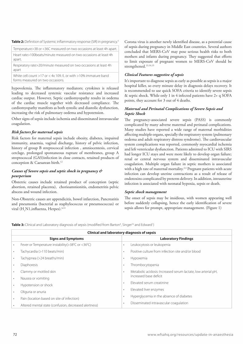
hypovolemia. The inflammatory mediators; cytokines is released leading to decreased systemic vascular resistance and increased cardiac output. However, Septic cardiomyopathy results in oedema of the cardiac muscle together with decreased compliance. The cardiomyopathy manifests as both systolic and diastolic dysfunction, increasing the risk of pulmonary oedema and hypotension. Other signs of sepsis include ischemia and disseminated intravascular coagulation.
Risk factors for maternal sepsisRisk factors for maternal sepsis include obesity, diabetes, impaired immunity, anaemia, vaginal discharge, history of pelvic infection, history of group B streptococcal infection , amniocentesis, cervical cerclage, prolonged spontaneous rupture of membranes, group A streptococcal (GAS)infection in close contacts, retained products of conception & Caesarean birth.12
Causes of Severe sepsis and septic shock in pregnancy & peurperiumObstetric causes include retained product of conception (septic abortion, retained placenta), chorioamnionitis, endometritis pelvic abscess and wound infection.
Non-Obstetric causes are appendicitis, bowel infarction, Pancreatitis and pneumonia (bacterial as staphylococcus or pneumococcus) or viral (H1N1,influenza, Herpes).4,11
Corona virus is another newly identified disease, as a potential cause of sepsis during pregnancy in Middle East countries. Several authors concluded that MERS-CoV may pose serious health risks to both mothers and infants during pregnancy. They suggested that efforts to limit exposure of pregnant women to MERS-CoV should be strengthened.13,14,15
Clinical Features suggestive of sepsisIt’s important to diagnose sepsis as early as possible as sepsis is a major hospital killer, so every minute delay in diagnosis delays recovery. It is recommended to use quick SOFA criteria to identify severe sepsis & septic shock. While only 1 in 4 infected patients have 2+ q SOFA points, they account for 3 out of 4 deaths.
Maternal and Perinatal Complications of Severe Sepsis and Septic ShockThe pregnancy-associated severe sepsis (PASS) is commonly complicated by many adverse maternal and perinatal complications. Many studies have reported a wide range of maternal morbidities affecting multiple organs, specially the respiratory system (pulmonary oedema and adult respiratory distress syndrome). The cardiovascular system complications was reported, commonly myocardial ischemia and left ventricular dysfunction. Patients admitted to ICU with SIRS had longer ICU stays and were more likely to develop organ failure; renal or central nervous system and disseminated intravascular coagulation. Multiple organ failure in septic mothers is associated with a high rate of maternal mortality.2,4 Pregnant patients with acute infection can develop uterine contractions as a result of release of endotoxins complicated by preterm delivery. In addition, intrauterine infection is associated with neonatal hypoxia, sepsis or death.
Septic shock managementThe onset of sepsis may be insidious, with women appearing well before suddenly collapsing, hence the early identification of severe sepsis allows for prompt, appropriate management. (Figure 1)
Clinical and laboratory diagnosis of sepsis
Signs and Symptoms Laboratory Findings
• Fever or Temperature instability(>380C or <360C)
• Tachycardia (>110 beats/min)
• Tachypnea (>24 breaths/min)
• Diaphoresis
• Clammy or mottled skin
• Nausea or vomiting
• Hypotension or shock
• Oliguria or anuria
• Pain (location based on site of infection)
• Altered mental state (confusion, decreased alertness)
• Leukocytosis or leukopenia
• Positive culture from infection site and/or blood
• Hypoxemia
• Thrombocytopenia
• Metabolic acidosis: Increased serum lactate, low arterial pH, increased base deficit
• Elevated serum creatinine
• Elevated liver enzymes
• Hyperglycemia in the absence of diabetes
• Disseminated intravascular coagulation
Table 3: Clinical and Laboratory diagnosis of sepsis (modified from Barton4, Singer16 and Edward1)
Temperature>38 or <36C measured on two occasions at least 4h apart.
Heart rate>100beats/minute measured on two occasions at least 4h apart.
Respiratory rate>20/minute measured on two occasions at least 4h apart
White cell count >17 or < 4x 10⁹ /L or with >10% immature band forms measured on two occasions.
Table 2: Definition of Systemic inflammatory response (SIR) in pregnancy.9
72 www.wfsahq.org/resources/update-in-anaesthesia

Two high profile campaigns, the Surviving Sepsis Campaign (SSC) and the Sepsis Six, have suggested “bundles” of care, though studies have shown that the tasks are rarely achieved. Neither of these care bundles has been specifically examined in pregnancy.5, 19, 20
Surviving Sepsis Campaign (SSC)The Surviving Sepsis Campaign (SSC) is a global effort to improve the care of patients with sepsis and septic shock, first published in 2004.- SSC is a multidisciplinary approach to treatment of sepsis based on 2 phases:
• Resuscitation phase• Management phase.20
Early screening and managementThe SSC recommend the following steps for early screening & management; 1. Screening and Management of Infection: The first step in screening should be identification of infection according to signs and Symptoms. Once the patient is identified as having infection, cultures and blood samples are obtained as indicated ,followed by administering appropriate antibiotics.
2. Screening for Organ Dysfunction and Management of Sepsis (formerly called Severe Sepsis): Patients with sepsis should be identified by the organ dysfunction criteria; the quick Sepsis-Related Organ Failure Assessment (q SOFA). 3. Identification and Management of Initial Hypotension: Patients with infection and hypotension or a lactate level >4mmol/L, are given 30mL/kg crystalloid with reassessment of volume responsiveness and tissue perfusion.9
Resuscitation bundleThe Severe Sepsis Bundles are a series of evidence-based therapies that, when implemented together, will achieve better outcomes.21
The SSC guidelines established goals of care across various therapeutic modalities for sepsis. These guidelines included recommendations on initial resuscitation; diagnostic goals; timing and regimens of antibiotic administration; vasopressor and inotropic use; corticosteroids; sedation strategies; ventilation strategies; blood product administration; and preventive measures against ventilator-acquired pneumonia, stress ulcers, and deep vein thrombosis (DVT).22
Figure 1: Initial management of sepsis in pregnancy. Quoted from Plante et al., 201911
Suspected sepsis
73
© World Federation of Societies of Anaesthesiologists 2019. This issue may be freely reproduced for the purposes of private research and study and extracts (or indeed, the full report) may be included in professional journals provided that suitable acknowledgement is made and the reproduction is not associated with any form of advertising. Applications for commercial reproduction should be addressed to: World Federation of Societies of Anaesthesiologists, Dean Bradley House, 52 Horseferry Rd, London SW1P 2AF, UK.

Maintain inspiratory plateau pressures at less than 30cmH2O for mechanically ventilate patients.26
Antimicrobial therapyThe new WHO guidance for treatment of maternal infections, recommends specific classes of antibiotics for the management of chorioamnionitis (ampicillin and gentamicin) and post-partum endometritis (clindamycin and gentamicin). However, they acknowledged that other simple, effective, and locally available antibiotics could be used as an alternative.27
In addition to antimicrobial therapy, the source of sepsis should be sought and dealt with.
Antimicrobial choices and its considerations• Co-amoxiclav: Does not cover MRSA or Pseudomonas and
may increase risk of neonatal necrotizing enterocolitis. It is commonly avoided in pregnancy
• Metronidazole: Only covers anaerobes• Clindamycin: Covers most streptococci and staphylococci
including MRSA.• Teicoplanin or linezolid : Covers MRSA.• Piperacillin-tazobactam (Tazocin) or meropenem : covers all
except MRSA• Gentamycin (3-5mg/kg single dose): Does not affect renal
function if given once.• cefuroxime or cefotaxime and metronidazole; with clarithromycin
or clindamycin and gentamicin as alternatives in those with penicillin allergy
• In critically unwell patients, piperacillin-tazobactem or meropenem or ciprofloxacin and gentamicin, may be preferred.9,28
The study of Patil and Jambulingappa in 2015 suggested the use of combination of ceftriaxone, sulbactam and disodium edetate (EDTA) for the treatment of multi-drug resistant MDR septicaemia associated with extended spectrum beta lactamse (ESBL) and metallo-beta lactamase (MBL) producing microbes.29
Fluid therapyThe Surviving Sepsis Campaign recommends an initial bolus of 30mL/kg of balanced crystalloids for initial resuscitation and maintenance volume replacement in patients with sepsis and septic shock.
When the patient require substantial amounts of crystalloids , balanced crystalloids or albumin can be given, while hydroxyethyl starches (HESs) and gelatin are not recommended for intravascular volume replacement in those patients.23 After initial fluid resuscitation, further fluid therapy should be guided by dynamic measures of preload; by using either pulse-pressure variation or passive leg raising.
Measurement of pulse-pressure variation is accomplished by analyzing the waveform of an arterial line, but it is reliable only in sedated individuals receiving positive-pressure ventilation, while passive leg raising to 30-45 can be used in spontaneously breathing patient. On
The international guidelines for management of sepsis and septic shock in 201623 emphasis that we should consider sepsis and septic shock as medical emergencies. Hence, the treatment and resuscitation should begin immediately. The evidence in the literature has demonstrated an association between compliance with bundles and improved survival in patients with sepsis and septic shock. The key elements in management according to SSC guidelines 2012, were divided into two bundles of care, the “resuscitation” and “management” bundle. The interventions are accomplished within 6hours and 24 hours, respectively.22 The most important recent change in the revised SSC bundles, is that the 3-h and 6-h bundles have been combined into a single “hour-1 bundle”.24
Initial Resuscitation Phase (first 6-h) - Figure 2:
1. Obtain blood cultures prior to antibiotic administration
2. Administer broad-spectrum antibiotic within one hour of recognition of severe sepsis
3. Measure serum lactate4. In the event of hypotension and/or a serum lactate >4mmol/l deliver an initial minimum 30ml/kg of crystalloid or an equivalent.5. Apply vasopressors (Norepinephrine) for hypotension that is not responding to initial fluid resuscitation to maintain mean arterial pressure (MAP) >65mmHg6. In the event of persistent hypotension despite fluid resuscitation (septic shock) and/or lactate >4mmol/l a. Achieve a central venous pressure (CVP) of ≥8mmHg b. Achieve a central venous oxygen saturation (ScvO2) ≥ 70% or mixed venous oxygen saturation (ScvO2) ≥ 65%. During the initial resuscitation of severe sepsis, pregnant women should be in the left lateral position to avoid aortocaval compression.11,24,25
Tasks to be performed as soon as possible and scored over 24 hours:Administer low-dose steroids per hospital policy Maintain glucose higher than lower limit of normal, but less than 180mg/dl (8.3mmol/l)
Lactate Culture AntibioticsFluids
30ml/kg
Lactate remeasure
Reassess Volume
Vasopressors (MAP-65)
6x
74 www.wfsahq.org/resources/update-in-anaesthesia
Figure 2: Resuscitation phase within 1st 6 hours

the other hand, some authors do not recommend static measures of preload (central venous pressure or pulmonary artery occlusion pressure), being poor predictors of fluid responsiveness. About 50% of hypotensive septic patients do not respond to fluid challenge, and are better treated by vasopressors.10 Aggressive fluid administration to all patients without assessment of their response, may result in pulmonary and cerebral edema, cerebral together with increased intra-abdominal pressure and increased mortality in sepsis.30
Role of corticosteroids:The indication of corticoids in septic patients is subject of controversy in the literature. The Surviving Sepsis Campaign 2012 & 201631, 23 recommended administration of intravenous infusion of hydrocortisone at a dose of 200mg per day. Hydrocortisone should be reserved only for septic patients with refractory shock (those who remain hypotensive following initial fluid resuscitation and vasopressors). Patients receiving corticosteroids should be cautiously monitored for hyperglycemia and hypernatremia. Corticoids are usually given within the first seven days of treatment and should be interrupted as soon as the patient shows signs of clinical improvement. A systematic review of Annane et al suggested that treatment with a long course of low-dose corticosteroids significantly reduced 28-day mortality.32 Another benefit of corticosteroids infusion in the case of sepsis in pregnant women is the need for corticoid to accelerate fetal lung maturation due to the risk of premature birth.
Deep venous thrombosis prophylaxis:Prevention of DVT is essential in septic pregnant patients as both pregnancy and sepsis are associated with hypercoagulability.33,
34 Methods of prophylaxis are the use of compression stockings, intermittent lower limb compression, and low molecular weight or unfractionated heparin.31
SSC recommend against the use of antithrombin for the treatment of sepsis and septic shock, but recommend the use of heparin as two systematic reviews showed increased survival when heparin is used for patients with sepsis without an increase in major bleeding.23, 35
Vasoactive DrugsSSC 2016 strongly recommend norepinephrine as the first choice vasopressor. Vasopressin or epinephrine to norepinephrine to norepinephrine for raising MAP to target.
Dopamine can be used as an alternative vasopressor to norepinephrine only in patients with low risk of tachyarrhythmias and bradycardia. However, low dose dopamine is not recommended for renal protection. Dobutamine can be used in patients with persistent hypoperfusion despite adequate fluid loading and the use of vasopressor agents.36-38
The role of intravenous immunoglobulin (IVIG):IVIG is recommended for severe invasive streptococcal or staphylococcal infection if other therapies have failed because of its immunomodulatory effect. High dose IVIG has been used in pregnant women and is effective in exotoxic shock caused by streptococci and staphylococci.28
Delivery decision:The decision of delivering the fetus or continuing the pregnancy is influenced by patient’s condition, gestational age, fetal status, presence of chorioamnionitis, and labour.
Generally, in a critically ill pregnant woman, birth of the baby may be considered if it would be beneficial to the mother or the baby or to both. During the intrapartum period, continuous electronic fetal monitoring is recommended. Attempting delivery of unstable mother maternal increases the maternal and fetal mortality rates unless the source of infection is intrauterine. A decision on the timing and mode of birth should be made by a senior obstetrician following discussion with the woman if her condition allows.39
Potential Indicators of Delivery are either maternal or foetal causes. Maternal causes are Intrauterine infection, DIC, hepatic/renal problems, heart Failure, compartmental syndrome, multifetal gestation and ARDS. While foetal factors include foetal demise and gestational age with viable fetus.4
Newborn complications from maternal sepsis:Maternal sepsis has a significant impact on neonatal mortality resulting in over one million infection-related neonatal deaths every year.40 The resultant Intra-amniotic infections cause neonatal sepsis, pneumonia and respiratory distress and long-term neurologic impairment in infants.41
Neonatal sepsis is a major complication that may result in death or major disability for 39% of those affected even with timely antimicrobial treatment.42
Anesthesia for delivery in sepsis:South Australian guidelines recommend against Epidural/spinal anaesthesia in women with sepsis and suggest that general anaesthetic is usually preferred for caesarean section. If preterm delivery is anticipated the use of antenatal corticosteroids for fetal lung maturity in the woman with sepsis can be considered.43
The decision process should involve a multidisciplinary team discussion among obstetrician, neonatologist, microbiologist, intensivist and anaesthetist. If surgical intervention is mandatory, the anaesthetist has to make the decision on regional or general anaesthesia. However, the decision to perform the surgery under regional or general anaesthesia must be individualized considering the risk–benefit ratio.44
Regional anaesthesiaNeuraxial block, is usually contraindicated in septic patients, as Septic hypotensive patients may not tolerate the sympathetic block and vasodilatation associated with spinal anaesthesia. There may be associated coagulopathy or thrombocytopaenia. The risk of epidural abscess or meningitis is very small in patients treated with antibiotics but it should be considered in these cases.
General anaesthesiaGeneral anaesthesia is highly likely to be required in a septic parturient. Induction and maintenance agents in patients with sepsis may increase their hemodynamic instability and should be
75
© World Federation of Societies of Anaesthesiologists 2019. This issue may be freely reproduced for the purposes of private research and study and extracts (or indeed, the full report) may be included in professional journals provided that suitable acknowledgement is made and the reproduction is not associated with any form of advertising. Applications for commercial reproduction should be addressed to: World Federation of Societies of Anaesthesiologists, Dean Bradley House, 52 Horseferry Rd, London SW1P 2AF, UK.

chosen with care. Ketamine and etomidate can be used safely in such cases and the use of a rapid-acting nondepolarizing agent, such as rocuronium, should be considered. Invasive monitoring including Intra – arterial pressure, CVP, and cardiac output monitoring are helpful, especially for the postoperative phase. The oxytocin bolus should be administered slowly by infusion over 5 min to avoid haemodynamic instability. The decision to extubate or transfer to critical care is judged by severity of sepsis and the altered physiology of pregnancy. After operation, oxygen is recommended to meet the increased demand. Analgesia should be maintained with paracetamol and opioids .Non-steroidal anti-inflammatory drugs are contraindicated because septic patients have deranged renal function and coagulation profile.45
Postoperative care and transfer to critical care:The decision & timing of transfer to intensive care should be decided by the critical care team in conjunction with the obstetric consultant and the consultant obstetric anaesthetist.
Transfer to critical care is needed if the patient is hemodynamically unstable and needs vasopressor support, mechanical ventilation is needed for pulmonary oedema, or altered conscious level, hemodialysis, multiple organ failure or hypothermia.46
Maternal sepsis in low-income countriesThe WHO meeting in 2018, announced that the global mortality rate shown in many studies is around 55%, and 60% in cases of septic shock. Meanwhile, in some African countries mortality from sepsis may reach 100%.47 Meanwhile, maternal mortality attributable to sepsis approach 33% in low-income countries,39 as these countries lack sufficient clean water, soap and clean birth-kits.
The health system of LMIC has limited access to timely diagnosis of septic shock (e.g. serum lactate level is not available) and shortage of health care professionals. The previous factors contributes to increase maternal sepsis.48
The WHO meeting recommended increasing awareness of the medical personnel of the preventive measures, early home visits and postnatal care of the newborn, in order to prevent maternal and neonatal sepsis.49
REFERENCES1. Prasad BGR, Sunanda GV. Sepsis. In: Johanson R, Cox C, Grady K, Howell C, editors. Managing obstetric emergencies and trauma. The MOET Course Manual. London: RCOG Press; 2003. p. 231-234
2. Knowles SJ, O’Sullivan NP, Meenan AM, Hanniffy R, Robson M. Maternal sepsis incidence, aetiology and outcome for mother and fetus: a prospective study. BJOG 2015; 122: 663–671.
3. Surgers L, Valin N, Carbonne B, Bingen E, Lalande V, Pacanowski J, et al. Evolving microbiological epidemiology and high fetal mortality in 135 cases of bacteremia during pregnancy and postpartum. Eur J Clin Microbiol Infect Dis 2013; 32: 107–13)
4. Barton J R and Sibai B M. Severe Sepsis and Septic Shock in Pregnancy. Obstet Gynecol. 2012 Sep; 120(3): 689-706. doi: 10.1097/AOG.0b013e318263a52d.
5. Singer M, Deutschman CS, Seymour CW, Shankar-Hari M, Annane D and Bauer M et al. The Third International Consensus Definitions for Sepsis and Septic Shock (Sepsis-3). JAMA. 2016; 315(8): 801-810. doi:10.1001/jama.2016.0287.
6. Verdonk F, Blet A, and Mebazaa A. The new sepsis definition: limitations and contribution to research and diagnosis of sepsis. Curr Opin Anesthesiol 2017, 30: 200–204.
7. Fee N., Hartigan L., McAuliffe F M., Higgins MF. Education in Sepsis: A Review for the Clinician of What Works, for Whom, and in What Circumstances. J Obstet Gynaecol Can 2017; 39(9): 772e780 https://doi.org/10.1016/j.jogc.2016.09.079.
8. Marik PE, Taeb AM: SIRS, qSOFA and new sepsis definition. J Thorac Dis. 2017, 9: 943-945.
9. Vinod P, Michael W., Wijayatilake, D S. Clinical Pearls of maternal critical care: part 1. Current Opinion in Anesthesiology 29(3): 304-316, June 2016.
10. Guo Y, Luan L, Patil NK, Wang J, Bohannon JK, Rabacal W, Fensterheim BA, Hernandez A, and Sherwood ER. Interleukin-15 Enables Septic Shock by Maintaining Natural Killer Cell Integrity and Function. J Immunol. 2017 February 01; 198(3): 1320–1333.
11. Plante L.A., Pacheco L.D., Louis J.M., Society for Maternal-Fetal Medicine (SMFM) Publications Committee. SMFM Consult Series #47: Sepsis during pregnancy and the puerperium. American Journal of Obstetrics and Gynecology 2019, 220 (4), pp. B2-B10.
12. Centre for Maternal and Child Enquiries (CMACE). Saving Mother’s Lives: reviewing maternal deaths to make motherhood safer: 2006-2008. BJOG 2011; 118(suppl. 1): 1-203.
13. Assiri A., Glen R. , Al Masri AM., Bin Saeed A , Gerber SI and Watson JT. Middle East Respiratory Syndrome Coronavirus Infection During Pregnancy: A Report of 5 Cases From Saudi Arabia. Clinical Infectious Diseases 2016; 63(7): 951–3. DOI: 10.1093/cid/ciw412.
14. Payne DC, Iblan I, Alqasrawi S, et al. Stillbirth during infection with Middle East respiratory syndrome coronavirus. J Infect Dis 2014; 209: 1870–2.
15. Malik A, El Masry KM, Ravi M, Sayed F. Middle East respiratory syndrome coronavirus during pregnancy, Abu Dhabi, United Arab Emirates, 2013. Emerg Infect Dis 2016; 22: 515–7.
16. Arulkumaran N, Singer M. Puerperal sepsis. Best Practice & Research Clinical Obstetrics and Gynaecology 27 (2013) 893–902.
17. Edwards SE, et al. Modified obstetric early warning scoring systems(MOEWS): alidating the diagnostic performance for severe sepsis in women with chorioamnionitis. Am J Obstet Gynecol. 2015; 212(4): 536.e1–8.
18. Dellinger RP, Carlet JM, Masur H, et al. Surviving Sepsis Campaign.guidelines for management of severe sepsis and septic shock. Intensive Care Med 2004; 30: 536e55.
19. Robson WP, Daniel R. The Sepsis Six: helping patients to survive sepsis. Br J Nurs 2008; 17: 16e21.
20. Simmonds M, Hutchison A, Chikhani M, et al. Surviving sepsis beyond intensive care: a retrospective cohort study of compliance with the international guidelines. J Intensive Care Soc 2008; 9: 124e7
21. Paul E Marik .Surviving sepsis: going beyond the guidelines: Annals of Intensive Care 2011; 1:17.
22. Jones AE, Shapiro NI, Trzeciak S, Arnold RClaremont HC, Kline JA. Lactate Clearance vs Central Venous Oxygen Saturation as Goals of Early Sepsis Therapy. A Randomized Clinical Trial. JAMA. 2010; 303(8): 739-746. doi:10.1001 JAMA. 2010.158.
23. Rhodes A, Evans L, Alhazzani W, Levy M, Antonelli M, Ferrer R, Kumar A, Jonathan Sevransky J, Sprung C, Nunnally M, Rochwerg B, Rubenfeld G, Angus D, Annane D, Beale R, Bellinghan G, Bernard G, Chiche JD, Coopersmith C, De Backer D, French C, Fujishima S, Gerlach H, Hidalgo J, Hollenberg S, Jones A, Karnad D, Kleinpell R, Koh Y, Lisboa T, Machado F, Marin J, Marshall J; Mazuski J, McIntyre L, McLean A, Mehta S, Moreno R, Myburgh J, Navalesi P, Nishida O, Osborn T, Perner A, Plunkett C, Ranieri M, Schorr C, Seckel M, Seymour C, Shieh L, Shukri K, Simpson S, Singer M, Thompson B, Townsend S, Van der Poll T, Vincent JL, Wiersinga W, Zimmerman Y, Dellinger R. Surviving Sepsis Campaign: International Guidelines for Management of Sepsis and Septic Shock: 2016. Intensive Care Med (2017) 43: 304–377.
76 www.wfsahq.org/resources/update-in-anaesthesia

24. Levy MM, Evans LE, Rhodes A. The Surviving Sepsis Campaign Bundle: 2018 Update Critical Care Medicine 2018; 46 (6): 797-1000.
25. Surviving Sepsis Campaign: Updated Bundle in response to new evidence. http://www.survivingsepsis.org/SiteCollectionDocuments/SSC_Bundle.pdf. [Accessed January 2016].
26. Dellinger RP, Levy MM, Rhodes A, et al. Surviving Sepsis Campaign: international guidelines for management of severe sepsis and septic shock, 2012. Intensive Care Med 2013; 39: 165e228.
27. Bonet M, Oladapo OT, Khan DN, Mathai M, Gülmezoglu AM. The lancet Global Health 2015; 3:e667, 668.http://dx.doi.org/10.1016/S2214-109X(15)00213-2.
28. Royal College of Obstetricians and Gynaecologists (RCOG). Bacterial sepsis following pregnancy – 64b April 2012. RCOG Green top Guidelines. London: RCOG Press; 2012b. Available from URL: http://www.rcog.org.uk/womens- health/clinical-guidance/sepsis-following-pregnancy-bacterial-green-top 64b.
29. Patil UN, Jambulingappa KL. A Combination Strategy of Ceftriaxone, Sulbactam and Disodium Edetate for the Treatment of Multi-Drug Resistant (MDR) Septicaemia: A Retrospective, Observational Study in Indian Tertiary Care Hospital. Journal of Clinical and Diagnostic Research. 2015 Nov, Vol-9(11): FC29-FC32.
30. Loflin R, Winters ME. Fluid Resuscitation in Severe Sepsis. Emerg Med Clin N Am 2017; 35: 59–74.
31. Cordioli RL, Cordioli E, Negrini R, Silva E.Sepsis and pregnancy: do we know how to treat this situation? Rev Bras Ter Intensiva. 2013; 25(4): 334-344.
32. Annane D, Bellissant E, Bollaert PE, Briegel J, Keh D, Kupfer Y (2015). Corticosteroids for treating sepsis. Cochrane Database Syst Rev 12:CD002243.
33. Vervloet MG, Thijs LG, Hack CE. Derangements of coagulation and fibrinolysis in critically ill patients with sepsis and septic shock. Semin Thromb Hemost. 1998; 24(1): 33-44.
34. Lockwood CJ. Pregnancy-associated changes in the hemostatic system. Clin Obstet Gynecol. 2006; 49(4): 836-43.
35. Zarychanski R, Abou-Setta AM, Kanji S et al. The efficacy and safety of heparin in patients with sepsis: a systematic review and metaanalysis. Crit Care Med 2015; 43(3): 511–518.
36. De Backer D, Creteur J, Silva E, Vincent JL. Effects of dopamine, norepinephrine, and epinephrine on the splanchnic circulation in septic shock: which is best? Crit Care Med 2003; 31(6): 1659–1667.
37. Martin C, Viviand X, Leone M, Thirion X .Effect of norepinephrine on the outcome of septic shock. Crit Care Med 2000; 28(8):2758–2765.
38. Hashmi M, Khan FH. A review of critical care management of maternal sepsis. Anaesth Pain & Intensive Care 2014; 18(4): 436-442.
39. Critical care in pregnancy. Practice Bulletin No. 100. American College of Obstetricians and Gynecologists. Obstet Gynecol 2009; 113: 443–50.
40. Arulkumaran N, Singer M. Puerperal sepsis. Best Pract Res Clin Obstet Gynaecol. 2013 Dec; 27(6): 893-902.
41. Seale AC, Mwaniki M, Newton CRJC, Berkley JA. Maternal and early onset neonatal bacterial sepsis: burden and strategies for prevention in sub-Saharan Africa. Lancet Infect Dis. 2009 Jul; 9(7): 428-38.)
42. Wynn JL, Wong HR, Shanley TP, Bizzarro MJ, Saiman L, Polin RA. Time for a neonatal–specific consensus definition for sepsis. Pediatr Crit Care Med. 2014; 15(6): 523–528.
43. South Australian Perinatal Practice Guidelines 2017.Clinical Guideline .Sepsis in Pregnancy. Policy developed by: SA Maternal, Neonatal & Gynaecology Community of Practice Approved SA Health Safety & Quality Strategic Governance Committee on: 01 March 2017
44. Platt F, Wray S. Role of anaesthetist in obstetric critical care. Best Pract Res Clin Obstet Gynaecol 2008; 22: 917– 35.
45. RJ Elton, S Chaudhari, Sepsis in obstetrics, Continuing Education in Anaesthesia Critical Care & Pain, Volume 15, Issue 5, October 2015, Pages 259–264.
46. Neligan PJ*and Laffey JG. Clinical review: Special populations - critical illness and pregnancy. Critical Care 2011, 15: 227
47. WHO Sepsis Technical Expert Meeting - Meeting report. Geneva: World Health Organization; 2018. Licence: CC BY-NC-SA 3.0 IGO.
48. Van Dillen J, Zwart J, Schutte J, van Roosmalen J. Maternal sepsis: epidemiology, etiology and outcome. Curr Opin Infect Dis. 2010 Jun; 23(3): 249-54.
49. Miller AE, Morgan C, Vyankandondera J. Causes of puerperal and neonatal sepsis in resource-constrained settings and advocacy for an integrated community-based postnatal approach. Int J Gynaecol Obstet. 2013 Oct; 123(1): 10-5.
77
© World Federation of Societies of Anaesthesiologists 2019. This issue may be freely reproduced for the purposes of private research and study and extracts (or indeed, the full report) may be included in professional journals provided that suitable acknowledgement is made and the reproduction is not associated with any form of advertising. Applications for commercial reproduction should be addressed to: World Federation of Societies of Anaesthesiologists, Dean Bradley House, 52 Horseferry Rd, London SW1P 2AF, UK.

Clin
ical
Art
icle
s
Madhusudan UpadyaKasturba Medical College
MangaloreKarnataka
INDIA
Mahesh Nayak MDDepartment of Anaesthesia
Kasturba Medical CollegeMangalore Manipal Academy of Higher
EducationMangalore
INDIA
SummaryPostpartum hemorrhage remains the leading cause of maternal mortality and morbidity.
Oxytocic drugs are the main stay in prevention as well as treatment. Oxytocin remains the drug of choice. The
use of 5 IU bolus dose has been questioned in recent times. Current evidence suggests infusion to be a preferred
choice than bolus dosage. Carbetocin is a newer alternative to oxytocin. Second line drugs include ergot alkaloids
(Methyl ergometrine) and prostaglandins (carboprost and misoprostol) which are used as adjuvants to oxytocin in
hemorrhage.
Oxytocics
Madhusudan Upadya* and Mahesh Nayak*Correspondence email: [email protected]:10.1029/WFSA-D-18-00033
INTRODUCTIONPostpartum Haemorrhage (PPH) remains a major cause of both maternal mortality and morbidity with an estimated mortality rate of 140,000 per year or one maternal death every four minutes.1 In the developing world, death from PPH occurs in approximately 1 out of 1,000 deliveries. Uterine atony is the commonest cause for postpartum hemorrhage accounting to 80% leading to blood transfusion and post partum hysterectomy.
Administration of Oxytocic drugs help in both prevention and treatment of postpartum haemorrhage. The American College of Obstetricians and Gynaecologists (ACOG) recommends prophylactic administration of uterotonic agents to prevent uterine atony. Active management of the third stage of labor includes oxytocin administration. ACOG recommends using Uterotonic agents as first-line treatment for postpartum hemorrhage caused by uterine atony.2 Three classes of drugs are in use and include oxytocin, ergot alkaloids, and prostaglandins.
OxytocinOxytocin is a nonapeptide hormone produced in the hypothalamus and later secreted into circulation by posterior pituitary gland. The word oxytocin was coined from the term oxytocic, Greek oxys, and toketos, meaning “quick birth”. It was first discovered by Sir Henry Dale in 1909 as extract from pituitary gland that caused uterine contractions in pregnant cat.3 Oxytocin became the first hormone to be synthesized in 1953 by the American biochemist, Vincent Du Vigneaud for which he received the Nobel Prize.4 It remains the first-line agent in the management and prevention of uterine atony after
vaginal or caesarean delivery. Not only does oxytocin play a role in uterine contraction, but this hormone is also involved in haemodynamic regulation. It can precipitate hypotension especially when given as a bolus, in addition to causing a release of atrial and brain natriuretic peptides. Due to its structural similarity to vasopressin/antidiuretic hormone (ADH), oxytocin may also cause water retention and hyponatremia. Several empirical regimens have been proposed for oxytocin administration during caesarean delivery and this has led to many different practices in its administration worldwide. Carvalho et al. in a report in 2004 stated the minimum effective initial dose of oxytocin in elective caesarean deliveries to be 0.35 IU, which was far less than the previous conventional dosages ranging from 5 to 10 IU.5 This sparked further research into the administration of oxytocics in rational and judicious manner to minimise the side effects while maintaining efficacy.Conventionally oxytocin has been used either as bolus administration or continuous infusion. The optimal dose, timing, and rate of administration for oxytocin during caesarean delivery remain ambiguous. Royal College of Obstetricians and Gynaecologists (UK) recommends slow intravenous bolus dose of 5 IU6, while the American College of Obstetricians and Gynaecologists (ACOG) recommends continuous infusion.2 WHO recommends 20 IU oxytocin in 1 litre crystalloid infusion at 60 drops per minute and to continue 20 IU in 1 litre crystalloid at 40 drops per hour if bleeding continues to not more than 3 litres of oxytocin containing IV fluid.7 WHO also recommends to avoid bolus IV dose oxytocin. The use of 5 IU of oxytocin as bolus dose has been questioned
78 www.wfsahq.org/resources/update-in-anaesthesia

in recent times. Oxytocin given prophylactically has known to cause haemodynamic instability including hypotension, tachycardia, arrhythmias and even myocardial ischemia.8 Other effects include headache, nausea, vomiting and flushing. Bolus dosages of 3 to 5 IU given rapidly have caused cardiovascular collapse and even death.9 Tsen and Balki10 in 2010 proposed a new protocol for safe oxytocin administration called “Rule of threes” (Table 1) which is evidence-based as well as easy to remember.
CarebetocinCarbetocin is a synthetic analogue of oxytocin with similar pharmacodynamic properties but longer acting. It is an octapeptide while oxytocin is nonapeptide and due to its structural difference carbetocin is more stable and is more resistant to degradation by disulphidase, aminopeptidase and oxidoreductase enzymes.10 It has a half life of about 40 minutes which is 10 times that of oxytocin. Side effects of carbetocin are similar to oxytocin including hypotension, flushing, headache and abdominal pain. A Cochrane database review in 2012 found lower incidence of PPH after caesarean deliveries with carbetocin compared with oxytocin.11 The ED90 dose of carbetocin during caesarean delivery to maintain uterine tone ranged from 14.8µg12 in non labouring women to 121µg13 in labouring women. The Society of Obstetricians and Gynaecologists of Canada recommends carbetocin 100µg be given as an intravenous bolus over one minute to prevent PPH.14 The current evidence in use of carbetocin is quite encouraging and further large scale studies are needed to know the effective dose and side effect profile.
Methyl ErgometrineErgot, derived from the fungus Claviceps purpurea, was the first effective oxytocic drug to be used in obstetrics. From 1582 to 1822 it was used to speed up delivery, it was no longer used after due to complications including uterine rupture, still birth, and maternal death.15 Methyl ergometrine is a semisynthetic ergot derivative. It causes sustained contraction of uterus and is the second line agent for treatment of PPH. WHO recommends 0.2mg intravenously or intramuscularly repeated every 15 min up to a maximum dosage of 1mg for PPH7. ACOG recommends 0.2mg intramuscularly every 2 to 4 hours.2 Side effects include hypertension with headache and even seizures, rarely coronary vasospasm and myocardial ischaemia.16 It is contraindicated in hypertensive patients and should be avoided in patients taking CYP3A4 inhibitors.
CarboprostCarboprost is a prostaglandin F2α analogue used as second line agent in uterine atony. Prostaglandins increase intramyometrial calcium concentrations and enhance uterine contraction. Their effects are
mediated via G-proteins and the activation of calcium channels.17 Prostaglandin F2α was first used by Takagi in 1976 as intramyometrial agent for PPH.18 The recommended dosage of carboprost is 250µg intramuscularly repeated every 15 to 30 minutes up to a maximum of 2mg i.e. 8 times. Butwick et al.19 in a retrospective study reported carboprost to be less effective than methyl ergometrine in treating PPH. Carboprost is preferred as second line agent in uterine atony when methyl ergometrine is contraindicated or ineffective. Side effects include nausea, vomiting and diarrhoea. Its use should be avoided in patients with bronchial asthma due to its potential to cause bronchospasm20.
MisoprostolAnother second line drug for treatment of PPH, misoprostol is a Prostaglandin E1 analogue. It can be used in various routes: Oral, sublingual, buccal, vaginal or rectal. WHO recommends sublingual misoprostol 200 - 800µg.7 Side effects include pyrexia, shivering and GI disturbances. In a meta-analysis misoprostol prevented postpartum hemorrhage by 24% and severe postpartum hemorrhage by 41% compared to placebo.21 But in other study there was no clear benefit of use of misoprostol as adjuvant to oxytocin in terms of major outcomes such as mortality and blood loss.22 Misoprostol is best used when all other methods have failed or when oxytocin and methyl ergometrine is unavailable.
Conclusion1 out of 1000 women die during delivery in developing world due to PPH. Oxytocic drugs remain an important intervention in both prevention and treatment of uterine atony. Oxytocin remains the drug of choice as well as the first line agent. But the dosage and administration remain ambiguous and require standardization. Slow infusion is preferred compared to bolus doses. WHO recommends 20 IU oxytocin in 1 litre crystalloid infusion at 60 drops per minute. Carbetocin is an alternative to oxytocin with similar profile but longer duration of action. Though current evidences are promising further research is needed to delineate proper dosage as well as efficacy. Second line of drugs include ergot alkaloids and prostaglandins with methyl ergometrine being superior than carboprost in terms of treating uterine atony. Prostaglandins are best suited if methyl ergometrine is contraindicated or when bleeding persists.
REFERENCES1. Abouzahr C. Global burden of maternal death and disability. Br. Med Bull. 2003; 67(1): 1–11.
2. American College of Obstetricians and Gynaecologists. Postpartum hemorrhage. Practice Bulletin No. 183. Obstet Gynecol 2017; 130: e168–86. doi:10.1097/aog.0000000000002351.
79
© World Federation of Societies of Anaesthesiologists 2019. This issue may be freely reproduced for the purposes of private research and study and extracts (or indeed, the full report) may be included in professional journals provided that suitable acknowledgement is made and the reproduction is not associated with any form of advertising. Applications for commercial reproduction should be addressed to: World Federation of Societies of Anaesthesiologists, Dean Bradley House, 52 Horseferry Rd, London SW1P 2AF, UK.
3 IU oxytocin intravenous loading dose (administered no faster than 15 seconds12)
3 min assessment intervals. If inadequate uterine tone, give 3-IU oxytocin intravenous rescue dose
3 total doses of oxytocin (Initial Load + 2 Rescue Doses)
3 IU oxytocin intravenous maintenance dose (3 IU/L at 100mL/h)
3 Pharmacologic options (e.g. ergometrine, carboprost and misoprostol) if inadequate uterine tone persists
Table 1: Oxytocin protocol for cesarean delivery: ‘‘Rule of threes’’

3. Dale HH. The action of extracts of the pituitary body. Biochem J 1909; 4: 427–47.
4. Du Vigneaud V, Ressler C, Swan JM, et al. Oxytocin: synthesis. J Am Chem Soc 1954; 76(12): 3115–8.
5. Carvalho JC, Balki M, Kingdom J, Windrim R. Oxytocin requirements at elective caesarean delivery: a dose-finding study. Obstet Gynecol 2004; 104: 1005–1010.
6. National Collaborating Centre for Women’s and Children’s Health. Caesarean section. Clinical guideline. RCOG Press, 2004.
7. World Health Organization. Managing complications in pregnancy and childbirth: a guide for midwives and doctors. World Health Organization;2007.
8. Dyer RA, Butwick AJ, Carvalho B. Oxytocin for labour and caesarean delivery: implications for the anaesthesiologist. Curr Opin Anaesthesiol 2011; 24: 255 261.
9. Vallera, C., Choi, L.O., Cha, C.M., Hong, R.W. Uterotonic medications oxytocin, methylergonovine, carboprost, misoprostol. Anesthesiol Clin. 2017; 35: 207 219.
10. Sweeney G, Holbrook AM, Levine M, Yip M, Alfredsson K, Cappi S et al. Pharmacokinetics of carbetocin, a longacting oxytocin analogue, in non pregnant women. Curr Ther Res Clin Exper 1990; 47; 528-39.
11. Su LL, Chong YS, Samuel M. Carbetocin for preventing postpartum haemorrhage. Cochrane Database of Systematic Reviews 2012, Issue 2. Art. No.: CD005457.
12. Khan M, Balki M, Ahmed I, et al. Carbetocin at elective caesarean delivery: a sequential allocation trial to determine the minimum effective dose. Can J Anaesth 2014; 61: 242–248
13. Nguyen-Lu N, Carvalho JC, Farine D, et al. Carbetocin at caesarean delivery for labour arrest: a sequential allocation trial to determine the effective dose. Can J Anaesth 2015; 62: 866–874.
14. Leduc D, Senikas V, Lalonde AB, Ballerman C, Biringer A, Delaney M, et al.Active management of the third stage of labour: prevention and treatment of postpartum hemorrhage. Journal of Obstetrics & Gynaecology Canada: JOGC 2009; 31(10): 980–93.
15. de Groot AN, van Dongen PW, Vree TB, et al. Ergot alkaloids. Current status and review of clinical pharmacology and therapeutic use compared with other oxytocics in obstetrics and gynaecology. Drugs 1998; 56(4): 523–35.
16. Kuczkowski KM. Myocardial ischemia induced by intramyometrial injection of methylergonovine maleate. Anesthesiology 2004; 100(4): 1043.
17. Dyer, R. A., van Dyk, D., & Dresner, A. (2010). The use of uterotonic drugs during caesarean section. International Journal of Obstetric Anesthesia; 19(3): 313 319. doi:10.1016/j.ijoa.2010.04.011.
18. Takagi S, Yoshida T, Togo Y et al. The effects of intramyometrial injection of prostaglandin F2alpha on severe post-partum hemorrhage. Prostaglandins 1976; 12: 565–79.
19. Butwick AJ, Carvalho B, Blumenfeld YJ, et al. Second-line uterotonics and the risk of hemorrhage-related morbidity. Am J Obstet Gynecol 2015; 212: 642 e1–642.e7.
20. O’Leary AM. Severe bronchospasm and hypotension after 15-methyl prostaglandin F2a in atonic postpartum haemorrhage. Int J Obstet Anesth 1994; 3: 42–4.
21. Oladapo OT. Misoprostol for preventing and treating postpartum hemorrhage in the community: a closer look at the evidence. Int J Gynaecol Obstet 2012; 119: 105–10.
22. Mousa HA, Blum J, Abou EL, et al. Treatment for primary postpartum hemorrhage. Cochrane Database Syst Rev 2014; (2): CD003249.
80 www.wfsahq.org/resources/update-in-anaesthesia

YooKuen Chan ffarcs
(Ireland)Universiti MalayaKuala Lumpur MALAYSIA
Carolyn Chue Wai Yim mbbs, manaes
Department of AnaesthesiologyFaculty of MedicineUniversity of MalayaMALAYSIA
Summary
Physiological changes in the parturient occur due to the needs of the developing foetus. Understanding these
changes is important when administering anaesthesia to the obstetric patient. Additionally, understanding the
physiological effects in the foetus allows for appropriate intrauterine monitoring and expedient delivery if there
are signs of foetal hypoxia.
Obstetric and foetal physiology – implications for clinical practice in obstetric analgesia and anaesthesia Yoo Kuen Chan* and Carolyn Chue Wai Yim*Correspondence email: [email protected]:10.1029/WFSA-D-18-00020
INTRODUCTIONPhysiological changes occur in the expectant mother very soon after conception. These changes ensure that her increased needs are met, as well as the needs of the zygote as it replicates into the complex foetus. All the physiological changes require additional energy, which for the developing foetus are completely met through placental transfer from the mother.
PHYSIOLOGICAL CHANGES IN THE PARTURIENT TO MEET THE FOETAL NEEDS1,2
Respiratory systemThe respiratory system ensures the increased oxygen demands from both the mother and foetus are met. Similarly, the increased amount of carbon dioxide from increased cell metabolism also needs to be excreted. The minute volume increases early on in pregnancy, achieved by a 60% increase in tidal volume and a 15% increase in respiratory rate. The increase in minute ventilation results in a decrease in arterial carbon dioxide tension to approximately 4kPa/30mmHg, causing a respiratory alkalosis with a compensatory increase in renal bicarbonate excretion. As the gravid uterus grows, there is significant reduction in functional residual capacity and compliance of the respiratory system resulting in an increased work of breathing. The functional residual capacity can be further reduced in the supine position hence, subjecting the mother to an increase risk of desaturation under anaesthesia as a result of decreased respiratory reserves.
Airway manipulation is fraught with risk in the pregnant patient. Oedema present throughout the body is also seen in the upper airways; resulting in a 10-fold likelihood of a difficult airway and hence
of a difficult intubation/failed airway during the induction of anesthesia for caesarean section. The increase in oxygen demand and the reduction of functional residual capacity further shortens the time to desaturation during periods of apnoea. Hence adequate time for pre-oxygenation with a well-sealed face mask needs to be observed and most would recommend up to 3 minutes, if time allows. Nursing the patient in a reverse Trendelenberg (head-up) position of between 15-20 degrees, ramping and supplemental oxygen via a nasal prong cannulae (in addition to the face mask) during pre-oxygenation are methods that can be considered to counteract these potential difficulties. Once the airway is secured, higher airway pressures are usually needed as a result of the decreased chest wall compliance.
The potential for a difficult airway and rapid desaturation on induction of anaesthesia have contributed to the worldwide change towards the use of regional anaesthesia instead of general anaesthesia for caesarean section.
Cardiovascular systemMaternal blood vessels dilate as a result of the high progesterone levels in pregnancy, resulting in a drop in systemic vascular resistance. Cardiac output increases by up to 40-50% at term. The increased cardiac output is accomplished by an increase in heart rate and stroke volume. Flow is increased to most organs including the kidneys and the uterus. Blood flow to the kidneys increases by up to 80%. The blood flow to the uterus is approximately 750mls/min at term and this is responsible for the likelihood of massive hemorrhage occurring from placenta previa/accreta or atonic uterus in the period around delivery.
81
© World Federation of Societies of Anaesthesiologists 2019. This issue may be freely reproduced for the purposes of private research and study and extracts (or indeed, the full report) may be included in professional journals provided that suitable acknowledgement is made and the reproduction is not associated with any form of advertising. Applications for commercial reproduction should be addressed to: World Federation of Societies of Anaesthesiologists, Dean Bradley House, 52 Horseferry Rd, London SW1P 2AF, UK.
Clin
ical
Art
icle
s

Blood volume increases due to activation of the renin-angiotensin system. As the red cell mass increase is less than plasma volume expansion, dilutional physiological anaemia occurs.
Gastrointestional systemThe progression of pregnancy with increasing fundal height results in displacement of the stomach altering the gastric axis and increasing intra-gastric pressure. Furthermore, decreased lower oesophageal sphincter tone due to progesterone compounds the risk of aspiration. Thus, all mothers must be managed as if they have a full stomach despite having an adequate fasting time. As the acidity of the gastric fluid is the main culprit of lung injury, the use of non-particulate antacids (e.g. Sodium Citrate) and H2 antagonists (such as Ranitidine) plus possibly a prokinetic (e.g. Metoclopramide) when general anaesthesia is being provided should be considered.
Coagulation system In preparation for delivery, the coagulation system produces a hypercoagulable state in order to prevent haemorrhage. Unfortunately, this hypercoagulable state predisposes the mother to an increased risk of thromboembolic disease. The gravid uterus can also impede the venous system in the lower limbs, further increasing the risk of deep vein thrombosis (DVT). Deep vein thrombosis and pulmonary embolism (PE) remain important causes of morbidity and mortality for all pregnancies.
The immune systemAn altered immune competence exists in pregnancy in order to allow “tolerance” of the presence of paternal antigen in foetoplacental tissue. This also results in higher predisposition to the development of sepsis and all care providers should be aware of this as a significant cause of morbidity or mortality in the parturient.
DIFFICULTIES WITH OXYGEN DELIVERY IN THE FOETUSEvery year 2.7 million babies die in utero as stillbirths. In comparison, approximately 303,000 parturients die during pregnancy or in the peripartum period.3 The disproportionately higher number of foetal deaths is due to the difficulty in oxygen transfer to the foetus from the mother. Oxygen inhaled by the mother crosses the placenta into the foetus and distributed via the foetal circulation.
Meeting the needs of the foetusAfter conception, before the embryo is attached to the uterus, the oxygen and energy needs of the blastomere stage of the embryo are met just by diffusion alone.4,5 After the embryo has attached to the uterus, the needs of the foetus are met by the growing placenta.6 The placenta supplies oxygen and nutrients to the foetus and removes carbon dioxide and other waste products.
The physiological needs of the growing foetus are usually completely met by the mother. However, parturients with impaired function of the respiratory7, cardiovascular8 or hematological9 systems find it more difficult to accommodate the physiological needs of delivering oxygen and energy to the foetus, especially towards term. If the oxygen and energy needs of the growing foetus are not met, it fails
to grow to its potential and may be small for gestational age. This is commonly seen in parturients with chronic problems with oxygen and energy delivery, such as pre-eclampsia, smoking, anaemia, respiratory or cardiac disease. It can also be seen in babies born to mothers living at high altitude.10 If the mothers and their ancestors live at high altitude over many generations, the ability to have normal sized babies improves – possibly as a result of adaptation over the generations to the chronic hypoxia.
Signs of poor oxygen and energy deliveries More acute deficiencies in delivery of oxygen and nutrients to the foetus can result in adverse outcomes and may be felt as decreased foetal movement.11 This can be seen nearer term when the blood flow in the placenta is decreased or when the parturient has multiple pregnancies and demand outstrips supply.
Foetal bradycardia represents an absolute sign of inadequate supply of oxygen (and energy) to the foetal myocardium. In those foetuses who are already at the extreme stages of coping with hypoxia, contraction of the uterus may inflict a further compromise in oxygen delivery12,13 as the uterine blood flow needs to traverse the uterine muscle before it arrives at the placenta. Foetal bradycardia especially when seen in early labour should alert the provider to be more vigilant in a suspected hypoxic baby and may warrant increased monitoring with a cardiotocograph. Foetal bradycardia which persists even without contraction of the uterus represents a reduced ability to cope with hypoxia than those who only develop bradycardia when there are uterine contractions.
MECHANIMS IN THE FOETUS TO COPE WITH HYPOXIA
Foetal hemoglobinDue to the relative hypoxia in which the foetus develops, there are several mechanisms that allow it to cope in such an environment.14,15 The foetus has hemoglobin with higher affinity for oxygen - HbF. Unlike HbA in adults, which has 2-α and 2-β globin chains, HbF has 2-α and 2-γ chains. Its higher affinity for oxygen allows oxygen to transfer from maternal haemoglobin to foetal haemoglobin even at very low differences in oxygen tensions. In addition, the newborn baby’s hemoglobin averages about 16-18g.dL-1 compared to the average adult’s haemoglobin concentration of 12-15g.dL-1. The higher hemoglobin level enables the foetus to have increased oxygen carrying capacity.
Double Bohr EffectOxygen diffuses across the uterine vessels of the parturient to the umbilical vessels of the foetus in the placenta. Unlike in adult lungs, the diffusion of oxygen across the placenta is less efficient as the surface area is smaller and the diffusion thickness is higher.16 The diffusion gradient for oxygen across the placenta is also relatively small, unlike the oxygen gradient found in the adult lung.
The “double Bohr” effect facilitates the transfer of oxygen across the placenta (see Figure 1). Carbon dioxide produced by the foetus diffuses across the placenta to the mother. The increased partial pressure of carbon dioxide on the maternal side shifts the maternal
82 www.wfsahq.org/resources/update-in-anaesthesia

oxyhaemoglobin dissociation curve to the right whilst the lower carbon dioxide levels on the foetal side shifts the HbF curve to the left. Thus, the transfer of oxygen across the placenta is increased from the mother to the foetus.
Redistribution of bloodHypoxia in the foetus like hypoxia in the adult will see redistribution of blood flow in the foetus in such a way that blood flow to the brain and the heart are prioritized whilst those of other less important organs, such as the gut and kidneys, get reduced blood flow.17
AVOIDING ACUTE AND CHRONIC HYPOXIA IN THE FOETUSAvoiding supine hypotension syndrome in the parturientKnowledge of foetal and maternal physiology allows us to maximize oxygen delivery to the foetus through the mother. By 20 weeks gestation, the uterus is large enough to compress the inferior vena cava if the parturient is placed in the supine position, which reduces venous return and cardiac output.18,19 The compression may not be limited to the vein as it may also compress the aorta. Compression of the aorta will further decrease flow to the placenta and aggravate the situation of poor oxygenation in the foetus. Supine hypotension most commonly occurs immediately pre-delivery following the administration of an epidural or a spinal injection (where the hypotension is more pronounced due to the vasodilation in the lower
limbs after the regional blockade. However, it can be a significant problem for most parturients in the later weeks of pregnancy. Therefore, all parturients should be encouraged to lie on the left or right side or with a left lateral tilt when lying down.
Avoiding severe anaemia in the parturientAnaemia is a burden in parturients from low resource countries, where iron deficiency may commonly occur.9 With reduced hemoglobin, the impact on the growth of the foetus is even more severe if they have the additional burden of preeclampsia – both of which can impair oxygen and energy delivery to the foetus.
Avoiding the use of excessive doses of uterotonicsWhilst uterotonics administered after the delivery of the shoulder is to be encouraged to prevent post delivery atonia of the uterus, excessive doses of uterotonics during the labor itself may predispose to hypertonia that can compromise the oxygen delivery of the foetus.20 When a parturient is administered uterotonics to accelerate and facilitate delivery, the foetal heart should be monitored. The foetal heart rate is a sensitive monitor to alert the care provider about the existence of foetal hypoxia.
HYPOXIA AND THE URGENCY TO EXTRICATE THE FOETUSAs the foetus is constantly in a state of relative hypoxia, a slight imbalance compromising its oxygen supply warrants a hastened delivery through the performance of an instrumental delivery or emergency lower segment caesarean section. Life threatening hypoxia causes profound fetal bradycardia and may be due to situations where the oxygen delivery is acutely interrupted e.g. placental abruption or prolapsed cord.
In utero resuscitationWhile waiting for urgent delivery, in utero resuscitation of the foetus should be performed. Such measures include nursing the mother in the left lateral position, which ensures a non-compromised blood supply to the foetus by preventing aortocaval compression.22 Supplemental oxygen provided to mother, indirectly increases the supply to the foetus. Tetanic or hyperstimulation of the uterus caused by uterotonics, which impedes blood flow to the placenta, should be stopped. Adequate hydration of the mother may also be beneficial.20
Foetal blood samplingThe sampling of foetal scalp blood during labor or cord blood during delivery gives an indication of the level of foetal compromise prior to delivery and at the time of delivery respectively.23 The uteroplacental status is primarily dependent on the maternal condition and is reflected by the umbilical vein sampling. Examples are the presence of maternal anaemia, hypoxia, hypertension, hypotension, ruptured
Figure 1: The Bohr effects are represented by the shift from the complete lines to the dotted lines. At the placenta level, the gradient for diffusion of oxygen (without the Bohr effects) is represented by the oxygen tension difference BA. With the Double Bohr effects, the gradient is increased to the oxygen tension difference DC.
Table 1: The accepted normal range of parameters of umbilical arterial blood sample.
pH PCO2 (mmHg) HCO3 (mmol/L) PO2(mmHg) Base Excess(mmol/L)
Newborn 7.18 – 7.38 32 – 66 17 – 27 6 – 31 - 8 – 0
Premature 7.14 – 7.4 32 – 69 16 – 27 - 7.3 – 1.3
*In the Premature infant the PO2 is based on gestation age
83
© World Federation of Societies of Anaesthesiologists 2019. This issue may be freely reproduced for the purposes of private research and study and extracts (or indeed, the full report) may be included in professional journals provided that suitable acknowledgement is made and the reproduction is not associated with any form of advertising. Applications for commercial reproduction should be addressed to: World Federation of Societies of Anaesthesiologists, Dean Bradley House, 52 Horseferry Rd, London SW1P 2AF, UK.

uterus, placental abruption or inadequate placental delivery of oxygen. On the other hand, umbilical artery sampling reflects foetal tissue oxygenation and the uteroplacental status. Examples of foetal conditions are heart failure and anaemia.
In 2012, Yeh et al24 concluded that “ideal” cord pH is between 7.26-7.30 (Table 1). Furthermore, the threshold pH for adverse neurological outcomes is 7.10. However, there is a weak association with adverse outcome above 7.00. Furthermore, a normal cord pH value also does not preclude the absence of neurological morbidity. Thus, other variables which can lead to an adverse outcome, should be taken into consideration.
Delivering the foetus rapidly when it is showing signs of compromise allows the pediatric provider to administer oxygen directly to the newborn. The blood gas sampling of the uterine artery at the time of delivery would serve as a very useful guide to the degree of compromise in the newborn so it is important that the blood gas is read immediately.
REFERENCES1. Bedson R, Riccoboni A. Physiology of pregnancy: clinical anaesthetic implications. Continuing Education in Anaesthesia, Critical Care & Pain. 2014; 14(2): 69-72
2. Kam P, Power I. Maternal and neonatal physiology. In: Kam P, Power I (ed) Principles of Physiology for the Anaesthetist. 3rd Ed. CRC Press (London) 2015.
3. WHO News Report 16th August 2016: True magnitude of stillbirths and maternal and neonatal deaths underreported. Available at http:// www.who.int/news-room/detail/16-08-2016-true-magnitude of-stillbirths-and-maternal-and-neonatal-deaths-underreported (last assessed 24th June 2018)
4. Kovacic B. Influence of atmospheric versus reduced oxygen concentration on development of human blastocysts in vitro: a prospective study on sibling oocytes. Reproductive BioMedicine Online 2008; 17(2): 229-36
5. Yang Y, Xu Y, Ding C, Khoudja RY, Lin M, Awonuga AO et al. Comparison of 2,5, and 20% on the development of post-thaw human embroyos. J Assist Reprod Genet. DOI 10.1007/s10815-016-01693-5
6. Hutter D, Kingdom, Jaeggi E. Causes and mechanisms of intrauterine hypoxia and its impact on the fetal cardiovascular system: a review. International Journal of Pediatrics Vol 2010, Article ID 401323; doi:10.1155/2010/401323
7. Wexler ID, Johannesson M, Edenborough FP, Sufian BS, Kerem E. Pregnancy and chronic progressive pulmonary disease. Am J Respir Crit Care Med 2007; 175: 300-5
8. Koutrolou-Sotiropoulou P, Parikh PB, Miller C, Lima FV, Butler J, Stergiopoulos K Impact of heart disease on maternal and fetal outcomes in pregnant women. Am J Cardiol 2015; 116(3): 474-80
9. Akhter S, Momen MA, Rahman MM. Parveen T, Karim RK. Effect of maternal anemia on fetal outcome. Mymensingh Med J 2010; 19(3): 391-8
10. Krampl E. Pregnancy at high altitude (editorial). Ultrasound Obstet Gynecol 2003; 19: 535-9
11. Green-top guideline No 57 (February 2011: Reduced fetal movements. Also available at https://www.rcog.org.uk/globalassets/documents guidelines/gtg_57.pdf ( last accessed 27th September 2018)
12. ACOG Practice Bulletin #106: Intrapartum fetal heart rate monitoring: nomenclature, interpretation and general management principles. Obstet Gynecol 2009; 114: 192-202
13. ACOG Practice Bulletin #116: Management of intrapartum feta heart rate tracings. Obstet Gynecol 2010; 116: 1232-40
14. Imagawa S, Goldberg MA, Doweiko J, Bunn HF. Regulatory elements of the erythropoietin gene. Blood 1991; 77: 278-85
15. Carter AM Placental oxygen transfer and the supply of oxygen to the fetus Fetal and Maternal Medicine Review 1999; 11(3): 151-61
16. Griffiths SK, Campbell JP Placental structure, function and drug transfer Continuing Education in Anaesthesia, Critical Care & Pain 2015; 15(2): 84-9
17. Vajiravelu V Maternal and fetal physiology. In: Chan YK & Ng KP (ed.) Physiological Basis of Acute Care. Saunders Elsevier 2012 Pg 145
18. Kerr MG, Scott DB, Samuel E. Studies of the inferior vena cava in late pregnancy BMJ 1964; 1: 532-3
19. Lee AJ, Landau R. Aortocaval compression syndrome: Time to revisit certain dogmas. Anesth Analg 2017; 125: 1975-85
20. Day LT, Hruschka D, Mussell F, Jeffers E, Saha SL, Alam S. Perinatal mortality associated with use of utertonics outside of comprehensive emergency obstetric and neonatal care: a cross-sectional study. Reproductive Health 2016; 13: 129. Also available at https://www ncbi.nlm.nih.gov/pmc/articles/PMC5054615/ ( last accessed at 30th September 2018)
21. Royal College of Obstetricians and Gynaecologist. Good Practice No 11, April 2010. Available from : https://www.rcog.org.uk/en guidelines-research-services/guidelines/good-practice-11/
22. Velayudhareddy S, KirankumarH Management of foetal asphyxia by intrauterine foetal resuscitation Indian J Anaesth 2010; 54: 394-9
23. Gemma L Malin, Rachel K Morris, Khalid S Khan. Strength of association between umbilical cord pH and perinatal and long term outcomes: systematic review and meta-analysis. BMJ 2010; 340: c1471
24. Yeh P, Emary K, Impey L. The relationship between umbilical cord arterial pH and serious adverse neonatal outcome: analysis of 51 519 consecutive validated samples. BJOG 2012; 119: 824–31.
84 www.wfsahq.org/resources/update-in-anaesthesia

Clin
ical
Art
icle
s
J O’Donohoe mb, mrcp, frcpch, msc Retired Paediatrician NORTHERN IRELAND
J Thomson bm, msc, frcpch, macadmed Consultant Paediatrician Homerton University Hospital NHS Foundation TrustLondonUNITED KINGDOM
C Robinson nremtp (USA), emtp (Illinois) PG Dip Resuscitation Practice Acting Lead Resuscitation Officer Western Health and Social Care Board NORTHERN IRELAND
SummaryDeaths of babies in the first 28 days of life in resource-limited countries can be significantly reduced with a
small number of low-tech interventions. This article summarises the core equipment and skills needed for the
resuscitation of newborns where equipment is limited and skilled paediatric support is not available. It draws
on the WHO Newborn Care Guidelines, updated in November 2017, to illustrate recent changes in resuscitation
practices and to emphasise the importance of keeping babies warm, and breastfeeding early. Anaesthetic
practitioners are well placed to promote good practice in all these aspects of newborn management.
Newborn resuscitation, assisted transition and on-going care in low resource settings
J O’Donohoe, J Thomson* and C Robinson*Correspondence email: [email protected]:10.1029/WFSA-D-18-00011
INTRODUCTIONWorldwide in 2016, 46% of deaths of children under five occurred in the newborn period.1 The global average neonatal mortality rate (deaths in the first 28 days of life) is 18.6 per 1000 live births. However there is a vast difference between rates in Europe which average 5 per 1000 and in the Eastern Mediterranean region and Africa which average 27.7 and 27.2 per 1000 respectively. The authors represent a group of health professionals from the UK who teach newborn care in Cameroon, West Africa, and train local instructors. Reduction of the infant mortality rate in Cameroon is our goal. Much can be gained by raising the standard of newborn resuscitation at birth. Unfortunately, many babies are beyond our help by the time they are born, and it may be that the most gain is to be had from learning, teaching and carrying out appropriate care of the surviving newborns in the first 28 days of their lives. The World Health Organisation (WHO) updated its Newborn Care Guidelines in 2017 and invites us to concentrate on keeping babies warm, supporting breastfeeding, recognising and treating infection. Medical and non-medical anaesthetic practitioners in low resource areas of the world are likely to be involved in neonatal resuscitation particularly in the context of Caesarean deliveries. As well as providing a skilled service to an individual baby at the time of resuscitation, it is likely anaesthesiologists will be looked to for examples of good practice in the immediate aftermath, particularly in the absence of skilled paediatric staff.
Imagine a situation where you are managing a primigravida woman with obstructed labour who is now having an emergency Caesarean delivery (under spinal or general anaesthesia). The baby is delivered, the cord is cut and the midwife has taken the baby to the resuscitation table. The baby is pale, floppy and not breathing. The mother is stable, the obstetrician is closing up and you offer to help the midwife with the baby.
Let’s use this scenario to look at a few issues surrounding stabilisation of the newborn infant.
Cutting the cord: Timing of this has changed recently to facilitate passive blood transfusion from placenta to baby. WHO now advises not cutting the cord for 1 to 3 minutes after delivery of the baby “unless the neonate is asphyxiated and needs immediate resuscitation”.2
Neonatal versus adult resuscitation: The change from intra-uterine to extra-uterine life involves extensive physiological changes. Some newborn babies require assistance with this adaptation rather than a need for resuscitation in the way an older child or adult might. Simple manoeuvres carried out promptly are likely to yield a rewarding response. In newborns, unlike adults, hypoxia is the primary problem and the Airway and Breathing must be prioritised. Cardiac compressions will not be effective unless there is oxygen in the blood supply.
Key words: neonate, neonatal mortality, cardio-pulmonary resuscitation, birth, stillbirth
85
© World Federation of Societies of Anaesthesiologists 2019. This issue may be freely reproduced for the purposes of private research and study and extracts (or indeed, the full report) may be included in professional journals provided that suitable acknowledgement is made and the reproduction is not associated with any form of advertising. Applications for commercial reproduction should be addressed to: World Federation of Societies of Anaesthesiologists, Dean Bradley House, 52 Horseferry Rd, London SW1P 2AF, UK.

Ethics: Resuscitation versus no or limited resuscitationIt is not always appropriate to start or continue resuscitation attempts. Consider the following situations:1. Preterm infants: babies of 1kg or more can breathe for themselves and have a good chance of survival if kept warm and any infections treated. If the estimated date of delivery is known, 1kg usually correlates with 28 weeks’ gestation or more. Weighing newborns (particularly if small) increases the risk of hypothermia and should not precede resuscitation. The midwife may be able to estimate weight by looking at the baby; use the whole team’s expertise. Smaller babies with shiny skin and fused eyes are likely to be too young to sustain their own lives after resuscitation without the help of ventilators in the neonatal unit. You will need to assess the appropriateness of trying to help these very small infants if you do not have advanced neonatal facilities.
2. When there is no response to resuscitative attempts: WHO suggests that if there is no heart beat after 10 minutes of adequate resuscitation there is unlikely to be any response to further efforts. If the heart beat is present but still less than 60 beats per minute at 20 minutes, there is also unlikely to be a positive outcome and resuscitation efforts should be stopped. It is important to teach this to healthcare providers so they do not try to resuscitate for too long and do not feel to blame if they cannot resuscitate the baby.
3. Congenital abnormalities: It would not be appropriate to resuscitate a child with obvious anencephaly (no brain development). One of our newly trained Cameroonian instructors used the immediacy of WhatsApp to contact one of us recently to check that she was correct not to resuscitate a baby with sirenomelia (fused, undeveloped lower part of the body leading to a mermaid-like appearance) she had just delivered. Other than these two examples, there are not many congenital abnormalities which can be so clearly recognised as to justify not attempting resuscitation.
Your role: An episode of resuscitation is one element of care that has the potential to reduce neonatal mortality. An anaesthetic practitioner aware of the broader context may be able to encourage good practice in a range of situations beyond resuscitation. The most readily available effective interventions besides resuscitation are temperature control, prevention of infection, kangaroo (skin to skin) mother care and breastfeeding. Ensure that the operating theatre is not too cold – turn off air-conditioning prior to delivery so that the temperature is not less than 25oC. Successful resuscitation may produce a “teachable moment” where advice about skin to skin care and breast feeding may be more readily accepted by relieved and grateful parents, and well modelled to other healthcare staff.
Maintenance of Temperature: Hypothermia is common in newborns in all parts of the world. Prevention of hypothermia is probably the single most useful intervention in the neonatal period. Hypothermia reduces the production of surfactant, lowers blood glucose and exacerbates acidosis. Mortality in the immediate post-partum hours is proportional to the degree of hypothermia. Ensure that two dry, warmed towels are available at a delivery - one to dry the baby and the other to cover it. The head should be covered with a hat. Once stabilised, put the baby skin to skin with its mother or another family member if the mother is not yet available. This allows the baby to
maintain its temperature, commence breastfeeding (if with mother) and start to be colonised by the family’s bacteria which will protect it from hospital acquired infections.
Apgar Score and Resuscitation: The Apgar score allows the midwife at a normal delivery to assess the baby’s condition at 1 and 5 minutes of age. Since a newborn baby should be helped to produce its first breath by 1 minute of age, the Apgar score is irrelevant to decision making and plays no part in any of the available resuscitation algorithms.
How to Judge the Need for Resuscitation: If a baby is not active and crying immediately after birth,4 things need to be assessed: colour, tone, breathing and heart rate. Peripheral cyanosis is a normal phenomenon in the newborn. However a pale, mottled, greyish colour suggests a serious problem. Reduced tone suggests that there is not enough oxygen going to the muscles or brain; a floppy baby is usually unconscious. If respiration is absent or the baby is gasping (about 12 deep, shuddering breaths per minute), bag-valve mask (BVM) ventilation should be started promptly.
How to Assess the Response to Intervention: A slow heart rate is one of the hallmarks of a need for ongoing respiratory support. Heart rate changes quickly in response to changes during resuscitation in newborns. Colour change, increase in tone and eventually spontaneous breathing will follow. Once the baby is crying, wrap him/her up and put skin to skin with the mother to establish breastfeeding. This is not the time for weighing, bathing, dressing or giving Vitamin K. These tasks can be completed after the first feed while the mother is being cleaned up. Anaesthetic practitioners are in a position to influence these early actions for the benefit of both baby and mother. Breastfeeding leads to the release of oxytocin and therefore the contraction of the uterus and a reduction in the risk of postpartum haemorrhage.
Physiology: If you have not seen this physiological diagram (Figure 1) from the Resuscitation Council (UK) Newborn Life Support course
86 www.wfsahq.org/resources/update-in-anaesthesia
Figure 1: Diagram showing the effects of sudden onset of hypoxia (time 0) and the response to resuscitation at birth indicated by the arrow at the top of the diagram. Reproduced with the kind agreement of the Northern Neonatal Network. All rights reserved.3

manual, it is worth taking a few minutes to try and understand it because it explains why some infants seem to resuscitate themselves, whilst others take a lot of effort to save and some resuscitation attempts – however expertly delivered - are unsuccessful. The data has been obtained from animal studies and represents what happens to various physiological parameters within the fetus when the oxygen supply is cut off in utero at time 0.
The most relevant features of Figure 1 from a clinical perspective are the top line (where each vertical line represents a breath) and the line in the lower third of the diagram representing heart rate. Lines representing PaO2 and pH (labelled Excess acid) are easily
understood from normal physiology but are often not measurable in resource-poor areas of the world.
The sudden hypoxia triggers breathing movements even in the womb. If these breathing movements are not followed by an increase in PaO2, they will stop. This is referred to as primary apnoea. After a further period of hypoxia, there will be irregular gasping breathing efforts and if these are not followed by an improvement in PaO2 these will also stop and a period called terminal apnoea occurs.
The difficulty clinically is when there is no breathing and a slow heart rate in a baby after birth, it is impossible to know whether this is
Figure 2: Newborn Life Support algorithm. Reproduced with the kind permission of the Resuscitation Council (UK). All rights reserved.4
87
© World Federation of Societies of Anaesthesiologists 2019. This issue may be freely reproduced for the purposes of private research and study and extracts (or indeed, the full report) may be included in professional journals provided that suitable acknowledgement is made and the reproduction is not associated with any form of advertising. Applications for commercial reproduction should be addressed to: World Federation of Societies of Anaesthesiologists, Dean Bradley House, 52 Horseferry Rd, London SW1P 2AF, UK.
(Antenatal counselling)Team briefing and equipment check
Birth
Dry the babyMaintain normal temperature
Start the clock or note the time
Assess (tone), breathing, heart rate
If gasping or not breathing:Open airway
Give 5 inflation breathsConsider SpO2 ± ECG monitoring
Re-assessIf no increase in heart rate look for chest movement
during inflation
If chest not moving:Recheck head position
Consider 2-person airway control and other airway manoeuvres
Repeat inflation breathsSpO2 ± ECG onitoringLook for a response
If no increase in heart rate look for chest movement
When the chest is moving:If heart rate is not detectable or very slow
(<60min-1) ventilate for 30 seconds
Reassess heart rateIf still<60min-1 start chest compressions:
coordinate with ventlation breaths (ratio 3:1)
Re-assess heart rate every 30 secondsIf heart rate is not detectable or very slow (<60min-1)
consider venous access and drugs
Update parents and debrief them
Acceptable pre-ductal SpO2
2 min 60%3 min 70%4 min 80%5 min 85%
10 min 90%
Incr
ease
oxy
gen
(gui
ded
by
oxim
etry
if
avai
labl
e)
60 s
AT
ALL
TIMES
ASK:
DO
YOU
NEED
HELP?
Mai
ntai
n te
mpe
ratu
re

primary or terminal apnoea, and if terminal apnoea, how long it has been going on for. The only way to differentiate is by response to resuscitation. If there is a response, it is almost certainly either primary apnoea or early terminal apnoea. If there is no response, it probably means it is long standing terminal apnoea and sufficient damage has been done in utero to prevent successful resuscitation. These are the cases where prolonged resuscitation is futile.
So, to get back to our clinical scenario: you approach the resuscitation table and start to work through whichever resuscitation algorithm your health facility uses. The below example Figure 2 is from the Resuscitation Council (UK). Yours may differ. However, the principles and evidence behind each version are similar and the important thing is that all members of the same team use the same algorithm. All the world’s principal resuscitation organisations base their resuscitation algorithms on the recommendations of the International Liaison Committee on Resuscitation (ILCOR) Consensus on Science and Treatment Recommendations (CoSTR).
We have already discussed drying and assessing the baby. This and the successful opening of the airway is the most that the majority of babies who are a bit floppy at birth require. Note there is no role for intubation, drugs and cardiac compressions until the last few boxes of the algorithm. Feedback from our Cameroonian paediatric, nursing and midwifery colleagues suggest that anaesthetic practitioners who have not been appropriately trained in newborn resuscitation tend to start at the bottom of the algorithm to the detriment of the baby. Consider displaying a cognitive aid such as a newborn resuscitation algorithm on your delivery room wall. These are available from the Resuscitation Council in the UK (https://www.resus.org.uk/resuscitation-guidelines), the European Resuscitation Council (https://cprguidelines.eu/), the American Academy of Paediatrics (http://pediatrics.aappublications.org/content/136/Supplement_2/S196 ) or the Helping Babies Breathe programme (https://www.aap.org/en-us/ImagesGen/hbs_2nded_actionplan.jpg).
Suction: Suction is nowadays generally regarded as being unnecessary and potentially harmful because of the vagal response provoked. In most newborns, the frothy secretions seen are a small fraction of the fluid left in the lungs from intra-uterine life. This fluid is cleared by a baby’s first breaths, or by effective bag-valve-mask (BVM) ventilation if the baby is not breathing spontaneously. Suction is still advocated for infants born through thick meconium who do not breathe at birth
Figure 3: Putting the baby’s head into the “neutral position” i.e. parallel to the surface on which the baby is lying
Figure 4: “C” position of thumb and index finger
Figure 5: “E” position of middle, ring and small finger
Figure 6: “C” grip with thumb and index finger pressing the mask onto the face and the “E” grip on the mandible pulling it up into the mask
88 www.wfsahq.org/resources/update-in-anaesthesia
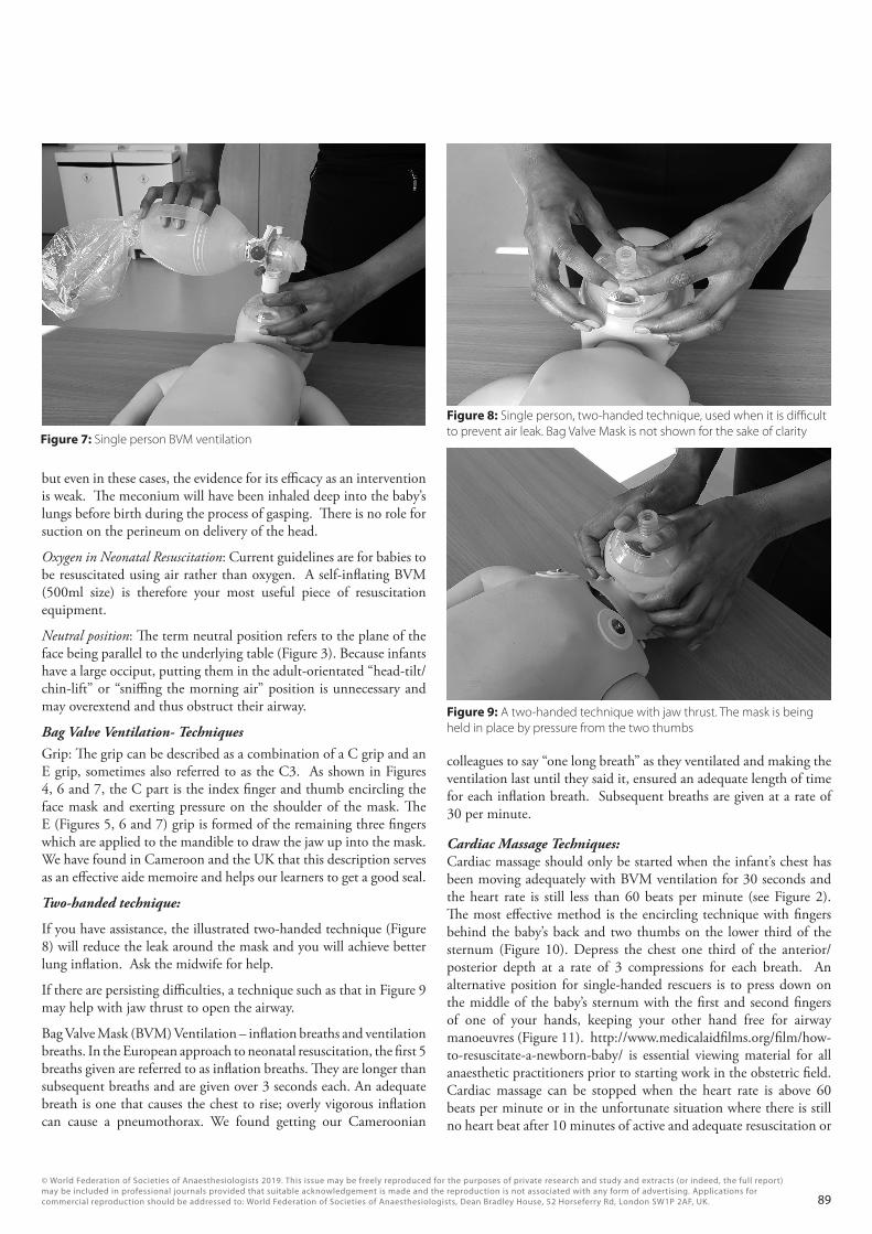
Figure 7: Single person BVM ventilation
Figure 8: Single person, two-handed technique, used when it is difficult to prevent air leak. Bag Valve Mask is not shown for the sake of clarity
Figure 9: A two-handed technique with jaw thrust. The mask is being held in place by pressure from the two thumbs
but even in these cases, the evidence for its efficacy as an intervention is weak. The meconium will have been inhaled deep into the baby’s lungs before birth during the process of gasping. There is no role for suction on the perineum on delivery of the head.
Oxygen in Neonatal Resuscitation: Current guidelines are for babies to be resuscitated using air rather than oxygen. A self-inflating BVM (500ml size) is therefore your most useful piece of resuscitation equipment.
Neutral position: The term neutral position refers to the plane of the face being parallel to the underlying table (Figure 3). Because infants have a large occiput, putting them in the adult-orientated “head-tilt/chin-lift” or “sniffing the morning air” position is unnecessary and may overextend and thus obstruct their airway.
Bag Valve Ventilation- Techniques Grip: The grip can be described as a combination of a C grip and an E grip, sometimes also referred to as the C3. As shown in Figures 4, 6 and 7, the C part is the index finger and thumb encircling the face mask and exerting pressure on the shoulder of the mask. The E (Figures 5, 6 and 7) grip is formed of the remaining three fingers which are applied to the mandible to draw the jaw up into the mask. We have found in Cameroon and the UK that this description serves as an effective aide memoire and helps our learners to get a good seal.
Two-handed technique:
If you have assistance, the illustrated two-handed technique (Figure 8) will reduce the leak around the mask and you will achieve better lung inflation. Ask the midwife for help.
If there are persisting difficulties, a technique such as that in Figure 9 may help with jaw thrust to open the airway.
Bag Valve Mask (BVM) Ventilation – inflation breaths and ventilation breaths. In the European approach to neonatal resuscitation, the first 5 breaths given are referred to as inflation breaths. They are longer than subsequent breaths and are given over 3 seconds each. An adequate breath is one that causes the chest to rise; overly vigorous inflation can cause a pneumothorax. We found getting our Cameroonian
colleagues to say “one long breath” as they ventilated and making the ventilation last until they said it, ensured an adequate length of time for each inflation breath. Subsequent breaths are given at a rate of 30 per minute.
Cardiac Massage Techniques:Cardiac massage should only be started when the infant’s chest has been moving adequately with BVM ventilation for 30 seconds and the heart rate is still less than 60 beats per minute (see Figure 2). The most effective method is the encircling technique with fingers behind the baby’s back and two thumbs on the lower third of the sternum (Figure 10). Depress the chest one third of the anterior/posterior depth at a rate of 3 compressions for each breath. An alternative position for single-handed rescuers is to press down on the middle of the baby’s sternum with the first and second fingers of one of your hands, keeping your other hand free for airway manoeuvres (Figure 11). http://www.medicalaidfilms.org/film/how-to-resuscitate-a-newborn-baby/ is essential viewing material for all anaesthetic practitioners prior to starting work in the obstetric field. Cardiac massage can be stopped when the heart rate is above 60 beats per minute or in the unfortunate situation where there is still no heart beat after 10 minutes of active and adequate resuscitation or
89
© World Federation of Societies of Anaesthesiologists 2019. This issue may be freely reproduced for the purposes of private research and study and extracts (or indeed, the full report) may be included in professional journals provided that suitable acknowledgement is made and the reproduction is not associated with any form of advertising. Applications for commercial reproduction should be addressed to: World Federation of Societies of Anaesthesiologists, Dean Bradley House, 52 Horseferry Rd, London SW1P 2AF, UK.

Figure 10: Cardiac compressions hand position. In the newborn cardiac massage is seldom of value if BVM ventilation is not being carried out well
Figure11: single rescuer cardiac massage
the heart beat remains under 60 beats per minute after 20 minutes of active and adequate resuscitation.
Vascular Access: Umbilical Venous Cannula and Intra Osseous Access: Volume replacement in resuscitation is rarely needed. Inserting a small nasogastric tube 5cms into an umbilical vein is probably the quickest method of establishing venous access at birth. It should be done as cleanly as possible, and fluid or medications given as needed. An intra-osseous needle, inserted into the bone marrow cavity of the baby’s proximal tibia can also be used. If you are not familiar with the use of an intraosseous needle, you may find the 25 minute video from OPENPediatrics (www.openpediatrics.org) helpful, available at https://www.youtube.com/watch?v=RTxbWkHKH-M. Needles specifically for this job may not be available in your setting but a sturdy hypodermic needle of 18g with a syringe attached will usually be an effective alternative.
Medications in Neonatal Resuscitation: Adrenaline, sodium bicarbonate and sometimes 10% dextrose are used in high resource countries but the evidence for their efficacy is poor and the outcome
for the baby is probably not improved greatly. In areas where there are no high level neonatal facilities, the role of medications in newborn resuscitation is unproven and probably best avoided.
You dry the baby, wrap him in a dry towel and put on his hat. You put his head in the neutral position, note that he is not breathing and his heart rate is slow. You apply a well-fitting face mask and, using a C and E type grip, you administer 5 slow inflation breaths. The chest rises satisfactorily and the midwife tells you that the baby’s heart rate has come up. The baby cries and you give him to his mother for skin to skin care and he goes to the breast immediately. A life saved, well done!
Training resources:Videos: basic neonatal life support in the community using mouth-to-mouth resuscitation techniques instead of bag and valve mask is well taught and explained at http://www.medicalaidfilms.org/film/what-to-do-when-a-newborn-baby-is-not-breathing/. A more detailed video from the same group, 20 minutes long and covering all aspects of newborn resuscitation is well worth watching at http://www.medicalaidfilms.org/film/how-to-resuscitate-a-newborn-baby/. All aspects of newborn delivery and care in resource poor areas are beautifully illustrated in the Global Health Media videos which we recommend to you highly (https://globalhealthmedia.org/videos/).
Training Courses: Probably the most widely available course in resource poor areas is Helping Babies Breathe (https://www.aap.org/en-us/advocacy-and-policy/aap-health-initiatives/helping-babies-survive/Pages/default.aspx). The training project we have been most directly involved in is the Newborn Care Course (http://www.nicheinternational.org.uk/newborn-care-course/), originally developed by Maternal and Child Advocacy International (http://www.mcai.org.uk/) and the Advanced Life Support Group (www.alsg.org), both based in the UK. The Newborn Care Course uses the UK Resuscitation Council guidelines for the life support teaching component (https://www.resus.org.uk/information-on-courses/newborn-life-support/).
Written material: The World Health Organisation published its updated guidelines on Management of the Newborn in 2017 (http://www.who.int/maternal_child_adolescent/documents/newborn-health-recommendations/en/).
Acknowledgements: We would like to thank Sabrina Bhandari and Sophie Bunce, medical students, Queens University, Belfast for their help in preparing the photographs.
OTHER REFERENCES:1. UNICEF. WHO, World Bank, UN DESA/Population Division. Levels and trends in Child Mortality 2017. UNICEF 2017. http://apps.who.int gho/data/node.sdg.3-2-viz-3?lang=en accessed 25.4.2018
2. Guidelines for Newborn Care. WHO 2017. Available at http://www. who.int/maternal_child_adolescent/documents/newborn-health- recommendations/en/ accessed on 20.04.2018
3. Northern Neonatal Network https://nornet.org.uk/ accessed 24.4.2018
4. Newborn Life Support algorithm from Resuscitation Council (UK) from https://www.resus.org.uk/resuscitation-guidelines/accessed/22.04.2018
90 www.wfsahq.org/resources/update-in-anaesthesia

Madhusudan UpadyaKasturba Medical College MangaloreKarnataka INDIA
Mahesh Nayak MDSenior ResidentKasturba Medical SchoolMangaloreINDIA
Summary
A significant number of women undergo anesthesia and surgery during pregnancy for procedures unrelated
to delivery. In order to provide safe anaesthesia for mother and fetus, it is essential for the anesthetist to have
thorough understanding of the physiological and pharmacological changes that characterize the three
trimesters of pregnancy. A multidisciplinary team approach involving the anesthetist, obstetrician, neonatologist
and surgeon is highly recommended to ensure an adequate standard of care. Anaesthesia management,
including post-operative analgesia, should be planned well to preserve the pregnancy and to ensure the safety
of the mother as well as the foetus. Once fetal viability is assumed (24–26 weeks), the fetal heart rate (FHR)
should be monitored. Regional anaesthesia minimizes fetal drug exposure, airway management is simplified,
blood loss may be decreased, and overall risks to the mother and fetus are less.
Anaesthesia for non obstetric surgery during pregnancy
Madhusudan Upadya* and Mahesh Nayak*Correspondence email: [email protected]:10.1029/WFSA-D-18-00020
INTRODUCTIONAnaesthesia for pregnant patients may not always be only for obstetric surgeries. These patients do present with surgical illness requiring surgery under general or regional anaesthesia. With advances in fetal surgeries the number is only likely to increase in future. Incidence of non-obstetric surgery being 1-2%.1 Surgery can be required during any stage of pregnancy depending on the urgency of the indication. In the largest single series concerning surgery and anaesthesia during pregnancy, 42% of surgery during pregnancy occurred during the first trimester, 35% during the second trimester, and 23% during the third.2 Appendicitis, ovarian disorders (torsion or neoplasm) and trauma constitute the most common non-obstetric conditions requiring surgery during pregnancy, appendicectomy being the most common. The incidence of Appendicitis is around 1 in 1500-2000 pregnancies.3
Non-obstetric surgery during pregnancy presents newer challenges to the anaesthetist. The anaesthetist has to take care of two lives. The goal being safe anaesthesia for both pregnant woman and the fetus. To ensure maternal safety, the anaesthetist must have a thorough understanding of the physiological and pharmacological adaptations to pregnancy. Fetal safety requires avoidance of potentially dangerous drugs at critical times during fetal development, assurance of continuation of adequate uteroplacental perfusion, and avoidance and/or treatment of preterm labour and delivery.4
Principles of anaesthetic managementThe anaesthetist has the following goals:5
(i) Optimize and maintain normal maternal physiological function;(ii) Optimize and maintain utero-placental blood flow and oxygen delivery; (iii) Avoid unwanted drug effects on the fetus; (iv) Avoid stimulating the myometrium (oxytocic effects); (v) Avoid awareness during general anaesthesia; (vi) Use regional anaesthesia, if possible.
Maternal safety According to American College of Obstetricians and Gynaecologists’ Committee on Obstetric Practice, regardless of trimester, pregnant woman should not be denied indicated surgery. Elective surgery should be postponed until after delivery. If possible, nonurgent surgery should be performed in the second trimester when preterm contractions and spontaneous abortion are least likely. The choice of anaesthetic technique(s), and the selection of appropriate drugs of anaesthesia should be guided by maternal indications for surgery and the location of the surgical procedure. Resuscitation, if required, should be vigorously performed following the standard advanced life support or advanced trauma life support protocols, with the addition of 150 left lateral tilt to avoid supine hypotension after 20 weeks.6
91
© World Federation of Societies of Anaesthesiologists 2019. This issue may be freely reproduced for the purposes of private research and study and extracts (or indeed, the full report) may be included in professional journals provided that suitable acknowledgement is made and the reproduction is not associated with any form of advertising. Applications for commercial reproduction should be addressed to: World Federation of Societies of Anaesthesiologists, Dean Bradley House, 52 Horseferry Rd, London SW1P 2AF, UK.
Clin
ical
Art
icle
s
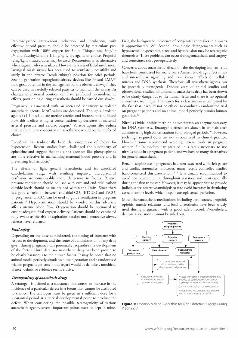
Rapid-sequence intravenous induction and intubation, with effective cricoid pressure, should be preceded by meticulous pre-oxygenation with 100% oxygen for 5min. Thiopentone 5mg/kg IV and Succinylcholine 1.5mg/kg iv are agents of choice. Propofol (2mg/kg iv titrated doses may be used. Rocuronium is an alternative when sugammadex is available. However, in cases of failed intubation, laryngeal mask airway has been used to ventilate successfully and safely in the reverse Trendelenburg’s position for brief periods. Second generation supraglottic airway devices like Proseal LMATM hold great potential in the management of the obstetric airway.7 They can be used in carefully selected patients to maintain the airway. As changes in maternal position can have profound haemodynamic effects, positioning during anaesthesia should be carried out slowly.
Pregnancy is associated with an increased sensitivity to volatile anaesthetic agents, MAC values are decreased. Though all volatile agents (<1.5 mac) dilate uterine arteries and increase uterine blood flow, this is offset at higher concentrations by decreases in maternal arterial pressure and cardiac output.8 Volatile agents also reduce uterine tone. Low concentration sevoflurane would be the preferred choice.
Ephedrine has traditionally been the vasopressor of choice for hypotension. Recent studies have challenged the superiority of ephedrine and suggest that the alpha agonists like phenylephrine are more effective in maintaining maternal blood pressure and in preventing fetal acidosis.9
The effects of light general anaesthesia and its associated catecholamine surge with resulting impaired uteroplacental perfusion are considerably more dangerous to foetus. Positive pressure ventilation should be used with care and end-tidal carbon dioxide levels should be maintained within the limits. Since there is a good correlation between end-tidal CO2 (ETCO2) and PaCO2 in pregnancy, ETCO2 can be used to guide ventilation in pregnant patients.10 Hyperventilation should be avoided as this adversely affects uterine blood flow. Oxygenation should be optimized to ensure adequate fetal oxygen delivery. Patients should be extubated fully awake as the risk of aspiration persists until protective airway reflexes have returned.
Fetal safety Depending on the dose administered, the timing of exposure with respect to development, and the route of administration of any drug given during pregnancy can potentially jeopardise the development of the foetus. Until date, no anaesthetic drug has been proven to be clearly hazardous to the human foetus. It may be noted that no animal model perfectly simulates human gestation and a randomised trial on pregnant patients in this regard would be definitely unethical. Hence, definitive evidence seems elusive.11
Teratogenicity of anaesthetic drugsA teratogen is defined as a substance that causes an increase in the incidence of a particular defect in a foetus that cannot be attributed to chance. The teratogen must be given in a sufficient dose for a substantial period at a critical developmental point to produce the defect. When considering the possible teratogenicity of various anaesthetic agents, several important points must be kept in mind.
First, the background incidence of congenital anomalies in humans is approximately 3%. Second, physiologic derangements such as hypoxaemia, hypercarbia, stress and hypotension may be teratogenic themselves. These problems can occur during anaesthesia and surgery and sometimes exist pre-operatively.
Concerns about anaesthetic effects on the developing human fetus have been considered for many years Anaesthetic drugs affect intra- and intercellular signalling and have known effects on cellular mitosis and DNA synthesis. Therefore, all anaesthetic agents can be potentially teratogenic. Despite years of animal studies and observational studies in humans, no anaesthetic drug has been shown to be clearly dangerous to the human fetus and there is no optimal anaesthetic technique. The search for a clear answer is hampered by the fact that it would not be ethical to conduct a randomized trial on pregnant patients and no animal model perfectly mimics human gestation.12
Nitrous Oxide inhibits methionine synthetase, an enzyme necessary for DNA synthesis. Teratogenic effects are shown in animals after administering high concentrations for prolonged periods.10 However, such high required doses are not encountered in clinical practice. However, some recommend avoiding nitrous oxide in pregnant women.13,14 In modern day practice, it is rarely necessary to use nitrous oxide in a pregnant patient, and we have so many alternatives for general anaesthesia.
Benzodiazepine use in pregnancy has been associated with cleft palate and cardiac anomalies. However, many recent controlled studies have countered this association.15,16 It is usually recommended to avoid benzodiazepine use throughout gestation and most especially during the first trimester. However, it may be appropriate to provide judicious pre-operative anxiolysis so as to avoid increases in circulating catecholamine levels, which impair uteroplacental perfusion.
Most other anaesthetic medications, including barbiturates, propofol, opioids, muscle relaxants, and local anaesthetics have been widely used during pregnancy with a good safety record. Nonetheless, delicate associations cannot be ruled out.
92 www.wfsahq.org/resources/update-in-anaesthesia
Pregnant surgical patient
Elective surgery
Essential surgery
Emergency surgery
Delay until postpartum
First trimester Second/third trimester
If no or minimal increased risk to mother, consider
delaying until midgestation
If greater than minimal increased risk to mother
proceed with surgery
Proceed with optimal anesthetic for mother, modified by considerations for maternal physiologic changes and fetal well-being
Consult a perinatologist or an obstetrician
Intraopertaive and postoperative fetal and uterine monitoring may be useful
Figure 1: Decision-Making Algorithm for Non-Obstetric Surgery During Pregnancy6

Timing of exposure is of crucial importance. During the first 15 days of human gestation an all or nothing phenomenon occurs: the fetus is lost or the fetus is preserved fully intact. During the time of organogenesis (15-56 days) structural abnormalities may occur. After this period, functional changes can be observed, but structural abnormalities are rare.
Decision-Making Algorithm for Non-Obstetric Surgery During PregnancyWhenever a pregnant woman undergoes nonobstetric surgery, consultations among her obstetrical team, surgeon(s), anesthesiologist(s), and neonatologist(s) are important to coordinate management (Figure 1).
General principles of anaesthesia management
Pre-operative preparation This should always involve close liaison with the obstetricians and include ultrasound assessment of the fetus when delivery is anticipated. Neonatologists will also need to be consulted. Many signs and symptoms often associated with cardiac disease, such as dyspnoea, heart murmurs and peripheral oedema are common during normal pregnancy. ECG changes during pregnancy include left axis deviation, premature beats and non-specific ST and T-wave changes.9 During radiological investigations, fetal exposure should be minimized. Results of relevant blood tests should be available and cross-matched blood must be ordered for all major surgery. Pregnant patients who require surgery should be evaluated pre-operatively in the same manner as non-pregnant patients. Laboratory and other testing should be performed as indicated by the patient’s comorbidities and the proposed surgery. In addition to standard pre-operative procedures, preparation of pregnant women takes into account risks of aspiration, difficult intubation, thromboembolism, and the well-being of the foetus. Standard adult fasting guidelines, i.e., 6-8 h for solid food, depending on the type of food ingested (e.g., fat content) are applicable to these patients.
Aspiration prophylaxis: The gastric emptying has recently been shown to be normal during pregnancy until the onset of labour. However, the risk of aspiration is still higher due to reduced gastric barrier pressure and lower oesophageal sphincter tone (a progesterone effect).17 The presence of additional risk for regurgitation and aspiration, including active reflux or obesity should be surveyed. Prophylaxis against aspiration pneumonitis should be administered from 16 weeks gestation with H2-receptor antagonists and non-particulate antacids.
Antibiotic prophylaxis: The need for antibiotic prophylaxis depends on the specific procedure. However, attention should be paid in selecting antibiotics with good safety profile in pregnancy.
Prophylactic glucocorticoids: Administration of a course of antenatal glucocorticoids 24-48hrs before surgery between 24 and 34 weeks of gestation can reduce perinatal morbidity/mortality if preterm birth occurs. Despite the potential benefits to the foetus, however, antenatal glucocorticoids are best avoided in the setting of systemic infection (such as sepsis or a ruptured appendix), because they may impair the ability of the maternal immune system to contain the infection.
Thromboprophylaxis: Pregnancy is a hypercoagulable state. The 2012 American College of Chest Physicians clinical practice guideline on prevention and treatment of thrombosis recommends mechanical or pharmacologic thromboprophylaxis for all pregnant patients undergoing surgery.
Prophylactic tocolytics: There is no proven benefit to routine administration of prophylactic perioperative tocolytic therapy. Minimising uterine manipulation may reduce the risk of development of uterine contractions and preterm labour. Tocolytics are indicated for the treatment of preterm labour until resolution of the underlying, self-limited condition that may have caused the contractions.
Conduct of AnaesthesiaNo studies have shown a beneficial effect on the outcome of pregnancy after regional compared with general anaesthesia. However, regional anaesthesia minimizes fetal drug exposure, airway management is simplified, blood loss may be decreased, and overall risks to the mother and fetus are less. The largest risk of regional anaesthesia is hypotension resulting from sympathetic nerve blockade, which reduces uterine blood flow and perfusion to the fetus. Attention to maternal fluid volume and blood pressure is critical. Regardless of the anaesthetic technique, steps to avoid hypoxemia, hypotension, acidosis, and hyperventilation are the most critical elements of anaesthetic management.
After 6–8 weeks gestation, cardiac, haemodynamic, respiratory, metabolic and pharmacological parameters are considerably altered. With the increase in minute ventilation and oxygen consumption and a decrease in oxygen reserve (decreased functional residual capacity and residual volume), pregnant women become hypoxaemic more rapidly. Supplementary oxygen must always be given during vulnerable periods to maintain oxygenation. Normal hyperventilation in pregnancy results in lowered expired CO2 (32–34mm Hg); this should be maintained during anaesthesia.
Aortocaval compression is a major hazard from 20 weeks onwards (and sometimes even earlier); this compromises uterine blood flow and, in some women, results in supine hypotension. This effect may be exacerbated by regional or general anaesthesia when normal compensatory mechanisms are attenuated or abolished. Aortocaval compression is only effectively avoided by the use of the 150 lateral position. It can be decreased by uterine displacement through wedging or manual displacement. Venacaval compression results in distension of the epidural venous plexus, increasing the risk of intravascular injection during regional blockade. The capacity of the epidural space is reduced, which probably contributes to the enhanced spread of local anaesthetics in pregnancy.
Pregnancy is associated with a hypercoagulable state because of increased pro-coagulant factors. The incidence of thromboembolic complications is at least five times greater during pregnancy; thromboprophylaxis is essential.18
During third trimester, delivery by caesarean section before major surgery is often recommended. Where possible, surgery should be delayed 48hr to allow steroid therapy to enhance fetal lung maturation. It may be appropriate to deliver the baby under
93
© World Federation of Societies of Anaesthesiologists 2019. This issue may be freely reproduced for the purposes of private research and study and extracts (or indeed, the full report) may be included in professional journals provided that suitable acknowledgement is made and the reproduction is not associated with any form of advertising. Applications for commercial reproduction should be addressed to: World Federation of Societies of Anaesthesiologists, Dean Bradley House, 52 Horseferry Rd, London SW1P 2AF, UK.

regional anaesthesia and then convert to a general anaesthesia for the definitive surgery. Anaesthesia post-delivery should be tailored to surgical requirements, with the precaution that volatile agents should be discontinued or used only in small doses.
Fetal monitoring Once fetal viability is assumed (24-26 weeks), the fetal heart rate (FHR) should be monitored. This may be difficult in the obese patient or during abdominal surgery. Inhalation agents typically cause a reduction in FHR variability, one of the changes indicative of fetal hypoxaemia. Intra-operative FHR monitoring requires skilled interpretation and an obstetrician with a plan of action should fetal distress be diagnosed. Uterine manipulation should be minimized to avoid preterm labour. Ketamine increases uterine tone in early pregnancy and should not be used. While some advocate the prophylactic use of tocolytic agents, they are not without risk themselves and there is no proof of efficacy.
The ACOG states, “although there are no data to support specific recommendations regarding nonobstetric surgery and anaesthesia in pregnancy, it is important for non-obstetric physicians to obtain obstetric consultation before performing non-obstetric surgery. The decision to use fetal monitoring should be individualized and each case warrants a team approach for optimal safety of the woman and her baby.19
General guidelines for fetal monitoring include the following:20
If the fetus is considered previable, it is generally sufficient to ascertain the fetal heart rate by Doppler before and after the procedure.
At a minimum, if the fetus is considered to be viable, simultaneous electronic fetal heart rate and contraction monitoring should be performed before and after the procedure to assess fetal well-being and the absence of contractions.
Intraoperative electronic fetal monitoring may be appropriate when all of the following apply:
• The fetus is viable.
• It is physically possible to perform intraoperative electronic fetal monitoring.
• A health care provider with obstetric surgery privileges is available and willing to intervene during the surgical procedure for fetal indications.
• When possible, the woman has given informed consent to emergency caesarean delivery.
• The nature of the planned surgery will allow the safe interruption or alteration of the procedure to provide access to perform emergency delivery.
Monitoring for uterine contractions When external tocodynamometer can be placed outside of the surgical field, uterine contractions may be monitored intraoperatively. If uterine contractions are detected, maternal haemodynamics should be improved by giving more intravenous fluids and also consider tocolytic treatment in consultation with the
perinatologist/obstetrician. Tocometry during post-operative period is useful as post-operative analgesia may mask awareness of mild early contractions and delay tocolysis.
Recovery from anaesthesia Recovery from anaesthesia requires close monitoring, particularly of the airway and respiratory system, because most severe anaesthetic complications due to hypoventilation or airway obstruction occur during emergence, extubation, or recovery.
Post-operative analgesia Provision of adequate analgesia is important in the post-operative period as well, since the pain has been shown to increase the risk of premature labour. Regional nerve or plexus blockade or epidural analgesia can provide excellent post-operative analgesia and reduce the risk of opioid-induced hypoventilation when compared with intravenous opioids. Ultrasound guided Transverse abdominis plane (TAP) block is an alternative to epidural analgesia. Though TAP block blocks only somatic component of pain while epidural blocks both somatic and visceral component, both provide adequate analgesia for abdominal studies. A randomised control study found no difference in pain scores, pain scores over time and opioid requirements between continuous TAP block and epidural analgesia.21 Opioids can be used, as needed, to control post-operative pain. Paracetamol is the analgesic of choice for the treatment of mild to moderate pain during any stage of pregnancy. NSAIDs should be avoided, especially after 32 weeks of gestation, because they may cause premature closure of the foetal ductus arteriosus (if given for more than 48hrs). They are also associated with oligohydramnios with reduced foetal renal function. NSAIDs can also inhibit uterine contraction.
Specific Procedures
LaparoscopyPregnancy is no longer considered a contraindication to laparoscopic surgery. Indications for laparoscopic treatment of acute abdominal processes are the same as for nonpregnant patients. Laparoscopy can be safely performed during any trimester of pregnancy. The advantages include less exposure of the fetus to possibly toxic agents, smaller incisions, decreased pain, less need for analgesics, and more rapid recovery and mobilization.22 Carbon dioxide pneumoperitoneum is associated with an increased risk of hypoxaemia, hypercarbia and hypotension because of the physiological and anatomical changes of pregnancy. Society of American Gastrointestinal Endoscopic Surgeons issued following guidelines.23 Fetal and uterine status should be monitored and also end-tidal PCO2 and maternal arterial blood gases. An open technique should be used to enter the abdomen. Aortocaval compression should be avoided. Low pneumoperitoneum pressures (between 10 and 15mmHg) should be used. Tocolytic agents should not be used prophylactically but should be considered when evidence of preterm labor is present.
Cardiac SurgeryThe cardiovascular changes of pregnancy include a 30-50% increase in blood volume and cardiac output. Although pregnant patients with heart disease are usually managed with medical therapy, those with severe decompensation and surgically correctable lesions might
94 www.wfsahq.org/resources/update-in-anaesthesia
come to surgery, in particular those with severe mitral or aortic valvular obstruction.24,25 Percutaneous balloon valvuloplasty seems to be a better alternative than surgical repair and is associated with a significant reduction in fetal and neonatal mortality. The use of cardiopulmonary bypass increases perioperative risk, particularly for the fetus. Factors related to cardiopulmonary bypass that can adversely affect fetal oxygenation include non-pulsatile perfusion, inadequate perfusion pressures, inadequate pump flow, embolic phenomena to the uteroplacental bed, and the release of renin and catecholamines. The use of intraoperative fetal monitoring can decrease the high fetal mortality rate. During cardiopulmonary bypass, a high pump flow (>2.5 litre min−1 m−2) and perfusion pressure (>70mmHg) are recommended to maintain uteroplacental blood flow.15 It is recommended that the maternal haematocrit be maintained >28% to optimize oxygen-carrying capacity.26,27
NeurosurgeryHaemorrhage from intracranial saccular aneurysm or arteriovenous malformation is unfortunately not uncommon during pregnancy. The risk of intracranial haemorrhage is increased by hypertensive conditions of pregnancy and their associated risk factors. The usual neurosurgical anaesthetic treatment of these patients can include controlled hypotension, hypothermia, hyperventilation, and diuresis, which must be undertaken carefully in the pregnant patient. Controlled hypotension can be induced with high-dose volatile anaesthetic, sodium nitroprusside, or nitroglycerin. Each carries its own potential hazards in addition to reduction in uteroplacental blood flow. All of these drugs cross the placenta and can induce hypotension in the fetus.28 When induced hypotension is deemed necessary, fetal heart rate monitoring should be used and the period of hypotension. Hyperventilation is commonly used in neuroanaesthesia to reduce and cerebral blood flow. Extreme hyperventilation (PaCO2 <3.3kPa) can cause uterine artery vasoconstriction and leftward shift of the maternal oxyhaemoglobin dissociation curve. Fetal heart rate monitoring should alert the anaesthesiologist to compromises in fetal condition and adjustments to maternal ventilation should be made accordingly. Diuresis is often accomplished with osmotic agents or loop diuretics to shrink the brain both intraoperatively and after operation. These can cause significant negative fluid shifts for the fetus. However, in individual case reports, mannitol in small doses of 0.25–0.5mg kg−1 has been used without ill effect to the fetus and appears safe if required.29 A loop diuretic provides an alternative but should also be used cautiously with fetal monitoring and only if necessary.
Fetal SurgeriesSurgery to the fetus while it is still in utero is used to treat an increasing number of lethal and non-lethal conditions. The problems of preterm labour and premature rupture of membranes associated with open surgery have led to the development of minimal access surgical techniques. Although fetal surgery is a new and fast-moving frontier of medicine, it is not one that all obstetric anaesthetists will encounter. The first successful human fetal operation was performed in 1983, but it is still only carried out in a limited number of specialist tertiary centres. There are basically three different type of surgeries – Minimally invasive, Midgestation Open procedures
or EXIT (Ex utero intrapartum) procedures. The broad challenges presented to the anaesthetist are: techniques used to prevent preterm labour, maintenance of maternal homeostasis in the face of tocolytic techniques, maintenance of fetal homeostasis and provision of fetal analgesia during surgery.30
REFERENCES1. Crowhurst JA. Anaesthesia for non-obstetric surgery during pregnancy. Acta Anaesthesiol Belg 2002; 53: 295-7
2. Mazze RI, Källén B. Reproductive outcome after anaesthesia and operation during pregnancy: a registry study of 5405 cases, Am J Obstet Gynecol, 1989, vol. 161 (pg. 1178-85)
3. Dietrich CS, Hill CC, Hueman M. Surgical diseases presenting in pregnancy. Surg Clin North Am 2008; 88: 403-419
4. Van De Velde M, De Buck F. Anaesthesia for non-obstetric surgery in the pregnant patient, Minerva Anestesiol, 2007, vol. 73 (pg. 235-40)
5. Nina Kylie Dorothy Walton, Venkata Krishnaker Melachuri; Anaesthesia for non obstetric surgery during pregnancy, Continuing Education in Anaesthesia Critical Care & Pain, Volume 6, Issue 2, 1 April 2006, Pages 83–85
6. Upadya M, Saneesh P J. Anaesthesia for non-obstetric surgery during pregnancy. Indian J Anaesth 2016; 60: 234-41
7. Dongare PA, Nataraj MS. Anaesthetic management of obstetric emergencies. Indian J Anaesth 2018; 62: 704-9
8. Ravindra G L, Madamangalam AS, Seetharamaiah S. Anaesthesia for non obstetric surgery in obstetric patients. Indian J Anaesth 2018; 62: 710-6
9. Walton NK, Melachuri VK. Anaesthesia for non-obstetric surgery during pregnancy. Contin Educ Anaesth Crit Care Pain. 2006; 6: 83–5
10. Bhavani-Shankar K, Steinbrook RA, Brooks DC, Datta S. Arterial to end-tidal carbon dioxide pressure difference during laparoscopic surgery in pregnancy. Anesthesiology 2000; 93: 370-3
11. Reitman E, Flood P. Anaesthetic considerations for non-obstetric surgery during pregnancy. Br J Anaesth 2011; 107 (Suppl. 1): i72–i78
12. Fujinaga M, Baden JM. Methionine prevents nitrous oxide-induced teratogenicity in rat embryos grown in culture. Anesthesiology 1994; 81: 184-9
13. Aldridge LM, Tunstall ME. Nitrous oxide and the fetus. A review and the results of a retrospective study of 175 cases of anaesthesia for insertion of Shirodkar suture. Br J Anaesth 1986; 58: 1348-56
14. Crawford JS, Lewis M. Nitrous oxide in early human pregnancy. Anaesthesia 1986; 41: 900-5
15. Safra MJ, Oakley GP Jr. Association between cleft lip with or without cleft palate and prenatal exposure to diazepam. Lancet 1975; 2: 478-80
16. Rosenberg L, Mitchell AA, Parsells JL, Pashayan H, Louik C, Shapiro S. Lack of relation of oral clefts to diazepam use during pregnancy. N Engl J Med 1983; 309: 1282-5
17. Wong CA, McCarthy RJ, Fitzgerald PC, Raikoff K, Avram MJ. Gastric emptying of water in obese pregnant women at term. Anesth Analg 2007; 105: 751-5
18. Barron WM. Medical evaluation of the pregnant patient requiring nonobstetric surgery. Clin Perinatol 1985; 12: 481–96
19. American College of Obstetricians and Gynaecologists: ACOG Committee Opinion no. 284, August 2003: Nonobstetric surgery in pregnancy, Obstet Gynecol 102: 431, 2003
20. Nonobstetric surgery during pregnancy. Committee Opinion No. 474. American College of Obstetricians and Gynaecologists. Obstet Gynecol 2011; 117: 420–1
21. Rao Kadam V, Van Wijk RM, Moran JI, Miller D. Epidural versus continuous transversus abdominis plane catheter technique for postoperative analgesia after abdominal surgery. Anaesth Intensive Care 2013; 41: 476-81.
22. Reedy MB, Kallen B, Kuehl TJ. Laparoscopy during surgery: a study of 5 fetal outcome parameters with use of the Swedish Health Registry, Am J Obstet Gynecol, 1997, vol. 177 (pg. 673-9)
95
© World Federation of Societies of Anaesthesiologists 2019. This issue may be freely reproduced for the purposes of private research and study and extracts (or indeed, the full report) may be included in professional journals provided that suitable acknowledgement is made and the reproduction is not associated with any form of advertising. Applications for commercial reproduction should be addressed to: World Federation of Societies of Anaesthesiologists, Dean Bradley House, 52 Horseferry Rd, London SW1P 2AF, UK.

23. Guidelines Committee of the Society of American Gastrointestinal and Endoscopic Surgeons, Yumi H. Guidelines for diagnosis, treatment, and use of laparoscopy for surgical problems during pregnancy. Surg Endosc 2008; 22: 849-61
24. Kuczkowski KM. Nonobstetric surgery during pregnancy: what are the risks of anaesthesia?, Obstet Gynecol Surv, 2004, vol. 59 (pg. 52-6)
25. Pomini F, Mercogliano D, Cavalletti C, Caruso A, Pomini P. Cardiopulmonary bypass in pregnancy, Ann Thorac Surg, 1996, vol. 61
26. Parry AJ, Westaby S. Cardiopulmonary bypass during pregnancy, Ann Thorac Surg, 1996, vol. 61 (pg. 1865-9)
27. John AS, Gurley F, Schaff HV, et al. Cardiopulmonary bypass during pregnancy, Ann Thorac Surg, 2011, vol. 91 (pg. 1191-6)
28. Nuevo FR. Birnbach D, Gatt SP, Datta S. Anaesthesia for nonobstetric surgery in the pregnant patient, Textbook of Obstetric Anaesthesia, 2000, New York Churchill Livingstone(pg. 289-98)
29. Bharti N, Kashyap L, Mohan VK. Anaesthetic management of a parturient with cerebellopontine-angle meningioma, Int J Obstet Anesth, 2002, vol. 11 (pg. 219-21)
30. Ritu Gupta, Mark Kilby, Griselda Cooper; Fetal surgery and anaesthetic implications, Continuing Education in Anaesthesia Critical Care & Pain, Volume 8, Issue 2, 1 April 2008, Pages 71–75
96 www.wfsahq.org/resources/update-in-anaesthesia

MISSION OF UIAUpdate in Anaesthesia (UIA) is an official journal of the World Federation of Societies of Anaesthesiologists (WFSA). UIA’s primary goal is to provide high-quality, peer-reviewed, clinically-relevant educational articles aimed at improving the quality and safety of anaesthesia care around the world.
UIA welcomes manuscripts focused on, and not limited to, anaesthesia that is relevant to resource-limited settings. UIA also serves to share knowledge from affiliated associations such as the World Health Organization (WHO) and surgical societies and educators that recognise safe anaesthesia as an essential part of safe surgery.
Over 16,000 anaesthetists around the world access UIA on the internet at http://www.wfsahq.org/resources/update-in-anaesthesia The printed version of UIA is sent free of charge to over 1000 English-speaking anaesthetists and has previously been translated into other languages. UIA is read by many others including anaesthesia trainees, nurses, surgeons and medical students.
Manuscripts should be submitted to:https://www.editorialmanager.com/wfsa/default.aspx
GENERAL CONSIDERATIONS FOR PREPARATION AND SUBMISSION OF A MANUSCRIPTA major focus of UIA is to provide educational materials for those working with limited resources. The types of manuscripts that are published in the journal reflect this. If you have an idea for an article and would like to discuss this further, please email the Editor-in-Chief with a proposed title and a brief (200 word) summary of the scope and content.
Manuscripts are screened by the editorial staff and are sent to independent reviewers who are not informed of the identity of the author(s). The reviewers’ comments and suggested revisions will be forwarded to the corresponding author. Manuscripts will be reviewed with plagiarism detection software. Manuscripts may not have been published in whole or any part in another journal.
Some readers of UIA may not use English as their first language. It is therefore important to keep text straightforward and to avoid long sentences and complex terminology. Explain words and abbreviations that may not be universally standardised.
Aim to include the full range of therapies available worldwide. Provide detailed descriptions of therapies available in resource-limited settings. When appropriate, discuss older medications, such as Halothane and Thiopentone that may still be used around the world.
Authors must acknowledge any financial support received or conflict of interest related to the contents of a submitted manuscript.
Contributions to the, 1) clinical care or research described in a manuscript or 2) preparation of a manuscript made by persons other than the authors may be acknowledged.
AnaesthesiaUpdate in
Guide for Contributors
Upon acceptance for publication copyright becomes vested with the journal.
Manuscripts based on clinical investigation of human subjects must adhere to the ethical principles of the Declaration of Helsinki developed by the World Medical Association. https://www.wma.net/policies-post/wma-declaration-of-helsinki-ethical-principles-for-medical-research-involving-human-subjects/
Manuscripts based on clinical investigation of human subjects must include the consent of the patients or study participants or appropriate person (parent/guardian) and a statement of approval from an appropriate Institutional Review Board (IRB)/Ethics Committee. The IRB/Ethics Committee must review and provide oversight or grant exemption from requiring patients’ or study participants’ consent for the performance of audits and this should be referred to in the text.
Manuscripts must protect patients and study participants from being identified. UIA adheres to the USA HIPAA Privacy Rule that defines 18 identifiers that must be removed to assure the protection of personal health information. https://privacyruleandresearch.nih.gov/pdf/HIPAA_Privacy_Rule_Booklet.pdf (Specifically refer to page 10)
Manuscripts should adhere to Uniform Requirements for Manuscripts Submitted to Biomedical Journals prepared by the International Committee of Medical Journal Editors. http://www.icmje.org/recommendations/browse/manuscript-preparation/preparing-for-submission.html
Manuscript Types As an official journal of the WFSA, UIA is the appropriate forum for publication of articles describing anaesthesia patient care, original research and audit conducted in, and specifically relevant to, low and middle income resource settings.
Manuscripts describing clinical care, research, procedures, techniques or equipment adapted by anaesthetists to the limited resource setting in which they care for patients are welcome in any of the following formats:
• Review article - Basic Science or Clinical (3000 words maximum)
• Original Research (3000 words maximum)
• Case Report/Clinical Overview (1500 words maximum)
• UIA Short Reports (750 words maximum)
• Editorials (1200 words maximum) - Editorials are usually by invitation of the Editor-in-Chief. Proposals for Editorials may be sent to the Editor-in-Chief for consideration and potential invitation.
• Review of Books or other Educational Materials (eg web app) (750 words maximum)
97
© World Federation of Societies of Anaesthesiologists 2019. This issue may be freely reproduced for the purposes of private research and study and extracts (or indeed, the full report) may be included in professional journals provided that suitable acknowledgement is made and the reproduction is not associated with any form of advertising. Applications for commercial reproduction should be addressed to: World Federation of Societies of Anaesthesiologists, Dean Bradley House, 52 Horseferry Rd, London SW1P 2AF, UK.

• Letters to the Editor (1000 words maximum) - Correspondence is welcome on any topic, article or editorial that has appeared in UIA or is of interest to individuals providing anaesthesia patient care.
COMPONENTS OF THE MANUSCRIPT
Title page that includes:
• Title of the article
• Name and affiliation of each author
• Email, postal address and telephone contact details of the corresponding author
• Key words using terms from the Medical Subject Headings (MeSH) of Index Medicus https://www.nim.nih.gov/mesh
• Abstract that includes no more than 200 words
For research manuscripts note Background, Methods, Results, and Conclusions
ILLUSTRATIONS / FIGURESTables, figures, graphs and illustrations should be submitted as electronic versions of drawings (black on white) or as black and white or color picture files in JPEG format. If computer or internet access is unavailable, print versions may be submitted. If facilities to produce drawings are unavailable, contact the Editor-in-Chief for assistance. Text accompanying tables, figures, graphs and illustrations should be supplied on a page separate from the image.
Tables, figures, graphs and illustrations copied from other publications must be accompanied by a statement that permission for reproduction has been obtained from the publisher. A footnote acknowledging permission to reproduce must accompany the copied table, figure, graph or illustration and the full reference of the original source included in the reference list.
If patients appear in an identifiable photo, a copy of their consent to the use of their image must be submitted.
REFERENCESA minority of UIA readers have extensive access to journals. References, therefore, should in general be limited to those that are considered ‘desirable supplemental reading’, and should not exceed 25 references. Number the references in the order they appear in the text, using the reference number as a superscript at the relevant point in the text.
Reference style should adhere to the format set forth in the Uniform Requirements for Manuscripts Submitted to Biomedical Journals prepared by the International Committee of Medical Journal Editors http://www.icmje.org
Abbreviations for journal titles should follow Index Medicus format https://www.ncbi.nlm.nih.gov/nlmcatalog/journals
Examples of the recommended format for references are noted below and can be found at https://www.nlm.nih.gov/bsd/uniform_requirements.html
1. Reynolds F, O ‘Sullivan G. Lumbar puncture and headache. ‘Atraumatic needle’ is a better term than ‘blunt needle’. Br Med J 1998; 316: 1018.
2. Costigan SN, Sprigge JS. Dural puncture: the patients’ perspective. A patient survey of cases at a DGH maternity unit 1983–1993. Acta Anaesthesiol Scand 1996; 40: 710–14.
3. Spriggs DA, Burn DJ, French J, Cartlidge NE, Bates D. Is bedrest useful after diagnostic lumbar puncture? Postgrad Med J 1992; 68: 581–3.
References to textbooks should give book title, place of publication, publisher and year; those of multiple authorship should also include chapter title, first and last page numbers, and names and initials of editors. For example:
1. Roberts F. Chapter 22: Ear, nose and throat surgery. In: Allman KG, Wilson IH, eds. Oxford handbook of Anaesthesia (1st edition) Oxford: Oxford University Press, 2001: 506-39.
Unpublished manuscripts in preparation or submitted yet not accepted for publication and personal communications and unpublished observations should be referred to as such in the text.
Articles accepted and not yet published should be included in the references, with the abbreviated journal name, followed by ‘(in press).
MEDICATIONSUse international units, e.g. mg.kg -1 rather than mg/kg. Use SI notation for g, mg, mcg etc. Use internationally accepted non-proprietary generic medication names, e.g. furosemide, epinephrine and avoid trade names.
Manuscripts should be submitted to:
https://www.editorialmanager.com/wfsa/default.aspx
UIA’s Editorial Team will be delighted to help with the preparation of articles. Please contact the Editors-in-Chief for any assistance.
• Christina Lundgren
• Victoria Howell
If you are unable to upload your manuscript to the online submission system, please email them directly to the editors.
Alternatively manuscripts may be sent to:
World Federation of Societies of Anaesthesiologists (WFSA)Dean Bradley House, 52 Horseferry Rd, LondonSW1P 2AFEngland
98 www.wfsahq.org/resources/update-in-anaesthesia

99
© World Federation of Societies of Anaesthesiologists 2019. This issue may be freely reproduced for the purposes of private research and study and extracts (or indeed, the full report) may be included in professional journals provided that suitable acknowledgement is made and the reproduction is not associated with any form of advertising. Applications for commercial reproduction should be addressed to: World Federation of Societies of Anaesthesiologists, Dean Bradley House, 52 Horseferry Rd, London SW1P 2AF, UK.

Volume 34 September 2019
Education for anaesthesia providers worldwide
4 Editors Notes
5 Editorial Obstetric anaesthesia in a resource limited settings Mauricio Vasco
Clinical Articles7 Obstetric airway management L Bordoni, K Parsons, MWM Rucklidge
14 General anesthesia for elective cesarean section in resource-limited) Hiroyuki Sumikura and Eichi Inada
18 Obstetric spinal anaesthesia S S Harsoor
22 Management of total spinal block in obstetrics Shiny Sivanandan and Anoop Surendran
24 Labour epidural the basics Marcel Durieux and Justin Ford
30 Labour epidrual troubleshooting Charlotte Kingsley and Alan McGlennan
35 Establishing an epidural service for labour analgesia in a variable resource environment Andrew Kintu, Hilary MacCormick, Ronald B. George
41 Emergency management of maternal collapse and arrest Elizabeth Yates and Richard Kaye
46 Pre-eclampsia - prevention, diagnosis and management Philip Hassell and Anoop Surendran
Design and printing: Sumographics Ltd. Email [email protected]
DisclaimerThe WFSA takes all reasonable care to ensure that the information contained in Update is accurate.
We cannot be held responsible for any errors or omissions and take no responsibility for the consequences of error or for any loss or damage which may arise from reliance on information contained.
51 Placental pathology: a review of placenta previa, placental abruption and placenta accreta Kristin Brennan
56 Anaesthetic implications of morbid obesity in pregnancy Yavor Metodiev and Mary Mushambi
63 Update in obstetric trauma management Nesrine Refai, Reham Abdel Rahman Ali and Hala Gomaa
71 Update of maternal sepsis Nesrine Refai, Vinod Patil and Hala Gomaa
78 Oxytocics Madhusudan Upadya and Mahesh Nayak
81 Obstetric and foetal physiology Yoo Kuen Chan and Carolyn Chue Wai Yim
85 Newborn resuscitation J O’Donohoe, J Thomson and C Robinson
91 Anaesthesia for non obstetric surgery during pregnancy Madhusudan Upadya and Mahesh Nayak
97 Guide for contributors


