Observer error in bone disease description: A cautionary note
9
RESEARCH ARTICLE Observer error in bone disease description: A cautionary note Lucie Biehler-Gomez 1 | Lara Indra 1 | Federica Martino 2 | Carlo Pietro Campobasso 2 | Cristina Cattaneo 1 1 LABANOF, Laboratorio Di Antropologia E Odontologia Forense, Sezione Di Medicina Legale, Dipartimento Di Scienze Biomediche per La Salute, Università Degli Studi Di Milano, Milan, Italy 2 Dipartimento di Medicina Sperimentale, Università degli Studi della Campania “Luigi Vanvitelli” , Naples, Italy Correspondence Lucie Biehler-Gomez, LABANOF, Laboratorio di Antropologia e Odontologia Forense, Sezione di Medicina Legale, Dipartimento di Scienze Biomediche per la Salute, Università degli Studi di Milano, Via Mangiagalli 37, Milan 20133, Italy. Email: [email protected] Abstract The aim of this paper is to examine the accuracy of pathological description on human bones. Ten participants (five forensic pathologists and five anthropologists) were asked to describe 30 bone lesions through observation of the real specimens and photographic images, including character of the lesion, the aspect of the margins and the presence of periosteal new bone, according to recognized and accepted pathological terminology on dry bone. Results were analysed using statistical analysis and interobserver and intraobserver agreements were tested. The anthropologists showed slightly more consistent and accurate results compared with the forensic pathologists, and overall results were better when assessed on the real specimens. Lesion descriptions showed important contradictions and inaccuracies, particularly in the evaluation of the character of the lesion and periosteal new bone, with dramatic potential consequences for the diagnosis of bone disease. This study shows the con- siderable pitfalls in the assessment of basic pathological bone manifestations and demonstrates the importance of continuing efforts in the standardization of patho- logical terminology on dry bone. KEYWORDS biological profile, bone disease, bone lesions, differential diagnosis, observer error, palaeopathology, terminology 1 | INTRODUCTION In the anthropology practice, one of the main objectives is the con- struction of an informative and reliable biological profile. The biologi- cal profile includes the estimations of sex, age, ancestry and stature, the recording of anatomical variants, and the analysis of traumatic injuries and pathological bone lesions. The macroscopic diagnosis of pathological conditions is based on the interpretation of the morpho- logical appearance of bone lesions, their position on the bone and their distribution on the skeleton compared with the clinical literature and previously published cases. The first step in the study of bone lesions consists in the recording of pathological bone abnormalities; indeed, as Roberts and Connell (2004, p. 9) mention, “the only way to attempt any form of classification or diagnosis of disease in skeletal remains is with clear and objective description.” Thus, a correct description of bone lesions using a comparable terminology under- stood by all is essential for an accurate diagnosis on dry bone, for the evaluation of proposed diagnoses by other researchers and for the archiving of the documentation of bone lesions, especially in cases where the skeletal material will no longer be available (e.g., skeletons may be reburied). From the 1980's, increased concern was raised relating to the use of inappropriate terms in describing normal and abnormal bone struc- tures. The Nomenclature in Paleopathology (Manchester, Ogden, & Storm, 2016), published in the Paleopathology Association Newslet- ter, represents one of the most important works aiming to standardize the terminology that may be used to describe bone structures to improve linguistic exactitude. Continuing efforts were realized to Institution from which the paper emanated: LABANOF, Laboratorio Di Antropologia E Odontologia Forense, Sezione Di Medicina Legale, Dipartimento Di Scienze Biomediche per La Salute, Università Degli Studi Di Milano, Milan, Italy Received: 12 February 2020 Revised: 10 April 2020 Accepted: 11 May 2020 DOI: 10.1002/oa.2885 Int J Osteoarchaeol. 2020;30:607–615. wileyonlinelibrary.com/journal/oa © 2020 John Wiley & Sons, Ltd. 607
Transcript of Observer error in bone disease description: A cautionary note

R E S E A R CH A R T I C L E
Observer error in bone disease description: A cautionary note
Lucie Biehler-Gomez1 | Lara Indra1 | Federica Martino2 |
Carlo Pietro Campobasso2 | Cristina Cattaneo1
1LABANOF, Laboratorio Di Antropologia E
Odontologia Forense, Sezione Di Medicina
Legale, Dipartimento Di Scienze Biomediche
per La Salute, Università Degli Studi Di Milano,
Milan, Italy
2Dipartimento di Medicina Sperimentale,
Università degli Studi della Campania “LuigiVanvitelli”, Naples, Italy
Correspondence
Lucie Biehler-Gomez, LABANOF, Laboratorio
di Antropologia e Odontologia Forense,
Sezione di Medicina Legale, Dipartimento di
Scienze Biomediche per la Salute, Università
degli Studi di Milano, Via Mangiagalli 37, Milan
20133, Italy.
Email: [email protected]
Abstract
The aim of this paper is to examine the accuracy of pathological description on
human bones. Ten participants (five forensic pathologists and five anthropologists)
were asked to describe 30 bone lesions through observation of the real specimens
and photographic images, including character of the lesion, the aspect of the margins
and the presence of periosteal new bone, according to recognized and accepted
pathological terminology on dry bone. Results were analysed using statistical analysis
and interobserver and intraobserver agreements were tested. The anthropologists
showed slightly more consistent and accurate results compared with the forensic
pathologists, and overall results were better when assessed on the real specimens.
Lesion descriptions showed important contradictions and inaccuracies, particularly in
the evaluation of the character of the lesion and periosteal new bone, with dramatic
potential consequences for the diagnosis of bone disease. This study shows the con-
siderable pitfalls in the assessment of basic pathological bone manifestations and
demonstrates the importance of continuing efforts in the standardization of patho-
logical terminology on dry bone.
K E YWORD S
biological profile, bone disease, bone lesions, differential diagnosis, observer error,
palaeopathology, terminology
1 | INTRODUCTION
In the anthropology practice, one of the main objectives is the con-
struction of an informative and reliable biological profile. The biologi-
cal profile includes the estimations of sex, age, ancestry and stature,
the recording of anatomical variants, and the analysis of traumatic
injuries and pathological bone lesions. The macroscopic diagnosis of
pathological conditions is based on the interpretation of the morpho-
logical appearance of bone lesions, their position on the bone and
their distribution on the skeleton compared with the clinical literature
and previously published cases. The first step in the study of bone
lesions consists in the recording of pathological bone abnormalities;
indeed, as Roberts and Connell (2004, p. 9) mention, “the only way to
attempt any form of classification or diagnosis of disease in skeletal
remains is with clear and objective description.” Thus, a correct
description of bone lesions using a comparable terminology under-
stood by all is essential for an accurate diagnosis on dry bone, for the
evaluation of proposed diagnoses by other researchers and for the
archiving of the documentation of bone lesions, especially in cases
where the skeletal material will no longer be available (e.g., skeletons
may be reburied).
From the 1980's, increased concern was raised relating to the use
of inappropriate terms in describing normal and abnormal bone struc-
tures. The Nomenclature in Paleopathology (Manchester, Ogden, &
Storm, 2016), published in the Paleopathology Association Newslet-
ter, represents one of the most important works aiming to standardize
the terminology that may be used to describe bone structures to
improve linguistic exactitude. Continuing efforts were realized to
Institution from which the paper emanated: LABANOF, Laboratorio Di Antropologia E
Odontologia Forense, Sezione Di Medicina Legale, Dipartimento Di Scienze Biomediche per
La Salute, Università Degli Studi Di Milano, Milan, Italy
Received: 12 February 2020 Revised: 10 April 2020 Accepted: 11 May 2020
DOI: 10.1002/oa.2885
Int J Osteoarchaeol. 2020;30:607–615. wileyonlinelibrary.com/journal/oa © 2020 John Wiley & Sons, Ltd. 607

increase scientific rigour, consensus and accuracy in the documenta-
tion and diagnosis of bone disease, in particular with guidelines for
the recording of pathological bone abnormalities (Brickley &
McKinley, 2004; Buikstra & Ubelaker, 1994; Mitchell &
Brickley, 2017) suggesting methodologies for an accurate description
of bone lesions and the adaptation of the “Istanbul Protocol Manual
on the Effective Investigation and Documentation of Torture and
other Cruel, Inhuman or Degrading Treatment or Punishment” (United
Nations, 2004), used in forensic sciences to indicate the degree of cer-
tainty of the diagnosis of trauma suffered during torture, to its appli-
cation in the diagnosis of bone disease (Appleby, Thomas, &
Buikstra, 2015). In addition, excellent articles have endeavored to
standardize the terminology used to describe bone lesions (Buikstra,
Cook, & Bolhofner, 2017; Grauer, 2008; Klaus, 2017; Lovell, 2000;
Mays, 2018; Miller, Ragsdale, & Ortner, 1996; Ortner, 2003, 2012;
Ragsdale, Campbell, & Kirkpatrick, 2018) and alerted on the impor-
tance of rigorous descriptions of skeletal lesions as the specific terms
selected “describe the interlinked factors of the morphology and
underlying pathological process (es) of abnormal new bone formation,
abnormal bone loss, and abnormal bone size/shape” (Klaus, 2017,
p.98) and thus constitute descriptive tools orienting the differential
diagnosis. Nonetheless, there is still considerable variation in the
description of bone lesions that may increase interobserver error and
ambiguity and ultimately lead to misdiagnoses and misinterpretations
among scientists. Consequently, continuing training in the recording
of bone lesions is needed.
In this article, we asked 10 participants (five forensic pathologists
and five anthropologists) to fill in a questionnaire for the description
of 30 bone lesions according to terminology used in
palaeopathological reference works (Aufderheide & Rodríguez-
Martín, 1998; Buikstra et al., 2017; Klaus, 2017; Manchester
et al., 2016; Mays, 2018; Ortner, 2003, 2011; Ragsdale et al., 2018;
Santos & Roberts, 2006). In addition, interobserver and intraobserver
errors were tested. It is important to note that the diagnosis of the
conditions responsible for the lesions was not evaluated. The aim of
this research was to observe the similarities and discrepancies in the
description of bone lesions.
2 | MATERIALS AND METHODS
The study consisted in the description of 30 bone lesions from
27 bones; in three instances, two lesions were located on the same
bone but in different positions. Twenty-one bones were selected from
skeletons of the CAL Milano Cemetery Skeletal Collection with ante-
mortem diagnoses of disease (Cattaneo et al., 2018), and six were
archaeological bones from the CAL archaeological collection com-
posed of about 5,000 skeletons from over 50 archaeological excava-
tion sites in Italy. The lesions were undamaged by taphonomic
alterations and clearly signalled on the bones, as the aim of this study
was not to assess the detection of bone lesions but the variation in
the description and classification of bone lesions according to recog-
nized and accepted pathological terminology on dry bone. These bone
alterations included lesions due to metastatic carcinoma, multiple
myeloma, rheumatoid arthritis and diabetes (documented from associ-
ated records), as well as tuberculosis, congenital syphilis, osteomyeli-
tis, rickets, antemortem trauma and periosteal new bone on long
bones (from archaeological remains). It is important to note that the
diagnoses of the conditions were never asked, only the description of
the lesions based on predefined answers.
Ten participants were asked to fill in a questionnaire assessing
the types of abnormalities of 30 bone lesions: five forensic patholo-
gists with practical experience in anthropology constituted the “foren-
sic pathologists” group and five anthropologists with experience in
forensic anthropology and/or bioarchaeology (varying between 3 and
10 years) formed the “anthropologists” (the experience of the partici-
pants is described inTable 1). Anthropologists are all graduates in biol-
ogy or natural sciences, who, for their MSc and/or PhD, have
specialized in forensic anthropology and bioarchaeology, forensic
genetics and anthropology or forensic sciences and anthropology
(Table 1). The years of experience in Table 1 refer to how long they
have been working (through research grants or professional case
work) in the field of physical anthropology after their last degree.
Forensic pathologists are graduates in medicine who have a speciali-
zation in forensic medicine and who have taken up anthropology dur-
ing their internship in forensic medicine. As with the anthropologists,
the years of experience in Table 1 refer to years of practice on dry
bone through research grants or casework in forensic anthropology
after the end of their specialization.
The participants were not specifically trained before the study on
the description of bone lesions so that the results could be an authen-
tic representation of real-life scenarios. Each participant was asked to
describe the 30 bone lesions twice, first based on photographic
images on the computer (referred to as “computer assessment”) and
after a few days through the visual observation of the real bone speci-
mens (“visual assessment”) to see if any difference was noted.
The description of bone lesions was based on a checklist with
multiple predefined answers and divided in various sections, including
character of the lesion, dimensions, degree of bone resorption (for
osteolytic lesions), margins of the lesion (for osteolytic lesions), perios-
teal new bone and articular involvement (the original checklist is pres-
ented in Supporting Information). For the purpose of this research,
the results of three categories will be considered here: character of
the lesion, margins and periosteal new bone. The questionnaire of the
categories selected for study and submitted to the participants with
their predefined answers is presented in Table 2, and an example is
provided in Figure 1. The description of bone abnormalities was based
on standard and published palaeopathological terminology
(Aufderheide & Rodríguez-Martín, 1998; Biehler-Gomez, Giordano, &
Cattaneo, 2019; Buikstra et al., 2017; Klaus, 2017; Mays, 2018;
Ortner, 2003, 2011; Ragsdale et al., 2018; Santos & Roberts, 2006).
These categories were selected because they are particularly informa-
tive on the disease process and are therefore of great interpretative
value. The correct recording of the character of the lesion (solely bone
forming, solely bone destruction or a combination of both compo-
nents), the presence or absence of bone remodelling on the margins
608 BIEHLER-GOMEZ ET AL.

of an osteolytic lesion and the presence and type of periosteal new
bone can not only indicate an attempt at bone healing but also
informs on the rapidity of the process. As Ortner (2012) explains, “in
general, the slower the process, the more clearly defined the margins
of a destructive lesion will be” on radiographs due to bone
remodelling. In osteolytic lesions, well-defined remodelled margins
evidence a slow and chronic process, whereas poorly defined margins
(without bone remodelling) suggest a rapid and aggressive process
(Lovell, 2000; Ortner, 2012). Similarly, in osteoblastic lesions, woven
bone (poorly organized porous fibre bone) is the result of a very rapid
bone formation, which remodels overtime into smooth and dense
compact bone, thus indicating a chronic and relatively slow process
(Lovell, 2000; Ortner, 2012). Finally, projecting spicules of bone reveal
a very rapid and aggressive process of bone formation (Lovell, 2000;
Ragsdale et al., 2018).
Although only one answer was expected in the “character of
lesion” and “margins” categories, several answers could be checked in
the “periosteal new bone” category. Based on paleopathological
literature (Aufderheide & Rodríguez-Martín, 1998;
Ortner, 2003, 2011; Ragsdale et al., 2018; Santos & Roberts, 2006),
one of the authors (LBG) elaborated the “correct” results, which were
then used as reference to calculate the accuracy rates of the
participants.
Results were analysed using statistical analysis. Interobserver
agreement was realized with Fleiss' Kappa in Excel (Microsoft®
Excel® 2016), intraobserver agreement was performed with Cohen
Kappa in SPSS (IBM SPSS Statistics for Windows, version 21) and
intraobserver agreements for periosteal new bone and accuracy rate
calculations were done with Excel (Microsoft® Excel® 2016).
3 | RESULTS
3.1 | Interobserver agreement
Based on Landis and Koch (1977), the results of the Fleiss' Kappa
interobserver agreements (Table 3) indicate that among the anthro-
pologists, the interobserver agreement is moderate (scoring
0.41–0.60) to substantial (scoring 0.61–0.80) in all categories except
for one: the “computer assessment” of the “character of the lesion,”
which can be classified as a “fair” agreement (scoring 0.21–0.40). In
addition, the agreement values are higher in all three categories of the
“visual assessment,” whereas the computer-based assessment shows
less agreement. Among the forensic pathologists, all values lie within
the fair agreement section. Overall, the Fleiss' Kappa values are higher
in the “visual assessment” than in the “computer assessment” and in
the group of anthropologists than among forensic pathologists
(Figure 2). Figure 2 also shows that the category “character of lesion”
shows the poorest agreement among participants.
3.2 | Intraobserver agreement
Similarly, the Cohen Kappa intraobserver agreement results (Table 4)
show a moderate to substantial intraobserver agreement between
“visual” and “computer” assessment for both groups with variation
TABLE 1 Description of the academic background and postacademic experience of the participants
Participants Group Degree Specialization Years of experience
Participant no. 1 Anthropologist MS Forensic Anthropology and Bioarchaeology 10
Participant no. 2 Anthropologist PhD Forensic Anthropology and Bioarchaeology 8
Participant no. 3 Anthropologist PhD Forensic Genetics and Anthropology 5
Participant no. 4 Anthropologist MS Forensic Sciences and Anthropology 3
Participant no. 5 Anthropologist MS Forensic Anthropology and Bioarchaeology 3
Participant no. 6 Forensic pathologist MD Forensic Pathology and Anthropology 3
Participant no. 7 Forensic pathologist MD Forensic Pathology and Anthropology 3
Participant no. 8 Forensic pathologist MD Forensic Pathology and Anthropology 5
Participant no. 9 Forensic pathologist MD Forensic Pathology and Anthropology 3
Participant no. 10 Forensic pathologist MD Forensic Pathology and Anthropology 10
TABLE 2 Categories and possible answers submitted during thetest and analysed in the present study
Character of
lesion
Only osteoblastic
Mostly osteoblastic, but osteolytic component
also present
Mostly osteolytic, but osteoblastic component
also present
Only osteolytic
Marginsa Rounded and remodelled
No remodelling
Periosteal new
bone
Absent
Woven bone deposition (young periosteal new
bone)
Remodelled lamellar bone (remodelled periosteal
new bone)
Spiculated new bone formation
aOnly applicable to osteolytic lesions.
BIEHLER-GOMEZ ET AL. 609
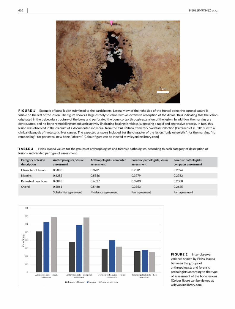
F IGURE 1 Example of bone lesion submitted to the participants. Lateral view of the right side of the frontal bone; the coronal suture isvisible on the left of the lesion. The figure shows a large osteolytic lesion with an extensive resorption of the diploe, thus indicating that the lesionoriginated in the trabecular structure of the bone and perforated the bone cortex through extension of the lesion. In addition, the margins aredenticulated, and no bone remodelling/osteoblastic activity (indicating healing) is visible, suggesting a rapid and aggressive process. In fact, this
lesion was observed in the cranium of a documented individual from the CAL Milano Cemetery Skeletal Collection (Cattaneo et al., 2018) with aclinical diagnosis of metastatic liver cancer. The expected answers included, for the character of the lesion, “only osteolytic”; for the margins, “noremodelling”; for periosteal new bone, “absent” [Colour figure can be viewed at wileyonlinelibrary.com]
TABLE 3 Fleiss' Kappa values for the groups of anthropologists and forensic pathologists, according to each category of description oflesions and divided per type of assessment
Category of lesion
description
Anthropologists, Visual
assessment
Anthropologists, computer
assessment
Forensic pathologists, visual
assessment
Forensic pathologists,
computer assessment
Character of lesion 0.5088 0.3781 0.2881 0.2594
Margins 0.6252 0.5856 0.3979 0.2782
Periosteal new bone 0.6843 0.6827 0.3200 0.2500
Overall 0.6061 0.5488 0.3353 0.2625
Substantial agreement Moderate agreement Fair agreement Fair agreement
F IGURE 2 Inter-observervariance shown by Fleiss' Kappa
between the groups ofanthropologists and forensicpathologists according to the typeof assessment of the bone lesions[Colour figure can be viewed atwileyonlinelibrary.com]
610 BIEHLER-GOMEZ ET AL.

between participants from fair to perfect agreements (Landis &
Koch, 1977). The average values indicate a slightly greater agreement
in the group of forensic pathologists and for the “periosteal new
bone” category.
3.3 | Accuracy rates
As seen in Table 5, the highest accuracies of both groups were
obtained in the “margins” category and the lowest in the “periosteal
new bone” category for the forensic pathologists and “character of
the lesion” category for the anthropologists, regardless of the type of
assessment used. Overall, “visual” assessment of bone lesions was
more often correct than when computer-based and evaluations made
by the anthropologists were slightly more accurate than in the group
of forensic pathologists. The accuracy rates of the positive identifica-
tion of the different types of periosteal new bone (Table 6) show poor
results, close to 65% (e.g., a periosteal new bone was described when
it was absent or vice versa or the wrong type of periosteal new bone
was noted as present). In addition, about one in two woven or lamellar
new bone depositions (the most commonly found types of periosteal
new bone) were wrongly identified (51.1% positive identification).
3.4 | Influence of the experience of the participants
Table 7 shows the correlation between the experience of the partici-
pants and their accuracy rates. Surprisingly, participants with less
experience showed an overall accuracy in the description of bone
lesions slightly higher than participants with 5 years of experience and
even 8–10 years of experience. Indeed, participants with 3 years of
experience showed a mean accuracy rate of 72.76% on visual assess-
ment and 69% on computer assessment, whereas the accuracy rate of
participants with 5 years of experience was 62.29% on real specimens
and 55.78% on photographs and that of the participants with
8–10 years of experience was 69.72% on visual assessment.
3.5 | Consistency in the assessment of the lesions
Inconsistencies between the assessment of character of the lesion
and their descriptions were noted. For instance, seven participants
(four anthropologists and three forensic pathologists) noted some
lesions as either osteolytic or osteoblastic (by opposition to man-
ifesting both components) and then listed the presence of periosteal
new bone/remodelled margins or bone resorption, respectively, which
contradicts the previous assessment of a lesion solely osteolytic or
osteoblastic.
4 | DISCUSSION
The interobserver agreement analysis shows that anthropologists
tended to agree more in their description of lesions than the forensic
pathologists in our study (Table 3). In addition, they were slightly more
correct in their description of bone lesions than the participants with
a medico-legal background (Table 5). These results were expected as
the anthropologists are more frequently confronted to skeletal
TABLE 4 Cohen Kappa values for all participants per category of description of lesions. All Cohen Kappa values were significant withα = 0.05
Category of lesion description
Participants of the group of anthropologists Participants of the group of forensic pathologistsa
1 2 3 4 5 Avg. 6 7 8 9 Avg.
Character of lesion 0.632 0.850 0.799 0.818 0.441 0.544 0.949 1.000 0.341 0.351 0.660
Margins 0.639 1.000 0.847 0.947 0.551 0.743 0.947 0.947 0.273 0.533 0.675
Periosteal new boneb 0.735 0.860 0.925 0.639 0.753 0.782 0.988 1.000 0.622 0.658 0.817
Total 0.690 0.717
aParticipant no. 10 did not perform the “visual assessment” of lesions and so the intraobserver agreement could not be calculated.bFor the Cohen Kappa calculation of multiple answers in the “periosteal new bone” category, partial agreement was not considered, the answers had to be
in the exact same combination to be seen as correct.
TABLE 5 Accuracy rates for the computer-based and “visual” assessments of the characteristics of bone lesions between the groups ofanthropologists and forensic pathologists
Category of lesion description
Anthropologists Forensic pathologists
Computer assessment (%) Visual assessment (%) Computer assessment (%) Visual assessment (%)
Character of lesion 64.33 65.83 66.67 66.67
Margins 80.67 80.00 70.67 80.67
Periosteal new bone 64.53 67.83 57.67 62.50
Total 69.84 71.22 65.00 69.95
BIEHLER-GOMEZ ET AL. 611

remains than forensic pathologists and so they have higher probabili-
ties to have been confronted to bone lesions, thus explaining a better
accuracy in their assessment.
Participants generally tended to describe lesions similarly whether
they performed the analysis based on photographs or by visual exami-
nation of the actual osseous elements (Table 4). Their answers were
more similar on “visual” assessment and were slightly more accurate
than when assessed on photographs (Tables 3 and 5). Again, these
results were expected because details of bone lesions may be more
difficult to recognize on photographs due to the constraint of defini-
tion and the lack of perspective. In addition, they demonstrate that
visual observation should be preferred to photographs when record-
ing bone lesions.
Participants tended to disagree on their assessment of the char-
acter of the lesions, without substantially changing their answers
between modalities of assessment (Tables 3 and 4). It is interesting to
see that this category, which is the initial interpretation of the lesion
and may significantly impact the diagnosis of the disease responsible
for the lesion, showed so much variation between participants and
such a low accuracy (varying between 64% and 67%; Table 5).
Another interesting finding was that in a total of 19 instances, the par-
ticipants evaluated lesions to be solely osteolytic or osteoblastic but
also recorded the presence of bone production or bone resorption,
respectively, in other categories. These variations, low accuracies and
contradictory assessments testify to a need for continuous training in
the description and recording of bone lesions.
The margin category was limited to a binary response: either
rounded and remodelled or not remodelled (Table 2); it is therefore
understandable that the results of this category were more similar
between participants and modalities (Tables 3 and 4) and the most
accurate results (Table 5). Nonetheless, they also show that the
descriptions of the pathological changes at the margin are the easiest
to interpret.
Contrary to the margin category, the interpretation of periosteal
new bone posed more difficulties. Although the participants showed
some variation in their results between themselves (in particular in the
group of forensic pathologists, which may be due to the fact that they
are less frequently confronted to pathologic lesions on dry bones than
the group of anthropologists, resulting in more uncertainty) and chan-
ged very little their results between modalities, this category is also
the one with the poorest accuracy (ranging between 58% and 68%
and with a 57% to 69% rate of positive identification). In fact, absent
periosteal new bone (i.e., the absence of a new bone deposition on
the cortical surface) was correctly identified in only 71% of cases.
Overall, woven and lamellar new bone (the most commonly encoun-
tered types of periosteal new bone) were correctly identified about
half of the time (51.1%; Table 6). In two lesions in particular, we could
understand that the participants mistook spongiosclerosis (observed
in the trabecular bone exposed by an osteolytic perforation of the
bone cortex) for the presence of a periosteal new bone. These results
demonstrate a poor understanding of the terminology associated to
the different types of periosteal new bone, which is particularly
alarming considering that these may considerably orient the under-
standing of the disease process and the diagnosis of the causative
agent responsible for the lesion. Ortner (2012, p. 252) said that “care-
ful attention to the characteristics of the abnormal features created
by the bone cells provides critical information basic to the description
and diagnosis of all skeletal disorders.” However, and as highlighted in
this study, there is still substantial variation, disagreement and inaccu-
racies in the description of bone lesions. Consequently, the under-
standing of the characteristics of bone lesions and the standardization
of their description remain a crucial topic to address for the correct
recording of bone lesions, the documentation of comparable data and
the accurate diagnosis of bone disease.
The study also considered the possible influence of the experi-
ence, specialization and training of the participants on the results
obtained. Indeed, one expects specialized observers with a long expe-
rience to fare much better in the description of bone lesions than
young practitioners confronted for the first time with this situation.
The study thus involved 10 participants with various experience on
dry bone (which can be divided in three groups: 3, 5 and 8–10 years),
trained in western Europe (Italy, England, France and Switzerland)
including five forensic pathologists (specialized in forensic pathology
and forensic anthropology) and five anthropologists (with specializa-
tion in forensic anthropology as well as other fields including bio-
archaeology, forensic genetics and forensic sciences; Table 1). As a
result of the study, the anthropologists showed substantial agreement
in their description of bone lesions on real specimens (Fleiss' Kappa
value: 0.6061) despite the variation in their years of experience and
fields of specialization, suggesting that these factors did not influence
their analysis. The forensic pathologists only showed a fair agreement
in their results (Fleiss' Kappa values: 0.3353 and 0.2625) in spite of
TABLE 6 Accuracy rates for the positive identifications in the subcategories of periosteal new bone between the groups of anthropologistsand forensic pathologists
Subcategories of periosteal new bone
Anthropologists Forensic pathologists
Computer assessment (%) Visual assessment (%) Computer assessment (%) Visual assessment (%)
Absent 77.50 76.25 60.00 70.31
Woven bone 46.67 55.56 40.00 44.44
Lamellar bone 57.78 62.22 57.78 44.44
Spiculated 80 80 70 100
Avg. 65.49 68.51 56.95 64.80
612 BIEHLER-GOMEZ ET AL.
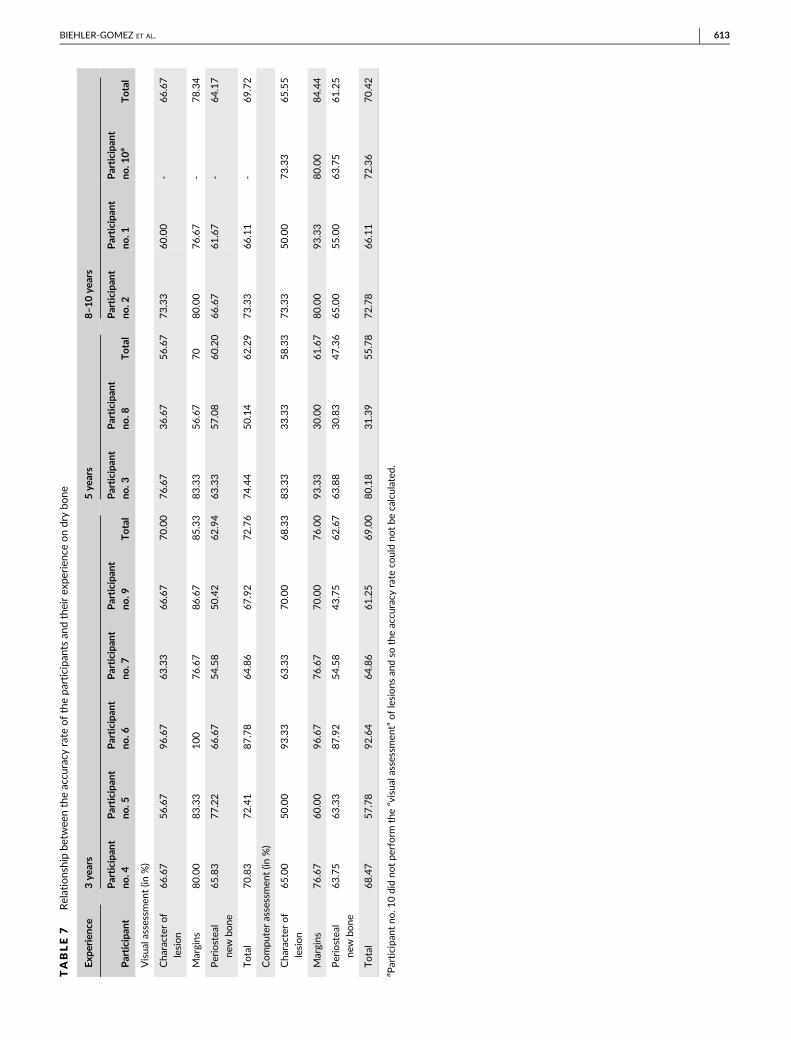
TABLE7
Relationshipbe
twee
ntheaccu
racy
rate
ofthepa
rticipan
tsan
dtheirex
perien
ceondrybo
ne
Exp
erienc
e3ye
ars
5ye
ars
8–1
0ye
ars
Participa
ntParticipa
ntno
.4Participa
ntno
.5Participa
ntno
.6Participa
ntno
.7Participa
ntno
.9Total
Participa
ntno
.3Participa
ntno
.8Total
Participan
tno.2
Participan
tno.1
Participan
tno.1
0a
Total
Visua
lassessm
ent(in
%)
Cha
racter
of
lesion
66.67
56.67
96.67
63.33
66.67
70.00
76.67
36.67
56.67
73.33
60.00
-66.67
Margins
80.00
83.33
100
76.67
86.67
85.33
83.33
56.67
70
80.00
76.67
-78.34
Periostea
l
new
bone
65.83
77.22
66.67
54.58
50.42
62.94
63.33
57.08
60.20
66.67
61.67
-64.17
Total
70.83
72.41
87.78
64.86
67.92
72.76
74.44
50.14
62.29
73.33
66.11
-69.72
Compu
terassessmen
t(in
%)
Cha
racter
of
lesion
65.00
50.00
93.33
63.33
70.00
68.33
83.33
33.33
58.33
73.33
50.00
73.33
65.55
Margins
76.67
60.00
96.67
76.67
70.00
76.00
93.33
30.00
61.67
80.00
93.33
80.00
84.44
Periostea
l
new
bone
63.75
63.33
87.92
54.58
43.75
62.67
63.88
30.83
47.36
65.00
55.00
63.75
61.25
Total
68.47
57.78
92.64
64.86
61.25
69.00
80.18
31.39
55.78
72.78
66.11
72.36
70.42
aParticipa
ntno
.10didno
tpe
rform
the“visua
lassessm
ent”oflesionsan
dso
theaccu
racy
rate
couldno
tbe
calculated
.
BIEHLER-GOMEZ ET AL. 613
having the same specialization. In this group, although the specializa-
tion cannot be a determining factor, the different level of experience
of the participants may explain the variation in the results. Yet Partici-
pants 8 and 10, with, respectively, 5 and 10 years of experience, did
not evidence a higher accuracy rate than Participant 6, with only
3 years of experience on dry bone (Table 7). In fact, Table 7 demon-
strates that the participants with less experience (3 years; Participants
4, 5, 6, 7 and 9; accuracy rates: 72.76% in real specimens and 69% on
photographs) had an overall slightly better accuracy in the description
of bone lesions than those with a longer experience (5 years; Partici-
pants 3 and 8; accuracy rates: 62.29% on real specimens and 55.78%
on photographs; and 8–10 years; Participants 1, 2 and 10; accuracy
rate: 69.72% on real specimens). Finally, Participants 6 and 8 are
forensic pathologists trained in Italy, yet the former shows the highest
accuracy rates of the study and the latter the poorest (Tables 1 and 7).
Therefore, the variation of results observed in this study cannot be
explained by differences in years of experience, fields of specialization
or even geographical place of training of the participants. It is proba-
ble that specific training in bone pathology would have increased the
accuracy rates and reduced the variation observed in our results.
Nonetheless, we decided not to train the participants beforehand in
order not to skew the results and give an authentic representation of
real-life scenarios, to which any of the participants selected for this
study could be confronted (as describing bone lesions forms the basis
of diagnosis in bone disease). Consequently, a possible explanation for
these results is that the description of bone lesions constitutes a spe-
cific skillset that is not entirely obtained through the experience of
the forensic pathologist or anthropologist or their specialty in forensic
anthropology or bioarchaeology, despite their ability, range of knowl-
edge and practice. The results of this study thus testify to the need
for more specific and thorough training in the description of bone
lesions to all practitioners on dry bone, regardless of their field of spe-
cialization or experience.
Previous studies (Bridges, 1993; Waldron & Rogers, 1991) have
already shown the existing interobserver disagreement in the scoring
and analysis of bone lesions (specifically, due to osteoarthritis) and
alerted on the need for a standardized methodology in the analysis of
bone lesions. In the present study, however, we also evidenced critical
issues in lesion recording, in particular when assessing the character
of lesion (bone forming or bone destruction) as well as the presence
of periosteal bone forming lesions (regardless of the exact sub-
category), namely, inconsistencies, confusions and low accuracies. The
results of this exploratory study thus argue in favour of continuing
efforts in the training on the description of the characteristics of bone
lesions.
5 | CONCLUSION
The accurate description of bone lesions is an essential step for the
correct diagnosis of bone disease. In this study, we submitted 30 bone
lesions to 10 anthropologist and forensic pathologist participants. As
a result, anthropologists showed slightly more consistent and accurate
results compared with the forensic pathologists, and overall results
were better when assessed by visual examination of the bone lesions.
Lesion descriptions, however, showed important inaccuracies and
contradictions, particularly in the evaluation of the character of the
lesion and periosteal new bone, with dramatic potential consequences
for the diagnosis of bone disease. We can only recommend continuing
training and communication on the identification of the characteristics
of bone lesions for the correct recording of bone lesions, the docu-
mentation of comparable data and the accurate diagnosis of bone dis-
ease. This study shows the considerable pitfalls in the description and
classification of basic pathological bone manifestations and demon-
strates the importance of continuing efforts in the standardization of
pathological terminology on dry bone.
CONFLICT OF INTEREST
None.
FUNDING
This research did not receive any specific grants from public, commer-
cial and not-for-profit funding agencies.
ORCID
Lucie Biehler-Gomez https://orcid.org/0000-0001-6674-7850
Lara Indra https://orcid.org/0000-0001-6644-2133
REFERENCES
Appleby, J., Thomas, R., & Buikstra, J. (2015). Increasing confidence in
paleopathological diagnosis–Application of the Istanbul terminological
framework. International Journal of Paleopathology, 8, 19–21. https://doi.org/10.1016/j.ijpp.2014.07.003
Aufderheide, A. C., & Rodríguez-Martín, C. (1998). The Cambridge encyclo-
pedia of human paleopathology. Cambridge, United Kingdom: Cam-
bridge University Press.
Biehler-Gomez, L., Giordano, G., & Cattaneo, C. (2019). The overlooked
primary: Bladder cancer metastases on dry bone. A study of the 20th
century CAL Milano Cemetery Skeletal Collection. International Journal
of Paleopathology, 24, 130–140. https://doi.org/10.1016/j.ijpp.2018.10.005
Brickley, M., & McKinley, J. I. (2004). Guidelines to the standards for record-
ing human remains. Southampton: British Association for Biological
Anthropology and Osteoarchaeology and Institute of Field
Archaeologists.
Bridges, P. S. (1993). The effect of variation in methodology on the out-
come of osteoarthritic studies. International Journal of
Osteoarchaeology, 3, 289–295. https://doi.org/10.1002/oa.
1390030407
Buikstra, J. E., Cook, D. C., & Bolhofner, K. L. (2017). Introduction: Scien-
tific rigor in paleopathology. International Journal of Paleopathology, 19,
80–87. https://doi.org/10.1016/j.ijpp.2017.08.005Buikstra, J. E., & Ubelaker, D. H. (1994). Standards for data collection from
human skeletal remains. Arkansas: Arkansas Archaeological Survey
Report Number 44: Fayetteville.
Cattaneo, C., Mazzarelli, D., Cappella, A., Castoldi, E., Mattia, M., Poppa, P.,
… Biehler-Gomez, L. (2018). A modern documented Italian identified
skeletal collection of 2127 skeletons: the CAL Milano Cemetery Skele-
tal Collection. Forensic Science International, 287, 219.e1–219.e5.https://doi.org/10.1016/j.forsciint.2018.03.041
Grauer, A. L. (2008). Macroscopic analysis and data collection in
palaeopathology. In R. Pinhasi, & S. Mays (Eds.), Advances in human
614 BIEHLER-GOMEZ ET AL.

palaeopathology (pp. 57–76). Chichester, England: Wiley Online
Library.
Klaus, H. D. (2017). Paleopathological rigor and differential diagnosis: Case
studies involving terminology, description, and diagnostic frameworks
for scurvy in skeletal remains. International Journal of Paleopathology,
19, 96–110. https://doi.org/10.1016/j.ijpp.2015.10.002Landis, J. R., & Koch, G. G. (1977). The measurement of observer agree-
ment for categorical data. Biometrics, 33, 159–174. https://doi.org/10.2307/2529310
Lovell, N. C. (2000). Paleopathological description and diagnosis. In
S. R. Saunders, & A. M. Katzenberg (Eds.), Biological anthropology of the
human skeleton (pp. 217–248). New York, USA: Wiley-Liss New York.
Manchester, M., Ogden, A., Storm, R. (2016). Nomenclature in paleopathol-
ogy. Paleopathology Association Newsletter.
Mays, S. (2018). How should we diagnose disease in palaeopathology?
Some epistemological considerations. International Journal of Paleopa-
thology, 20, 12–19. https://doi.org/10.1016/j.ijpp.2017.10.006Miller, E., Ragsdale, B. D., & Ortner, D. J. (1996). Accuracy in dry bone
diagnosis: A comment on palaeopathological methods. International
Journal of Osteoarchaeology, 6, 221–229. https://doi.org/10.1002/(
SICI)1099-1212(199606)6:3<221::AID-OA267>3.0.CO;2-2
Mitchell PD, Brickley M. 2017. Updated guidelines to the standards for
recording human remains. Chartered Institute for Archaeologists.
Ortner, D. J. (2003). Identification of pathological conditions in human skele-
tal remains (3rd ed.). New York: Smithsonian Institution Press.
Ortner, D. J. (2011). What skeletons tell us. The story of human paleopa-
thology. Virchows Archiv, 459, 247–254. https://doi.org/10.1007/
s00428-011-1122-x
Ortner, D. J. (20120. Differential diagnosis and issues in disease
classification. In A. L. Grauer (Ed.). A companion to paleopathology
(pp. 250–267). Chichester, England: Wiley Online Library. https://doi.
org/10.1002/9781444345940.ch14
Ragsdale, B. D., Campbell, R. A., & Kirkpatrick, C. L. (2018). Neoplasm or
not? General principles of morphologic analysis of dry bone
specimens. International Journal of Paleopathology, 21, 27–40. https://doi.org/10.1016/j.ijpp.2017.02.002
Roberts, C. A., & Connell, B. (2004). Guidance on recording
palaeopathology. In Guidelines to the standards for recording human
remains (Vol. 102) (pp. 34–39). Southampton: British Association for
Biological Anthropology and Osteoarchaeology and Institute of Field
Archaeologists. https://doi.org/10.1063/1.2756072
Santos, A. L., & Roberts, C. A. (2006). Anatomy of a serial killer: Differential
diagnosis of tuberculosis based on rib lesions of adult individuals from
the Coimbra identified skeletal collection, Portugal. American Journal of
Physical Anthropology, 130, 38–49. https://doi.org/10.1002/ajpa.
20160
United Nations. (2004). Istanbul protocol manual on the effective investiga-
tion and documentation of torture and other cruel, inhuman or degrading
treatment or punishment. New York/Geneva: Office of The United
Nations High Commissioner for Human Rights.
Waldron, T., & Rogers, J. (1991). Inter-observer variation in coding osteo-
arthritis in human skeletal remains. International Journal of
Osteoarchaeology, 1, 49–56. https://doi.org/10.1002/oa.1390010107
SUPPORTING INFORMATION
Additional supporting information may be found online in the
Supporting Information section at the end of this article.
How to cite this article: Biehler-Gomez L, Indra L, Martino F,
Campobasso CP, Cattaneo C. Observer error in bone disease
description: A cautionary note. Int J Osteoarchaeol. 2020;30:
607–615. https://doi.org/10.1002/oa.2885
BIEHLER-GOMEZ ET AL. 615