Observations on the innervation of the macula sacculi in man
16
OBSERVATIONS ON THE INNERVATION OF THE AIACULA SACCULI IN RZAN MARY HARDY Otological Research Laborator?/, Jo7~is.s Hopkins Univcrsit!/ NINE FIGURES INTRODUCTION The early anatomists described thc sawulns of mammals as being innervated entirely by the R. sii(*cularis,the major branch of the inferior division of the vestibular nerve. In 1907 Voit described a second nerve to the sacculus coming from the superior division of the vestibular nerve. De Burlet ( '24) and l'oliak ( '27) confirm Voit's description of a double innervation of the sacculus. We find, in man, a third nerve to the macula sacculi that separates from the cochlear nerve in the region of the spiral ganglion near its basal end. Our observations indicate that this third nerve to the macnla sac- culi is a true branch of the cochlear division of the VIIIth nerve, but do not entirely rule out the possibility that it may be an aberrant branch of the vestibular nerve. It is defi nitely distinct from the vestibnlo-coclilear anastomosis oi' Oort ( 'IS), and apparently has not been previously described. De Burlct's schema of the distribution of the acoustic nerve in the labyrinth is given as figure 1, with the addition of this third saccular nerve. This paper is concerned primarily with a description of this branch, which we have designated as the cochleo-saccnlar nerve. MATERIAL AND METIIOD The material for this study consists of serial sections of adult human temporal bones. large group of cases with every fifth section stained (haematoxylin and eosiii, 24 p) was previously used for a study of the spiral ganglion. It was in the course of counting the ganglion cells in these sec- 403
-
Upload
mary-hardy -
Category
Documents
-
view
214 -
download
2
Transcript of Observations on the innervation of the macula sacculi in man

OBSERVATIONS ON THE INNERVATION OF THE AIACULA SACCULI I N RZAN
MARY HARDY Otological Research Laborator?/, Jo7~is.s Hopkins Univcrsit!/
NINE FIGURES
INTRODUCTION
The early anatomists described thc sawulns of mammals as being innervated entirely by the R. sii(*cularis, the major branch of the inferior division of the vestibular nerve. In 1907 Voit described a second nerve to the sacculus coming from the superior division of the vestibular nerve. De Burlet ( '24) and l'oliak ( '27) confirm Voit's description of a double innervation of the sacculus. W e find, in man, a third nerve to the macula sacculi that separates from the cochlear nerve in the region of the spiral ganglion near its basal end. Our observations indicate that this third nerve to the macnla sac- culi is a true branch of the cochlear division of the VIII th nerve, but do not entirely rule out the possibility that it may be an aberrant branch of the vestibular nerve. It is defi nitely distinct from the vestibnlo-coclilear anastomosis oi' Oort ( 'IS), and apparently has not been previously described. De Burlct's schema of the distribution of the acoustic nerve in the labyrinth is given as figure 1, with the addition of this third saccular nerve. This paper is concerned primarily with a description of this branch, which we have designated as the cochleo-saccnlar nerve.
MATERIAL AND METIIOD
The material for this study consists of serial sections of adult human temporal bones. large group of cases with every fifth section stained (haematoxylin and eosiii, 24 p ) was previously used for a study of the spiral ganglion. It was in the course of counting the ganglion cells in these sec-
403

404 MARY T1AT:DY
tioiis that tlie coclileo-saccnlar nerve m i b first obstxrvcd. Tlicsc wcrc all sectioned in tlic vertical plane. Series cut horizontally and otlicrs in a plaiic ~)erpcndicular t o tlic modi- olar axis were also examined. Iii vertical scries tlie buiiclles of this nerve a re freqixiitly cut loiigitndiiially. Six cases with the most favornl~lc oriciitatioii were selected for detailed study. Iiitermediatc scc+tioiis w ~ r e stained with Weigcrt 's hematoxylin.
/ 1 hmn hor I / I I \ \
N. Ve~tibular;~
N.Cochlrdr
Fig. 1 De Burlet's sclienia of the innerration of the mcmhranous lnl)p5iith, with the :iilditioii of the third iierve to the iiiaeuln saceuli (iiidiwted by tlie arrow). The iierves desc+ril)cd bp Voit and bp Oort are labeled 4 and 5, rcspce- tivelg.
Draiviiigs of altcriiatc sections, magnified to 6 i diameters, were made oil tracing pwper wit11 the aid of a projectioii laii- tern. By superimposing these tracings the courses of all nerve bundles in the region were followed.
OBSERVATTONS
Sections in the verticwl plane a millimeter or less from the vestibular end of thcl cochlca sliow small bundles of nervc
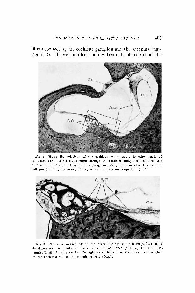
fibres coiiiicctiiig tlic cochlear ganglion ant1 the sacciilns (figs. 2 aiid 3 ) . Tlic~sr biiiiclles, comiiig from tho clirectioii of tlic
Fig. 2 Shows tire relatioiis of tlic eochlco-saecular nerve to other parts of tlie iniicr ear in a vertical section tlirougli the anterior margiii of the footi)late of tlici stapes (St.). C.G., cochlear ganglion; Sac., saeculus (the free i~:iIl is collapsed) ; Utr., utriculus; R.p.a., iierve t o posterior ampulla. x 15.
F ig .3 The area marked off iii the precetling figure, a t :I iiiagiiifieation of 44 diameters. A bundle of tlic coclileo-saceular iierre (C. S.13.) is cu t ;rlniost 1oiigitudiii:illy iu this scictioii tlirough its eutire course from coclilcar ganglion t o the posterior t ip of the macula saceuli (M.s.).

406 MARY HARDY
cocahlea, pass under the posterior t ip of the macula saccnli. Here they spread out and the fibres mingle with those from the R. saccularis iiifcrior. Recanse of this plexus formatioil arid because with our tecliiiique single fibres cannot he traced to hair cell terminations, it is not possible to state tlie csact arcs of the macula supplied by the eoclileo-saccnl~ir nerve. To reach the saccnlus tlie nerve bundles coiirse tllrongh qmall chwnncls in tlie ridge of 1)oiie between coclilca and recc spliaericns ; the caiialis reuiiieris lies on the siirface of this ridge slightly posterior to these nerve canals. The general course of the nerve is upward aiid m e d i a l ~ ~ r d i i i the itlalie of the vertical sections. There arc usually several ver? small hmiclles of fibres in discrete osseoiis canals. The length in these caiials is about 4 to 3 mm. Some sectioiis slioiv huiidlcs exteiidiiig on into tlie membranons wall of the sncculns for. ail additional fraction of a millimeter before changing direc- tion. The longest bmidle \ re hare seen in oiie section is about I A mm. I n tlie ganglionic canal the fibres of these bundles caii be seen passing among the scattered ganglion cells of this par t of the spiral gaiiglioii, wliere they mingle wit11 tlie fibres that snpply the organ of ('orti. Some gaiiglioii cells liavc pcriplieral 1)roeesses extending toward the osseoiis channels through wliicli tlie bundlcs leave tlie gaiiglioiiica c~anal, bnt it is, of course, not possible to follow any oiie fibre from ganglion cell to macnla. Since the term is ann- tomically descriptive, this g1-0111) of small nerve bundles which
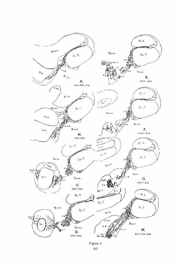
Fig. 4 Outline drnwiiig\ showing the courses of the several rierves of the regiou, as fo l low4 iii scrinl sections (vc,rtical plaiic) of the right w r of it
.>a-year-oltl mau. Outliiies A to G werc c:i(.Ii niade by superinipo\iiig tllc tr:rciiigs of five scetioirs (tlie alteriintr OIICS for a block thickness of 240 p ) ; 11 is a composite f rom nltcw:itc scc*tioiii for :L 1)loc.k thickiicw of 720 p. The eight outliiirs wprwoiit a total I)lock tliickirrss of 0.4 mm. iii the region of tlie baq:il c~ntl of c o d i l r : ~ ant1 the sacculn.;. c . , small bundle of corhlear nerve ; C.G., coch1e:ir gaiiglioii ; C.N., cochle:ir nerve, loivcr basal braiich ; (".-S.B., cochlro- siccular buiidlc ; T).("., tluctus coclilcnris ; T A M . , intcriinl auditory nicntus ; M.C.M., niac~ul:~ cri1,io.a iricrlia ; 0 's.R., Oort 's huiitlle ; R.P.A., rnmuq posterior iimpullaris ; K.R., minus s:icwlaris ; r.s.', Y.s.', ramus sncculnris, s~nall br:lnclles; R.S:I(~.SII~P., T:IIUIIS s:rccularis superior (Voit) ; Sac., saccnlus; SC.~'., srala tgm- pani ; Rc.V., scala rrstibuli; V., vcii:i spirale posterior ; Vest., pcrilymphatic s1~ac.c of \ c~t i l )u l r .
- -

408 MARY HARDY
coiirse from coclilear ganglion region to niacnla sacculi has been dcsigna ted as the cochlco-saccular nerve.
Although it is not possible to trace individual fibres in our material, it is fairly easy, b~ the method of superimposiiig tracings, to follow the general course of tlic nerve bnnd lc~ from the saccnlus back to the internal meatus. By this method i t can he demonstrated, 1) that the coclilco-s~iccnlar nerve is apparently a branch of the cochlear nerve, and, 2) that it is distinct from the aiiastomosis of Oort. From the region of the ganglionic canal centralward the fibres of the cochlco-saccular bundles are so mixed with those of the cochlear nerve proper that wc are nnable to distingnish them as separate bundles. On the other hand i t is easy to follow the hundlcs of Oort ’s vestibulo-cochlear aiiastomosis from the region of S c a r p ’ s ganglion to the coclilear ganglion. Figlire 4 illustrates both these points. Tlie coclileo-sacc.nlar fibres are seen in 9 and B passing through small bony canals from the saccnlns to the cochlear gaiiglion. In C the gang- lionic canal which these fibres have entered is sepnratccl from the saccvlus by a portion of the bony cnpsnlc. From this point the course of the iierve may be followed medialwird into the internal auditory meatus. The composite tracings partially misrepresent thc actual coiirse of the individual twigs because it is impossible to express clearly in a line drawing their devious pathways through narrow bony chan- nels from the ganglionic canal to tlic basal end of the area cribrosa spiralis. One bundle of fibres is laheled c (D-H) as a landmark to carry over from one figure to another until it joins the lower basal branch of the cochlear nerve. I+om this material and method of study, we judge the cochleo- saccular nerve to be a branch of the cochlear nerve.
The vcstibnlo-cochlear anastomosis which Oort ( ’1 8) cle- scribed for the rabbit and other mammals and also for a 7-montli lluman fetus, is in the internal auditory meatus and estcwls from Scarpa’s ganglion, pars inferior, to the area cribrosa spiralis. Here i t mingles with bundles of the coch- lear nerve as they enter the g::anglionic canal. I t is easy to

I N N E R V A T I O N O F MACULA SACCULI I N M A N 409
identify this anastomosis in most of our series of adult human temporal bone sections. Figure 4 H shows the course of Oort’s bundle which, for this particular ear, was traced through thirty sections. In some series the aiiastomosis is so cut that the greater part is caught in a single section, as illustrated in figure 2. The bundles of Oort ’S aiiastomosis enter the ganglionic canal in a region about 3 mm. from the
Fig. 5 I’liotoniicrogr:1~~1i of a vertical section through the posterior part of fundus of internal auditory meatus. X 15. This section shows Oort ’8 allasto- mosis (0.a.) as it passes from the pars iiiferior of Sc:irp:i’s ganglion (SC.~ . ) t o join the coclilear iicrve bundles. Rsac., rainus saccularis of the inferior division of the vestibular nerve; the superior division of this nerve is seen in the upper right-liand part of the figure.
basal end of the cochlea, which is 2 mm. or more anterior to that part of the gaiiglion associated with the cochleo- saccular nerve. Beyond their entrance into the ganglionic canal the fibres of Oort’s nerve are indistinguishable in our preparations from those of the cochlear nerve. It is very possible, as Lorerite de No (’26) maintains, that Oort’s anas- tomosis is an aberrant branch of the cochlear nerve. Cer- tainly it is quite distinct from the cochlco-saccnlar nerve.

410 MARY H A R D Y
The main saccular iierve, the R. snccnlnris of the inferior tlivisioii of the vcstibnlar, caii be followed in tlic sei-ics of seclioiis traced in figure 4 from the sacculns through the mncula crihrosa media to the internal auditory moatiis and Scarpa ’s ganglion. The small hnndles, r.s.’ aiicl i ~ . s . ~ , are labeled for lanclmai~lts to carry over fiaom O I I C figui~> to another. The bundle r.s.’ is a11 example of a coml);irat ively long branch of the saccnlar nerve that separates from the
main triiiik and reaches tlie saccnlus through a separate bony canal. l’oliak (’27) gives a good description of the varions and many ramifications, such as this one, of the R. sac,cnlaris.
The distribution in the macula sacculi of Voit’s iierre, the saccular branch of the pars superior, is indicated in fignre 4, C and D. A photomicrograph of a section passing throiigh both anterior and 1)osterior portions of tlie macula is givcn as figure 6. A bundle of T’oit ’s 11e1~vc and fibres from thc R. sac-

INNERVATION O F MACULA SACCLTIJ I N MAN 411
ciilaris inferior are seen terminating under the anterior and posterior portions of the macula, respectively. We arc able to iclciilify Voit's nerve in cvcry series of sections, and thus can confirm for the adult liuman Voit 's descriptioii of tlie innervation of the mammalian saccnliis from thc suljerior as me11 a s from the inferior division of the vestibnlar nerve.
The drawing by Alr. Mas Brodel (fig. 7 ) shows in a semi- diagrammatic form the iniiervation of the inner ear in man. The figure is based not oiily on the study of serial sections, but also on casts of the bony labyrinth and on special clis- sections made by Mi.. Briitlel f o r this pinpose. Tlie honnd- arics of the internal auditory meatus arc not sliomi, nor is the tlivision of the nerves into small bundles a s tlicy j x ~ s s through the cribrosal areas. Slight inaccuracies of relations of some par ts to each other have heen introduced in order to show clearly Ilie three iiervcs to the sacculiis ;ind also Oort's ~.estihulo-cochlcar anastomosis. Tlie macnla of the sawilus is oiitliiied by Ilie eiidiiigs shown for the three nei'ves: N.sac.maj., from tlie inferior division of the vestihnlar nerve ; R.sa<*.sup. (Voit) , from the superior division of the vestihnlai.; and IZ.cocli.-sacc., from the cochlear nerve. Tlie lai*gei. p i a t
of the macula, the posterior portion slipplied by tlie main saccular nerve, lies from this direction of view just under the sacculus, and is represented as seen through the saccular wall. Tlie coclileo-saccular iierve (R.coc2il.-sacc.) is seen, as described in this paper, connecting the posterior t ip of the macula sacculi with the spiral ganglion (gang1.spir.coch.) near the basal end of the cochlea. The drawing shows the location of this nerve with reference to canalis reuniens, Oort 's anastomosis (R.vest.-cochl.) and the labyrinth as a whole.
The fact that Voit's nerve enters the anterior portion of the macula, the N. saccnlaris thc larger posterior portion, and the cochleo-saccnlar nerve the t ip of the 1)osterior por- tion, suggests a possible relation hetween the three zone's of different-sized nerve fibres in the macula described by Loreiite de KO ('at;), and these three diff'erent ncrvc bundles

Fig
. 7
A
scni
i-tli
agra
inm
natir
dra
wii
ig o
f th
e ii
iiic
rvat
ioii
of
the
lahy
riii
tli.
Tlie
or
ieii
tati
oii
of
tlie
th
rcc
nerv
vs t
o th
e ni
ncnl
a sa
wul
i is
ciii
phas
ized
; &
o tl
ic i
iidcp
cnde
itw
ot
Oor
t 's
iier
vc :
nid
tlie
eoc
lileo
- s:
rcc.
ular
iie
rve.

INNERVATION OF MACULA SACCULI I N MAN 413
supplying the sacculus. Figure 8 is de No’s schema of tlic fibre pattern of the mammalian maciila saccnli. There is somc intermingling of the three liinds of fibrcs, as Poliali (’27) also poiiits ou t ; tliat is, the boundaries of thc zoiies are not wcll defiiied. B u t , in general, there are thick fibres ( a ) in the aiitcrior par t of the macula, medinm-sized fibres ( h ) iii the main par t of tlie largcr posterior port ion, and tliiii fibres ( c ) in tlie posterior t ip and snlwrior mai*giri of tlic posterior part . hleasnr-emciits of the iiei*re fibres in our see- tioiis (human) show that on the average tliosc in Voit’s iicrve a rc l a i p r than those iii tlic niaiii snccnlar nerve, a i d the fibrcs of tlic coc~hleo-saccnlar iicrvcl are tliimier than those in (lither of tlic other nei*ves to the macula.
X
Fig. 8 Lorcnte tle No’s scl~ema of the tlistribution of nerve fibres of cliffcrcnt a, tlrick fibres; b, medium; c, thin. s izci in tlic mac.nla ~ : i t ~ c . n l i .
Tlie coclilco-saccular nerve varics in size and has iiot been idciitified in every case. I n a group of 85 ears, 20 of which had every tenth section stained and 65 cvcry fifth, one or more sections of 36 cars sliowcd a t least a tracc of this iiervc, mhilc in 49 it w a s iiot foiiiid in the sections examined. That is, evi- dence of the presciicc of the cochlco-saccnlar iierve was fouiid in about 40 per cent of this group of cases. T h e e concln- sioiis are permissible: 1 ) tliat tlic nerve is iiot of coiistaiit oecurrence; 2 ) that i t is only an aberrant braiicli of tlie inferior division of the vestibular iicrve; or, 3 ) that i t is normally present but because of i ts small size is frequently missed nnless all scctioiis of the series bc examined.

DISCUSSION
The sacculus is generally considerecl a par t of the vcstibu- lar, as distinct from the auditory, apparatus of the inner. car. The morphologic conacetion between the cochlear nerve and the sacdi i s , however, as described in this paper, calls to mind other observations whicli suggest that the sacculus is perhaps more closely associated with the cochlea than with the ntriculns and the semicircular canals. The specific role played by tlie sacculus in the whole laybyrinthine function has been consictcred auditory by some observers and equilib- rial by otlicrs. The exact fnnction of the sacculiis iii the higher vertebrates has long 1)nzzled the physiologist, and is still an unsettled question.
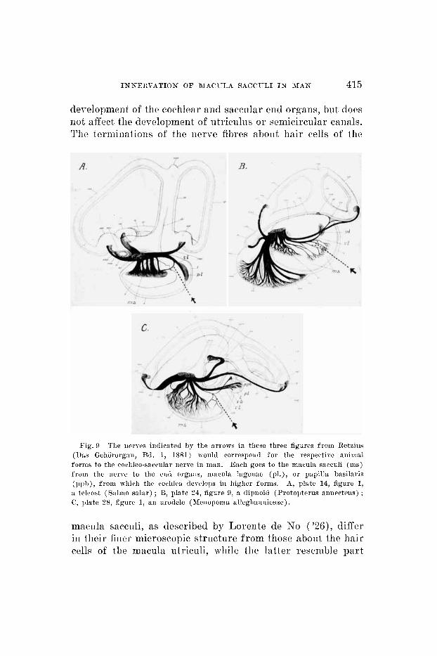
Although a cochleo-saccular iicrve has not previously been dcscribctl for mammals a cwrresponcling branch is foiind in some species of tlic lower forms of wrtebrates. Figure 9 is a reproduction of three drawings from Rctziiis (1881) of the labprinths of a tcleost, a lung fish and an urodelc, rcspec- tivcly. The nerves we have indicated by arrows bccnnse they are riot labeled or mciitioned in tlie test by Rctzins, hraiich to the macula sacculi (ms) from the ramulus lageiiae (1-1) or from the ramulns basilaris ( rb) . Since the lageiia ant1 the papilla basilaris are the phylogenic precursors of t h ~ mammalian coclilea, the analogy between these branches and the coclileo-saccnlar nerve found in man is apparent.
Ontogcneticallp, as well as ph;vlogcnetically, tlie saccnlus is tlie primary strnctnrc. I n one direction the utriculus and the canals develop and finally become only indirectly coii- nected through the t lwtus cndolympliaticus ; in aiiothcir direc- tion the cochlea develop, ant1 maintains n direct commiinica- tion through tlic canalis i*eiiniens (fig. 1). The close relationship between the end organs of sacculus mid coclilea is illustrated in tlic case of the albino cat (Alesander, '00; Howe and Cxnild, '33, and others). The cristae of tlic canals and the macula ntricnli a i ~ noi*mal but both organ of Corti and macnla saccnli arc abnormal. The interesting point is that some factor, p ~ ~ ) h a b l p genetic, interferes with normal
INNF,RVATION O F M A C U L A S A C C U L I I N MAN 415
development of the cochlear and saccular end organs, but does not affect tlie development of utriculus or semicircular canals. Tlic terminations of the nerve fibres about hair cells of the
Fig. 9 l’lie nerves iiirlicatcd by the arrows in these three figurcs f rom Retzius (Ilas Gehiirorg:iii, Ed. 1, 1881) would correspond f o r the respective aiiiiiial fornis t o the coclileo-sawular nerve in man. E:ieh goes t o the macula sacculi (ins) froin the iicrve to tlie cut1 organs, inacnla hgenae (pl.), or pnpilla hasilaris (p l )h) , from which the cochlea (1eLclops in Iiiglicr forms. A, plate 14, figure 1, a tcleost (Salmo salar) ; G, plate 24, figure 9, a dipnoid (Protoptcrus ainiectciis) : C’, plate 28, figure 1, an urodele ( ~ k i i o p o m a allcghaiiiiiciisc) .
macula sacculi, as described by Lorcnte de No (’26)’ differ in their finer microscopic structure from those abont the hair cells of the mncula utriculi, ~ h i l e the latter resemble part

416 MARY HARDY
of the nerve endings in the cristae. Werner ( ' 3 3 ) , working with labyrinth preparations of guinea pigs, reports that tho thicliness of the macula sacculi varies with atmospheric pres- sure while the macula of the utriculus does not. This ITIIC-
tion of the macula sacculi to pressure changes is suggestive of a possible basic relationship between saccnlus and cochlea, in that the cochlear end organ has a specific sensitivity t o sound, and sound may be defined physically as a rhytlimic variation in pressure of a medium.
Interest in the function of tlic sacculus, whether it is con- cerned with sound vibrations or, like the canals and utricnlus, with equilibrium, has recently been revived. ;\lcNally and Tait ('as), in their ablation experiments on the frog, find absolutely no change in either static or kinetic eqiiilibiGiim when the saccular macula is destroyed. Ablation of the utricuhis or of any one of the canals docs cause definite dis- turbance of balance. Versteegh ( '27), working with rabbits, and Benjamins and Huizinga ( 'as), with pigeons, report simi- lar findings after extirpation of the sacculus. McXally ( '29) in a review on the physiology of the labyrinth states that, 1) there is no experimental evidence to show that tlie macula sacculi has any vestibular or equilibria1 function, either in mammals or in tlie lower vertebrates; and, 2 ) there is some experimental evidence that in fish the macula sacculi may be concerned with hearing. Tait ( ' 3 2 ) maintains that in fishes both the macula sacculi and the lagcna are auditory re- ceptors.
The qixestioii of the function of the sacculus is still nnset- tlcd; the eviclence to date has not proved i t t o h a v c h an equilibria1 function, nor has i t disproved the possibility of an auditory function. It may be that in tlie higher verte- brates the presence of the sacculus is only of phylogenetic and chmbryologic significance.
From ihe surgical viewpoint, the partial innervation in man of tlic macula sacculi by a branch from the cochlear nerve and of the organ of Corti by an anastomotic branch (Oort 's) from the vestibular nerve, is of 1)ract ical interest i n co1]11e('-

INNEItVATION O F MACULB SACCULI IN MAN 41 ’7
tion with Dandy’s ( ’33) o1)erativc proceclurc for the relief of MhniGre’s disease by section of part of the eighth nerve.
SUMMARY
A description is given of small bnndlcs of nerve fibres that from tlie cochlear ganglion, in a region near its basal
ciid, to tlie posterior til) of the macula sacciili, as seen in serial sections of adult liiiman temporal bones. The course of tliesc bundles is through small bony cliaiiiiels ahout a milli- meter in length. The gwera l direction from ganglion to mac- nla is iip\x-ard and medialwaid and approximately in the 1)Iaiic of the ‘vertical ’ sections. Tlie term coclileo-saccular nerve is proposed fo r this groiip of ncr re hi idles . T h e helief is es- pressed that t h i s nerve to the saccnlus is a triic braiicli o f the cochlear nerve, and not merely ail aiiastomotic twig from the vcstibiilar nerve.
The saccnlar iierve from tlic superior vestihular division described by Voit for mammals is of constant oeciirrencc in tlic adiilt liumari ear. Oort ’s vcstibnlo-cochlear anastomosis is identified in some but not in all series observed. The gcn- era1 course and location relative to the labpi*iiitli as a mhole is clescrihed for these two iierve hraiiches, as w-cll a s for tlie coclileo-s;icciilar iicrvc.
The macula sacciili in man, therefore, is inncrvatecl hy brancbhes from all tlirec divisions of tlic eighth nerve. The ramiis saccnlaris o f tlic inferior division of the vestibular nerve is milch larger than either Voit’s nerve or the cochleo- saccular nerve; ilic latter is the smallest of tlic three.
In coneinsion I wish to tliaiilc Dr. Stacy R. Criiilcl for his helpful suggestions in tlie 1)rcparation of this paper.
418 MARY JTAIWY
LITERATURE CITED
ALEXANDER, G. 1900 Zur vcrgl(,icliciidrii, ~~ntliologisclicn A i i : ~ toiriic dcs Gellor- organs. I. Geli5rorg:iii iiiitl Geliirii cii1c.r uiirollkomnicii alhiiiotisclicn, weissen Katze. Archiv f . Ol~rc~ilicilkuiitlr, Rd. 50, S. 159-181.
BENJAJIINS, C. E., AND E. IIVIZINGA 1927 IJiitersucliuiigeii iihcr die Funktion des Vestibii1nrapl)aratcs bci dcr Taube. I’fliigcrs Arvh. f . (1. ges. Plrysiol., Bd. 217, S. 105-123.
DE BURLET, 11. M. 1924 Zur I~iiicrvatioii der M: icu l :~ sacculi bei SBugeticrcn. Aiiat. Anz., Bd. 58, S. 2(i-32.
DANDY, W. E. 1933 Treatment of NBiiiPrc’s disease by scctioii of only the vestibular portion of the acoustic iierre. Bull. Johns TIopkiiis Hosp., vol. 53, pp. 52-55.
1933 A1)sciicc of tlie org:in of (lorti aiid i ts possible relation t o clect,rie auditory rcs1)oiiscs. Aiiat. Rec., rol. 55,
LORENTE DE KO, R. 1926 Qtudcs sur l’aiiatoniie ct la 1)liysiologie du Iaby- riiitlie dr l’oreillc ct du TIII‘ nerf. Trnr. (1. lab. (10 Rech. biol. M:idrid, vol. 24, pp. 53-153.
MCNALLY, W. J. 1929 Tlic physiology of tlie saccule :nid s:rccus eiidolyiii- p1i:it.ieiis. Aiiii. Otol. Rliiii. nird Laryii., vol. 38, pp. 1180-1196.
MCNALLY, W. .T., A N D J. TAIT Abkrtioii espcriiiieiits oii tlic 1:il)yriiitli of thc frog. Ain. J. Pliysiol., vol. 75, 1)p. 135-179.
OORT, H. 1918 h e r die Vcriistclung dcs Nrrvus octaruw I)ri Siingctitww. Aiiat. Aiiz., Litl. 51, 8. 272-280.
POLIAR, S. jil)rr dic tlol)l~r~ltc Tiiiwrviitioii tler Maciiln s:icc*nli niitl iilxr das coc.lilco-rcstibu1iil.e I3iiiitlcl 1Jci tlon Siiugc.ticrcw. Zci tsclir. f . tl. p s . Aiiat., 1. AM., Jitl. 84, 8. 145-152.
HOWE, €1. A., AND R. It. GTJILI)
suppl., p. 20.
1985
1927
R.ETZIIJS, G. 1881 1)as Gehiirorgaii dcr Wirl)eltliiere, rol. I. Stockliohii. TAIT, J. 1932 Ts $111 1ic:niiig coclilcar? Aiiii. Otol. Rhiii. a n d L n ~ y i . , rol. 41,
VERSTEEGII, R. 1927 Jhgcbiiisse particller 1~:il)yriiitlicsstirp:itinii h i l~ :~ r i i~ i c l i c~~ i .
VOIT, M. Zur Frngc dcr ITeriistcluiig des X-erviis acusticus 1.wi dcii Sicuge-
WERNER, C.1,. F. 1933 Zel1re:tktioiieii iiii iiiiicreii Ohr hci ITeriiiidcruiigcii des Zeitsclir. f . Ilals- Nascn- 11. Ohrciih., l3d. 32, S. 544-549.
1J)l. 681-704.
Acta Oto-L:iryiigol., vol. 11, pi). 393-408. 1907 ticren.
Luftdruckes.
Aiiat. Aim, B(1. 31, s. 635-640.