Objectives Colonoscopy and Polypectomy
23
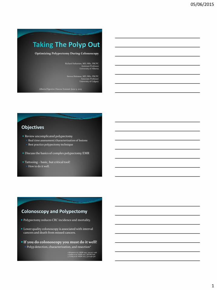
05/06/2015 1 Optimizing Polypectomy During Colonoscopy Richard Sultanian, MD, MSc, FRCPC Assistant Professor University of Alberta Steven Heitman, MD, MSc, FRCPC Associate Professor University of Calgary Alberta Digestive Disease Summit: June 5, 2015 Objectives Review uncomplicated polypectomy Real time assessment/characterization of lesions Best practice polypectomy technique Discuss the basics of complex polypectomy/EMR Tattooing – basic, but critical tool! How to do it well. Colonoscopy and Polypectomy Polypectomy reduces CRC incidence and mortality. Lower quality colonoscopy is associated with interval cancers and death from missed cancers. If you do colonoscopy you must do it well! Polyp detection, characterization, and resection* 1.Winawer et al. NEJM 1993. 329:1977-1981 2.Zauber et al. NEJM 2012. 366:687-696 3. Corley et al. NEJM 2014. 370:1298-306
Transcript of Objectives Colonoscopy and Polypectomy
05/06/2015
1
Optimizing Polypectomy During Colonoscopy
Richard Sultanian, MD, MSc, FRCPC Assistant Professor
University of Alberta
Steven Heitman, MD, MSc, FRCPC Associate Professor
University of Calgary
Alberta Digestive Disease Summit: June 5, 2015
Objectives
Review uncomplicated polypectomy
Real time assessment/characterization of lesions
Best practice polypectomy technique
Discuss the basics of complex polypectomy/EMR
Tattooing – basic, but critical tool!
How to do it well.
Colonoscopy and Polypectomy
Polypectomy reduces CRC incidence and mortality.
Lower quality colonoscopy is associated with interval cancers and death from missed cancers.
If you do colonoscopy you must do it well! Polyp detection, characterization, and resection*
1.Winawer et al. NEJM 1993. 329:1977-1981 2.Zauber et al. NEJM 2012. 366:687-696 3. Corley et al. NEJM 2014. 370:1298-306
05/06/2015
2
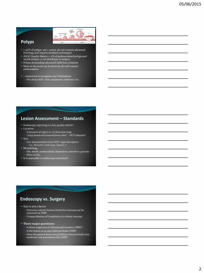
Polyps
> 90% of polyps are < 10mm, do not contain advanced histology and require standard techniques
ASGE Quality Metric: < 2% of pedunculated polyps and sessile polyps <2 cm should go to surgery
Polyps demanding advanced skills less common.
Most of the work can be done by all well trained endoscopists.
…important to recognize one’s limitations Not always skill – time, equipment, assistance, etc.
Lesion Assessment – Standards Endoscopy reporting is a key quality metric!
Location Estimation of region vs. cm from anal verge
“polyp found and removed from colon” – NOT adequate!
Size Use measurements (mm) NOT vague descriptors
(e.g.. diminutive, small, large, “gigantic”…)
Morphology Flat, sessile, pedunculated, depressions, smooth vs. granular
Is it amenable to endoscopic resection?
Endoscopy vs. Surgery
Size is not a factor Extensive colonic lesions limited to mucosa can be
removed via EMR
Unique absence of lymphatics in colonic mucosa
Three major questions: Is there suspicion of submucosal invasion (SMI)?
Is the lesion in an area that precludes EMR?
Does the patient have comorbidities that preclude even moderate risk procedures like EMR?
05/06/2015
3
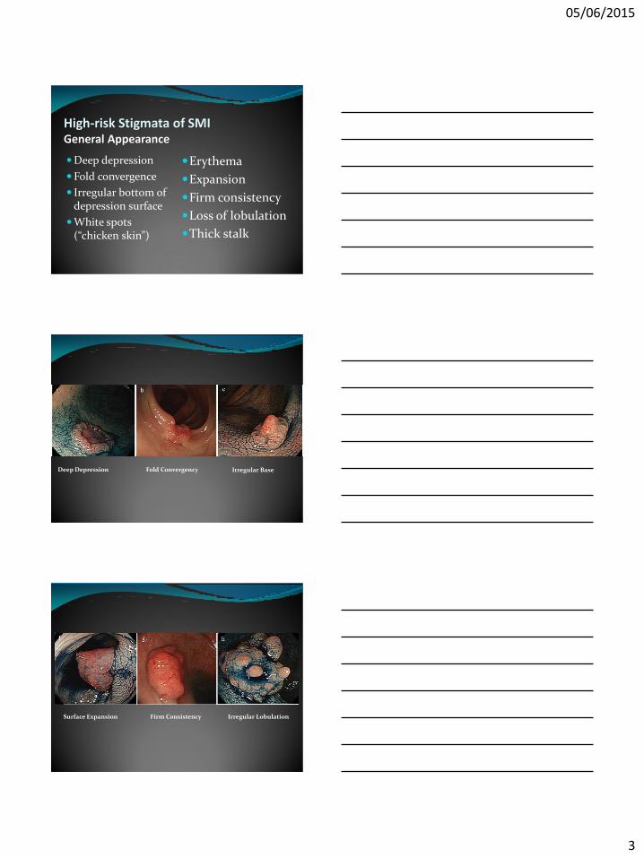
High-risk Stigmata of SMI General Appearance
Deep depression
Fold convergence
Irregular bottom of depression surface
White spots (“chicken skin”)
Erythema
Expansion
Firm consistency
Loss of lobulation
Thick stalk
Deep Depression Fold Convergency Irregular Base
Surface Expansion Firm Consistency Irregular Lobulation
05/06/2015
4
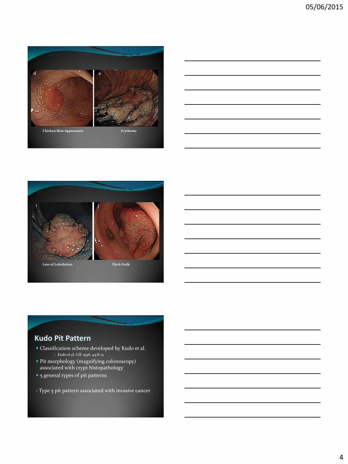
Chicken Skin Appearance Erythema
Loss of Lobulation Thick Stalk
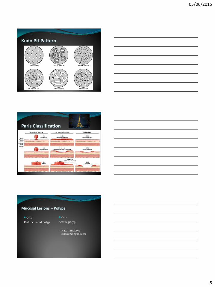
Kudo Pit Pattern Classification scheme developed by Kudo et al.
Kudo et al. GIE 1996. 44:8-14
Pit morphology (magnifying colonoscopy) associated with crypt histopathology
5 general types of pit patterns
- Type 5 pit pattern associated with invasive cancer
05/06/2015
5
Kudo Pit Pattern
Paris Classification
Mucosal Lesions – Polyps
0-Ip
Pedunculated polyp
0-Is
Sessile polyp
- > 2.5 mm above surrounding mucosa
05/06/2015
6
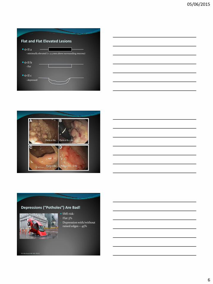
Flat and Flat Elevated Lesions
0-II a - minimally elevated (< 2.5 mm above surrounding mucosa)
0-II b - flat
0-II c
- depressed
Paris 0-IIa Paris 0-Is + IIa
Paris 0-IIa Paris 0-IIa + 0-IIc
Depressions (“Potholes”) Are Bad!
SMI risk:
- Flat 3%
- Depression with/without raised edges ~ 45%
DG MacIntosh MD MSc FRCPC
05/06/2015
7
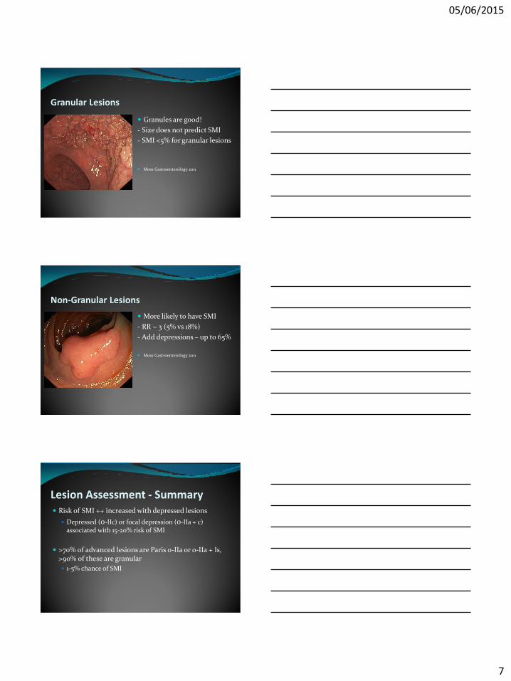
Granular Lesions
Granules are good!
- Size does not predict SMI
- SMI <5% for granular lesions
Moss Gastroenterology 2011
Non-Granular Lesions
More likely to have SMI
- RR ~ 3 (5% vs 18%)
- Add depressions – up to 65%
Moss Gastroenterology 2011
Lesion Assessment - Summary Risk of SMI ++ increased with depressed lesions
Depressed (0-IIc) or focal depression (0-IIa + c) associated with 15-20% risk of SMI
>70% of advanced lesions are Paris 0-IIa or 0-IIa + Is, >90% of these are granular
1-5% chance of SMI
05/06/2015
8
What About Location? “location, location, location”
Anorectal Junction
Increased risk of pain with lesions adjacent to dentate line
Long acting analgesia injection (Marcaine 1%) or topical lidocaine (1%) may be needed post-procedure.
Prophylactic antibiotics
Increased risk of systemic bacteremia from repeated SM injections with involvement of porto-systemtic collaterals bypassing portal circulation and reticuloendothelial system
Recommended in recent polypectomy technical review.
Burgess et al. GIE 2015. 813-35.
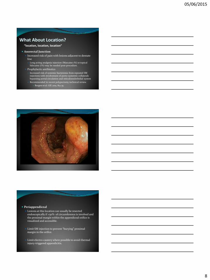
Periappendiceal
Lesions at this location can usually be resected endoscopically if <50% of circumference is involved and the proximal margin within the appendiceal orifice is visualized and accessible.
Limit SM injection to prevent “burying” proximal margin in the orifice.
Limit electro-cautery where possible to avoid thermal injury-triggered appendicitis.
05/06/2015
9
Ileocecal Valve (ICV)
Advanced lesions here have a higher risk of reoccurrence
Factors associated with failure and recurrence: ileal infiltration and involvement of both ICV lips
Nanda et al. Endoscopy 2015. [Epub ahead of print]
Both antegrade and retroflexed positions often necessary
Theoretical risk of ICV stenosis post-resection
clinically rare occurrence
Polyp found and characterized … so now what?
Uncomplicated polypectomy
Polyps < 10mm,
No high risk stigmata and favorable location
Wide variety of tools available…
Choice of the tool based on situation and personal preference
As long as it’s a snare!
05/06/2015
10
Small Polyp Removal Cold biopsy forceps
Quick, easy to use and cheap
Associated with significant rates of incomplete polyp removal, increased recurrence rates and interval CRC
Efthymiou et al. conducted en bloc snare resection of surrounding mucosa of 5mm polyps removed with cold biopsy forceps1
61% of these sites had residual adenomatous tissue!
In general DO NOT USE! Only for diminutive (1-2mm) polyps not amenable to
snare removal
1. Efthymiou et al. Endoscopy 2011. 43(4): 312-316
Small Polyp Removal Hot Biopsy forceps
Once popular, now out of favor.
Increased complication rates compared to snares
Poor quality of specimen histology due to cautery artifact
Same (POOR) quality of polyp eradication as cold biopsy forceps1,2
DO NOT USE
1. Monkemuller,KE et al. Endoscopy. 2004. 36(5) 432-436 2. Paspatis GA et al. Colorectal Dis. 2011. 13 (10): 345-348
Small Polyp Removal Snare polypectomy – gold standard
Technique: Polyp position 6 O’clock
Aim to capture 1-2mm of normal tissue around polyp
Hot vs. Cold? No significant difference in removal rate
Cold for polyps <8mm, hot snare for larger
Increased non-important immediate bleeding with cold compared to increased delayed bleeding and post-polypectomy syndrome rates with hot snare.
COLD IS BECOMING THE STANDARD

05/06/2015
11
Cold Snare Technique
Cold Snare Polypectomy
Less Optimal Technique
05/06/2015
12
Key points: Small Polyps
Majority of polyps < 10mm
Cold forceps biopsy is associated with high rates of incomplete removal - AVOID
Hot forceps should also be avoided - associated with high complication rates, incomplete removal
Cold snare polypectomy is gold standard
What if it’s Large?
Large Polyps
Advanced mucosal neoplasia (AMN)
>10mm, components of villous (tubulovillous or villous) or serrated histology or evidence of high-grade dysplasia (HGD)
Can be pedunculated or sessile
~10% of adenomas detected are sessile lesions >10mm1
Sessile lesions have greater frequency of HGD and early invasive disease compared with polypoid lesions of equivalent size
1. Rotondano et al. Endoscopy. 2011. 43: 856-861

05/06/2015
13
Laterally Spreading Tumors - LST
Grow laterally along the surface of the bowel
Size doesn’t matter
May reach an enormous size before demonstrating invasive features
Common AMN
Paris 0-II (a – minimally elevated & b - flat) and O-Is (elevated) polyps often >20mm in size
Treatment of AMN Surgical excision
Endoscopic mucosal resection (EMR), endoscopic submucosal dissection (ESD)
EMR First described in 19731
Multiple prospective multicenter trials have demonstrated that wide field EMR is safe and effective
Prospective data demonstrates a net health care savings of US $10,000 and 6 days in hospital per patient in comparison to ideal surgical outcome without complication1
1. Swan et al. Gastrointest Endosc 2009;70:1128–1136.
EMR Outcomes Large multi-centered, prospective trial (479 patients1)
EMR effective (complete, single session) in 89.2%
No associated mortality
Risk factors associated with EMR failure:
prior attempted EMR (OR 3.8, p = 0.001)
ICV involvement (OR 3.4, p = 0.021)
Predictors of recurrence:
Size >40mm (OR 4.37, p < 0.001)
APC of residual tissue (OR 3.51, p = 0.0017).
Moss et al. Gastroenterol 2011: 1909-1918.
05/06/2015
14
EMR Technique Submucosal injection
Fluid “cushion” between the mucosa and muscularis propria (MP)
Reduces risk of perforation and transmural thermal injury
“Lift sign” to identify SMI
Ideally inexpensive, easy to use while providing sustained, well-circumscribed mucosal elevation
Normal saline most common but colloid solutions reported to be superior in studies
EMR Technique Submucosal injection solution
Methylene blue / Indigo carmine
Biologically inert blue dyes that are avid for loose areolar tissue in the SM layer
Confirms resection in the correct tissue plane
Helps delineate polyp borders
Dilute epinephrine (1:50-100,000) Added to injectate by some physicians
Bloodless resection field, but higher risk of delayed polypectomy bleeding
05/06/2015
15
Methylene Blue
0.1 ml in 50 cc minibag
EMR Technique Resection Technique
Inject and resect
As few pieces as safely possible
En bloc resection for lesions up to 20mm right colon and 25mm in the left colon More accurate histology, reduced risk of recurrence
Include 2-3mm margin of normal mucosa
Exam borders with white light endoscopy and NBI to ensure complete resection of polyp
Endocut Electrosurgical current controlled by microprocessor
EMR Technique Snares
Evaluation of the lesion size, morphology and location allows for the selection of the most appropriate snare

05/06/2015
16
EMR Technique Other Important Considerations
Give yourself enough time! (1 hour)
Don’t do during index procedure – consent?
Ensure you have an experienced nurse
2 RNs extremely helpful at times
Ensure equipment readily available
Injection solution (colloid + dye)
Devices for complications – clips, ? Hemospray
Cases should be done with CO2
EMR Technique Technique equally applicable to lesions 1-2cm in size
Proper technique minimizes need for repeat attempts at polypectomy and optimizes patient outcomes
05/06/2015
17
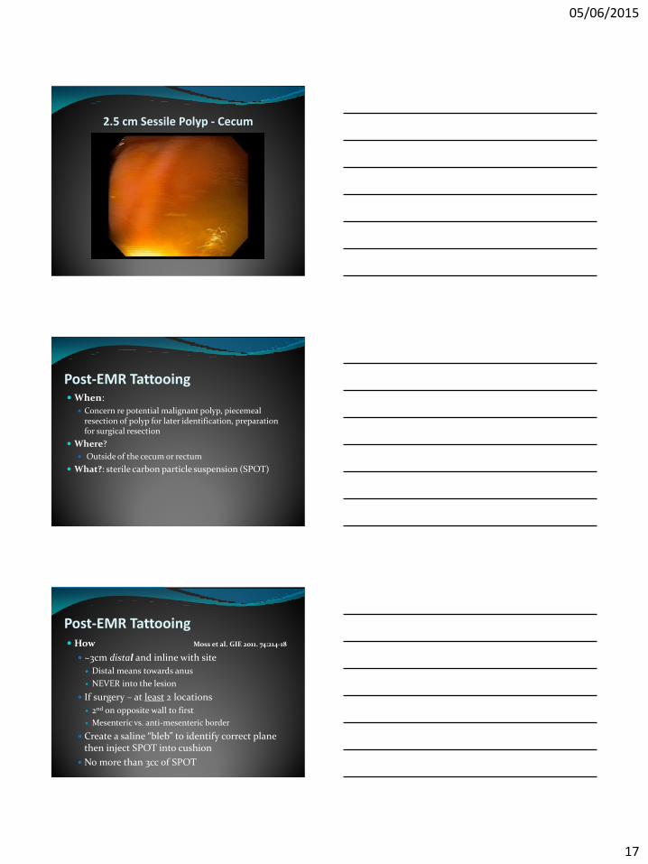
2.5 cm Sessile Polyp - Cecum
Post-EMR Tattooing When:
Concern re potential malignant polyp, piecemeal resection of polyp for later identification, preparation for surgical resection
Where?
Outside of the cecum or rectum
What?: sterile carbon particle suspension (SPOT)
Post-EMR Tattooing How Moss et al. GIE 2011. 74:214-18
~3cm distal and inline with site
Distal means towards anus
NEVER into the lesion
If surgery – at least 2 locations
2nd on opposite wall to first
Mesenteric vs. anti-mesenteric border
Create a saline “bleb” to identify correct plane then inject SPOT into cushion
No more than 3cc of SPOT
05/06/2015
18
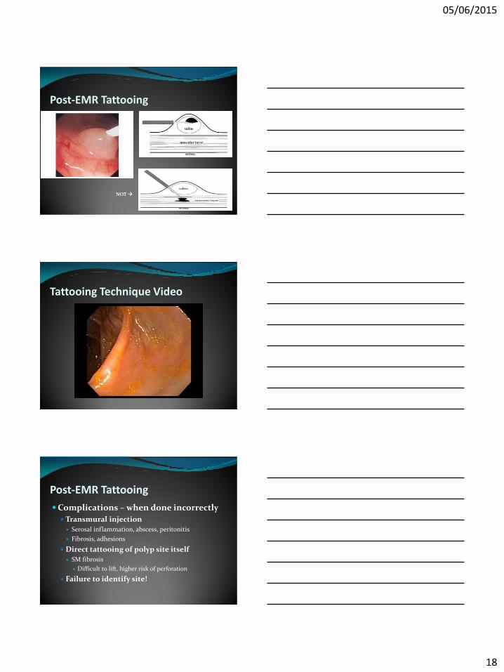
Post-EMR Tattooing
NOT
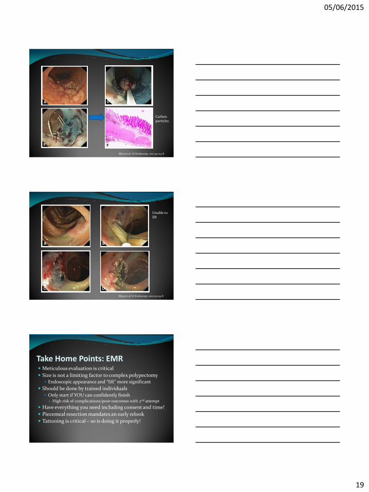
Tattooing Technique Video
Post-EMR Tattooing
Complications – when done incorrectly
Transmural injection
Serosal inflammation, abscess, peritonitis
Fibrosis, adhesions
Direct tattooing of polyp site itself
SM fibrosis
Difficult to lift, higher risk of perforation
Failure to identify site!
05/06/2015
19
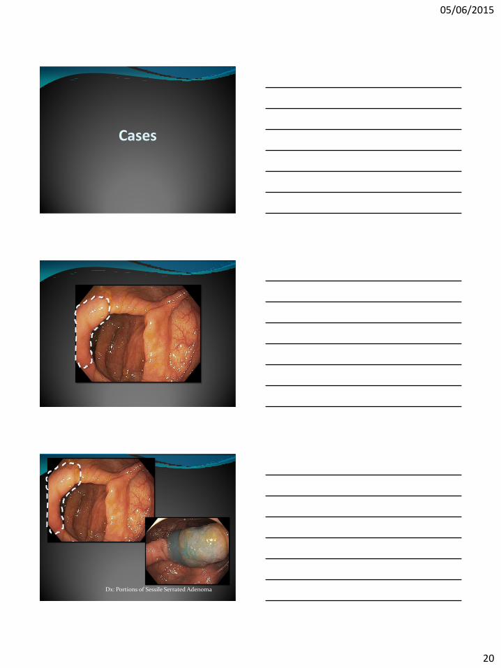
Moss et al. GI Endoscopy 2011;74:214-8
Carbon particles
Moss et al GI Endoscopy 2011;74:214-8
Unable to lift
Take Home Points: EMR Meticulous evaluation is critical
Size is not a limiting factor to complex polypectomy Endoscopic appearance and “lift” more significant
Should be done by trained individuals Only start if YOU can confidently finish
High risk of complications/poor outcomes with 2nd attempt
Have everything you need including consent and time!
Piecemeal resection mandates an early relook
Tattooing is critical – so is doing it properly!
05/06/2015
20
Cases
Dx: Portions of Sessile Serrated Adenoma
05/06/2015
21
What about this?
61
Large Pedunculated Polyp – Descending
05/06/2015
22
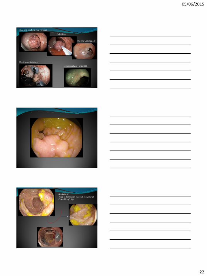
Base and head injected with epi
Debulking
This one was clipped!
Don’t forget to tattoo!
4 months later – with NBI
- Kudo IV/V - Area of depression (not well seen in pic) - “Non lifting” sign
05/06/2015
23
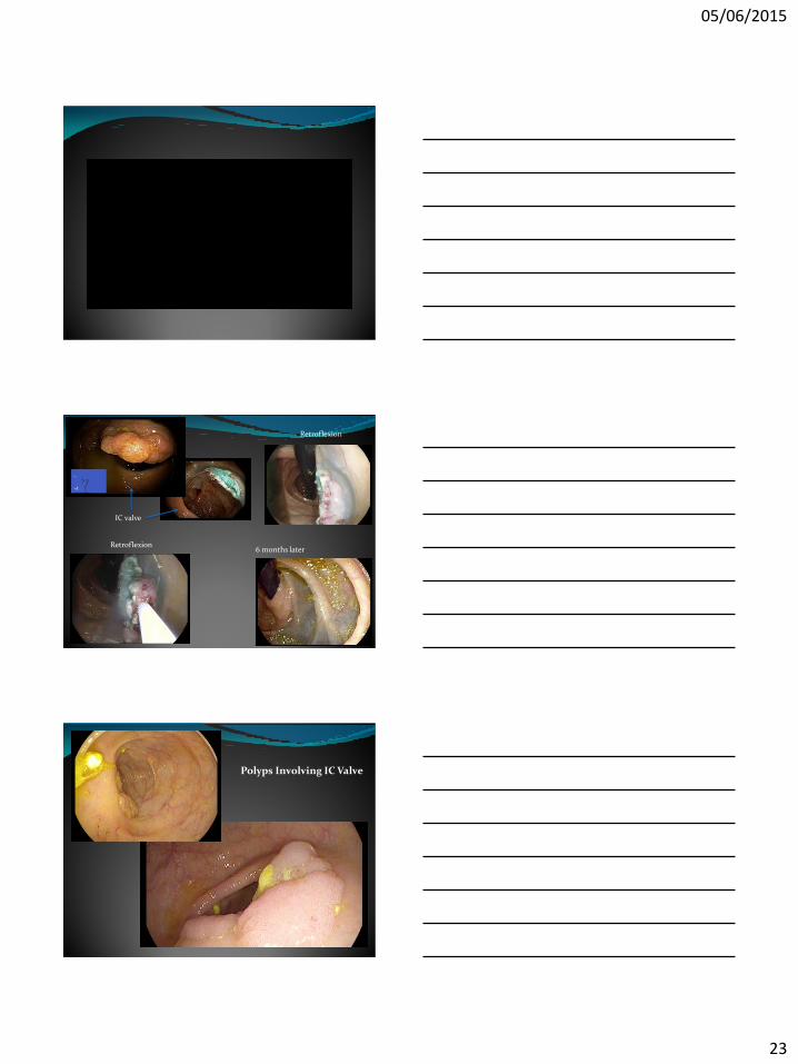
IC valve
Retroflexion
Retroflexion 6 months later
Polyps Involving IC Valve