OBJECTIVE SHEET THE NERVOUS SYSTEM - SD5 · OBJECTIVE SHEET THE NERVOUS SYSTEM 1. ... •...
55
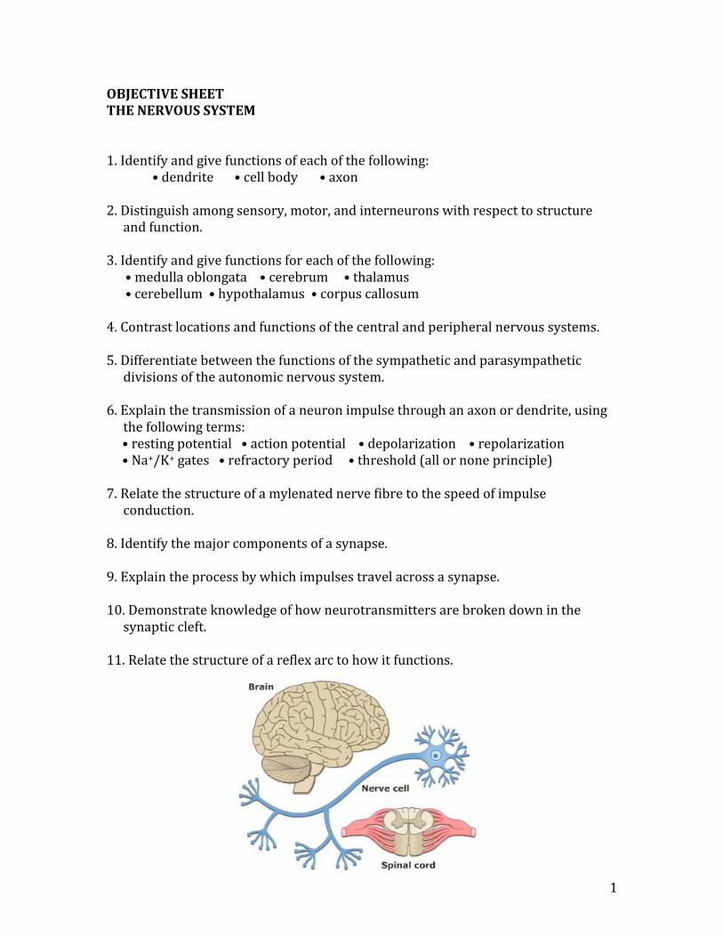
1 OBJECTIVE SHEET THE NERVOUS SYSTEM 1. Identify and give functions of each of the following: • dendrite • cell body • axon 2. Distinguish among sensory, motor, and interneurons with respect to structure and function. 3. Identify and give functions for each of the following: • medulla oblongata • cerebrum • thalamus • cerebellum • hypothalamus • corpus callosum 4. Contrast locations and functions of the central and peripheral nervous systems. 5. Differentiate between the functions of the sympathetic and parasympathetic divisions of the autonomic nervous system. 6. Explain the transmission of a neuron impulse through an axon or dendrite, using the following terms: • resting potential • action potential • depolarization • repolarization • Na + /K + gates • refractory period • threshold (all or none principle) 7. Relate the structure of a mylenated nerve fibre to the speed of impulse conduction. 8. Identify the major components of a synapse. 9. Explain the process by which impulses travel across a synapse. 10. Demonstrate knowledge of how neurotransmitters are broken down in the synaptic cleft. 11. Relate the structure of a reflex arc to how it functions.
Transcript of OBJECTIVE SHEET THE NERVOUS SYSTEM - SD5 · OBJECTIVE SHEET THE NERVOUS SYSTEM 1. ... •...
1
OBJECTIVE SHEET THE NERVOUS SYSTEM 1. Identify and give functions of each of the following:
• dendrite • cell body • axon 2. Distinguish among sensory, motor, and interneurons with respect to structure
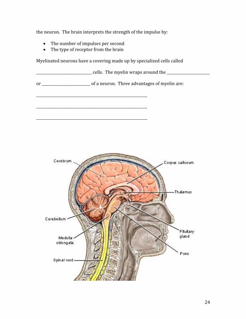
and function. 3. Identify and give functions for each of the following: • medulla oblongata • cerebrum • thalamus • cerebellum • hypothalamus • corpus callosum 4. Contrast locations and functions of the central and peripheral nervous systems. 5. Differentiate between the functions of the sympathetic and parasympathetic
divisions of the autonomic nervous system. 6. Explain the transmission of a neuron impulse through an axon or dendrite, using
the following terms: • resting potential • action potential • depolarization • repolarization • Na+/K+ gates • refractory period • threshold (all or none principle) 7. Relate the structure of a mylenated nerve fibre to the speed of impulse
conduction. 8. Identify the major components of a synapse. 9. Explain the process by which impulses travel across a synapse. 10. Demonstrate knowledge of how neurotransmitters are broken down in the
synaptic cleft. 11. Relate the structure of a reflex arc to how it functions.

2
Label the following neurons:

3
A Typical Neuron 1. Cell Body: contains the nucleus and much of the cytoplasm. the site of the greatest metabolic activity in the cell. 2. Dendrites: a fibre, branched extensions leading toward the cell body. always carries impulses toward the cell body from the environment or from
another neuron. 3. Axon: a fibre, that carries impulses away from the cell body toward an effector such as a
muscle or gland. axons end in axon terminals or axon bulbs. 3 Types of Neurons 1. Sensory Neuron (afferent)
carries impulses from sense organs to the brain or spinal cord (CNS). a long dendrite and short axons.
2. Motor Neuron (efferent)
carries impulses from the (CNS) brain or spinal cord to a muscle or gland. short dendrites and a long axon.
3. Interneurons
connect sensory neurons to motor neurons and receive impulse from other interneurons
short dendrites and short axons located in the spinal cord all interneurons are unmyelinated and they sum up all the messages received
from all the neurons before they communicate with motor neurons.
4

5
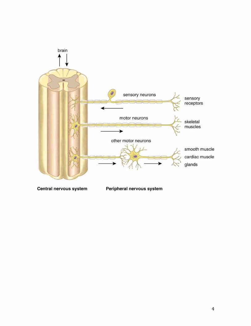
Nervous System
Central Nervous System Peripheral Nervous System
Brain Spinal Cord Autonomic Somatic
Cerebrum Dorsal Root Sympathetic Parasympathetic Thalamus Ventral Root Hypothalamus Cerebellum Sensory nerves Medulla Oblongata
Motor nerves *Limbic System
ORGANIZATIONAL LADDER
OF THE NERVOUS SYSTEM

6

7
The Central Nervous System The brain and spinal cord make up the central nervous system. The brain is covered with three membranes called meninges. A liquid called the cerebrospinal fluid, cushions the CNS against shock. When a disease is suspected, this fluid is extracted by a spinal tap and analyzed. The spinal cord has two main functions:
the centre for many reflex actions causing skeletal and smooth muscles to contract.
Provides a means of communication between the brain and spinal nerves. One pair of spinal nerves leaves the spinal cord between each vertebra. The spinal cord is made up of gray and white matter. The CNS has two types of nervous tissue – Gray matter is gray in colour because it contains cell bodies and short, unmyelinated fibres. White matter is white in colour because it contains myelinated axons that run in bundles called tracts. Brain Anatomy text reference pgs. 326-330 1. Cerebrum The largest part of the brain, composed of two hemispheres. The cerebrum is the last centre to receive sensory input before it commands voluntary responses. The outer cerebral cortex has gray matter that is highly folded. There are four lobes in each hemisphere:
Lobes of the Cerebrum Function Frontal
The area of conscious thought, the location of personality, motor areas that control skeletal muscle, motor control of
speech Temporal Smelling and hearing Parietal Has sensory areas called the sensory
strip that receives information from skin and from muscle receptors.
Occipital Responsible for vision The two hemispheres of the cerebrum are bridged by the Corpus Callosum. This bridge of tracts allows the left and right side of the cerebrum to share information. The Limbic System lies just beneath the cerebral cortex and is intimately involved in our emotions causing sensations of pain, pleasure, rage, sexual interest, fear and sorrow. It is also involved with learning and memory.

8
2. Thalamus The thalamus is often called the “gatekeeper” to the brain, because it receives all sensory impulses (except smell) and channels them to the appropriate parts of the cerebrum for you to make a conscious decision about. It is connected to the medulla oblongata by the ARAS (ascending reticular activating system) that is involved in controlling awareness and consciousness levels in situations. The thalamus sorts out stimuli from the sense organs and passes on those that need immediate action. 3. Hypothalamus It is sometimes called the “brain within the brain”. It is located below the thalamus and is involved with maintaining homeostasis. It controls the pituitary gland (linking the nervous and endocrine systems). The hypothalamus has osmoreceptors that control the OP of your blood; Chemoreceptors that detect chemical and hormone changes in your blood, and temperature receptors to control body temperature. 4. Cerebellum This is the second largest part of your brain. The functions include control of fine muscle co-ordination, allowing smooth coordinated movement, and the sense of balance. 5. Medulla Oblongata It is part of the brain stem. It is often considered to “boss of the ANS”. It lies closest to the spinal cord and coordinates your heartbeat, breathing rate and blood pressure. It has reflex centres that include vomiting, sneezing and swallowing.

9
The Human Brain

10
This diagram illustrates the amount of neurons in the brain dedicated to sensing the environment (somatosensory) and moving (motor) the following body parts in a person. (interest only)

11
Autonomic Nervous System Functions
SYMPATHETIC N.S. PARASYMPATHETIC N.S. (fight or flight) (normal times) Cranial region
Thoracic region Lumbar region
Saccral region

12
Autonomic Nervous System Functions

13
Neuron Impulse Transmission The Resting Potential The axon or dendrite of a neuron is simply referred to as a fibre. These fibres carry impulses. The transmission of an impulse is made possible by the position of Na+ and K+ ions surrounding the membrane of an axon or dendrite. If we look at the ion distribution around a section of an axon, we would find a large amount of Na+ outside the membrane and a large amount of K+ ions inside the membrane. There are also a number of negatively charged protein molecules (-ve) inside the neuron. -ve -ve -ve -ve -65mV The inside of the neuron is more negative than the outside of the neuron because of the way the ions are distributed. The membrane is polarized. This is referred to as a resting potential. At rest, small amounts of K+ inside the axon slowly leak outside where they are pumped back into the neuron by the Na+/ K+ pump. This pump maintains the resting potential. The resulting inside membrane potential of a neuron is about -65 to -70 mV of charge, slightly more negative than outside. The Action Potential If a strong enough stimulus occurs, the threshold is reached and an impulse occurs. There is an exchange of ions across the membrane. A stimulus that reaches the “threshold” causes the Na+ gated channels on the membrane of the axon to open. Large amounts of Na+ diffuse from outside the axon to inside the axon of the neuron. The axon is now said to be “depolarized” and the inside membrane charge increases from -65mV to +40mv of charge. This reversal of a small part of the membrane results in a flow of an electric current that affects the permeability of the adjacent area of the axon allowing the inward movement of Na+ there. In this way, the wave of depolarization travels down the length of the axon. (think of it like a lit fuse.)

14
An oscilloscope records an impulse: In the area of the impulse, the high permeability of the membrane to Na+ lasts only a fraction of a millisecond before the Na+ gates close. It takes longer for the K+ gated channels to open. When they do open, K+ rushes out of the axon, this is referred to as “repolarization” and the polarity of the membrane looks the same as it was during the resting potential. The only problem is that the ions are distributed in the opposite location from where they started. The “refractory period” begins as the neuron cannot fire another action potential until the Na+/ K+ pump in the membrane restores the ions to their original positions, pumping Na+ outside the membrane and K+ back inside. This restores the original resting potential and the neuron can “fire” again. The Rate of Impulse Conduction The speed of conduction varies according to the fibre size and whether or not myelin is present on the axon or dendrite. Small unmyelinated fibres = 2 m/sec Large myelinated fibres = 100-200 m/sec

15
Check your understanding of a Neuron Impulse… This section of axon illustrated below all represent the same section of an axon at the same location, just at different times. The axon starts at a resting potential… Interpret the diagram below to answer the following questions:
Interstitial fluid (outside axon)
Intracellular fluid (inside axon)
1. Which direction is the impulse moving? Brain to hand or hand to brain? ______________________________________________ 2. What type of neuron does the diagram represent? ___________________________________ 3. Which section(s) would active transport of ions be the most active? _______________ 4. Which section(s) is diffusion of ions occurring the most? ____________________________ 5. Explain specifically what is occurring in each of the sections: A. _____________________________________________________________________________________________ B. _____________________________________________________________________________________________ C. _____________________________________________________________________________________________ D. _____________________________________________________________________________________________ E. _____________________________________________________________________________________________
16
Check your understanding of a Neuron Impulse… Write a paragraph or two to describe how an impulse travels along a neuron. Use your oscilloscope recording as a tool to help you write your description. _________________________________________________________________________________________________ _________________________________________________________________________________________________ _________________________________________________________________________________________________ _________________________________________________________________________________________________ _________________________________________________________________________________________________ _________________________________________________________________________________________________ _________________________________________________________________________________________________ _________________________________________________________________________________________________ _________________________________________________________________________________________________ _________________________________________________________________________________________________ _________________________________________________________________________________________________ _________________________________________________________________________________________________ _________________________________________________________________________________________________ _________________________________________________________________________________________________ _________________________________________________________________________________________________ _________________________________________________________________________________________________ _________________________________________________________________________________________________ _________________________________________________________________________________________________ _________________________________________________________________________________________________ _________________________________________________________________________________________________
17
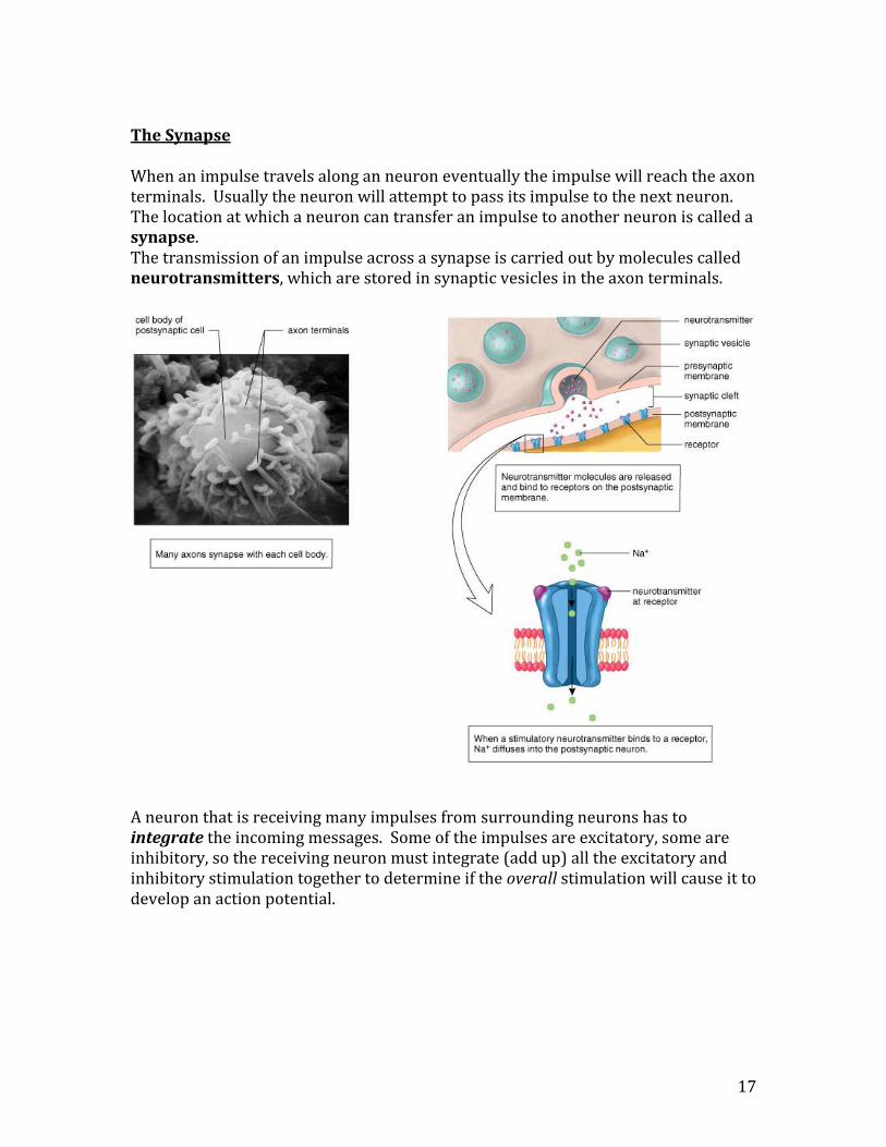
The Synapse When an impulse travels along an neuron eventually the impulse will reach the axon terminals. Usually the neuron will attempt to pass its impulse to the next neuron. The location at which a neuron can transfer an impulse to another neuron is called a synapse. The transmission of an impulse across a synapse is carried out by molecules called neurotransmitters, which are stored in synaptic vesicles in the axon terminals.
A neuron that is receiving many impulses from surrounding neurons has to integrate the incoming messages. Some of the impulses are excitatory, some are inhibitory, so the receiving neuron must integrate (add up) all the excitatory and inhibitory stimulation together to determine if the overall stimulation will cause it to develop an action potential.

18
The Synapse
Step 1: The impulse reaches the end of the axon terminal, gated channels for calcium ions open up in the pre-synaptic membrane. Step 2: Ca ++ present in the synaptic cleft diffuse into the axon terminal where they interact with the microfilaments in the axon. Step 3: The microfilaments contract in the presence of Ca++ pulling the synaptic vesicles and their neurotransmitters toward the pre-synaptic membrane. The vesicles fuse with the membrane and the NT is released into the synaptic cleft. Step 4: Most of the Ca++ are pumped back out into the cleft. The neurotransmitters bind with the receptors on the post-synaptic membrane of the adjacent dendrite from the second neuron. Step 5: When the receptors are activated by the neorotransmitter, the gated sodium channels of the dendrite open up and Na+ diffuse into the dendrite. This creates an action potential as the membrane is now depolarized. Step 6: The enzymes present in the synaptic cleft will destroy the NT on the receptors or cause it to move back to the endoplasmic reticulum in the axon terminal to recycle the NT into a new vesicle.

19
Drug Action reference pg. 338-339 When a person takes illicit drugs such as cocaine, heroin, or meth, they run the risk of developing a physical addiction along with all the other negative effects of the drug abuse. The hypothalamus maintains homeostasis by continually monitoring various levels of substances in the body. Many illicit drugs mimic the effect of our natural opiates in the body as well as neurotransmitters that may be excitatory in nature. With higher than normal levels in the body due to drug abuse, the body shuts down production of the natural opiates and NT that may be excitatory in nature. Abusers will “crash” when the drug is removed and they experience the feeling of having little to no natural opiates or excitatory NT left in the body. In other words, they are physically addicted to their drug. Because their bodies are not making these substances, they go in search of more drugs to obtain the “high” they physically crave. Neuron impulses can be either excitatory or inhibitory. Drugs act on us in one of two ways:
Drugs that are mood-altering can affect the ARAS (the ascending reticular activating system) these are cell bodies and fibres extending from the medulla to the thalamus and then cerebrum.
Drugs that promote or inhibit the action of NT

20
Drugs affect Synapses: Using the diagram below, label the circles to indicate the 6 ways that drugs can affect a synapse: a). drugs can cause a leakage of NT out of the synaptic vesicles. b) can prevent the release of the NT into the synaptic cleft. c) can promote the release of NT into the synaptic cleft. d) can prevent uptake of the NT by the pre-synaptic membrane. e) can block the enzyme that causes the breakdown of the NT. f) can bind to a receptor, mimicking the action or preventing the uptake of an NT.

21
The Reflex Arc Label the diagram
22
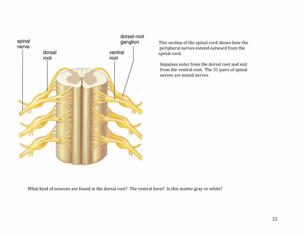
This section of the spinal cord shows how the
peripheral nerves extend outward from the spinal cord.
Impulses enter from the dorsal root and exit from the ventral root. The 31 pairs of spinal nerves are mixed nerves. What kind of neurons are found in the dorsal root? The ventral horn? Is this matter gray or white?
23
Neuron Impulse/Synapse (review…) When a neuron is at rest, there are more ______________ ions outside and more _____________ ions and negatively charged _______________________ inside. The result is a membrane that is ________________________________ with +ve outside and –ve inside. The _________________________________ maintains this difference as K+ tend to “leak” out of the axoplasm. The “upswing” is caused by ____________________________________________________ _________________ making the inside ______________________________. The “downswing” is caused by the _____________________________________________________________ moving out making the outside ____________________________ again. The ________________________________ puts the ions back where they started. The time it takes this to happen is called the ______________________________________ and the neuron cannot fire until this is done. When the impulse or __________________________________________ reaches the end of the axon, ___________________________ ions flow in the pre-synaptic membrane contracting the __________________________________ which pull the _________________________________________ so they can ______________________ with the pre-synaptic membrane dumping out the _________________________________________________ to diffuse across the synaptic cleft. There it binds with ______________________________ on the post-synaptic membrane of the ____________________________ causing ____________________________________ to open up and and Na+ diffuse in. This develops an __________________________________ in the new dendrite. __________________________________ present in the synapse break down or recycle ________________________________. Neurons follow the ____________________________________ principle and the speed of the impulse (varies/does not vary) ____________________________________ as it travels along
24
the neuron. The brain interprets the strength of the impulse by:
The number of impulses per second The type of receptor from the brain
Myelinated neurons have a covering made up by specialized cells called _______________________________ cells. The myelin wraps around the ________________________ or ___________________________ of a neuron. Three advantages of myelin are: ______________________________________________________________ ______________________________________________________________ ______________________________________________________________

25
Number the following in the order in a reflex arc. Synapses occur more than once. Effector __________ axon of motor neuron __________ Dendrite of motor neuron __________ Axon of interneuron __________ Dorsal root __________ Dendrite of interneuron __________ Dendrite of sensory neuron __________ Ventral root __________ Cell body of motor neuron __________ Receptor __________ Synapse ____________________ Number the following in the order that they occur during an impulse. Fill in any blanks (on the right) with appropriate words. __________ potassium diffuses _____________ __________ resting potential is in place __________ sodium diffuses ____________ __________ gated sodium channels open __________ Na+/K+ pump will pump Na+ ____________ and K+ ____________ in by the process of _________________________________________. __________ stimulus __________ depolarization occurs __________ end of the refractory period __________ threshold

26
Nervous System Matching a) integration __________ 1. runs toward cell body b) inhibitory__________ 2. controls the ANS c) node of Ranvier __________ 3. higher [axoplasm] d) cerebellum __________ 4. released by sympathetic nerves e) hypothalamus __________ 5. released by parasympathetic nerves f) depolarization __________ 6. made up of cell bodies g) sympathetic __________ 7. time when a neuron can’t fire h) refractory period __________ 8. neurons fire at a constant speed i) medulla __________ 9. no Schwann cells on this part j) norepinephrine __________ 10. when Na+ gates open k) threshold __________ 11. controls the pituitary gland l) all or none __________ 12. increases speed of transmission m) thalamus __________ 13. type of NT; Stops impulse n) axon __________ 14. controls balance and coordination o) motor neuron __________ 15. what happens when Na+ diffuses p) myelin __________ 16. needed for microfilaments to work q) gray matter __________ 17. excitatory + inhibitory summation r) active transport __________ 18. regulates incoming nerve impulses s) K+ __________ 19. have axons running to effectors t) Ca++ __________ 20. leave via the ventral root
21. leave out the thoracic and lumbar regions
22. establishes the resting potential
27
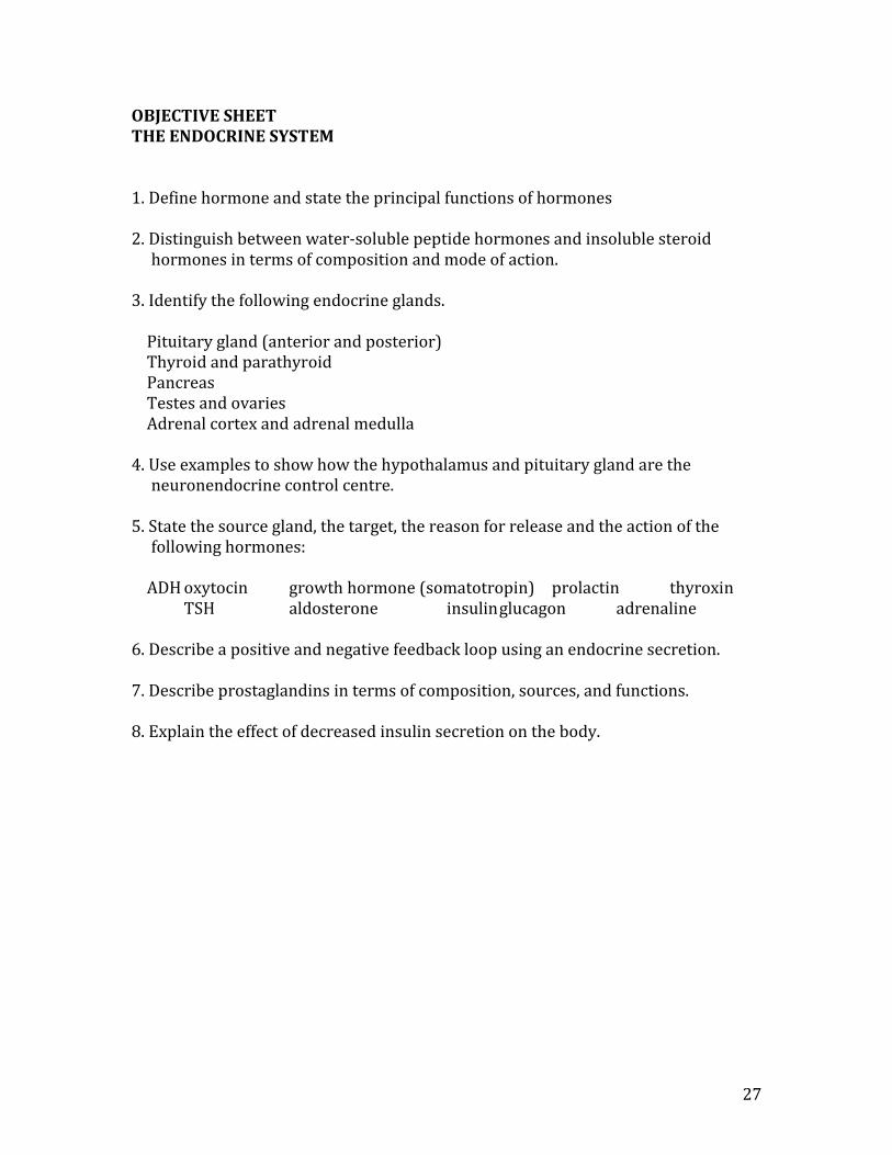
OBJECTIVE SHEET THE ENDOCRINE SYSTEM 1. Define hormone and state the principal functions of hormones 2. Distinguish between water-soluble peptide hormones and insoluble steroid
hormones in terms of composition and mode of action. 3. Identify the following endocrine glands. Pituitary gland (anterior and posterior) Thyroid and parathyroid Pancreas Testes and ovaries Adrenal cortex and adrenal medulla 4. Use examples to show how the hypothalamus and pituitary gland are the
neuronendocrine control centre. 5. State the source gland, the target, the reason for release and the action of the
following hormones: ADH oxytocin growth hormone (somatotropin) prolactin thyroxin
TSH aldosterone insulin glucagon adrenaline 6. Describe a positive and negative feedback loop using an endocrine secretion. 7. Describe prostaglandins in terms of composition, sources, and functions. 8. Explain the effect of decreased insulin secretion on the body.
28
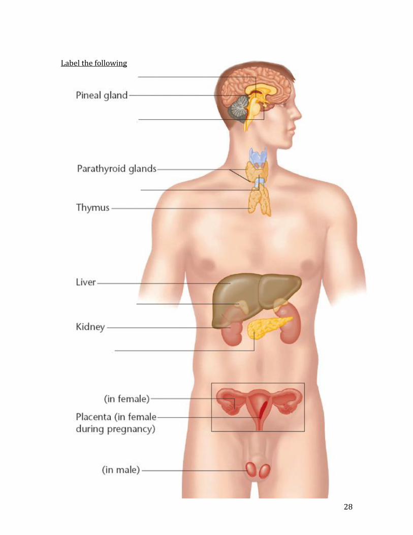
Label the following

29
Hormone Regulation The nervous system acts quickly and on specific body parts. The endocrine system operates by “chemical messengers” called hormones. The effects of hormones are longer lasting but slower to take effect than neurotransmitters. Hormones are released directly into the blood by ductless or endocrine glands. By contrast, exocrine glands dump their secretions through ducts (ie: salivary, liver, pancreas-when secreting digestive juices) Hormones are only recognized by specific tissues in the body called targets. The hormone may stimulate the target to increase or decrease its activity. Hormones can control:
Overall metabolism Maintain homeostasis Growth and reproduction
Hormones fall into two classes: 1. Peptide hormones (water soluble): These hormones activate receptors on the target cell surface. They work quickly (have a lot of “pep”) to affect the target cell but the effects don’t last very long. These hormones activate enzymes already present in the target cell. Examples include: insulin, glucagon, oxytocin, ACTH.
30
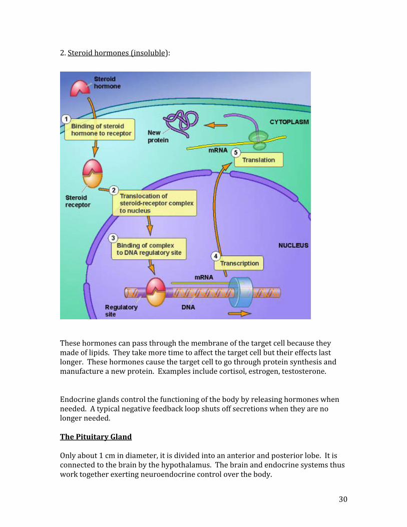
2. Steroid hormones (insoluble): These hormones can pass through the membrane of the target cell because they made of lipids. They take more time to affect the target cell but their effects last longer. These hormones cause the target cell to go through protein synthesis and manufacture a new protein. Examples include cortisol, estrogen, testosterone. Endocrine glands control the functioning of the body by releasing hormones when needed. A typical negative feedback loop shuts off secretions when they are no longer needed. The Pituitary Gland Only about 1 cm in diameter, it is divided into an anterior and posterior lobe. It is connected to the brain by the hypothalamus. The brain and endocrine systems thus work together exerting neuroendocrine control over the body.

31
Diabetes Mellitus 1. Type 1 Diabetes (juvenile diabetes) This type of diabetes is an autoimmune disease. The immune system attacks the beta cells in the pancreas that produce insulin. A high level of sugar in the blood causes sugar to appear in the urine. This causes your cells to lack sugar, and you feel hungry. Water follows the sugar in the urine and you have increased urination. This causes you to feel thirsty. The current treatment is to deliver daily injections of insulin. 2. Type 2 Diabetes (adult onset diabetes) This type of diabetes usually occurs around the age of 40, although it is appearing in younger individuals in Canada. Insulin is being produced normally by the pancreas, however, the cells insulin receptors on the body cells no longer work. The symptoms are identical to type 1 diabetes. The current treatment is to control one’s sugar intake. Prostaglandins These are lipid hormones. They were named after the prostate gland as they were first discovered in semen. We now know that most tissues make prostaglandins. Prostaglandins are produced when injury occurs in tissues. They are involved in the inflammatory response and they act locally. Prostaglandins produce pain and swelling in injured areas and can be blocked by taking aspirin. Prostaglandins are believed to cause painful cramping during the early days of the menstrual cycle.

32
Islets of Langerhans cells in the pancreas produce alpha and beta cells that produce glucagon and insulin respectively.
33
OBJECTIVE SHEET THE CIRCULATORY SYSTEM 1. Identify the five types of blood vessels. 2. Identify and describe the function of the following:
subclavian arteries/veins jugular veins carotid arteries mesenteric artery vena cava hepatic vein pulmonary veins/arteries hepatic portal vein renal veins/arteries iliac artery/vein coronary veins/arteries aorta septum chordae tendinae left/right atria left/right ventricles atrioventricular valves (tricuspid and mitral) semilunar valves (pulmonary and aortic)
3. Distinguish between pulmonary and systemic circulation. 4. Identify and describe the differences between adult and fetal circulation. 5. Trace a blood cell out of the aorta, through the body and back to the left ventricle. 6. List the major components of blood plasma. 7. Identify and describe functions of lymph capillaries, veins, and lymph nodes. 8. Describe capillary-tissue fluid exchange. 9. Describe the shape, function and origin of red/white blood cells and platelets. 9. Explain the role of blood antigens and antibodies. 10. Describe the location and functions of the SA node, AV node and Purkinje fibres. 11. Explain the following equations: BP = CO x AR and CO = HR x SV 12. Distinguish between systolic and diastolic pressure. 13. Demonstrate the proper technique in the use of a sphygmomanometer.

34
14. Identify factors that help return venous blood back to the heart. 15. Relate the factors that affect and regulate blood pressure to hyper/hypotension
using feedback loops. 16. List the dangers of hypertension.

35
Arteries and Veins
endothelium
layer elastin smooth muscle fibrous coat epithelial cells
36
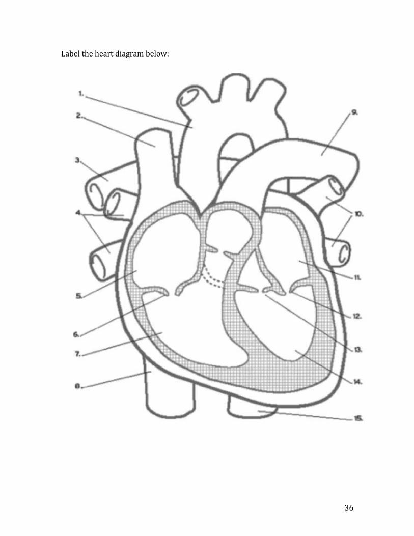
Label the heart diagram below:
37
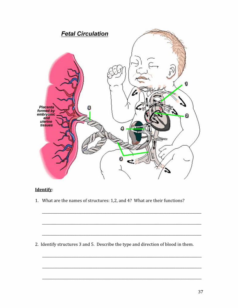
Identify: 1. What are the names of structures: 1,2, and 4? What are their functions?
____________________________________________________________________________________________ ____________________________________________________________________________________________ ____________________________________________________________________________________________
2. Identify structures 3 and 5. Describe the type and direction of blood in them. ____________________________________________________________________________________________ ____________________________________________________________________________________________ ____________________________________________________________________________________________

38

39
Major Arteries of the Body * * * * * *
*
*

40
Major Veins of the Body * * * * * * * * *

41
Cardiovascular System Using your textbook pg. 246, label the following simplified diagram of blood flow: 1. How many capillary beds would a blood cell travel through from the right atrium to the liver? _________________________________ 2. Which blood vessels would have more blood in them after birth than
before birth? __________________________________________________________________________
3. Which blood vessel would contain the most nutrients after a meal? ________________________________________________________

42
43
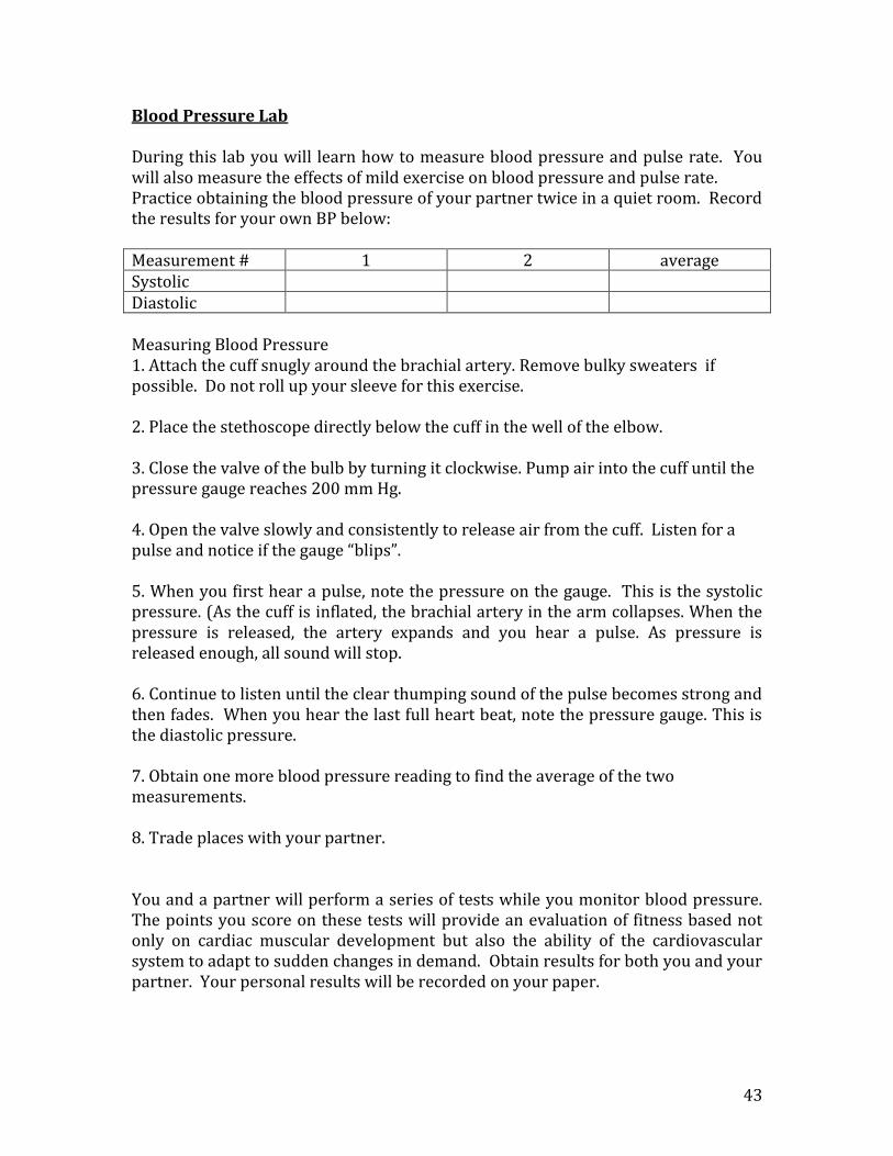
Blood Pressure Lab During this lab you will learn how to measure blood pressure and pulse rate. You will also measure the effects of mild exercise on blood pressure and pulse rate. Practice obtaining the blood pressure of your partner twice in a quiet room. Record the results for your own BP below: Measurement # 1 2 average Systolic Diastolic Measuring Blood Pressure 1. Attach the cuff snugly around the brachial artery. Remove bulky sweaters if possible. Do not roll up your sleeve for this exercise. 2. Place the stethoscope directly below the cuff in the well of the elbow. 3. Close the valve of the bulb by turning it clockwise. Pump air into the cuff until the pressure gauge reaches 200 mm Hg. 4. Open the valve slowly and consistently to release air from the cuff. Listen for a pulse and notice if the gauge “blips”. 5. When you first hear a pulse, note the pressure on the gauge. This is the systolic pressure. (As the cuff is inflated, the brachial artery in the arm collapses. When the pressure is released, the artery expands and you hear a pulse. As pressure is released enough, all sound will stop. 6. Continue to listen until the clear thumping sound of the pulse becomes strong and then fades. When you hear the last full heart beat, note the pressure gauge. This is the diastolic pressure. 7. Obtain one more blood pressure reading to find the average of the two measurements. 8. Trade places with your partner. You and a partner will perform a series of tests while you monitor blood pressure. The points you score on these tests will provide an evaluation of fitness based not only on cardiac muscular development but also the ability of the cardiovascular system to adapt to sudden changes in demand. Obtain results for both you and your partner. Your personal results will be recorded on your paper.

44
Test 1: Standing Systolic compared with Reclining Systolic 1. Find somewhere clean and comfortable (somewhat) to reclinefor at least five minutes. At the end of this time measure the systolic and diastolic pressure while they are still lying down and record their values: reclining systolic pressure reclining diastolic pressure __________ __________ 2. Remain reclined for two minutes, then stand and IMMEDIATELY take their blood pressure again. (arms down) Record their values: standing systolic pressure standing diastolic pressure __________ __________ 3. Determine the change in systolic pressure from reclining to standing by subtracting the standing measurement from the reclining measurement. Assign fitness points from the table below and record on the fitness data table.
mm Hg points rise of 8 or more 3 rise of 2-7 2 no rise 1 change _______________ fall of 2-5 0 fall of 6 or more -1
Test 2: Standing Pulse Rate 1. After test 1 stand at ease for two minutes then take your partner’s pulse for 30 sec. and double it to get the beats per minute and record the fitness points on the fitness data table.
Beats/minute points
60-70 3 71-80 3 81-90 2 91-100 1 101-110 1 pulse rate _______________ 111-120 0 121-130 0 131-140 -1
Test 3: Reclining Pulse Rate 1. Have your partner recline for five minutes then take their pulse for 30 sec. and double it to get the beats per minute and record the fitness points on the fitness data table.
Beats/minute points
50-60 3 61-70 3 71-80 2 81-90 1 reclining pulse 91-100 0 rate _______________ 101-110 -1

45
Test 4: Barorreceptor (stretch receptor) reflex 1. The reclining person should stand up and immediately count the number of beats for 30 sec. and double it. The observed increase in the pulse rate is initiated by the stretch receptors in the carotid artery and aortic arch. When the stretch receptors detect a drop in blood pressure, they signal the medulla oblongata to increase the heart rate. Pulse immediately upon standing ______________ beats/min. 2. Subtract the reclining pulse rate (Test 3) from the pulse rate immediately upon standing to determine the pulse rate increase upon standing. Record the fitness points on the fitness data table. Reclining Pulse Pulse Rate Increase on Standing (#beats) (beats/min) 0-10 11-18 19-26 27-34 35-43 _______________________________________________________________________________________ 50-60 3 3 2 1 0 61-70 3 2 1 0 -1 pulse 71-80 3 2 0 -1 -2 increase upon 81-90 2 1 -1 -2 -3 standing 91-100 1 0 -2 -3 -3 101-110 0 -1 -3 -3 -3 _______________
Test 5: Endurance 1. Quietly run up and down the stairwell 3 times. I said QUIETLY. 2. Immediately after you are done, measure your pulse for 15 seconds and have your partner record below; start counting again for another 15 seconds and record; continue recounting and recording your pulse rates at 60, 90, and 120 second intervals. Number of beats in the 0 – to -15 second interval __________ x 4 = ___________ beats/min Number of beats in the 16 – to -30 second interval __________ x 4 = ___________ beats/min Number of beats in the 31 – to -60 second interval __________ x 2 = ___________ beats/min Number of beats in the 61 – to -90 second interval __________ x 2 = ___________ beats/min Number of beats in the 91 – to -120 second interval __________ x 2 = ___________ beats/min Record the time it takes for your pulse to return to the same pulse rate as Test 2 pulse rate. Record the fitness points on the fitness data table. Seconds points 0-30 4 31-60 3 61-90 2 91-120 1 121+ 1 1-10 beats above standing rate = assign 0 points 11-30 beats above standing rate = assign -1 points 3. Subtract your normal standing pulse rate (Test 2) from your pulse rate immediately after exercise (the 0-15 sec. interval) to obtain your pulse rate increase. Record your fitness points from the following chart on your data sheet:
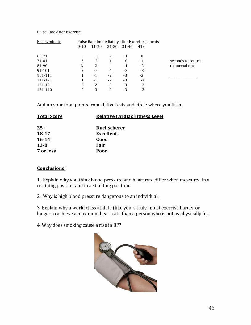
46
Pulse Rate After Exercise Beats/minute Pulse Rate Immediately after Exercise (# beats) 0-10 11-20 21-30 31-40 41+ 60-71 3 3 2 1 0 71-81 3 2 1 0 -1 seconds to return 81-90 3 2 1 -1 -2 to normal rate 91-101 2 0 -1 -3 -3 101-111 1 -1 -2 -3 -3 _________________ 111-121 1 -1 -2 -3 -3 121-131 0 -2 -3 -3 -3 131-140 0 -3 -3 -3 -3
Add up your total points from all five tests and circle where you fit in. Total Score Relative Cardiac Fitness Level 25+ Duchscherer 18-17 Excellent 16-14 Good 13-8 Fair 7 or less Poor Conclusions: 1. Explain why you think blood pressure and heart rate differ when measured in a reclining position and in a standing position.
2. Why is high blood pressure dangerous to an individual. 3. Explain why a world class athlete (like yours truly) must exercise harder or longer to achieve a maximum heart rate than a person who is not as physically fit. 4. Why does smoking cause a rise in BP?
47
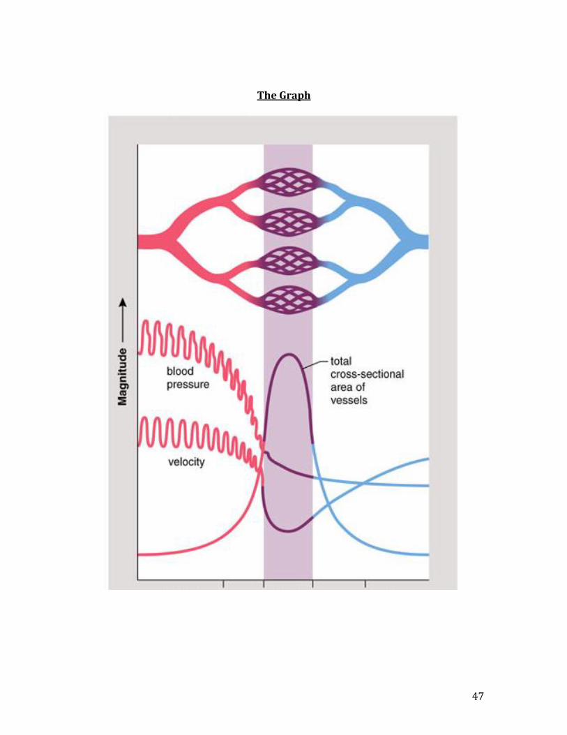
The Graph

48
Factors that Return Blood Back to the Heart Blood pressure in the vena cava is extremely low. The blood in the vena cava is further from the heart than any other blood vessel, so the blood pressure can approach zero. Several factor come into play to help return blood back to the heart. 1. Veins have one-way valves:
2. Gravity above the heart
3. Muscle contractions that squeeze on veins help push the blood toward the heart.
4. Respirtatory Pump
As the chest cavity expands prior to inhalation, the pressure drops inside the chest cavity. Blood will always flow to an area of lower pressure drawing the blood in the vena cava toward the heart.

49
Tissue Capillary Exchange

50
Tissue Capillary Exchange At the arteriole end of the capillary bed the blood pressure (the force pushing fluid out) is greater than the osmotic pressure (the force pulling water back in), so water is forced out the capillary bed. In this water is glucose, amino acids, and oxygen. This water is now called interstitial fluid. The substances diffuse into the cells. Halfway along the capillary bed the BP equals the OP because the BP is dropping as we get further from the heart. Because of this, there is no net movement of water but substances diffuse along their concentration gradient. Oxygen will diffuse from the capillary to the cells while carbon dioxide diffuses from the interstitial fluid into the capillary.
At the venous end of the capillary bed, the OP is now greater than the BP and water is drawn back into the capillary bed carrying in wastes from the cells. Any excess interstitial fluid enters the lymph capillary and becomes lymph fluid. The lymph capillaries join to become lymph veins. These veins have nodes located along them to trap foreign substances like bacteria, cancer cells and viruses. Lymphocytes reside in lymph nodes to fight these substances. Eventually the clean lymph is returned back into the blood at the subclavian and jugular veins.
51
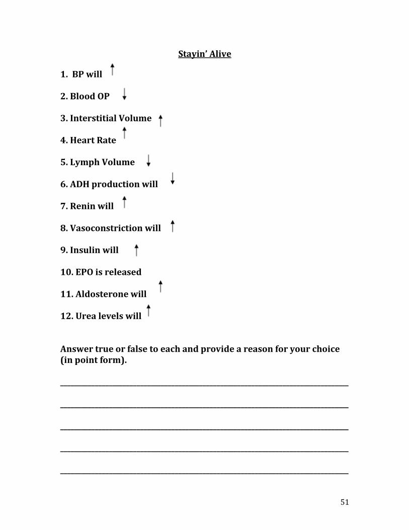
Stayin’ Alive
1. BP will 2. Blood OP 3. Interstitial Volume 4. Heart Rate 5. Lymph Volume 6. ADH production will 7. Renin will 8. Vasoconstriction will 9. Insulin will 10. EPO is released 11. Aldosterone will 12. Urea levels will Answer true or false to each and provide a reason for your choice (in point form). ___________________________________________________________________________________ ___________________________________________________________________________________ ___________________________________________________________________________________ ___________________________________________________________________________________ ___________________________________________________________________________________

52
___________________________________________________________________________________ ___________________________________________________________________________________ ___________________________________________________________________________________ ___________________________________________________________________________________ ___________________________________________________________________________________ ___________________________________________________________________________________ ___________________________________________________________________________________ ___________________________________________________________________________________ ___________________________________________________________________________________ ___________________________________________________________________________________ ___________________________________________________________________________________ ___________________________________________________________________________________ ___________________________________________________________________________________ ___________________________________________________________________________________ ___________________________________________________________________________________ ___________________________________________________________________________________ ___________________________________________________________________________________ ___________________________________________________________________________________ ___________________________________________________________________________________ ___________________________________________________________________________________
53
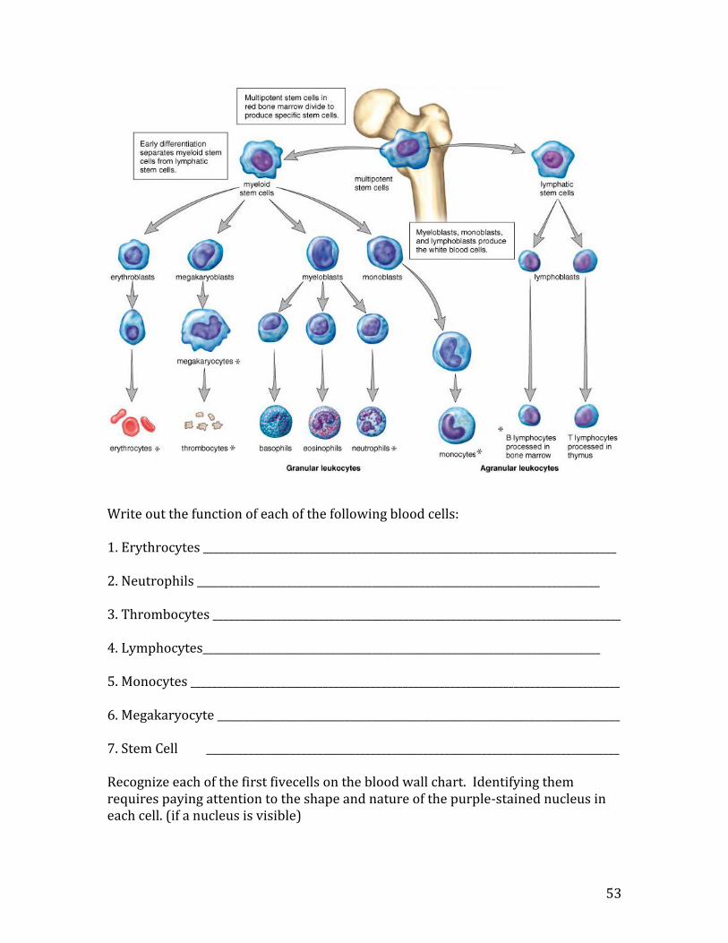
Write out the function of each of the following blood cells: 1. Erythrocytes ______________________________________________________________________________ 2. Neutrophils ____________________________________________________________________________ 3. Thrombocytes _____________________________________________________________________________ 4. Lymphocytes___________________________________________________________________________ 5. Monocytes _________________________________________________________________________________ 6. Megakaryocyte ____________________________________________________________________________ 7. Stem Cell ______________________________________________________________________________ Recognize each of the first fivecells on the blood wall chart. Identifying them requires paying attention to the shape and nature of the purple-stained nucleus in each cell. (if a nucleus is visible)

54
Hemoglobin A globular protein capable of carrying oxygen, carbon dioxide, or H+. Hemoglobin works hard to keep the blood and tissues clean. Hemoglobin contains four heme groups with an iron atom in the middle capable of attracting oxygen.

55
The Intrinsic Beat of the Heart Individual heart cells will beat rhythmically on their own when isolated. When cardiac cells are functioning as an organ the cardiac cells will beat together as a unit. The cells with the fastest rate determine the pace of the rest of the cells. The SA node or sino-atrial node is a special group of nerve cells in the right atrium that determine the rate for the rest of the heart. The SA node transmits its impulse as a wave of contraction across the atria and then to the ventricles. To transmit the impulse to the ventricles another relay collection of nerve cells called the AV node or atrial-ventricular node in the right atrium passes the impulse to the Purkinje fibres which innervate the ventricular muscle. This allows the two ventricles to contract as the two atria are relaxing.
SA AV Purkinje fibres