
Obg seminar
79
BLEEDING DISORDERS IN LATE PREGNANCY DEEPTHY P. THOMAS II YEAR M.S.c. NURSING GOVT. COLLEGE OF NURSING ALAPPUZHA
-
Upload
deepthy-philip-thomas -
Category
Healthcare
-
view
442 -
download
2
Transcript of Obg seminar

BLEEDING
DISORDERS IN LATE
PREGNANCY
DEEPTHY P. THOMAS
II YEAR M.S.c. NURSING
GOVT. COLLEGE OF NURSING
ALAPPUZHA

ANTEPARTUM HAEMORRHAGE:
Antepartum haemorrhage is defined as bleeding from or into the genital tract after the 28 week of pregnancy but before the birth of the baby.

CAUSES
APH
Placental bleeding unexplained extra placental
Placenta abruptio local cervicoprevia placenta vaginal lesions
-Cervical polyp-Carcinoma cervix-Varicose vein-Local trauma
PLACENTA PRAEVIA
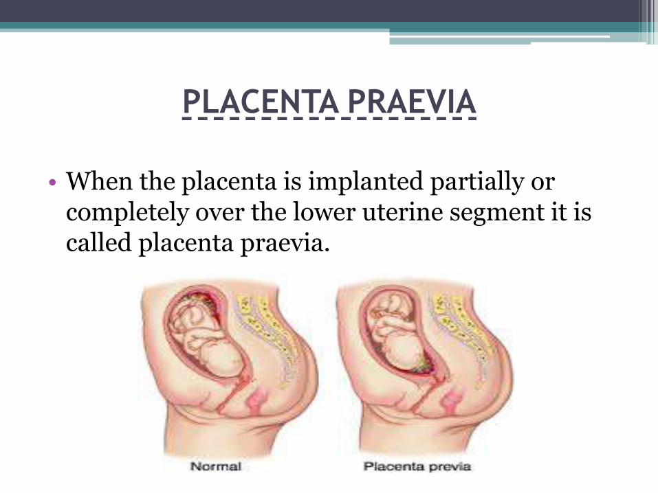
• When the placenta is implanted partially or completely over the lower uterine segment it is called placenta praevia.

INCIDENCE
0.5-1% amongst hospital deliveries.
One third of all cases of antepartumhaemorrhage are due to placenta previa.
Higher incidences have been reported in multiparous, multiple pregnancies, elderly woman [ over 35 years]

ETIOLOGY
Dropping theory
• The fertilized ovum drops down and implanted in the lower uterine segment. Poor decidualreaction in the upper uterine segment may be the cause. Failure of zona pellucida to disappear at time can be hypothetical possibility. This explains the formation of central placenta previa.

Persistence of chorionic activity
• Chorionic activity in the deciduas capsularis and its subsequent development into capsular placenta which comes in contact with deciduavera of the lower segment can explain the formation of lesser degrees of placenta previa.
Defective deciduas
• It results in the spreading of the chorionic villiover wide area in the uterine wall to get nourishment. During this process not only the placenta become membraneous but encroaches onto lower segment. Such a placenta may invade the underlying decidua or myometrium to cause placenta accrete, increta or percreta.
Big surface area of the placenta
• Big placenta as in twins may encroach onto lower segment.
PREDISPOSING FACTORS
Multiparity
Increased maternal age
History of previous caesarean section or any other scars in the uterus.
Placental size
Smoking
Leiomyomas distorting uterine cavity
Congenital malformations of the uterus.

PATHOLOGICAL ANATOMY
Placenta
• The placenta may be large and thin. This is often tongue shaped extension from the main placental mass. Extensive areas of degeneration with infarction and calcification may evident. The placenta may be morbidly adherent due to poor decidual formation in the lower segment.

Umbilical cord
• The cord may be attached to the margin or into the membranes. The insertion of the cord may be close to the internal os or the fetus blood vessels may run across the internal os in velamentous insertion due to poor placental formation

Lower uterine segment
• Due to increased vascularity, the lower uterine segment and the cervix becomes soft and more friable.

TYPES OR DEGREES
Type I (low lying)
Type II (marginal)

Type III (incomplete or partial central)
Type IV (central or total)

Dangerous placenta previa is the name given to the type-II placenta previa.
• Because of curved birth canal major thickness of the placenta overlies the sacral promontory, thereby diminishing the antero-posterior diameter of the inlet and prevents engagement of the presenting part. This hinders effective compression of the separated placenta to stop bleeding.
• Placenta is more likely to compressed if vaginal delivery is allowed.
• More chance of cord compression or cord prolapsed.

CAUSE OF BLEEDING
• Thrombosis of the open sinuses.
• Mechanical pressure by the presenting part .
• Placental infarction.

Placental Migration
The term placental migration could be explained in two ways:
• With progressive increase in the length of lower uterine segment, the lower placental edge relocates away from the cervical os .
• Due to trophotropism (growth of trophoblastictissue towards the fundus), there is resolution of placenta previa.
CLINICAL FEATURES
Symptoms
• Vaginal bleeding
sudden onset, pain less, apparently causeless and recurrent

Signs
Abdominal examination
• Size of the uterus is proportionate to the period of gestation.
• The uterus feels relaxed, soft and elastic without any localized areas of tenderness.
• Persistence of malpresentations
• The head is floating in contrast to the period of gestation.

• The FHS is usually present unless there is major separation of the placenta. Slowing of the FHS on pressing the head down into the pelvis which soon recovers promptly as the pressure is released is suggestive of the presence of low lying placenta specially of posterior type it is known as Stallworthy’s sign.

Vulval inspection :
• Only inspection is to be done to check whether bleeding is still occurring or has ceased
• In placenta praevia the blood is bright red as the bleeding occurs from the separated uteroplacental sinuses close to the cervical opening and escape out immediately.
• Vaginal examination must not be done outside the operation theatre in the hospital.

DIAGNOSIS
History and clinical features
• Painless and recurrent vaginal bleeding in the second half of pregnancy should be taken as placenta praevia unless provoked otherwise.

Placentography
• Sonography
Transabdominal
• The accuracy after 30th week of gestation is about 98%.
Transvaginal (TVS)
• The probe is very close to the target area. it is more accurate than TAS.
Transperineal (TPS)
• It is well accepted by patients. Internal os is visualized in 97-100% of cases.

Color doppler flow study
• Prominent venous flow in the hypo-echoic areas near the cervix is consistent with the diagnosis of placenta praevia.
MRI
• It is a non invasive method without any risk of ionizing radiation.

Clinical confirmation of placenta preavia
• Double set up examination (vaginal examination)
• it is less frequently done these days. Indications are
• inconclusive USG report
• USG reveals type I placenta preavia
• USG facilities not available

Visualization of the placental implantation on the lower segment can be confirmed during caesarean section.
Examination of the placenta following vaginal delivery reveals;
• a tongue shaped, thin segment of placental tissue projecting beyond the main placental mass with evidences of degeneration.
• Rent on the membranes on the margin of the placenta.
• Abnormal attachment of the cord.

DIFFERENTIAL DIAGNOSIS
• Vasa preavia
• The local cervical lesions
• Circumvallate placenta
COMPLICATIONS
Maternal
During pregnancy
• APH with varying degrees of shock.
• Malpresentation is common.
• Premature labour: either spontaneous or induced.

During labour
• Early rupture of membranes.
• Cord prolase.
• Slow dilatation of the cervix.
• Intrapartum haemorrhage.
• Increased incidence of operative interference.
• Post partum haemorrhage
• Retained placenta
During puerperium
• Sepsis is increased due to operative interference.
▫ Placental site near the vagina
▫ Anemia and devitalized state of the patient
• Subinvolution
• Embolism

• Fetal
• Low birth weight
• Asphyxia: Early separation of placenta
Compression of the placenta or
Compression of the cord
• Intrauterine death
• Birth injuries
• Congenital malformation

PROGNOSISMaternal • Early diagnosis• Omission of internal examination outside the
hospital.• Free availability of blood transfusion facilities.• Potent antibiotics.• Wider use of caesarean section with expert
anaesthetist.• Skill and judgement with which the cases are
managed.

fetal
judicious extension of expectant treatment thereby reducing the loss from prematurity
liberal use of caesarean section which greatly lessens the loss from anoxia
improvement in the neonatal care unit.

MANAGEMENT
Prevention
• Adequate antenatal care.
• Antenatal diagnosis of low lying placenta.
• Significance of “ warning haemorrhage”.
• Family planning and limitation of births.
Management at home
• The patient is immediately put to bed.
• Assess the blood loss.
• Quick but gentle abdominal examination.
• Vaginal examination must not be done.

• Hospital treatment
• Shift the patient to an equipped.
• An intravenous dextrose saline drip.
• Patient should be accompanied by 2/3 persons fit for donation of blood if necessary.
• All cases of APH should be

TREATMENT ON ADMISSION
1. Immediate attention
• Amount of blood loss.
• Blood samples are taken for grouping, cross matching and Hb.
• A large bore IV cannula is sited and an infusion of normal saline is started.
• Gentle abdominal palpation.
• Inspection of the vulva.
• Confirmation of diagnosis.

• Formulation of line of treatment
Expectant management
Active management

Expectant management
• Vital prerequisites
• Availability of blood for transfusion
• Facilities for caesarean section
• Selection of cases
• Mother is in good health status.(Hb >10 gm%, haematocrit >30%)
• Duration of pregnancy is <37 weeks.
• Active vaginal bleeding is absent.
• Fetal well being is assured by USG.

Expectant management
• Bed rest.
• Investigations.
• Periodic inspection.
• Supplementary haematinics.
• When the patient is allowed out of bed a gentle speculum examination is made to exclude local cervical and vaginal lesions for bleeding.
• Use of tocolytics and cervical encirclage are not helpful.

• Expectant management is done in home
• Patient lives close to hospital
• 24 hour transportation is available
• Bed rest assured
• Patient is well motivated to understand the risks

Termination of the expectant treatment
• The expectant management is carried upto 37 weeks of pregnancy. By this time baby become mature.
• Premature termination is done in conditions such as Presence of brisk haemorrhage and which is continuing.
The fetus is dead.
The fetus is found congenitally malformed on investigation.
Steroid therapy
• It is indicated when duration of pregnancy is <34 weeks. Betamethasone reduces the risk of respiratory distress of the new born when preterm delivery is considered.

Active management (Delivery)
• Bleeding occurs at or after 37 weeks of pregnancy.
• Patient is in labour
• Patient in exsanguinated state on admission.
• Bleeding is continuing and of moderate degree.
• Baby is dead/congenitally deformed.

DEFINITIVE MANAGEMENT
• Vaginal examination in OT followed by Lower rupture of membranes or
Caesarean section
• Caesarean section without internal examination
Vaginal examination
• Contraindications
• Patient in exsanguinated state
• Diagnosed cases of major degrees of placenta preavia confirmed by USG.
• Associated complications like malpresentations, elderly primigravida, pregnancy with h/o previous caesarean section, contracted pelvis etc.

Low rupture of membranes
• Low rupture of membrane is done using long Kocher’s forceps in lesser degrees of placenta praevia (type I & type II anterior).
Precautions during vaginal delivery
• steps to restore the blood volume.
• Methergine 0.2 mg.
• Proper examination of the cervix should be done soon following delivery.
• Baby’s blood Hb level is to be checked.

Caesarean section
Indications
• Severe degree of placenta praevia .
• Lesser degree of placenta praevia where amniotomy fails to stop bleeding or fetal distress appears.
• Complicating factors associated with lesser degrees of placenta praevia where vaginal delivery is unsafe.
Caesarean section without internal examination
• If precise location of placenta.
• It should be performed by senior obstetrician with the help of a senior anesthetist.
• Regional anaesthesia is generally avoided.
• If patient is in shock, and the bleeding continues, operation has to be performed immediately along with restorative measures.
• Low transverse abdominal incision is to be avoided; intra umbilical longitudinal incision is preferred.

NURSING CONSIDERATIONS
• strict bed rest
• Teaching
• home visits for comprehensive fetal and maternal assessment
• specific, information about the condition of the fetus.
• Make the family understands

In patient care
• Nursing assessments
• Periodic electronic fetal monitoring
• Immediate delivery

ABRUPTIO PLACENTAE-DEFINITION
• It is one form of anteparum hemorrhage where the bleeding occurs due to premature separation of normally situated placenta.

VARIETIES• Concealed
• Mixed
• Revealed

ETIOLOGY
• High birth order pregnancies with gravid 5 and above- three times more common than in first birth.
• Advancing age of the mother.
• Poor socio economic condition.
• Malnutrition.
• Smoking

• Hypertension in pregnancy
• Trauma
• Sudden uterine decompression
• Short cord
• Supine hypotension syndrome
• Placental anomaly

PATHOGENESIS
• premature separation is initiated by haemorrhage into the deciduas basalis. The collected blood (decidual haematoma)
• The decidual hematoma may be small and self limited
• If major spiral artery ruptures, a big hematoma is formed.
• As the uterus remains distended by the conceptus, it fails to contract and therefore fails to compress the torn bleeding points.

• Couvelaire Uterus( Utero placental apoplexy )
It is a pathological entity in association with severe form of concealed abruption placentae. There is massive intravasation of blood into the uterine musculature upto the serous coat. The condition can only be diagnosed on laprotomy.

• Changes in other organs
• In the liver presence of fibrin knots in the hepatic sinusoids.
• Kidneys may show acute cortical necrosis or acute tubular necrosis.
• Shock protenuria

Blood coagulopathy:
• Blood coagulopathy is due to excess consumption of plasma fibrinogen due to disseminated intravascular coagulation and retro placental bleeding .There is overt hypo fibrinogenemia (< 150 mg / dl) and elevated levels of fibrin degradation products and D- dimer

Clinical Classification
• Grade -0 :
• Clinical features may be absent .The diagnosis is made after inspection of placenta following delivery.
• Grade -1 (40% ) :
• Vaginal bleeding is sight .
• Uterus : Irritable , tenderness may be minimal or absent.
• Maternal BP and fibrinogen levels unaffected.
• FHS is good.

• Grade -2 (45% ):
• Vaginal bleeding mild to moderate.
• Uterine tenderness is always present .
• Maternal pulse is always increased, BP maintained .
• Fibrinogen level may be decreased.
• Shock or even death occurs.
• Grade -3 (15% ):
• Bleeding is moderate to severe or may be concealed
• Uterine tenderness is marked .
• Shock is pronounced .
• Fetal death is the rule .
• Associated coagulation defect or anuria may complicate.

CLINICAL FEATURES OF ABRUPTIO
PLACENTAE
The clinical features depend on :
• Degree of separation of placenta ,
• Speed at which separation occurs and
• Amount of blood concealed inside the uterine cavity

DIFFERENTIAL DIAGNOSIS
Revealed type:
• Confusion with intermediate causes of APH is difficult to eliminate.
Mixed or concealed type:
• Rupture uterus, rectus sheath hematoma, appendicular or intestinal perforation, Twisted ovarian tumour, Volvulus, acute hyramnios, tonic uterine contraction.

COMPLICATIONS OF ABRUPTIO
PLACENTAMaternal• In revealed type • maternal risk is proportional to the visible
blood loss and maternal death is rare.• In concealed variety• Haemorrhage• Shock• Blood coagulation disorders • Oliguria and anuria• Postpartum haemorrhage• puerperal sepsis.

Fetal :
• In revealed type, the fetal death is to extent of 25- 30% .
• In concealed type, however, the fetal death is appreciably high, ranging from 50-100%.

MANAGEMENT OF ABRUPTIO
PLACENTAEPrevention:
• Elimination of known factors likely to produce placental separation.
• Correction of anemia during antenatal period so that the patient can withstand blood loss.
• Prompt detection and institution of the therapy to minimize the grave complication namely shock, blood coagulation disorders and renal failure.

• Prevention of known factors likely to cause placental separation are :
• Early detection and effective therapy of pre-eclampsia
• Needle puncture during amniocentesis should be under ultra sound guidance.
• Avoidance of trauma–specially forceful external cephalic version under anesthesia.
• To avoid sudden decompensation of the• To avoid supine hypotension• Routine administration of folic

• Treatment:
• At home
• In the hospital :
▫ Revealed type
• Amount of blood loss.
• Maturity of the fetus
• Whether the patient is in labor or not (usually labor starts).
• Presence of any complication.
• Type and grade of placental abruption.

• Emergency measures:
• Blood is sent.
• Ringer solution drip is started with a wide bore cannula and arrangement for blood transfusion is made for resuscitation.
• Close monitoring of maternal and fetal condition is done.

1.Definitive treatment
(immediate delivery):
• The patient is in labour:
▫ Vaginal delivery is favored in cases with :
Limited placental abruption .
FHR tracing is reassuring .
Facilities for continuous (electronic ) fetal monitoring is available .
Prospect of vaginal delivery is soon as or
Placental abruption of a dead fetus.

The patient is not in labour:
Pregnancy 37 weeks or greater:
Induction is done by low rupture of membranes with or without oxytocin.
Indications of caesarean section are:
Appearance of fetal distress.
Amniotomy couldn’t be done or amniotomyfails to control.
Associated complicating factors.
• Pregnancy less than 37 weeks:
Bleeding more to severe and continuing- low rupture of membranes is quite effective. Oxytocin drip may be added.
Bleeding slight or has stopped- the patient is put on conservative treatment as outlined in placenta praevia.

Induction of labour is done by low rupture of membrane
• Oxytocin may be added to expedite delivery.
• Placenta with varying amount of retro-placental clot is expelled in most often simultaneously with the delivery of the baby.
• Inj. oxytocin 10.IU IV slowly or IM or Inj. methergin 0.2 mg IV is given with the delivery of the baby to minimize postpartum blood loss.

Caesarean Section :
• Severe abruption of with live fetus.
• Amniotomy could not be done (un-favorable cervix).
• Prospect of immediate vaginal delivery despite amniotomy failed to arrest the process of abruption (rising fundal height).
• Appearance of adverse (fetal distress, falling fibrinogen level, oliguria).
• Anesthesia during caesarean section :
• Regional anesthesia is generally avoided when there is significant hemorrhage.

• 2.Expectant management in case of placental abruption is an exception and not the rule :
• Cases where bleeding is slight and has stopped (grade I abruption), fetus reactive (CTG ) and remote from term, may be considered.
• Continuous electric fetal monitoring • Patient should be observed in the labour ward
for 24-48 hours.• betamethasone is given to accelerate fetal lung
maturity in the event preterm delivery has to be contemplated.

Management of complications :
The major complications of placental abruption are :
• Haemorrhagic shock .
• DIC
• Renal failure.
• Uterine atony and post partum haemorrhage.
NURSING MANAGEMENT OF BLEEDING
IN LATE PREGNANCY
Nursing assessment:
• Amount and nature of blood
• Pain type and location
• Maternal vital signs.
• Uterine contractions.
• Obstetric history: gravid, para, previous abortions, preterm infants, previous pregnancy outcomes.
• Length of gestation:LMP, fundal height, co-orelationof fundal height with estimated gestation.
• Laboratory data

Nursing diagnosis
• Deficient fluid volume related to excessive vascular lose as evidenced by hypotension, increased pulse rate, decreased/concentrated urine.
• Ineffective utero-placental tissue perfusion related to hypovolemia as evidenced by changes in fetal heart rate and activity.
• Activity Intolerance related to enforced bed rest during pregnancy secondary to potential for hemorrhage
• Anxiety related to unknown effects of bleeding and lack of knowledge of predicted course of management

THANK YOU…..


