Obesity - publications.iarc.fr€¦ · Obesity is multifactorial Generally defined, obesity is a...
12
Unit 5 • Chapter 24. Obesity 441 UNIT 5 CHAPTER 24 UNIT 5. APPLICATION OF BIOMARKERS TO DISEASE CHAPTER 24. Obesity Salma Musaad and Erin Haynes Summary The adverse effects of obesity support the use of biomarkers to help elucidate disease mechanism, therapeutic interventions, and preventive strategies. Emerging biomarkers for obesity-associated cardiovascular disease (CVD), type 2 diabetes and cancer play diverse roles in biological pathways including immune modulation and fat metabolism. Animal and in vitro data support the association of these biomarkers with obesity-associated diseases, but evidence in humans is still lacking. In humans, plasma levels of biomarkers are widely used to determine risk, but many studies are limited by ethnicity/race, gender or sample size. In this chapter, the use of biomarkers in obesity research and in the context of CVD, type 2 diabetes and cancer will be discussed. Markers of exposure (adipokines), effect (resulting metabolic abnormalities), and susceptibility (genetic determinants for obesity and related disorders) are covered for each of the three diseases. Introduction Obesity epidemiology has typically relied upon long-established markers, such as blood cholesterol, triglycerides and blood pressure. It is now recognized that novel, non-traditional biomarkers have the potential to augment the utility of traditional markers. Emerging as a more formative tool in obesity epidemiology, the majority of non- traditional biomarkers act in relation to fat cells, or adipocytes. Acting as endocrine organs, adipocytes produce a variety of peptides and metabolites that result in a cascade of events leading to inflammation and oxidative stress. These products are being explored as biomarkers for the prevention, diagnosis, risk stratification and control of obesity co-morbidities, such as cardiovascular disease (CVD) (1,2), type 2 diabetes (3,4) and certain cancers (5,6). Despite the dangerous health effects of obesity, little is known regarding the clinical utility of adipokines in modifying disease risk, especially cancer. This chapter focuses on the use of non-traditional biomarkers in obesity research and more specifically, in
Transcript of Obesity - publications.iarc.fr€¦ · Obesity is multifactorial Generally defined, obesity is a...
Unit 5 • Chapter 24. Obesity 441
Un
it 5
Ch
ap
ter
24
unit 5.application of biomarkers to disease
chapter 24.
ObesitySalma Musaad and Erin Haynes
Summary
The adverse effects of obesity support the use of biomarkers to help elucidate disease mechanism, therapeutic interventions, and preventive strategies. Emerging biomarkers for obesity-associated cardiovascular disease (CVD), type 2 diabetes and cancer play diverse roles in biological pathways including immune modulation and fat metabolism. Animal and in vitro data support the association of these biomarkers with obesity-associated diseases, but evidence in humans is still lacking. In humans, plasma levels of biomarkers are widely used to determine risk, but many studies are limited by ethnicity/race, gender or sample size. In this chapter, the use of biomarkers in obesity research and in the context of CVD,
type 2 diabetes and cancer will be discussed. Markers of exposure (adipokines), effect (resulting metabolic abnormalities), and susceptibility (genetic determinants for obesity and related disorders) are covered for each of the three diseases.
Introduction
Obesity epidemiology has typically relied upon long-established markers, such as blood cholesterol, triglycerides and blood pressure. It is now recognized that novel, non-traditional biomarkers have the potential to augment the utility of traditional markers. Emerging as a more formative tool in obesity epidemiology, the majority of non-
traditional biomarkers act in relation to fat cells, or adipocytes. Acting as endocrine organs, adipocytes produce a variety of peptides and metabolites that result in a cascade of events leading to inflammation and oxidative stress. These products are being explored as biomarkers for the prevention, diagnosis, risk stratification and control of obesity co-morbidities, such as cardiovascular disease (CVD) (1,2), type 2 diabetes (3,4) and certain cancers (5,6). Despite the dangerous health effects of obesity, little is known regarding the clinical utility of adipokines in modifying disease risk, especially cancer.
This chapter focuses on the use of non-traditional biomarkers in obesity research and more specifically, in
442
the context of CVD, type 2 diabetes and cancer. Adipokines will be referred to as markers of exposure and the resulting metabolic abnormalities as markers of effect. Genetic determinants for obesity and related disorders are referred to as markers of susceptibility. Markers of exposure, effect and susceptibility are discussed for each of the three co-morbidities.
Obesity is multifactorial
Generally defined, obesity is a state of excess weight gain and increased body fat that is disproportionate to the individual’s height. Obesity is a prevalent disorder adversely impacting quality of life (7,8) and life expectancy (9). In the USA it is estimated that 32% of adults and 17% of children and adolescents aged 2–19 years were obese in 2003–2004, a dramatic increase from the previous two decades (10), justifying the need for prevention of obesity and related disorders. Caused by a combination of genetic, metabolic and environmental factors, obesity is characterized by an imbalance between energy intake and energy expenditure. This imbalance is closely regulated by signals emanating from and controlled by the central nervous system.
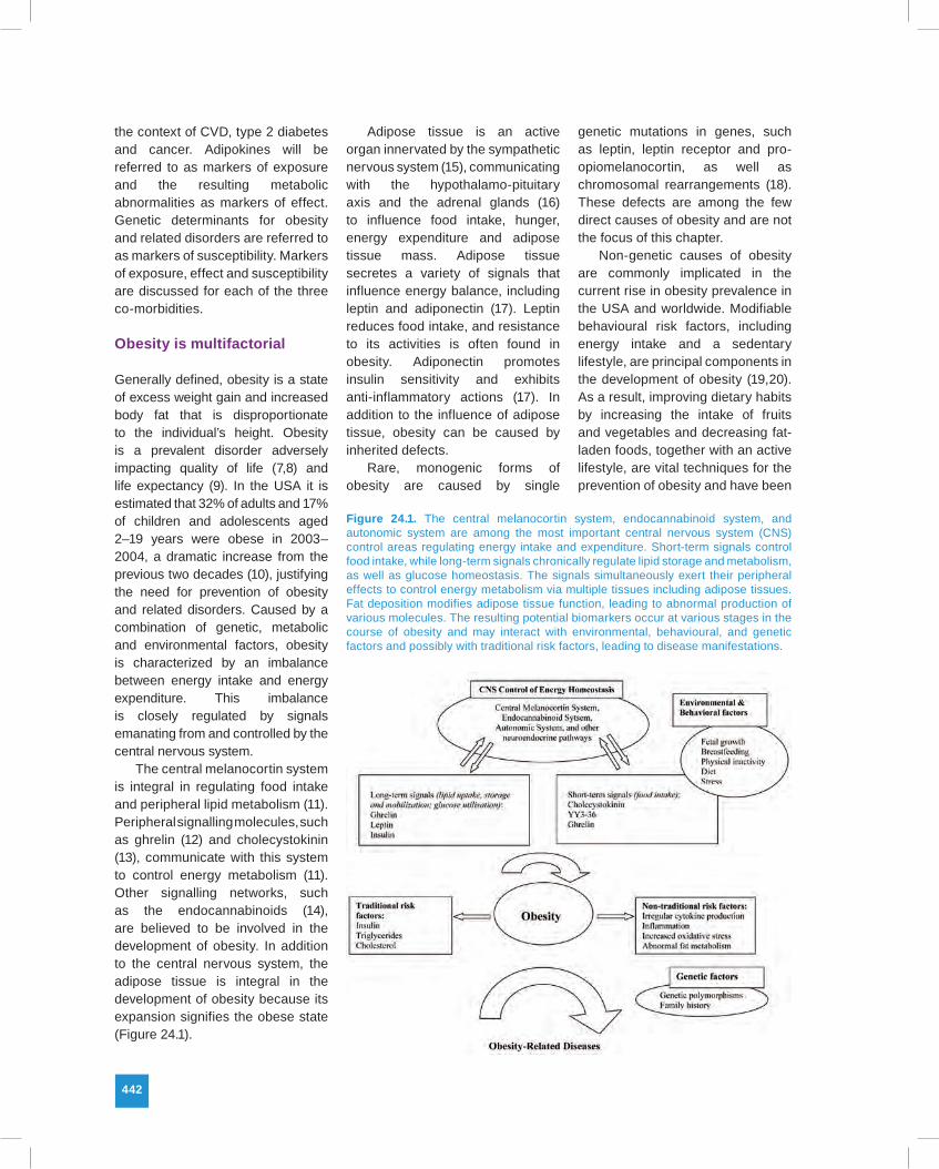
The central melanocortin system is integral in regulating food intake and peripheral lipid metabolism (11). Peripheral signalling molecules, such as ghrelin (12) and cholecystokinin (13), communicate with this system to control energy metabolism (11). Other signalling networks, such as the endocannabinoids (14), are believed to be involved in the development of obesity. In addition to the central nervous system, the adipose tissue is integral in the development of obesity because its expansion signifies the obese state (Figure 24.1).
Adipose tissue is an active organ innervated by the sympathetic nervous system (15), communicating with the hypothalamo-pituitary axis and the adrenal glands (16) to influence food intake, hunger, energy expenditure and adipose tissue mass. Adipose tissue secretes a variety of signals that influence energy balance, including leptin and adiponectin (17). Leptin reduces food intake, and resistance to its activities is often found in obesity. Adiponectin promotes insulin sensitivity and exhibits anti-inflammatory actions (17). In addition to the influence of adipose tissue, obesity can be caused by inherited defects.
Rare, monogenic forms of obesity are caused by single
genetic mutations in genes, such as leptin, leptin receptor and pro-opiomelanocortin, as well as chromosomal rearrangements (18). These defects are among the few direct causes of obesity and are not the focus of this chapter.
Non-genetic causes of obesity are commonly implicated in the current rise in obesity prevalence in the USA and worldwide. Modifiable behavioural risk factors, including energy intake and a sedentary lifestyle, are principal components in the development of obesity (19,20). As a result, improving dietary habits by increasing the intake of fruits and vegetables and decreasing fat-laden foods, together with an active lifestyle, are vital techniques for the prevention of obesity and have been
Figure 24.1. The central melanocortin system, endocannabinoid system, and autonomic system are among the most important central nervous system (CNS) control areas regulating energy intake and expenditure. Short-term signals control food intake, while long-term signals chronically regulate lipid storage and metabolism, as well as glucose homeostasis. The signals simultaneously exert their peripheral effects to control energy metabolism via multiple tissues including adipose tissues. Fat deposition modifies adipose tissue function, leading to abnormal production of various molecules. The resulting potential biomarkers occur at various stages in the course of obesity and may interact with environmental, behavioural, and genetic factors and possibly with traditional risk factors, leading to disease manifestations.
Unit 5 • Chapter 24. Obesity 443
Un
it 5
Ch
ap
ter
24
the focus of large obesity prevention trials (21).
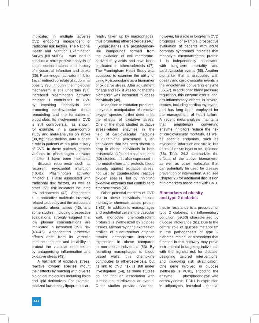
Just as obesity is a complex disorder, molecular biomarkers that occur in obesity and that help identify individuals most at risk for subsequent cardiovascular disease, diabetes or cancer are complex. There is a significant degree of functional overlap among the biomarkers, and in some cases it is hard to discern the order in which they first appear in the body. For simplicity, however, the biomarkers have been broadly classified into exposure, effect or susceptibility (Table 24.1).
Overview of inflammation and oxidative stress
Abnormal adipokine production with consequent inflammation and oxidative stress may play an instrumental role in obesity-related disorders. Increased accumulation of adipose tissue, particularly visceral obesity, causes abnormal cytokine release and macrophage recruitment, all of which induce systemic inflammation (22).
Accumulation of adipose tissue also leads to increased oxidative stress partly via the oxidant effects of free fatty acids (23). Leukocytes derived from obese individuals and healthy individuals infused
with free fatty acids generate reactive oxygen species (24,25). Consequently, reactive oxygen species induce a pro-inflammatory state and promote adverse metabolic complications, such as insulin resistance (23). Despite the association of oxidative stress with CVD independent of traditional risk factors (26), prospective clinical trials of antioxidant supplementation for reducing CVD risk provide conflicting evidence (27,28), possibly due to lack of a strong effect of oxidative stress on atherosclerosis, choice of antioxidant therapy, confounding by other dietary and non-dietary factors, or unsuitable choice of the biomarker. Nevertheless, oxidative stress is increasingly recognized as a potential mechanism for obesity-related disorders, hence it is included in this chapter.
Owing to the clear link between fat accumulation, inflammation and oxidative stress, the following three sections focus on these mechanisms in the context of CVD, diabetes and cancer. The sections are not intended to be all-inclusive, but aim to introduce the reader to major biomarkers of potential benefit in disease prevention and early detection. A brief introduction to the major epidemiologic study designs used to evaluate the biomarkers
and common methodological issues then follows.
Biomarkers of obesity and subsequent cardiovascular disease
For the purposes of this section, cardiovascular disease (CVD) outcomes comprise cerebrovascular disease (cerebral embolism, thrombosis and haemorrhage), peripheral arterial disease, coronary heart disease, and atherosclerosis. Different mechanisms are implicated in the link between obesity and CVD. For example, the fetal origin of metabolic risk (29), epigenetic gene regulation (30), and the “pup in a cup” model (31) are potential causes of increased CVD risk in obesity. In addition, cardiovascular injury is promoted by adipokines, cytokines and other molecules that affect multiple pathways, such as lipid metabolism and immune modulation, eventually leading to inflammation or oxidative stress (32).
Inflammatory biomarkers that are elevated in obesity include leptin, plasminogen activator inhibitor 1, and adiponectin. Beyond regulation of energy expenditure, leptin induces a myriad of inflammatory mediators (33) and alters myocardial structure (34). Leptin has extensive regulatory functions and has been
Table 24.1. Classification of molecular biomarkers for obesity and subsequent cardiovascular disease, diabetes or cancer
Class Molecular biomarkers Examples of studies
Exposure Adipokines: surrogates for adipose tissue deposition Characterise adipose tissue type and activity; association with disease outcome; assess change with weight loss
Effect Markers of inflammation and oxidative stress: mechanisms by which obesity may exert its toxic effects
Monitor disease progression; association with disease outcome; disease prognosis; disease intervention
Genetic susceptibility Gene polymorphisms: account for variation in susceptibility to obesity-related disorders
Gene-phenotype relations; heritable variations in the quantity of systemic biomarkers; risk stratification
444
implicated in multiple adverse CVD endpoints independent of traditional risk factors. The National Health and Nutrition Examination Survey (NHANES) III was used to conduct a retrospective analysis of leptin concentrations and history of myocardial infarction and stroke (35). Plasminogen activator inhibitor 1 is an indirect correlate of abdominal obesity (36), though the molecular mechanism is still uncertain (37). Increased plasminogen activator inhibitor 1 contributes to CVD by impairing fibrinolysis and promoting cardiovascular tissue remodeling and the formation of blood clots. Its involvement in CVD is still controversial, as shown, for example, in a case–control study and meta-analysis on stroke (38,39); nevertheless, data suggest a role in patients with a prior history of CVD. In these patients, genetic variants in plasminogen activator inhibitor 1 have been implicated in disease recurrence such as recurrent myocardial infarction (40,41). Plasminogen activator inhibitor 1 is also associated with traditional risk factors, as well as other CVD risk indicators including low adiponectin (42). Adiponectin is a protective molecule inversely related to obesity and the associated metabolic abnormalities (43), and some studies, including prospective evaluations, strongly suggest that low plasma concentrations are implicated in increased CVD risk (43–45). Adiponectin’s protective effects arise from its versatile immune functions and its ability to protect the vascular endothelium by antagonising inflammation and oxidative stress (43).
A hallmark of oxidative stress, reactive oxygen species mount their effects by reacting with diverse biological molecules including lipids and lipid derivatives. For example, oxidized low density lipoproteins are
readily taken up by macrophages, thus promoting atherosclerosis (46). F2-isoprostanes are prostaglandin-like compounds formed from the oxidation of cell membrane-derived fatty acids and have been implicated in atherosclerosis (47). The Framingham Heart Study was accessed to examine the utility of using F2-isoprostane as a biomarker of oxidative stress. After adjustment for age and sex, it was found that the biomarker was increased in obese individuals (48).
In addition to oxidation products, enzymatic manipulation of reactive oxygen species further determines the effects of oxidative stress. One of the most studied oxidative stress-related enzymes in the field of cardiovascular medicine is glutathione peroxidase 1, an antioxidant that has been shown to drop in obese individuals in both prospective (49) and cross-sectional (50) studies. It is also expressed in the endothelium and protects blood vessels against oxidative stress, not just by counteracting reactive oxygen species, but by inhibiting oxidative enzymes that contribute to atherosclerosis (51).
Other potential markers of CVD risk in obese individuals include monocyte chemoattractant protein 1 (52). In addition to macrophages and endothelial cells in the vascular wall, monocyte chemoattractant protein 1 is synthesized by adipose tissues. Microarray gene expression profiles of subcutaneous adipose tissues demonstrate increased expression in obese compared to non-obese individuals (53). By recruiting macrophages to blood vessel walls, this chemokine contributes to atherosclerosis, but its link to CVD risk is still under investigation (54), as some studies do not find an association with subsequent cardiovascular events. Other studies provide evidence,
however, for a role in long-term CVD prognosis. For example, prospective evaluation of patients with acute coronary syndromes indicates that monocyte chemoattractant protein 1 is independently associated with long-term mortality and cardiovascular events (55). Another biomarker that is associated with obesity and cardiovascular events is the angiotensin converting enzyme (56,57). In addition to blood pressure regulation, this enzyme exerts local pro-inflammatory effects in several tissues, including cardiac myocytes, and has long been employed for the management of heart failure. A recent meta-analysis maintains that angiotensin converting enzyme inhibitors reduce the risk of cardiovascular mortality, as well as specific endpoints, such as myocardial infarction and stroke, but the mechanism is yet to be explained (58). Table 24.2 summarizes the effects of the above biomarkers, as well as other molecules that can potentially be used for disease prevention or intervention. Also, see Chapter 20 for additional discussion of biomarkers associated with CVD.
Biomarkers of obesity and type 2 diabetes
Insulin resistance is a precursor of type 2 diabetes, an inflammatory condition (59,60) characterized by glucose intolerance (61). Due to the central role of glucose metabolism in the pathogenesis of type 2 diabetes, molecular biomarkers that function in this pathway may prove instrumental in targeting individuals with the highest risk for disease, designing tailored interventions, and improving risk stratification. One gene involved in glucose synthesis is PCK1, encoding the enzyme phosphoenolpyruvate carboxykinase. PCK1 is expressed in adipocytes, intestinal epithelia,
Unit 5 • Chapter 24. Obesity 445
Un
it 5
Ch
ap
ter
24
and hepatocytes. Hepatic overexpression is associated with a diabetic phenotype in mice and overexpression in adipose tissues causes obesity. Among the few human studies performed to date, some show a link between PCK1 variants with type 2 diabetes (62). Another gene, ectonucleotide pyrophosphatase/phosphodiesterase 1, has shown conflicting associations with type 2 diabetes and obesity, but its role as an inhibitor of insulin signalling warrants further examination as a potential candidate gene for obesity-associated type 2 diabetes. In some populations, polymorphisms in this gene are associated with childhood and adult obesity, as well as type 2 diabetes in obese individuals (63–65) (see Chapter 7). Further population-based studies are needed to validate its use as a predictive biomarker. Glucose homeostasis is also regulated by several obesity-associated adipokines that control multiple immune pathways. Low adiponectin concentrations (66,67) and high
tumour necrosis factor-α levels (68–70) have often been associated with insulin resistance. Despite conflicting evidence, promoter polymorphisms in the tumour necrosis factor-α gene have been implicated in increased insulin resistance, particularly in obese adults with type 2 diabetes (71–73). Further, one large (n = 809) population-based cross-sectional study of unrelated Caucasians showed that this gene interacts with adiponectin resulting in lower adiponectin levels and higher glucose and insulin concentrations two hours after glucose administration (74).
In addition to glucose, perturbed fatty acid metabolism, uptake and transport has been implicated in insulin resistance and diabetes. Uptake of fatty acids is partly controlled by fatty acid translocase (CD36), which regulates long chain fatty acid transport in skeletal muscles and adipose tissue (75). Subcutaneous adipose tissue expression of this binding protein was increased in obese individuals and further increased in those with
type 2 diabetes (75). A promoter polymorphism in CD36 was also linked with insulin resistance and type 2 diabetes (76). Interestingly, CD36 is linked to oxidative stress, because it is a scavenger receptor for oxidized lipoproteins on the surface of macrophages, rendering them insulin-resistant (77).
Other genetic candidates include stearoyl-coenzyme A desaturase type 1 (SCD1) and 11β-hydroxysteroid dehydrogenase type 1 (11HSD1), a glucocorticoid-amplifying enzyme. Genetic variants in the fatty acid metabolizing enzyme SCD1 have been linked with decreased waist circumference and improved insulin sensitivity in adults (78). The glucocorticoid-amplifying enzyme 11HSD1 is an intriguing molecule with varying roles ranging from regulation of adipocyte differentiation to possible amplification of macrophage-driven adipose tissue inflammation in obesity (79). It stimulates lipid synthesis in the intra-abdominal fat depots of diet-induced obese mice (80). Several studies find
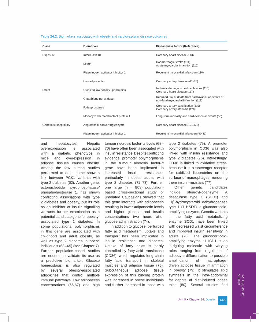
Table 24.2. Biomarkers associated with obesity and cardiovascular disease outcomes
Class Biomarker Disease/risk factor (Reference)
Exposure Interleukin 18 Coronary heart disease (113)
Leptin Haemorrhagic stroke (114) Acute myocardial infarction (115)
Plasminogen activator inhibitor 1 Recurrent myocardial infarction (116)
Low adiponectin Coronary artery disease (43-45)
Effect Oxidized low density lipoproteins Ischemic damage in cortical lesions (115)Coronary heart disease (117)
Glutathione peroxidase Reduced risk of death from cardiovascular events or non-fatal myocardial infarction (118)
F2-Isoprostanes Coronary artery calcification (119)Coronary artery stenosis (120)
Monocyte chemoattractant protein 1 Long-term mortality and cardiovascular events (55)
Genetic susceptibility Angiotensin-converting enzyme Coronary heart disease (121,122)
Plasminogen activator inhibitor 1 Recurrent myocardial infarction (40,41)
446
dysregulated adipose tissue activity in obesity (81) and type 2 diabetes (82), including one prospective study (83). Lipid storage and adipocyte differentiation is partially regulated by peroxisome proliferator-activated receptor gamma (PPAR-γ). This nuclear receptor is highly expressed in adipose tissues and favourably controls the release of several adipokines, such as adiponectin, leptin, resistin, interleukin 6 and monocyte chemoattractant protein 1, mounting anti-inflammatory and anticoagulant actions that intensely counteract the adipose tissue dysfunction plaguing obesity (84,85). Notably, PPAR-γ improves glucose uptake and insulin sensitivity, as evidenced by the actions of receptor agonists for treatment of type 2 diabetes. According to multiple investigations, a Pro12Ala polymorphism that decreases receptor activity, protects against hyperinsulinemia, type 2 diabetes (86), and high free fatty
acid concentrations (87). The Ala12 carriers show up to a 19% risk reduction, and the protective effect is greatest at a lower body mass index (BMI) (88). It is important to note that, as in the case with any gene, the effects of this variant can be modified by other genetic influences, such as variants within the same or other genes, and by environmental factors, such as diet, BMI or physical activity. Indeed, physical activity was found to modify its effect in one follow-up study (89). LDL receptor-related protein 1 is another vital regulator of systemic lipid transport and absorption in liver, muscle, heart and adipocytes. This receptor plays a role in the uptake and hydrolysis of triglyceride-rich lipoproteins. Adipose-specific knockout mice are protected from diet-induced obesity and exhibit increased metabolic rate and glucose tolerance (90). By understanding the potential role of LDL receptor-related protein
1 in humans, this receptor can potentially serve as a valuable marker for conferring susceptibility to obesity and type 2 diabetes in individuals at risk. In addition to the above biomarkers, other candidates for obesity-associated insulin resistance and type 2 diabetes are highlighted in Table 24.3.
Biomarkers of obesity and cancer
Cancer is a condition of uncontrolled cell growth triggered by a variety of factors. It is estimated that one third of all cancer deaths in 2006 were related to physical inactivity, nutrition and obesity (91). Compared to other known genetic or environmental risk factors for cancer, obesity may play a minor role. Nevertheless, its effect can be magnified in susceptible individuals, such as those with a family history of cancer or who belong to a particular race or gender.
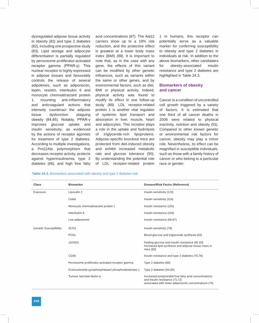
Table 24.3. Biomarkers associated with obesity and type 2 diabetes risk
Class Biomarker Disease/Risk Factor (Reference)
Exposure Lipocalin 2 Insulin sensitivity (123)
Cideb Insulin sensitivity (124)
Monocyte chemoattractant protein 1 Insulin resistance (125)
Interleukin 8 Insulin resistance (126)
Low adiponectin Insulin resistance (66,67)
Genetic Susceptibility SCD1 Insulin sensitivity (78)
PCK1 Blood glucose and triglyceride synthesis (62)
11HSD1 Fasting glucose and insulin resistance (82,83)Increased lipid synthesis and adipose tissue mass in mice (80)
CD36 Insulin resistance and type 2 diabetes (75,76)
Peroxisome proliferator-activated receptor gamma Type 2 diabetes (86)
Ectonucleotide pyrophosphatase/ phosphodiesterase 1 Type 2 diabetes (64,65)
Tumour necrosis factor-α Increased postprandial free fatty acid concentrations and insulin resistance (71,72)associated with lower adiponectin concentrations (74)
Unit 5 • Chapter 24. Obesity 447
Un
it 5
Ch
ap
ter
24
Defined using BMI or a high upper body (central) fat distribution, obesity modified risk for the development and progression of cancers affecting multiple target organs, including the gastrointestinal system (92–94), ovaries (95), breasts (96) and prostate (97). For example, women with a high BMI exhibited cytological abnormalities in the breast that may predispose to cancer (98). A few of the many possible mechanisms implicated in these findings is detection bias, hormonal imbalance (e.g. sex hormones or insulin) or genetic predisposition. Notably, abnormal adipokine regulation is another potential mechanism for this predisposition.
In the obese environment, insulin stimulates leptin activity in breast cancer cells (99), and an imbalance in leptin and adiponectin secretion is highly implicated as one mechanism for breast cancer development (100). Leptin exerts mitogentic and antiangiogenic effects that appear to be counteracted by adiponectin, which is decreased in obesity and protects against breast cancer (101). High leptin or low adiponectin levels are also implicated in other malignancies, such as non-Hodgkin
lymphoma (102) and endometrial cancer (103).
In addition to adipokines, genetic variants in lipid metabolizing genes are associated with breast cancer, such as the leptin receptor and the paraoxonase gene (PON1), which prevents low density lipoprotein oxidation. Polymorphisms in these genes are protective against breast cancer development in postmenopausal Caucasian women with benign breast disease (104) (these findings should be interpreted with caution due to the small number of cases (61 cases out of a total of 994)). The aforementioned molecules and other molecules putatively implicated in obesity-linked cancer are summarized in Table 24.4.
Common epidemiological study designs and methodological issues
Multiple epidemiologic study designs have been used to examine putative biomarkers in human populations (see Chapter 14). Case–control and cross-sectional studies are among the most common designs. Relatively cheap and rapid, the
designs are a sound stepping stone for collecting background information on the desired criteria for any biomarker, including average plasma concentrations, inter-individual and intra-individual variability, effect size in cases compared to controls, stability, half-life, circadian variation and ethnic/racial differences, as well as age and gender effects. Several biomarkers illustrated in this chapter have been preliminarily identified and repeatedly investigated using these designs to justify further study in more demanding prospective evaluations. More difficult to conduct, population-based prospective, longitudinal studies and randomized controlled trials greatly help strengthen the predictive role of the biomarker and support its predictive and clinical utility.
Conclusions and future directions
The adverse effects of obesity propagate through many human generations, begging the use of biomarkers that help elucidate disease mechanism, therapeutic interventions and preventive strategies. Emerging biomarkers
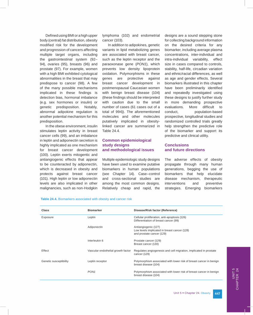
Table 24.4. Biomarkers associated with obesity and cancer risk
Class Biomarker Disease/Risk factor (Reference)
Exposure Leptin Cellular proliferation, anti-apoptosis (126)Differentiation of breast cancer (99)
Adiponectin Antiangiogenic (127)Low levels implicated in breast cancer (128)and prostate cancer (129)
Interleukin 6 Prostate cancer (129)Breast cancer (130)
Effect Vascular endothelial growth factor Regulates angiogenesis and cell migration, implicated in prostate cancer (129)
Genetic susceptibility Leptin receptor Polymorphism associated with lower risk of breast cancer in benign breast disease (104)
PON1 Polymorphism associated with lower risk of breast cancer in benign breast disease (104)
448
for obesity-associated CVD, type 2 diabetes or cancer play diverse roles in biological pathways including immune modulation and fat metabolism. Support for the association of these biomarkers with obesity-associated diseases stems from animal and in vitro data, but evidence in humans is still lacking. In humans, plasma levels of biomarkers or genetic polymorphisms are widely used to determine risk, but many studies are limited by ethnicity/race, gender or sample size. These deficits are perhaps the most challenging to overcome in future studies, but it is the only way by which a biomarker can be validated for reliable use in human populations. Along these lines, there has recently been a series of large-scale genome-wide association studies of BMI that are
uncovering a substantial number of new loci associated with obesity (105–111).
In the field of chronic inflammatory disorders exist acute indicators, such as C-reactive protein, an acute phase protein that rises in any inflammatory condition, and chronic prognostic indicators, such as monocyte chemoattractant protein 1 in the case of CVD. Management of obesity-associated disorders may benefit from the use of acute indicators augmented with chronic markers to better predict disease progression. In fact, multiple biomarkers may be required to complement standard traditional risk factors to enhance risk stratification and to develop measurable therapeutic targets.
Additional measures of obesity aside from the typical BMI are
required to better characterize obesity in the context of other chronic disorders. Other non-invasive measures (e.g. waist circumference, waist-to-height ratio and the conicity index) are surrogates of abdominal (central) obesity. Central obesity is often found to predict disease outcome better than BMI (112). One major cause of central obesity is a large visceral adipose tissue distribution. Visceral adipose tissue actively expresses and secretes a myriad of adipokines and other agents that act locally and systemically to promote obesity-associated disorders. Therefore, central obesity should be fully investigated in the context of relevant biomarkers, for it may add to their predictive utility and clinical validity.
References
1. Wilson PW, D’Agostino RB, Sullivan L et al. (2002). Overweight and obesity as determinants of cardiovascular risk: the Framingham experience. Arch Intern Med, 162:1867–1872.doi:10.1001/archinte.162. 16.1867 PMID:12196085
2. Fox CS, Massaro JM, Hoffmann U et al. (2007). Abdominal visceral and subcutaneous adipose tissue compartments: association with metabolic risk factors in the Framingham Heart Study. Circulation, 116:39–48.doi:10. 1161/CIRCULATIONAHA.106.675355 PMID: 17576866
3. Jiang Y, Chen Y, Mao Y; CCDPC Obesity Working Group (2008). The contribution of excess weight to prevalent diabetes in Canadian adults. Public Health, 122:271–2 76 . d o i :10 .1016 / j . p u h e . 2 0 07. 0 6 . 0 0 2 PMID:17931673
4. Krishnan S, Rosenberg L, Djoussé L et al. (2007). Overall and central obesity and risk of type 2 diabetes in U.S. black women. Obesity (Silver Spring), 15:1860–1866.doi:10.1038/oby.2007.220 PMID:17636105
5. Stoll BA (2002). Upper abdominal obesity, insulin resistance and breast cancer risk. Int J Obes Relat Metab Disord, 26:747–753. PMID:12037643
6. Takahashi H, Yoneda K, Tomimoto A et al. (2007). Life style-related diseases of the digestive system: colorectal cancer as a life style-related disease: from carcinogenesis to medical treatment. J Pharmacol Sci, 105:129–132.doi:10.1254/jphs.FM0070022 PMID:17928742
7. Schwimmer JB, Burwinkle TM, Varni JW (2003). Health-related quality of life of severely obese children and adolescents. JAMA, 289:1813–1819.doi:10.1001/jama.289.14.1813 PMID:12684360
8. Varni JW, Limbers CA, Burwinkle TM (2007). Impaired health-related quality of life in children and adolescents with chronic conditions: a comparative analysis of 10 disease clusters and 33 disease categories/severities utilizing the PedsQL 4.0 Generic Core Scales. Health Qual Life Outcomes, 5:43.doi:10.1186/1477-7525-5-43 PMID:17634123
9. Fontaine KR, Redden DT, Wang C et al. (2003). Years of life lost due to obesity. JAMA, 289:187–193.doi:10.1001/jama.289.2.187 PMID:12517229
Unit 5 • Chapter 24. Obesity 449
Un
it 5
Ch
ap
ter
24
10. Ogden CL, Carroll MD, Curtin LR et al. (2006). Prevalence of overweight and obesity in the United States, 1999–2004. JAMA, 295:1549–1555.doi:10.1001/jama.295.13. 1549 PMID:16595758
11. Nogueiras R, Wiedmer P, Perez-Tilve D et al. (2007). The central melanocortin system directly controls peripheral lipid metabolism. J Clin Invest, 117:3475–3488.doi:10.1172/JCI31743 PMID:17885689
12. Gil-Campos M, Aguilera CM, Cañete R, Gil A (2006). Ghrelin: a hormone regulating food intake and energy homeostasis. Br J Nutr, 96:201–226.doi:10.1079/BJN20061787 PMID:16923214
13. Date Y, Toshinai K, Koda S et al. (2005). Peripheral interaction of ghrelin with cholecystokinin on feeding regulation. Endocrinology, 146:3518–3525.doi:10.1210/en.2004-1240 PMID:15890776
14. Matias I, Di Marzo V (2007). Endocannabinoids and the control of energy balance. Trends Endocrinol Metab, 18:27–37.doi:10.1016/ j.tem.2006.11.006 PMID:17141520
15. Bartness TJ, Song CK (2007). Brain-adipose tissue neural crosstalk. Physiol Behav, 91:343–351.doi:10.1016/j.physbeh.2007.04. 002 PMID:17521684
16. Roberge C, Carpentier AC, Langlois MF et al. (2007). Adrenocortical dysregulation as a major player in insulin resistance and onset of obesity. Am J Physiol Endocrinol Metab, 293:E1465 –E1478.doi:10.1152/ajpendo. 00516.2007 PMID:17911338
17. Trayhurn P, Bing C, Wood IS (2006). Adipose tissue and adipokines–energy regulation from the human perspective. J Nutr, 136 Suppl;1935S–1939S. PMID:16772463
18. Farooqi IS (2005). Genetic and hereditary aspects of childhood obesity. Best Pract Res Clin Endocrinol Metab, 19:359–374.doi:10.1016/j.beem.2005.04.004 PMID:16150380
19. Hill JO (2006). Understanding and addressing the epidemic of obesity: an energy balance perspective. Endocr Rev, 27:750–761.PMID:17122359
20. Sallis JF, Glanz K (2006). The role of built environments in physical activity, eating, and obesity in childhood. Future Child, 16:89–108.doi:10.1353/foc.2006.0009 PMID:16532660
21. Flodmark CE, Marcus C, Britton M (2006). Interventions to prevent obesity in children and adolescents: a systematic literature review. Int J Obes (Lond), 30:579–589.doi:10. 1038/sj.ijo.0803290 PMID:16570086
22. Fantuzzi G (2005). Adipose tissue, adipokines, and inflammation. J Allergy Clin Immunol, 115:911–919, quiz 920.doi:10.1016/j.jaci.2005.02.023 PMID:15867843
23. Furukawa S, Fujita T, Shimabukuro M et al. (2004). Increased oxidative stress in obesity and its impact on metabolic syndrome. J Clin Invest, 114:1752–1761. PMID:15599400
24. Dandona P, Mohanty P, Ghanim H et al. (2001). The suppressive effect of dietary restriction and weight loss in the obese on the generation of reactive oxygen species by leukocytes, lipid peroxidation, and protein carbonylation. J Clin Endocrinol Metab, 86:355–362.doi:10.1210/jc.86.1.355 PMID:11 232024
25. Tripathy D, Mohanty P, Dhindsa S et al. (2003). Elevation of free fatty acids induces inflammation and impairs vascular reactivity in healthy subjects. Diabetes, 52:2882–2887.do i:10.2337/d iabetes.52.12.2882 PMID:14633847
26. Stephens JW, Gable DR, Hurel SJ et al. (2006). Increased plasma markers of oxidative stress are associated with coronary heart disease in males with diabetes mellitus and with 10-year risk in a prospective sample of males. Clin Chem, 52:446–452.doi:10.1373/clinchem.2005.060194 PMID:16384883
27. Yusuf S, Dagenais G, Pogue J et al.; The Heart Outcomes Prevention Evaluation Study Investigators (2000). Vitamin E supplementation and cardiovascular events in high-risk patients. N Engl J Med, 342:154–160. PMID:10639540
28. Stephens NG, Parsons A, Schofield PM et al. (1996). Randomised controlled trial of vitamin E in patients with coronary disease: Cambridge Heart Antioxidant Study (CHAOS). Lancet, 347:781–786.doi:10.1016/S0140-6736(96)90866-1 PMID:8622332
29. Barker DJ, Eriksson JG, Forsén T, Osmond C (2002). Fetal origins of adult disease: strength of effects and biological basis. Int J Epidemiol, 31:1235–1239.doi:10.1093/ije/31. 6.1235 PMID:12540728
30. Waterland RA, Jirtle RL (2003). Transposable elements: targets for early nutritional effects on epigenetic gene regulation. Mol Cell Biol, 23:5293–5300.doi:10.1128/MCB.23.15.5293-5300.2003 PMID:12861015
31. Patel MS, Srinivasan M (2002). Metabolic programming: causes and consequences. J Biol Chem, 277:1629–1632.doi:10.1074/jbc.R 100017200 PMID:11698417
32. Musaad S, Haynes EN (2007). Biomarkers of obesity and subsequent cardiovascular events. Epidemiol Rev, 29:98–114.doi:10.10 93/epirev/mxm005 PMID:17494057
33. Hekerman P, Zeidler J, Korfmacher S et al. (2007). Leptin induces inflammation-related genes in RINm5F insulinoma cells. BMC Mol Biol, 8:41.doi:10.1186/1471-2199-8-41 PMID: 17521427
34. Karmazyn M, Purdham DM, Rajapurohitam V, Zeidan A (2007). Leptin as a cardiac hypertrophic factor: a potential target for therapeutics. Trends Cardiovasc Med, 17:206–211.doi:10.1016/j.tcm.2007.06.001 PMID:17662916
35. Sierra-Johnson J, Romero-Corral A, Lopez-Jimenez F et al. (2007). Relation of increased leptin concentrations to history of myocardial infarction and stroke in the United States population. Am J Cardiol, 100:234–239.doi:10.1016/ j .amjcard.2007.02.088 PMID:17631076
36. Appel SJ, Harrell JS, Davenport ML (2005). Central obesity, the metabolic syndrome, and plasminogen activator inhibitor-1 in young adults. J Am Acad Nurse Pract, 17:535–541.doi:10.1111/j.1745-7599.2005.00083.x PMID: 16293162
37. Lindeman JH, Pijl H, Toet K et al. (2007). Human visceral adipose tissue and the plasminogen activator inhibitor type 1. Int J Obes (Lond), 31:1671–1679.doi:10.1038/sj.ijo. 0803650 PMID:17471294
38. Saidi S, Slamia LB, Mahjoub T et al. (2007). Association of PAI-1 4G/5G and -844G/A gene polymorphism and changes in PAI-1/tPA levels in stroke: a case-control study. J Stroke Cerebrovasc Dis, 16:153–159.doi:10.1016/j.jstrokecerebrovasdis.2007.02.002 PMID:1768 9411
39. Tsantes AE, Nikolopoulos GK, Bagos PG et al. (2007). Plasminogen activator inhibitor-1 4G/5G polymorphism and risk of ischemic stroke: a meta-analysis. Blood Coagul Fibrinolysis, 18:497–504.doi:10.1097/MBC.0b013e3281ec4eee PMID:17581326
40. Corsetti JP, Ryan D, Moss AJ et al. (2008). Plasminogen activator inhibitor-1 polymorphism (4G/5G) predicts recurrence in nonhyperlipidemic postinfarction patients. Arterioscler Thromb Vasc Biol, 28:548–554.doi:10.1161/ATVBAHA.107.155556 PMID:180 96824
41. Morange PE, Saut N, Alessi MC et al. (2007). Association of plasminogen activator inhibitor (PAI)-1 (SERPINE1) SNPs with myocardial infarction, plasma PAI-1, and metabolic parameters: the HIFMECH study. Arterioscler Thromb Vasc Biol, 27:2250–2257.doi:10.1161/ATVBAHA.107.149468 PMID:17656673
42. Mertens I, Ballaux D, Funahashi T et al. (2005). Inverse relationship between plasminogen activator inhibitor-I activity and adiponectin in overweight and obese women. Interrelationship with visceral adipose tissue, insulin resistance, HDL-chol and inflammation. Thromb Haemost, 94:1190–1195. PMID:16411393
43. Goldstein BJ, Scalia R (2004). Adiponectin: A novel adipokine linking adipocytes and vascular function. J Clin Endocrinol Metab, 89:2563–2568.doi:10.1210/jc.2004-0518 PMID:15181024
44. Cavusoglu E, Ruwende C, Chopra V et al. (2006). Adiponectin is an independent predictor of all-cause mortality, cardiac mortality, and myocardial infarction in patients presenting with chest pain. Eur Heart J, 27:2300–2309.doi:10.1093/eurheartj/ehl153 PMID:16864609
450
45. Wolk R, Berger P, Lennon RJ et al. (2007). Association between plasma adiponectin levels and unstable coronary syndromes. Eur Heart J, 28:292–298.doi:10.1093/eurheartj/ehl361 PMID:17090613
46. Weinbrenner T, Schröder H, Escurriol V et al. (2006). Circulating oxidized LDL is associated with increased waist circumference independent of body mass index in men and women. Am J Clin Nutr, 83:30–35, quiz 181–182. PMID:16400046
47. Morrow JD (2005). Quantification of isoprostanes as indices of oxidant stress and the risk of atherosclerosis in humans. Arterioscler Thromb Vasc Biol, 25:279–286.doi:10.1161/01.ATV.0000152605.64964.c0 PMID:15591226
48. Keaney JF Jr, Larson MG, Vasan RS et al.; Framingham Study (2003). Obesity and systemic oxidative stress: clinical correlates of oxidative stress in the Framingham Study. Arterioscler Thromb Vasc Biol, 23:434–439.doi:10.1161/01.ATV.0000058402.34138.11 PMID:12615693
49. Bougoulia M, Triantos A, Koliakos G (2006). Plasma interleukin-6 levels, glutathione peroxidase and isoprostane in obese women before and after weight loss. Association with cardiovascular risk factors. Hormones (Athens), 5:192–199. PMID:16950753
50. Ustundag B, Gungor S, Aygün AD et al. (2007). Oxidative status and serum leptin levels in obese prepubertal children. Cell Biochem Funct, 25:479–483.doi:10.1002/cbf. 1334 PMID:16874844
51. Espinola-Klein C, Rupprecht HJ, Bickel C et al.; AtheroGene Investigators (2007). Glutathione peroxidase-1 activity, atherosclerotic burden, and cardiovascular prognosis. Am J Cardiol, 99:808–812.doi:10. 1016/j.amjcard.2006.10.041 PMID:17350371
52. Christiansen T, Richelsen B, Bruun JM (2005). Monocyte chemoattractant protein-1 is produced in isolated adipocytes, associated with adiposity and reduced after weight loss in morbid obese subjects. Int J Obes (Lond), 29:146–150.doi:10.1038/sj.ijo.0802839 PMID: 15520826
53. Dahlman I, Kaaman M, Olsson T et al. (2005). A unique role of monocyte chemoattractant protein 1 among chemokines in adipose tissue of obese subjects. J Clin Endocrinol Metab, 90:5834–5840.doi:10. 1210/jc.2005-0369 PMID:16091493
54. Frangogiannis NG (2007). The prognostic value of monocyte chemoattractant protein-1/CCL2 in acute coronary syndromes. J Am Coll Cardiol, 50:2125–2127.doi:10.1016/j.jacc. 2007.08.027 PMID:18036448
55. de Lemos JA, Morrow DA, Blazing MA et al. (2007). Serial measurement of monocyte chemoattractant protein-1 after acute coronary syndromes: results from the A to Z trial. J Am Coll Cardiol, 50:2117–2124.doi:10.1016/j.jacc. 2007.06.057 PMID:18036447
56. Kramer H, Wu X, Kan D et al. (2005). Angiotensin-converting enzyme gene polymorphisms and obesity: an examination of three black populations. Obes Res, 13:823–828.doi:10.1038/oby.2005.94 PMID: 15919834
57. Moran CN, Vassilopoulos C, Tsiokanos A et al. (2005). Effects of interaction between angiotensin I-converting enzyme polymorphisms and lifestyle on adiposity in adolescent Greeks. Obes Res, 13:1499–1504.doi:10.1038/oby.2005.181 PMID:16222048
58. Saha SA, Molnar J, Arora RR (2007). Tissue ACE inhibitors for secondary prevention of cardiovascular disease in patients with preserved left ventricular function: a pooled meta-analysis of randomized placebo-controlled trials. J Cardiovasc Pharmacol Ther, 12:192–204.doi:10.1177/1074248407304791 PMID:17875946
59. Homo-Delarche F, Calderari S, Irminger JC et al. (2006). Islet inflammation and fibrosis in a spontaneous model of type 2 diabetes, the GK rat. Diabetes, 55:1625–1633.doi:10.2337/db05-1526 PMID:16731824
60. Herder C, Peltonen M, Koenig W et al. (2006). Systemic immune mediators and lifestyle changes in the prevention of type 2 diabetes: results from the Finnish Diabetes Prevention Study. Diabetes, 55:2340–2346.doi:10.2337/db05-1320 PMID:16873699
61. McLaughlin T, Abbasi F, Cheal K et al. (2003). Use of metabolic markers to identify overweight individuals who are insulin resistant. Ann Intern Med, 139:802–809. PMID:14623617
62. Beale EG, Harvey BJ, Forest C (2007). PCK1 and PCK2 as candidate diabetes and obesity genes. Cell Biochem Biophys, 48:89–95.doi:10.1007/s12013-007-0025-6 PMID:17 709878
63. Matsuoka N, Patki A, Tiwari HK et al. (2006). Association of K121Q polymorphism in ENPP1 (PC-1) with BMI in Caucasian and African-American adults. Int J Obes (Lond), 30:233–237.doi:10.1038/sj.ijo.0803132 PMID: 16231022
64. Bochenski J, Placha G, Wanic K et al. (2006). New polymorphism of ENPP1 (PC-1) is associated with increased risk of type 2 diabetes among obese individuals. Diabetes, 55:2626 –2630.do i:10.2337/db06 - 0191 PMID:16936213
65. Böttcher Y, Körner A, Reinehr T et al. (2006). ENPP1 variants and haplotypes predispose to early onset obesity and impaired glucose and insulin metabolism in German obese children. J Clin Endocrinol Metab, 91:4948–4952.doi:10.1210/jc.2006-0540 PMID:16968801
66. Blüher M, Fasshauer M, Tönjes A et al. (2005). Association of interleukin-6, C-reactive protein, interleukin-10 and adiponectin plasma concentrations with measures of obesity, insulin sensitivity and glucose metabolism. Exp Clin Endocrinol Diabetes, 113:534–537.doi:10.1055/s-2005-872851 PMID:16235156
67. Singhal A, Jamieson N, Fewtrell M et al. (2005). Adiponectin predicts insulin resistance but not endothelial function in young, healthy adolescents. J Clin Endocrinol Metab, 90:4615–4621.doi:10.1210/jc.2005-01 31 PMID:15886241
68. Kempf K, Hector J, Strate T et al. (2007). Immune-mediated activation of the endocannabinoid system in visceral adipose tissue in obesity. Horm Metab Res, 39:596– 600.doi:10.1055/s-2007-984459 PMID:17712725
69. Serino M, Menghini R, Fiorentino L et al. (2007). Mice heterozygous for tumor necrosis factor-alpha converting enzyme are protected from obesity-induced insulin resistance and diabetes. Diabetes, 56:2541–2546.doi:10.23 37/db07-0360 PMID:17646208
70. Krogh-Madsen R, Plomgaard P, Møller K et al. (2006). Influence of TNF-alpha and IL-6 infusions on insulin sensitivity and expression of IL-18 in humans. Am J Physiol Endocrinol Metab, 291:E108–E114.doi:10.1152/ajpendo. 00471.2005 PMID:16464907
71. Fontaine-Bisson B, Wolever TM, Chiasson JL et al. (2007). Tumor necrosis factor alpha -238G>A genotype alters postprandial plasma levels of free fatty acids in obese individuals with type 2 diabetes mellitus. Metabolism, 56:649–655.doi:10.1016/j.metabol.2006.12. 013 PMID:17445540
72. Sookoian SC, González C, Pirola CJ (2005). Meta-analysis on the G-308A tumor necrosis factor alpha gene variant and phenotypes associated with the metabolic syndrome. Obes Res, 13:2122–2131.doi:10.1038/oby.2005.263 PMID:16421346
73. Zeggini E, Groves CJ, Parkinson JR et al. (2005). Large-scale studies of the association between variation at the TNF/LTA locus and susceptibility to type 2 diabetes. Diabetologia, 48:2013–2017.doi:10.1007/s00125-005-1902-4 PMID:16132956
74. González-Sánchez JL, Martínez-Calatrava MJ, Martínez-Larrad MT et al. (2006). Interaction of the -308G/A promoter polymorphism of the tumor necrosis factor-alpha gene with single-nucleotide polymorphism 45 of the adiponectin gene: effect on serum adiponectin concentrations in a Spanish population. Clin Chem, 52:97–103.doi:10.1373/clinchem.2005.049452 PMID:16254197
75. Bonen A, Tandon NN, Glatz JF et al. (2006). The fatty acid transporter FAT/CD36 is upregulated in subcutaneous and visceral adipose tissues in human obesity and type 2 diabetes. Int J Obes (Lond), 30:877–883.doi:10.1038/sj.ijo.0803212 PMID:16418758
76. Corpeleijn E, van der Kallen CJ, Kruijshoop M et al. (2006). Direct association of a promoter polymorphism in the CD36/FAT fatty acid transporter gene with Type 2 diabetes mellitus and insulin resistance. Diabet Med, 23:907–911.doi:10.1111/j.1464-5491.2006.01888.x PMID:16911630
Unit 5 • Chapter 24. Obesity 451
Un
it 5
Ch
ap
ter
24
77. Handberg A, Levin K, Højlund K, Beck-Nielsen H (2006). Identification of the oxidized low-density lipoprotein scavenger receptor CD36 in plasma: a novel marker of insulin resistance. Circulation, 114:1169–1176.doi:10.1161/CIRCULATIONAHA.106.626135 PMID:16952981
78. Warensjö E, Ingelsson E, Lundmark P et al. (2007). Polymorphisms in the SCD1 gene: associations with body fat distribution and insulin sensitivity. Obesity (Silver Spring), 15:1732–1740.doi:10.1038/oby.2007.206 PMID:17636091
79. Ishii T, Masuzaki H, Tanaka T et al. (2007). Augmentation of 11beta-hydroxysteroid dehydrogenase type 1 in LPS-activated J774.1 macrophages–role of 11beta-HSD1 in pro-inflammatory properties in macrophages. FEBS Lett, 581:349–354.doi:10.1016/j.febslet.2006.11.032 PMID:17239856
80. Berthiaume M, Laplante M, Festuccia W et al. (2007). Depot-specific modulation of rat intraabdominal adipose tissue lipid metabolism by pharmacological inhibition of 11beta-hydroxysteroid dehydrogenase type 1. Endocrinology, 148:2391–2397.doi:10.1210/en.2006-1199 PMID:17272400
81. Desbriere R, Vuaroqueaux V, Achard V et al. (2006). 11beta-hydroxysteroid dehydrogenase type 1 mRNA is increased in both visceral and subcutaneous adipose tissue of obese patients. Obesity (Silver Spring), 14:794–798.doi:10.1038/oby.2006.92 PMID:16855188
82. Alberti L, Girola A, Gilardini L et al. (2007). Type 2 diabetes and metabolic syndrome are associated with increased expression of 11beta-hydroxysteroid dehydrogenase 1 in obese subjects. Int J Obes (Lond), 31:1826–1831.doi:10.1038/sj.ijo.0803677 PMID:17593901
83. Koska J, de Courten B, Wake DJ et al. (2006). 11beta-hydroxysteroid dehydrogenase type 1 in adipose tissue and prospective changes in body weight and insulin resistance. Obesity (Silver Spring), 14:1515–1522.doi:10. 1038/oby.2006.175 PMID:17030962
84. Sharma AM, Staels B (2007). Review: Peroxisome proliferator-activated receptor γ and adipose tissue–understanding obesity-related changes in regulation of lipid and glucose metabolism. J Clin Endocrinol Metab, 92:386–395.doi:10.1210/jc.2006-1268 PMID:17148564
85. Cole SA, Mitchell BD, Hsueh WC et al. (2000). The Pro12Ala variant of peroxisome proliferator-activated receptor-γ2 (PPAR-γ2) is associated with measures of obesity in Mexican Americans. Int J Obes Relat Metab Disord, 24:522–524.doi:10.1038/sj.ijo.0801210 PMID:10805513
86. Tönjes A, Stumvoll M (2007). The role of the Pro12Ala polymorphism in peroxisome proliferator-activated receptor gamma in diabetes risk. Curr Opin Clin Nutr Metab Care, 10:410–414.doi:10.1097/MCO.0b013e3281e389d9 PMID:17563457
87. Tan GD, Neville MJ, Liverani E et al. (2006). The in vivo effects of the Pro12Ala PPARgamma2 polymorphism on adipose tissue NEFA metabolism: the first use of the Oxford Biobank. Diabetologia, 49:158–16 8 .d o i :10 .10 07/s 0 012 5 - 0 0 5 - 0 0 4 4 -z PMID:16362285
88. Ludovico O, Pellegrini F, Di Paola R et al. (2007). Heterogeneous effect of peroxisome proliferator-activated receptor γ2 Ala12 variant on type 2 diabetes risk. Obesity (Silver Spring), 15:1076–1081.doi:10.1038/oby.2007. 617 PMID:17495182
89. Kilpeläinen TO, Lakka TA, Laaksonen DE et al. (2008). SNPs in PPARG associate with type 2 diabetes and interact with physical activity. Med Sci Sports Exerc, 40:25–33. PMID:18091023
90. Hofmann SM, Zhou L, Perez-Tilve D et al. (2007). Adipocyte LDL receptor-related protein-1 expression modulates postprandial lipid transport and glucose homeostasis in mice. J Clin Invest, 117:3271–3282.doi:10. 1172/JCI31929 PMID:17948131
91. American Cancer Society. Cancer facts and figures 2006. Atlanta (GA): American Cancer Society; 2006.
92. Dai Z, Xu YC, Niu L (2007). Obesity and colorectal cancer risk: a meta-analysis of cohort studies. World J Gastroenterol, 13: 4199–4206. PMID:17696248
93. Larsson SC, Wolk A (2007). Obesity and colon and rectal cancer risk: a meta-analysis of prospective studies. Am J Clin Nutr, 86:556–565. PMID:17823417
94. Merry AH, Schouten LJ, Goldbohm RA, van den Brandt PA (2007). Body mass index, height and risk of adenocarcinoma of the oesophagus and gastric cardia: a prospective cohort study. Gut, 56:1503–1511.doi:10.1136/gut.2006.116665 PMID:17337464
95. Olsen CM, Green AC, Whiteman DC et al. (2007). Obesity and the risk of epithelial ovarian cancer: a systematic review and meta-analysis. Eur J Cancer, 43:690–709.doi:10.1016/j.ejca.2006.11.010 PMID:17223544
96. Daling JR, Malone KE, Doody DR et al. (2001). Relation of body mass index to tumor markers and survival among young women with invasive ductal breast carcinoma. Cancer, 92:720–729.doi:10.1002/10970142(20010815)92:4<720::AID-CNCR1375>3.0.CO;2-T PMID:11550140
97. Freedland SJ, Platz EA (2007). Obesity and prostate cancer: making sense out of apparently conflicting data. Epidemiol Rev, 29:88–97.doi:10.1093/epirev/mxm006 PMID:17478439
98. Seewaldt VL, Goldenberg V, Jones LW et al. (2007). Overweight and obese perimenopausal and postmenopausal women exhibit increased abnormal mammary epithelial cytology. Cancer Epidemiol Biomarkers Prev, 16:613–616.doi:10.1158/1055-9965.EPI-06-0878 PMID: 17372261
99. Garofalo C, Koda M, Cascio S et al. (2006). Increased expression of leptin and the leptin receptor as a marker of breast cancer progression: possible role of obesity-related stimuli. Clin Cancer Res, 12:1447–1453.doi:10.1158/1078-0432.CCR-05-1913 PMID:16533767
100. Vona-Davis L, Rose DP (2007). Adipokines as endocrine, paracrine, and autocrine factors in breast cancer risk and progression. Endocr Relat Cancer, 14:189–206.doi:10.1677/ERC-06-0068 PMID:17639037
101. Vona-Davis L, Howard-McNatt M, Rose DP (2007). Adiposity, type 2 diabetes and the metabolic syndrome in breast cancer. Obes Rev, 8:395–408.doi:10.1111/j.1467-789X.2007.00396.x PMID:17716297
102. Skibola CF, Holly EA, Forrest MS et al. (2004). Body mass index, leptin and leptin receptor polymorphisms, and non-hodgkin lymphoma. Cancer Epidemiol Biomarkers Prev, 13:779–786. PMID:15159310
103. Soliman PT, Wu D, Tortolero-Luna G et al. (2006). Association between adiponectin, insulin resistance, and endometrial cancer. Cancer, 106:2376–2381.doi:10.1002/cncr.218 66 PMID:16639730
104. Gallicchio L, McSorley MA, Newschaffer CJ et al. (2007). Body mass, polymorphisms in obesity-related genes, and the risk of developing breast cancer among women with benign breast disease. Cancer Detect Prev, 31:95–101.doi:10.1016/j.cdp.2007.02.004 PMID:17428620
105. Frayling TM, Timpson NJ, Weedon MN et al. (2007). A common variant in the FTO gene is associated with body mass index and predisposes to childhood and adult obesity. Science, 316:889–894.doi:10.1126/science.1141634 PMID:17434869
106. Dina C, Meyre D, Gallina S et al. (2007). Variation in FTO contributes to childhood obesity and severe adult obesity. Nat Genet, 39:724–726.doi:10.1038/ng2048 PMID:17496892
107. Scuteri A, Sanna S, Chen WM et al. (2007). Genome-wide association scan shows genetic variants in the FTO gene are associated with obesity-related traits. PLoS Genet, 3:e115.doi:10.1371/journal.pgen.0030115 PMID:17658951
108. Loos RJ, Lindgren CM, Li S et al.; Prostate, Lung, Colorectal, and Ovarian (PLCO) Cancer Screening Trial; KORA; Nurses’ Health Study; Diabetes Genetics Initiative; SardiNIA Study; Wellcome Trust Case Control Consortium; FUSION (2008). Common variants near MC4R are associated with fat mass, weight and risk of obesity. Nat Genet, 40:768–775.doi:10.1038/ng.140 PMID:18454148
109. Willer CJ, Speliotes EK, Loos RJ et al.; Wellcome Trust Case Control Consortium; Genetic Investigation of ANthropometric Traits Consortium (2009). Six new loci associated with body mass index highlight a neuronal influence on body weight regulation. Nat Genet, 41:25–34.doi:10.1038/ng.287 PMID:19079261
452
110. Thorleifsson G, Walters GB, Gudbjartsson DF et al. (2009). Genome-wide association yields new sequence variants at seven loci that associate with measures of obesity. Nat Genet, 41:18–24.doi:10.1038/ng.274 PMID:19079260
111. Speliotes EK, Willer CJ, Berndt SI et al.; MAGIC; Procardis Consortium (2010). Association analyses of 249,796 individuals reveal 18 new loci associated with body mass index. Nat Genet, 42:937–948.doi:10.1038/ng.686 PMID:20935630
112. Musaad SM, Patterson T, Ericksen M et al. (2009). Comparison of anthropometric measures of obesity in childhood allergic asthma: central obesity is most relevant. J Allergy Clin Immunol, 123:1321–1327, e12.doi: 10.1016/j.jaci.2009.03.023 PMID:19439348
113. Blankenberg S, Luc G, Ducimetière P et al.; PRIME Study Group (2003). Interleukin-18 and the risk of coronary heart disease in European men: the Prospective Epidemiological Study of Myocardial Infarction (PRIME). Circulation, 108:2453–2459.doi: 10.1161/01.CIR.0000099509.76044.A 2 PMID:14581397
114. Söderberg S, Ahrén B, Stegmayr B et al. (1999). Leptin is a risk marker for first-ever hemorrhagic stroke in a population-based cohort. Stroke, 30:328–337. PMID:9933268
115. Söderberg S, Ahrén B, Jansson JH et al. (1999). Leptin is associated with increased risk of myocardial infarction. J Intern Med, 246:409–418.doi:10.1046/j.1365-2796.1999. 00571.x PMID:10583712
116. Wiman B, Andersson T, Hallqvist J et al. (2000). Plasma levels of tissue plasminogen activator/plasminogen activator inhibitor-1 complex and von Willebrand factor are significant risk markers for recurrent myocardial infarction in the Stockholm Heart Epidemiology Program (SHEEP) study. Arterioscler Thromb Vasc Biol, 20:2019–2023. PMID:10938026
117. Meisinger C, Baumert J, Khuseyinova N et al. (2005). Plasma oxidized low-density lipoprotein, a strong predictor for acute coronary heart disease events in apparently healthy, middle-aged men from the general population. Circulation, 112:651–657.doi:10. 1161/CIRCULATIONAHA.104.529297 PMID: 16043640
118. Blankenberg S, Rupprecht HJ, Bickel C et al.; AtheroGene Investigators (2003). Glutathione peroxidase 1 activity and cardiovascular events in patients with coronary artery disease. N Engl J Med, 349: 1605 –1613.doi:10.1056/NEJMoa030535 PMID:14573732
119. Gross M, Steffes M, Jacobs DR Jr et al. (2005). Plasma F2-isoprostanes and coronary artery calcification: the CARDIA Study. Clin Chem, 51:125–131.doi:10.1373/clinchem.2004.037630 PMID:15514100
120. Shishehbor MH, Zhang R, Medina H et al. (2006). Systemic elevations of free radical oxidation products of arachidonic acid are associated with angiographic evidence of coronary artery disease. Free Radic Biol Med, 41:1678–1683.doi:10.1016/j.freeradbiomed.2006.09.001 PMID:17145556
121. Riera-Fortuny C, Real JT, Chaves FJ et al. (2005). The relation between obesity, abdominal fat deposit and the angiotensin-converting enzyme gene I/D polymorphism and its association with coronary heart disease. Int J Obes (Lond), 29:78–84.doi:10. 1038/sj.ijo.0802829 PMID:15520830
122. Sekuri C, Cam FS, Ercan E et al. (2005). Renin-angiotensin system gene polymorphisms and premature coronary heart disease. J Renin Angiotensin Aldosterone Syst, 6:38–42.doi:10.3317/jraas.2005.005 PMID:16088850
123. Yan QW, Yang Q, Mody N et al. (2007). The adipokine lipocalin 2 is regulated by obesity and promotes insulin resistance. Diabetes, 56:2533–2540.doi:10.2337/db07-0007 PMID:17639021
124. Li JZ, Ye J, Xue B et al. (2007). Cideb regulates diet-induced obesity, liver steatosis, and insulin sensitivity by controlling lipogenesis and fatty acid oxidation. Diabetes, 56:2523–2532.doi:10.2337/db07-0040 PMID:17646209
125. Kim CS, Park HS, Kawada T et al. (2006). Circulating levels of MCP-1 and IL-8 are elevated in human obese subjects and associated with obesity-related parameters. Int J Obes (Lond), 30:1347–1355.doi:10.1038/sj.ijo.0803259 PMID:16534530
126. Chen C, Chang YC, Liu CL et al. (2007). Leptin induces proliferation and anti-apoptosis in human hepatocarcinoma cells by up-regulating cyclin D1 and down-regulating Bax via a Janus kinase 2-linked pathway. Endocr Relat Cancer, 14:513–529.doi:10.1677/ERC-06-0027 PMID:17639064
127. Bråkenhielm E, Veitonmäki N, Cao R et al. (2004). Adiponectin-induced antiangiogenesis and antitumor activity involve caspase-mediated endothelial cell apoptosis. Proc Natl Acad Sci USA, 101:2476–2481.doi:10.1073/pnas.0308671100 PMID:14983034
128. Miyoshi Y, Funahashi T, Kihara S et al. (2003). Association of serum adiponectin levels with breast cancer risk. Clin Cancer Res, 9:5699–5704. PMID:14654554
129. Mistry T, Digby JE, Desai KM, Randeva HS (2007). Obesity and prostate cancer: a role for adipokines. Eur Urol, 52:46–53.doi:10.1016/j.eururo.2007.03.054 PMID:17399889
130. Lorincz AM, Sukumar S (2006). Molecular links between obesity and breast cancer. Endocr Relat Cancer, 13:279–292.doi:10.1677/erc.1.00729 PMID:16728564