NUTRITIONAL DISORDERS II Myrna D.C. San Pedro, MD, FPPS.
37
NUTRITIONAL NUTRITIONAL DISORDERS II DISORDERS II Myrna D.C. San Pedro, MD, FPPS Myrna D.C. San Pedro, MD, FPPS
-
Upload
derick-conley -
Category
Documents
-
view
246 -
download
2
Transcript of NUTRITIONAL DISORDERS II Myrna D.C. San Pedro, MD, FPPS.
NUTRITIONAL NUTRITIONAL DISORDERS IIDISORDERS II
Myrna D.C. San Pedro, MD, FPPSMyrna D.C. San Pedro, MD, FPPS

TheTheFat-Soluble Fat-Soluble
VitaminsVitamins
Vitamin AVitamin A
Active forms are retinol, Active forms are retinol, retinaldehyde, and retinoic acidretinaldehyde, and retinoic acid
Plants synthesize the more complex Plants synthesize the more complex carotenoids which are cleaved to carotenoids which are cleaved to retinol by most animals and stored in retinol by most animals and stored in the liver as retinyl palmitate the liver as retinyl palmitate
N retinol plasma values: 15-30 mcg/dl N retinol plasma values: 15-30 mcg/dl in infants & 30-90 mcg/dl in adultsin infants & 30-90 mcg/dl in adults
Vitamin A (Retinol) DeficiencyVitamin A (Retinol) DeficiencyFunctions:Functions:1.1. Retinal is the prosthetic Retinal is the prosthetic
group of photosensitive group of photosensitive pigment in both rods pigment in both rods (rhodopsin) & cones (rhodopsin) & cones (iodopsin), major (iodopsin), major difference lies in the difference lies in the nature of protein boundnature of protein bound
2.2. Needed in lysosomal Needed in lysosomal membrane stabilitymembrane stability
3.3. Plays a role in Plays a role in keratinization, keratinization, cornification, bone cornification, bone development & cell development & cell growth & reproductiongrowth & reproduction
Etiology:Etiology: Absence in the diet Absence in the diet
common by 2-3 yrs oldcommon by 2-3 yrs old Poor fetal storagePoor fetal storage Poor absorption as in Poor absorption as in
low-fat diet, low-fat diet, malabsorption malabsorption syndromes, etc.syndromes, etc.
Low protein intake Low protein intake resulting in deficient resulting in deficient carrierscarriers
Increased excretion as Increased excretion as in cancer & UTIin cancer & UTI
Clinical Manifestations of Clinical Manifestations of Hypovitaminosis AHypovitaminosis A
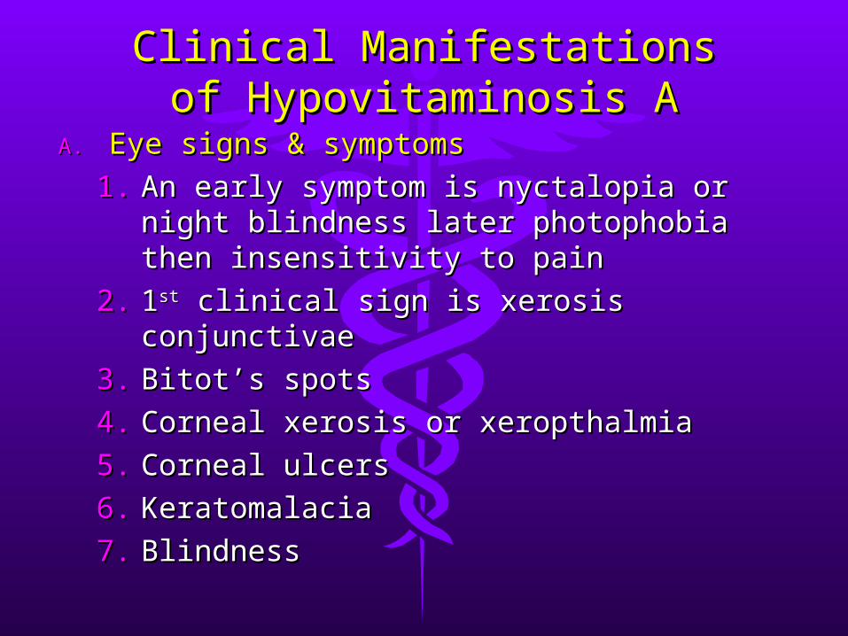
A.A. Eye signs & symptomsEye signs & symptoms
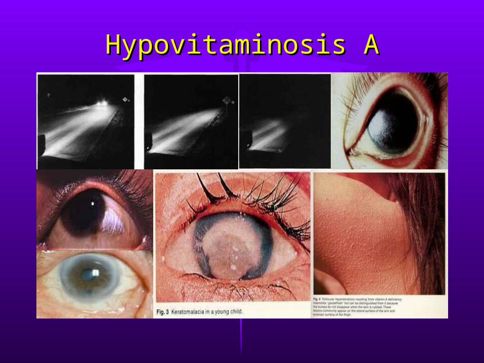
1.1. An early symptom is nyctalopia or An early symptom is nyctalopia or night blindness later photophobia night blindness later photophobia then insensitivity to painthen insensitivity to pain
2.2. 11stst clinical sign is xerosis conjunctivae clinical sign is xerosis conjunctivae
3.3. Bitot’s spotsBitot’s spots
4.4. Corneal xerosis or xeropthalmiaCorneal xerosis or xeropthalmia
5.5. Corneal ulcersCorneal ulcers
6.6. KeratomalaciaKeratomalacia
7.7. Blindness Blindness
Hypovitaminosis AHypovitaminosis A
B.B. Skin signs:Skin signs: xerosis of xerosis of the skin & follicular the skin & follicular hyperkeratosis or hyperkeratosis or phrynodermaphrynoderma
C.C. Others:Others: apathy, apathy, retardation of physical retardation of physical & mental growth, faulty & mental growth, faulty epiphyseal bone epiphyseal bone formation, defective formation, defective teeth enamel & signs of teeth enamel & signs of benign increased ICPbenign increased ICP
Diagnosis:Diagnosis:
1.1. Routine PERoutine PE
2.2. Biophysical exam, Biophysical exam, dark adaptation dark adaptation test to detect test to detect nyctalopianyctalopia
3.3. Absorption testAbsorption test
4.4. Conjunctival Conjunctival impression cytology impression cytology to evaluate early to evaluate early xeropthalmiaxeropthalmia
Hypovitaminosis AHypovitaminosis A
RDA: RDA: 1800 IU/day (1 IU vitamin 1800 IU/day (1 IU vitamin A = 0.3 mcg retinol)A = 0.3 mcg retinol)
Prevention:Prevention:1.1. Pregnant in last trimester Pregnant in last trimester
given 5000 IU p.o.given 5000 IU p.o.2.2. Every 6 months, infants <1 Every 6 months, infants <1
yr given retinol palmitate yr given retinol palmitate 55mg or 33mg retinol 55mg or 33mg retinol acetate (100,000 IU) p. o.acetate (100,000 IU) p. o.
3.3. Every 4-6 months, older Every 4-6 months, older children given 110mg children given 110mg retinol palmitate or 66 mg retinol palmitate or 66 mg retinol acetate (200,000 retinol acetate (200,000 IU) p. o.IU) p. o.
4.4. In areas where prevalent, In areas where prevalent, 100,000 IU p. o. q 3 mo100,000 IU p. o. q 3 mo
5.5. For malnourished children For malnourished children 1-6 yrs, 250,000 IU p. o. q 1-6 yrs, 250,000 IU p. o. q 6 months6 months
Treatment:Treatment:1.1. One yr of age or over:One yr of age or over:
110mg retinol palmitate or 110mg retinol palmitate or 66mg retinol acetate (200,000 66mg retinol acetate (200,000 IU) orally or preferably IU) orally or preferably 33mg(100,000 IU) of water-33mg(100,000 IU) of water-miscible vitamin A (retinyl miscible vitamin A (retinyl palmitate) by IM injectionpalmitate) by IM injection
2.2. The oral dose should be The oral dose should be repeated on 2nd day and repeated on 2nd day and again on discharge from again on discharge from hospital 7-30 days after 1st hospital 7-30 days after 1st dosedose
3.3. Above doses halved for Above doses halved for infantsinfants
4.4. For corneal involvement, For corneal involvement, apply antibiotic ointment like apply antibiotic ointment like topical topical bacitracinbacitracin to both to both eyes 6x/day and administer eyes 6x/day and administer also systemic antibioticsalso systemic antibiotics
Hypovitaminosis AHypovitaminosis A
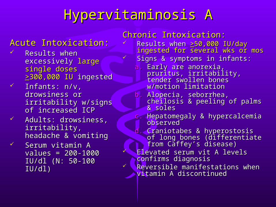
Hypervitaminosis AHypervitaminosis A
Acute Intoxication:Acute Intoxication: Results when Results when
excessively excessively large large single doses single doses >>300,000 300,000 IUIU ingested ingested
Infants: n/v, Infants: n/v, drowsiness or drowsiness or irritability w/signs of irritability w/signs of increased ICPincreased ICP
Adults: drowsiness, Adults: drowsiness, irritability, headache & irritability, headache & vomitingvomiting
Serum vitamin A Serum vitamin A values = 200-1000 values = 200-1000 IU/dl (N: 50-100 IU/dl)IU/dl (N: 50-100 IU/dl)
Chronic Intoxication:Chronic Intoxication: Results when Results when >>50,000 IU/day 50,000 IU/day
ingested for several wks or mosingested for several wks or mos Signs & symptoms in infants:Signs & symptoms in infants:
a.a. Early are anorexia, pruritus, Early are anorexia, pruritus, irritability, tender swollen irritability, tender swollen bones w/motion limitationbones w/motion limitation
b.b. Alopecia, seborrhea, Alopecia, seborrhea, cheilosis & peeling of palms cheilosis & peeling of palms & soles& soles
c.c. Hepatomegaly & Hepatomegaly & hypercalcemia observedhypercalcemia observed
d.d. Craniotabes & hyperostosis Craniotabes & hyperostosis of long bones (differentiate of long bones (differentiate from Caffey’s disease)from Caffey’s disease)
Elevated serum vit A levels Elevated serum vit A levels confirms diagnosisconfirms diagnosis
Reversible manifestations when Reversible manifestations when vitamin A discontinuedvitamin A discontinued
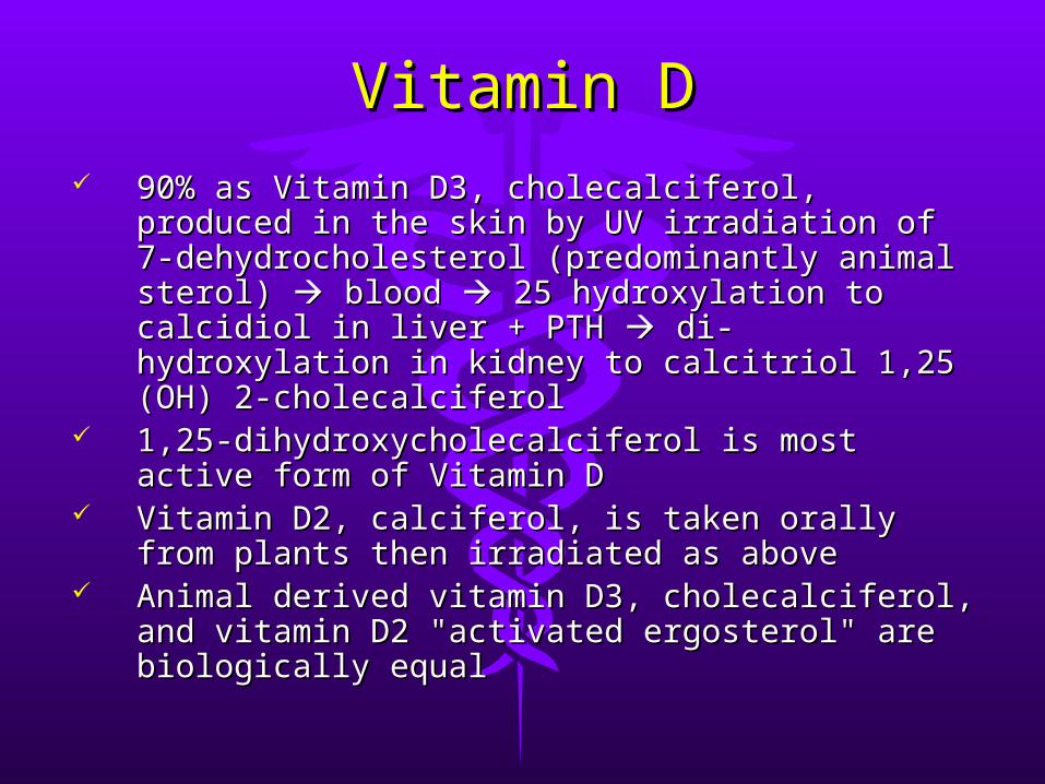
Vitamin DVitamin D 90% as Vitamin D3, cholecalciferol, produced in 90% as Vitamin D3, cholecalciferol, produced in
the skin by UV irradiation of 7-the skin by UV irradiation of 7-dehydrocholesterol (predominantly animal dehydrocholesterol (predominantly animal sterol) sterol) blood blood 25 hydroxylation to calcidiol 25 hydroxylation to calcidiol in liver + PTH in liver + PTH di-hydroxylation in kidney to di-hydroxylation in kidney to calcitriol 1,25 (OH) 2-cholecalciferolcalcitriol 1,25 (OH) 2-cholecalciferol
1,25-dihydroxycholecalciferol is most active 1,25-dihydroxycholecalciferol is most active form of Vitamin Dform of Vitamin D
Vitamin D2, calciferol, is taken orally from Vitamin D2, calciferol, is taken orally from plants then irradiated as aboveplants then irradiated as above
Animal derived vitamin D3, cholecalciferol, and Animal derived vitamin D3, cholecalciferol, and vitamin D2 "activated ergosterol" are vitamin D2 "activated ergosterol" are biologically equalbiologically equal
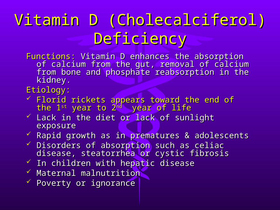
Vitamin D (Cholecalciferol) Vitamin D (Cholecalciferol) DeficiencyDeficiency
Functions:Functions: Vitamin D enhances the absorption of Vitamin D enhances the absorption of calcium from the gut, removal of calcium from calcium from the gut, removal of calcium from bone and phosphate reabsorption in the bone and phosphate reabsorption in the kidney.kidney.
Etiology:Etiology: Florid rickets appears toward the end of the 1Florid rickets appears toward the end of the 1stst
year to 2year to 2ndnd year of life year of life Lack in the diet or lack of sunlight exposureLack in the diet or lack of sunlight exposure Rapid growth as in prematures & adolescentsRapid growth as in prematures & adolescents Disorders of absorption such as celiac disease, Disorders of absorption such as celiac disease,
steatorrhea or cystic fibrosissteatorrhea or cystic fibrosis In children with hepatic diseaseIn children with hepatic disease Maternal malnutritionMaternal malnutrition Poverty or ignorancePoverty or ignorance
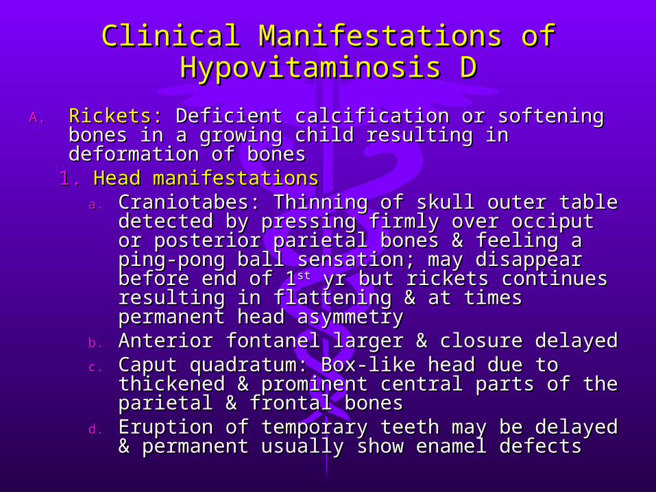
Clinical Manifestations of Clinical Manifestations of Hypovitaminosis DHypovitaminosis D
A.A. Rickets:Rickets: Deficient calcification or softening bones in Deficient calcification or softening bones in a growing child resulting in deformation of bonesa growing child resulting in deformation of bones
1.1. Head manifestationsHead manifestationsa.a. Craniotabes: Thinning of skull outer table Craniotabes: Thinning of skull outer table
detected by pressing firmly over occiput or detected by pressing firmly over occiput or posterior parietal bones & feeling a ping-pong posterior parietal bones & feeling a ping-pong ball sensation; may disappear before end of 1ball sensation; may disappear before end of 1stst yr but rickets continues resulting in flattening & yr but rickets continues resulting in flattening & at times permanent head asymmetryat times permanent head asymmetry
b.b. Anterior fontanel larger & closure delayedAnterior fontanel larger & closure delayedc.c. Caput quadratum: Box-like head due to Caput quadratum: Box-like head due to
thickened & prominent central parts of the thickened & prominent central parts of the parietal & frontal bonesparietal & frontal bones
d.d. Eruption of temporary teeth may be delayed & Eruption of temporary teeth may be delayed & permanent usually show enamel defectspermanent usually show enamel defects
Clinical Manifestations of Clinical Manifestations of Hypovitaminosis DHypovitaminosis D
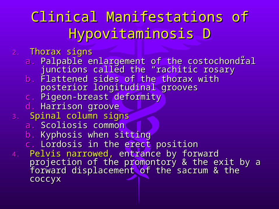
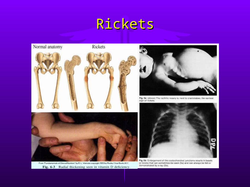
2.2. Thorax signsThorax signsa.a. Palpable enlargement of the costochondral Palpable enlargement of the costochondral
junctions called the “rachitic rosary”junctions called the “rachitic rosary”b.b. Flattened sides of the thorax with posterior Flattened sides of the thorax with posterior
longitudinal grooveslongitudinal groovesc.c. Pigeon-breast deformityPigeon-breast deformityd.d. Harrison grooveHarrison groove
3.3. Spinal column signsSpinal column signsa.a. Scoliosis commonScoliosis commonb.b. Kyphosis when sittingKyphosis when sittingc.c. Lordosis in the erect positionLordosis in the erect position
4.4. Pelvis narrowed, Pelvis narrowed, entrance by forward projection of entrance by forward projection of the promontory & the exit by a forward the promontory & the exit by a forward displacement of the sacrum & the coccyxdisplacement of the sacrum & the coccyx
Clinical Manifestations of Clinical Manifestations of Hypovitaminosis DHypovitaminosis D
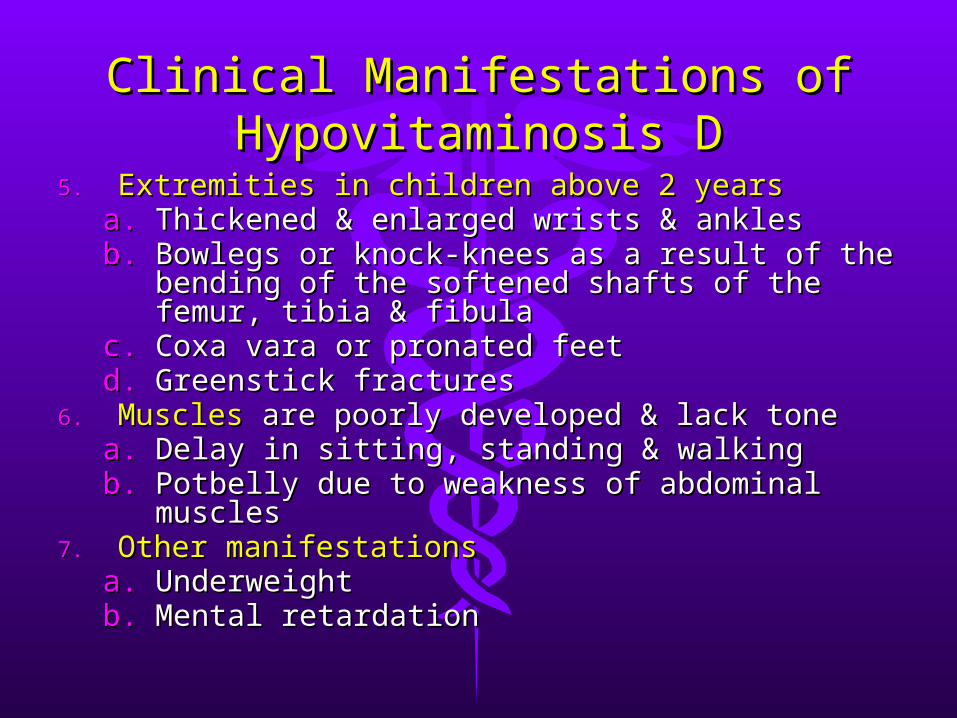
5.5. Extremities in children above 2 yearsExtremities in children above 2 yearsa.a. Thickened & enlarged wrists & anklesThickened & enlarged wrists & anklesb.b. Bowlegs or knock-knees as a result of the Bowlegs or knock-knees as a result of the
bending of the softened shafts of the femur, bending of the softened shafts of the femur, tibia & fibulatibia & fibula
c.c. Coxa vara or pronated feetCoxa vara or pronated feetd.d. Greenstick fracturesGreenstick fractures
6.6. MusclesMuscles are poorly developed & lack tone are poorly developed & lack tonea.a. Delay in sitting, standing & walkingDelay in sitting, standing & walkingb.b. Potbelly due to weakness of abdominal Potbelly due to weakness of abdominal
musclesmuscles7.7. Other manifestationsOther manifestations
a.a. UnderweightUnderweightb.b. Mental retardationMental retardation

Clinical Manifestations of Clinical Manifestations of Hypovitaminosis DHypovitaminosis D
B.B. Osteomalacia:Osteomalacia: Accumulation of uncalcified Accumulation of uncalcified osteoid tissue in rib joints of an adult resulting inosteoid tissue in rib joints of an adult resulting in
1.1. Pain in the pelvis, lower back and legsPain in the pelvis, lower back and legs2.2. Tenderness felt in the shins and in other Tenderness felt in the shins and in other
bonesbones3.3. Waddling gaitWaddling gait4.4. Deformities of the pelvisDeformities of the pelvis5.5. Tetany may occur manifested by involuntary Tetany may occur manifested by involuntary
twitching of the facial muscles or by twitching of the facial muscles or by carpopedal spasmcarpopedal spasm
6.6. Spontaneous fractures may be a featureSpontaneous fractures may be a featureC.C. Osteomalacia should not be confused with Osteomalacia should not be confused with
osteoporosisosteoporosis, a disease of ageing, in which , a disease of ageing, in which decalcification is also a feature decalcification is also a feature
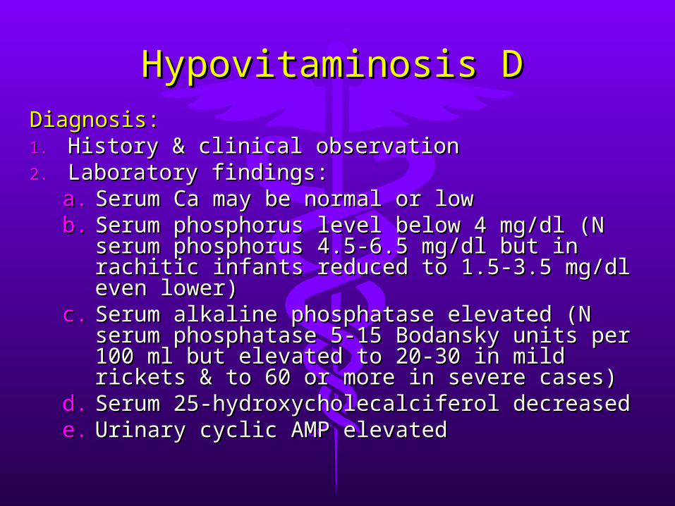
Hypovitaminosis DHypovitaminosis DDiagnosis:Diagnosis:1.1. History & clinical observationHistory & clinical observation2.2. Laboratory findings:Laboratory findings:
a.a. Serum Ca may be normal or lowSerum Ca may be normal or lowb.b. Serum phosphorus level below 4 mg/dl (N Serum phosphorus level below 4 mg/dl (N
serum phosphorus 4.5-6.5 mg/dl but in rachitic serum phosphorus 4.5-6.5 mg/dl but in rachitic infants reduced to 1.5-3.5 mg/dl even lower)infants reduced to 1.5-3.5 mg/dl even lower)
c.c. Serum alkaline phosphatase elevated (N serum Serum alkaline phosphatase elevated (N serum phosphatase 5-15 Bodansky units per 100 ml phosphatase 5-15 Bodansky units per 100 ml but elevated to 20-30 in mild rickets & to 60 or but elevated to 20-30 in mild rickets & to 60 or more in severe cases)more in severe cases)
d.d. Serum 25-hydroxycholecalciferol decreasedSerum 25-hydroxycholecalciferol decreasede.e. Urinary cyclic AMP elevatedUrinary cyclic AMP elevated
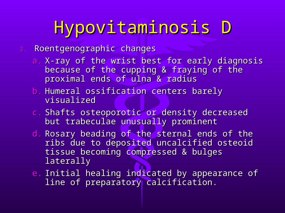
Hypovitaminosis DHypovitaminosis D3.3. Roentgenographic changesRoentgenographic changes
a.a. X-ray of the wrist best for early diagnosis X-ray of the wrist best for early diagnosis because of the cupping & fraying of the because of the cupping & fraying of the proximal ends of ulna & radiusproximal ends of ulna & radius
b.b. Humeral ossification centers barely visualizedHumeral ossification centers barely visualized
c.c. Shafts osteoporotic or density decreased but Shafts osteoporotic or density decreased but trabeculae unusually prominenttrabeculae unusually prominent
d.d. Rosary beading of the sternal ends of the ribs Rosary beading of the sternal ends of the ribs due to deposited uncalcified osteoid tissue due to deposited uncalcified osteoid tissue becoming compressed & bulges laterally becoming compressed & bulges laterally
e.e. Initial healing indicated by appearance of line Initial healing indicated by appearance of line of preparatory calcification.of preparatory calcification.
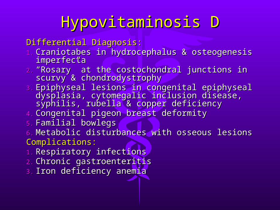
Hypovitaminosis DHypovitaminosis DDifferential Diagnosis:Differential Diagnosis:1.1. Craniotabes in hydrocephalus & osteogenesis Craniotabes in hydrocephalus & osteogenesis
imperfectaimperfecta2.2. ““Rosary” at the costochondral junctions in Rosary” at the costochondral junctions in
scurvy & chondrodystrophyscurvy & chondrodystrophy3.3. Epiphyseal lesions in congenital epiphyseal Epiphyseal lesions in congenital epiphyseal
dysplasia, cytomegalic inclusion disease, dysplasia, cytomegalic inclusion disease, syphilis, rubella & copper deficiencysyphilis, rubella & copper deficiency
4.4. Congenital pigeon breast deformityCongenital pigeon breast deformity5.5. Familial bowlegsFamilial bowlegs6.6. Metabolic disturbances with osseous lesionsMetabolic disturbances with osseous lesionsComplications:Complications:1.1. Respiratory infectionsRespiratory infections2.2. Chronic gastroenteritisChronic gastroenteritis3.3. Iron deficiency anemiaIron deficiency anemia
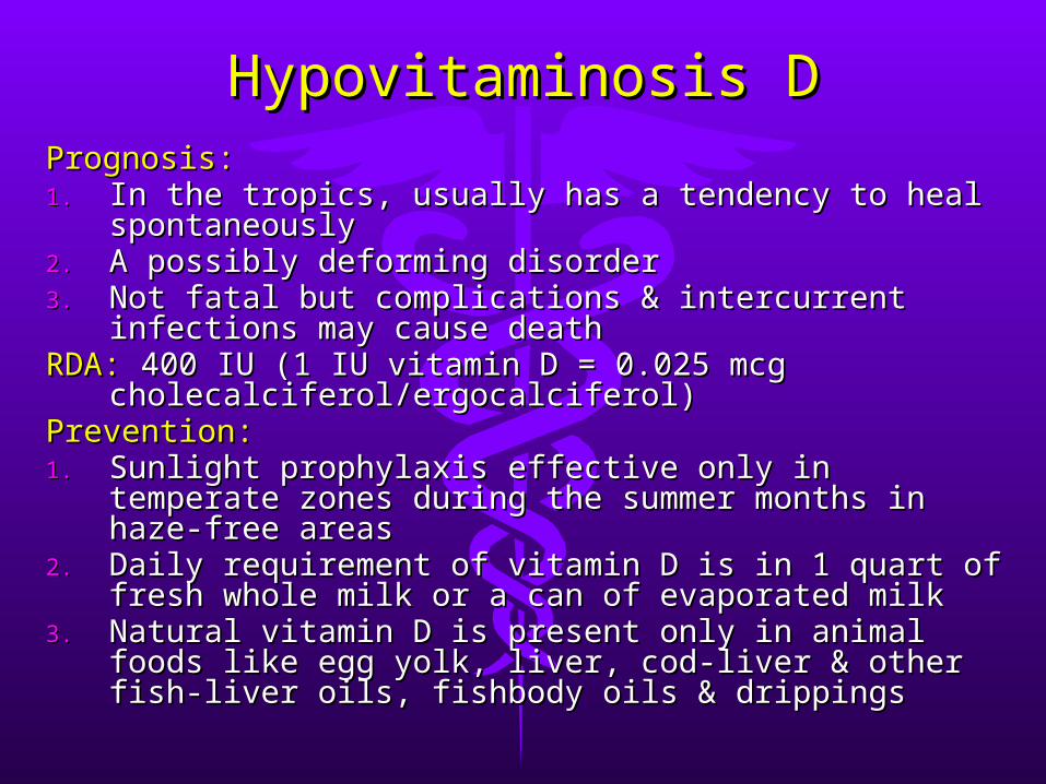
Hypovitaminosis DHypovitaminosis DPrognosis:Prognosis:1.1. In the tropics, usually has a tendency to heal In the tropics, usually has a tendency to heal
spontaneouslyspontaneously2.2. A possibly deforming disorderA possibly deforming disorder3.3. Not fatal but complications & intercurrent Not fatal but complications & intercurrent
infections may cause deathinfections may cause deathRDA:RDA: 400 IU (1 IU vitamin D = 0.025 mcg 400 IU (1 IU vitamin D = 0.025 mcg
cholecalciferol/ergocalciferol)cholecalciferol/ergocalciferol)Prevention:Prevention:1.1. Sunlight prophylaxis effective only in temperate Sunlight prophylaxis effective only in temperate
zones during the summer months in haze-free zones during the summer months in haze-free areasareas
2.2. Daily requirement of vitamin D is in 1 quart of Daily requirement of vitamin D is in 1 quart of fresh whole milk or a can of evaporated milkfresh whole milk or a can of evaporated milk
3.3. Natural vitamin D is present only in animal foods Natural vitamin D is present only in animal foods like egg yolk, liver, cod-liver & other fish-liver oils, like egg yolk, liver, cod-liver & other fish-liver oils, fishbody oils & drippingsfishbody oils & drippings

Hypovitaminosis DHypovitaminosis D4.4. Prematures or breast-fed infants should receive Prematures or breast-fed infants should receive
supplemental vitamin D daily because milk is a supplemental vitamin D daily because milk is a poor source unless fortifiedpoor source unless fortified
5.5. Vitamin D should also be administered to Vitamin D should also be administered to pregnant & lactating motherspregnant & lactating mothers
Treatment:Treatment:1.1. Daily administration of 50-150 mcg of vitamin D3 Daily administration of 50-150 mcg of vitamin D3
or 0.5-2 mcg of 1,25-dihydroxycholecalciferol will or 0.5-2 mcg of 1,25-dihydroxycholecalciferol will produce healing seen on X-ray within 2-4 wksproduce healing seen on X-ray within 2-4 wks
2.2. Vitamin D 15,000 mcg in a single dose w/o Vitamin D 15,000 mcg in a single dose w/o further therapy for several months may be further therapy for several months may be advantageousadvantageous
3.3. After healing is complete, the dose of vitamin D After healing is complete, the dose of vitamin D should be lowered to 10 mcg/dayshould be lowered to 10 mcg/day
4.4. If no healing occurs, rickets is probably resistant If no healing occurs, rickets is probably resistant to vitamin D or non-nutritional ricketsto vitamin D or non-nutritional rickets
RicketsRickets
RicketsRickets
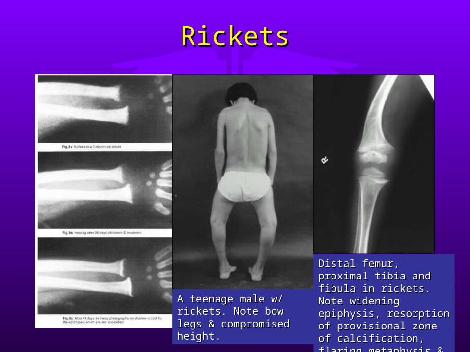
A teenage male w/ A teenage male w/ rickets. Note bow legs & rickets. Note bow legs & compromised height. compromised height.
Distal femur, proximal Distal femur, proximal tibia and fibula in tibia and fibula in rickets. Note widening rickets. Note widening epiphysis, resorption of epiphysis, resorption of provisional zone of provisional zone of calcification, flaring calcification, flaring metaphysis & bone metaphysis & bone deformity. deformity.
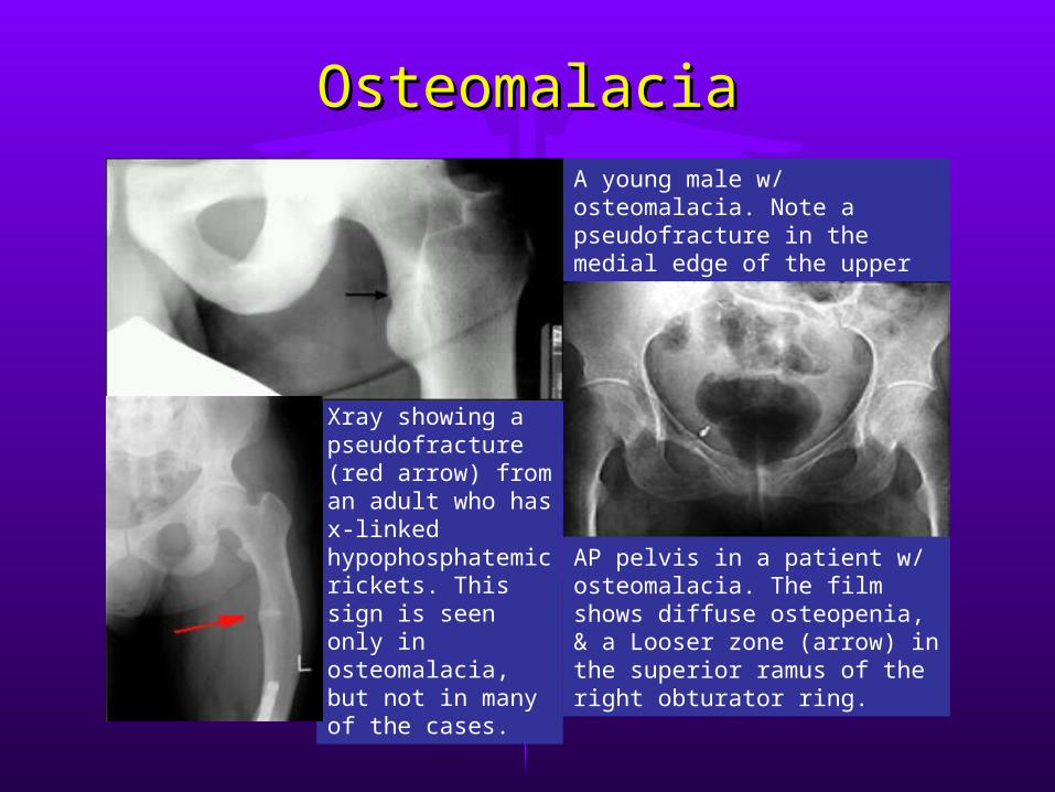
OsteomalaciaOsteomalaciaA young male w/ osteomalacia. Note a pseudofracture in the medial edge of the upper femoral shaft (arrow).
Xray showing a pseudofracture (red arrow) from an adult who has x-linked hypophosphatemic rickets. This sign is seen only in osteomalacia, but not in many of the cases.
AP pelvis in a patient w/ osteomalacia. The film shows diffuse osteopenia, & a Looser zone (arrow) in the superior ramus of the right obturator ring.
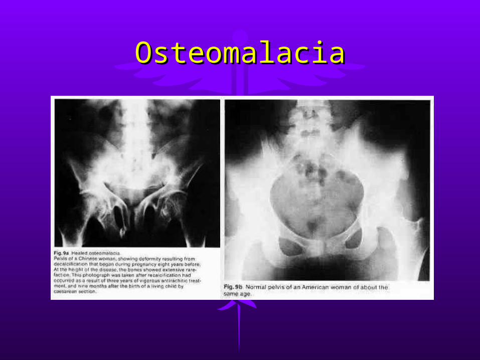
OsteomalaciaOsteomalacia
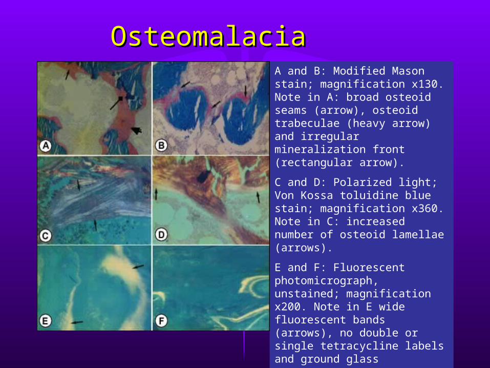
OsteomalaciaOsteomalaciaA and B: Modified Mason stain; magnification x130. Note in A: broad osteoid seams (arrow), osteoid trabeculae (heavy arrow) and irregular mineralization front (rectangular arrow).
C and D: Polarized light; Von Kossa toluidine blue stain; magnification x360. Note in C: increased number of osteoid lamellae (arrows).
E and F: Fluorescent photomicrograph, unstained; magnification x200. Note in E wide fluorescent bands (arrows), no double or single tetracycline labels and ground glass appearance.
Hypervitaminosis DHypervitaminosis D
Etiology:Etiology:Excessive intakes from Excessive intakes from Inadvertently substituting Inadvertently substituting
concentrated form for concentrated form for dilutedilute
Parents’ increasing Parents’ increasing prescribed doseprescribed dose
Inadequately controlling Inadequately controlling dosages for children dosages for children receiving large amounts receiving large amounts of vitamin D for chronic of vitamin D for chronic hyperphosphatemic hyperphosphatemic statesstates
Clinical Manifestations:Clinical Manifestations:Symptoms after 1-3 monthsSymptoms after 1-3 months1.1. Hypotonia, anorexia, Hypotonia, anorexia,
irritability, constipation, irritability, constipation, polydipsia, polyuria & polydipsia, polyuria & pallorpallor
2.2. Dehydration usually Dehydration usually presentpresent
3.3. Aortic valvular stenosis, Aortic valvular stenosis, vomiting, hypertension, vomiting, hypertension, retinopathy & clouding of retinopathy & clouding of cornea & conjunctiva may cornea & conjunctiva may occuroccur
Hypervitaminosis DHypervitaminosis D
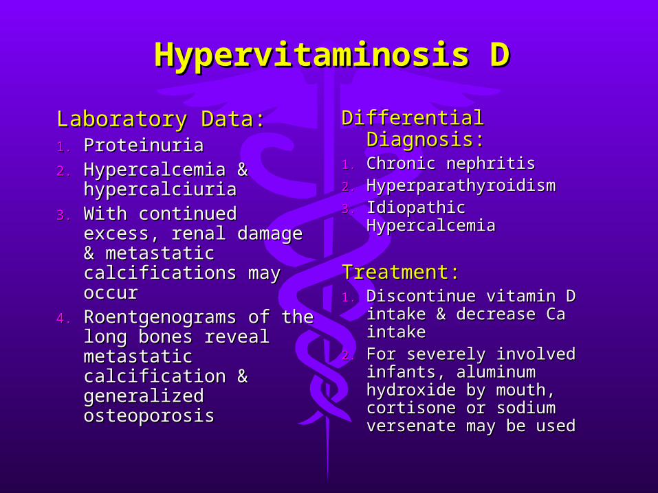
Laboratory Data:Laboratory Data:1.1. ProteinuriaProteinuria2.2. Hypercalcemia & Hypercalcemia &
hypercalciuriahypercalciuria3.3. With continued excess, With continued excess,
renal damage & renal damage & metastatic calcifications metastatic calcifications may occurmay occur
4.4. Roentgenograms of the Roentgenograms of the long bones reveal long bones reveal metastatic calcification & metastatic calcification & generalized osteoporosisgeneralized osteoporosis
Differential Diagnosis:Differential Diagnosis:1.1. Chronic nephritisChronic nephritis2.2. HyperparathyroidismHyperparathyroidism3.3. Idiopathic HypercalcemiaIdiopathic Hypercalcemia
Treatment:Treatment:1.1. Discontinue vitamin D Discontinue vitamin D
intake & decrease Ca intake & decrease Ca intakeintake
2.2. For severely involved For severely involved infants, aluminum infants, aluminum hydroxide by mouth, hydroxide by mouth, cortisone or sodium cortisone or sodium versenate may be usedversenate may be used
Vitamin KVitamin K
Vitamin KVitamin K11,, naturally occurring vitamin K, naturally occurring vitamin K, is abundant in pork, liver, soybeans & is abundant in pork, liver, soybeans & green leafy vegetablesgreen leafy vegetables
Intestinal microorganisms synthesizeIntestinal microorganisms synthesize Required for normal clotting of bloodRequired for normal clotting of blood Vitamin K-dependent clotting factors Vitamin K-dependent clotting factors
made in the liver: prothrombin, made in the liver: prothrombin, proconvertin (Factor VII), plasma proconvertin (Factor VII), plasma thromboplastin component or PTC (Factor thromboplastin component or PTC (Factor IX) & Stuart-Prower factor (Factor X)IX) & Stuart-Prower factor (Factor X)
Vitamin K Deficiency Vitamin K Deficiency (Hypoprothrombinemia)(Hypoprothrombinemia)
Etiology:Etiology: The fetus depends on the mother for supply & at The fetus depends on the mother for supply & at
birth, the bacterial flora of the GIT not yet producebirth, the bacterial flora of the GIT not yet produce Exclusively breast-fed infants lower vitamin K Exclusively breast-fed infants lower vitamin K
compared to formula-fedcompared to formula-fed Faulty intestinal absorption as in diarrhea, celiac Faulty intestinal absorption as in diarrhea, celiac
disease, gastrointestinal malformation & disease, gastrointestinal malformation & steatorrheasteatorrhea
Obstructive jaundice, biliary fistula, insufficient Obstructive jaundice, biliary fistula, insufficient production of bile acids or pancreatic insufficiency production of bile acids or pancreatic insufficiency lead to inadequate intestinal absorptionlead to inadequate intestinal absorption
Administration of antibiotics which inhibit intestinal Administration of antibiotics which inhibit intestinal bacteriabacteria
In sepsis, deficiency result s from disease affecting In sepsis, deficiency result s from disease affecting hepatobiliary functions & therapyhepatobiliary functions & therapy
Drugs like Coumarin, Salicylates & anticonvulsantsDrugs like Coumarin, Salicylates & anticonvulsants
Vitamin K Deficiency Vitamin K Deficiency (Hypoprothrombinemia)(Hypoprothrombinemia)
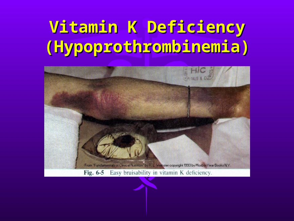
Clinical Manifestations:Clinical Manifestations:1.1. Hemorrhagic manifestations are the hallmarkHemorrhagic manifestations are the hallmark2.2. Bleeding in the newborn from the cord or Bleeding in the newborn from the cord or
circumcision sitecircumcision site3.3. GIT bleeding, hematuria & intracranial GIT bleeding, hematuria & intracranial
hemorrhage more serioushemorrhage more serious4.4. Anemia & shock may ensue from severe Anemia & shock may ensue from severe
blood lossblood loss
Laboratory Test: Laboratory Test: The most useful test is the The most useful test is the 1-1-stage prothrombin time test (Quick)stage prothrombin time test (Quick), , prolongation indicates presumptive evidence prolongation indicates presumptive evidence deficiencydeficiency
Vitamin K Deficiency Vitamin K Deficiency (Hypoprothrombinemia)(Hypoprothrombinemia)
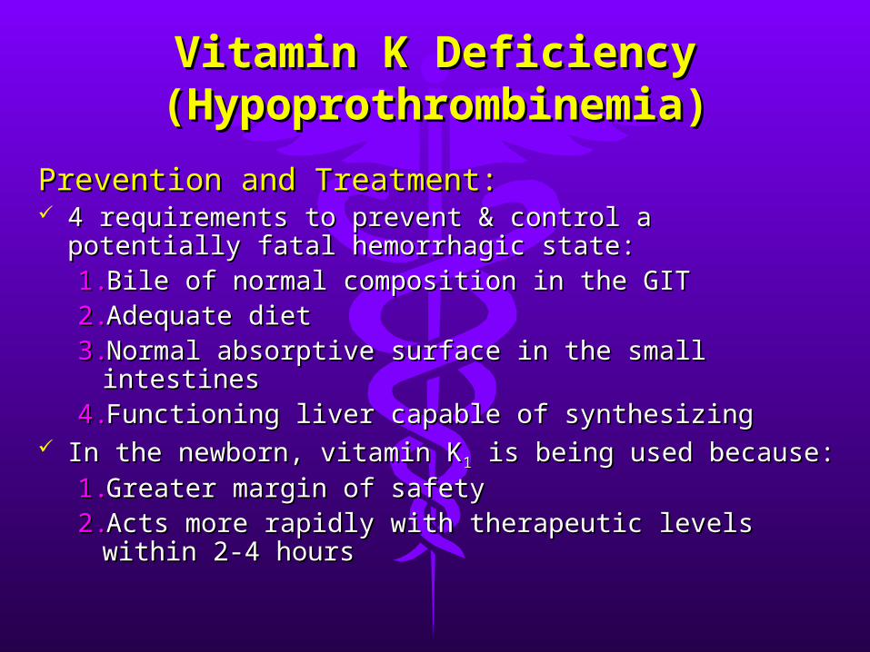
Prevention and Treatment:Prevention and Treatment: 4 requirements to prevent & control a potentially 4 requirements to prevent & control a potentially
fatal hemorrhagic state:fatal hemorrhagic state:1.1.Bile of normal composition in the GITBile of normal composition in the GIT2.2.Adequate dietAdequate diet3.3.Normal absorptive surface in the small intestinesNormal absorptive surface in the small intestines4.4.Functioning liver capable of synthesizingFunctioning liver capable of synthesizing
In the newborn, vitamin KIn the newborn, vitamin K11 is being used because: is being used because:1.1.Greater margin of safety Greater margin of safety 2.2.Acts more rapidly with therapeutic levels within Acts more rapidly with therapeutic levels within
2-4 hours2-4 hours
Vitamin K Deficiency Vitamin K Deficiency (Hypoprothrombinemia)(Hypoprothrombinemia)
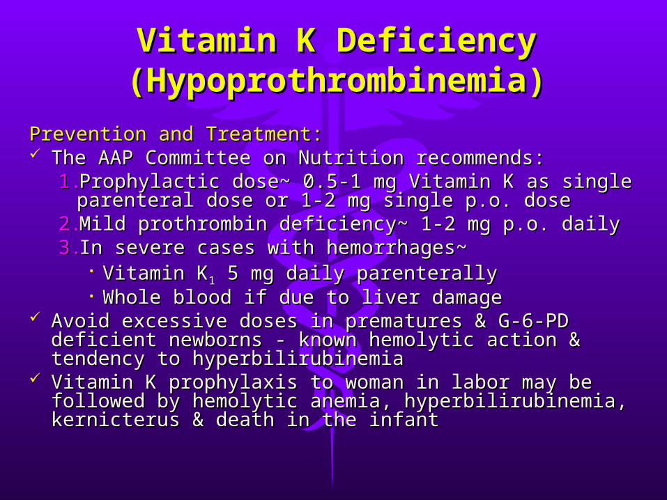
Prevention and Treatment:Prevention and Treatment: The AAP Committee on Nutrition recommends:The AAP Committee on Nutrition recommends:
1.1.Prophylactic dose~ 0.5-1 mg Vitamin K as single Prophylactic dose~ 0.5-1 mg Vitamin K as single parenteral dose or 1-2 mg single p.o. doseparenteral dose or 1-2 mg single p.o. dose
2.2.Mild prothrombin deficiency~ 1-2 mg p.o. dailyMild prothrombin deficiency~ 1-2 mg p.o. daily3.3.In severe cases with hemorrhages~In severe cases with hemorrhages~
• Vitamin KVitamin K11 5 mg daily parenterally 5 mg daily parenterally• Whole blood if due to liver damageWhole blood if due to liver damage
Avoid excessive doses in prematures & G-6-PD Avoid excessive doses in prematures & G-6-PD deficient newborns - known hemolytic action & deficient newborns - known hemolytic action & tendency to hyperbilirubinemiatendency to hyperbilirubinemia
Vitamin K prophylaxis to woman in labor may be Vitamin K prophylaxis to woman in labor may be followed by hemolytic anemia, hyperbilirubinemia, followed by hemolytic anemia, hyperbilirubinemia, kernicterus & death in the infantkernicterus & death in the infant
Vitamin K Deficiency Vitamin K Deficiency (Hypoprothrombinemia)(Hypoprothrombinemia)
Tocopherol (Vitamin E) Tocopherol (Vitamin E) DeficiencyDeficiency
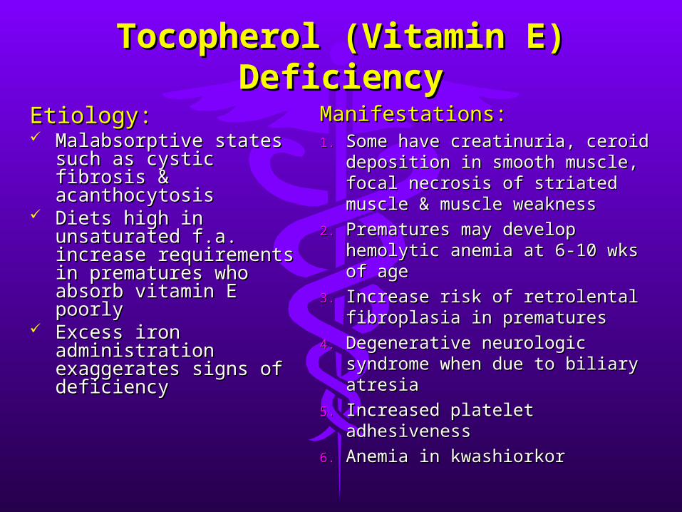
Etiology:Etiology: Malabsorptive states Malabsorptive states
such as cystic fibrosis & such as cystic fibrosis & acanthocytosisacanthocytosis
Diets high in Diets high in unsaturated f.a. increase unsaturated f.a. increase requirements in requirements in prematures who absorb prematures who absorb vitamin E poorlyvitamin E poorly
Excess iron Excess iron administration administration exaggerates signs of exaggerates signs of deficiencydeficiency
Manifestations:Manifestations:1.1. Some have creatinuria, ceroid Some have creatinuria, ceroid
deposition in smooth muscle, deposition in smooth muscle, focal necrosis of striated focal necrosis of striated muscle & muscle weaknessmuscle & muscle weakness
2.2. Prematures may develop Prematures may develop hemolytic anemia at 6-10 wks hemolytic anemia at 6-10 wks of ageof age
3.3. Increase risk of retrolental Increase risk of retrolental fibroplasia in prematuresfibroplasia in prematures
4.4. Degenerative neurologic Degenerative neurologic syndrome when due to biliary syndrome when due to biliary atresiaatresia
5.5. Increased platelet adhesivenessIncreased platelet adhesiveness
6.6. Anemia in kwashiorkorAnemia in kwashiorkor

Tocopherol (Vitamin E) Tocopherol (Vitamin E) DeficiencyDeficiency
Prevention & Treatment:Prevention & Treatment: 1.1. RDA not known but 0.7mg/g of RDA not known but 0.7mg/g of
unsaturated fat in the diet adequateunsaturated fat in the diet adequate
2.2. Premature infants may be given 15-Premature infants may be given 15-25 IU/24 hrs25 IU/24 hrs
3.3. Large oral or parenteral doses may Large oral or parenteral doses may prevent permanent neurologic prevent permanent neurologic abnormalities in biliary atresia or abnormalities in biliary atresia or abetalipoproteinemiaabetalipoproteinemia


Challenges can be stepping stones or stumbling Challenges can be stepping stones or stumbling blocks. blocks.
It’s just a matter of how you view them.It’s just a matter of how you view them.