Nucleic Acids – Basic Concepts - University College Dublin d murray dna rna slides 09.pdf ·...
136
Nucleic Acids Nucleic Acids – – Basic Concepts Basic Concepts David Murray PhD UCD|Mater Clinical Research Centre UCD School of Medicine and Medical Sciences Mater Misericordiae University Hospital Dublin
Transcript of Nucleic Acids – Basic Concepts - University College Dublin d murray dna rna slides 09.pdf ·...
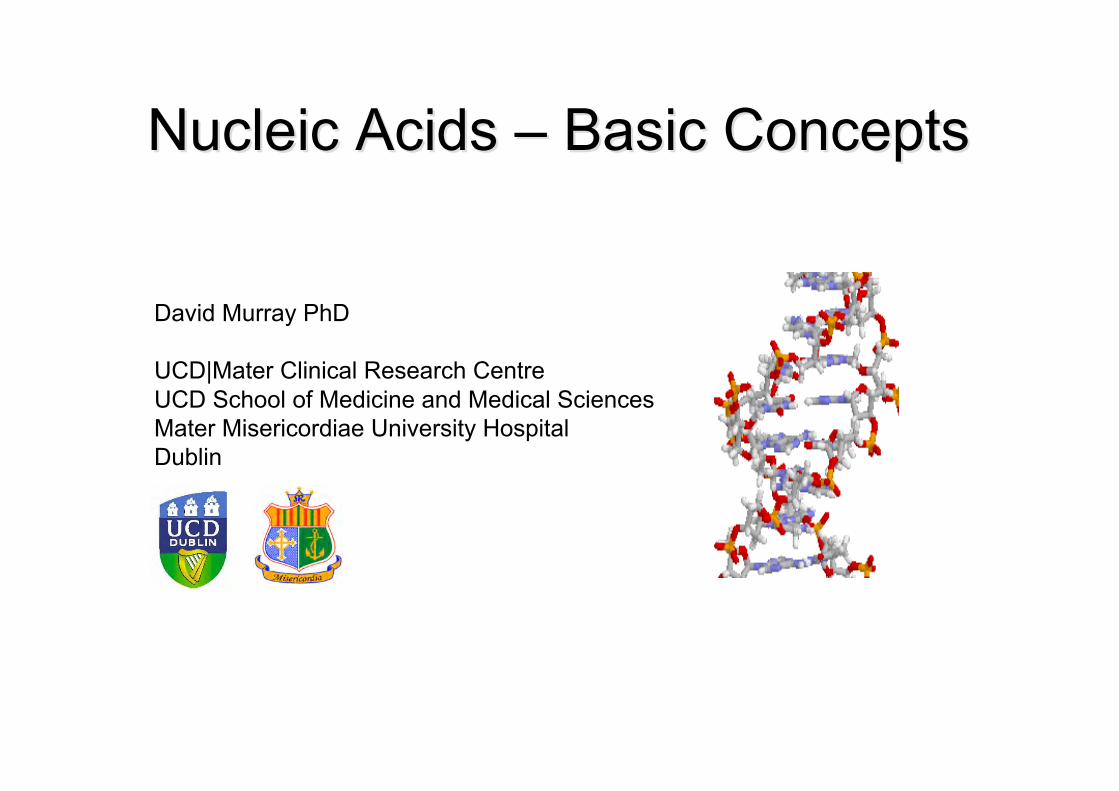
Nucleic Acids Nucleic Acids –– Basic Concepts Basic Concepts
David Murray PhD
UCD|Mater Clinical Research CentreUCD School of Medicine and Medical SciencesMater Misericordiae University HospitalDublin
DNA and RNA are NucleicAcids
Outline
• What are DNA and RNA ?• The Structure and Function of DNA and
RNA• What is a Gene ?• What is a Genome ?
DNA and RNAWhat's the Big Deal ?
• Hereditary Genetics• Importance of Genetics in Disease• Predisposition• Mutations• Loss of Genetic Control
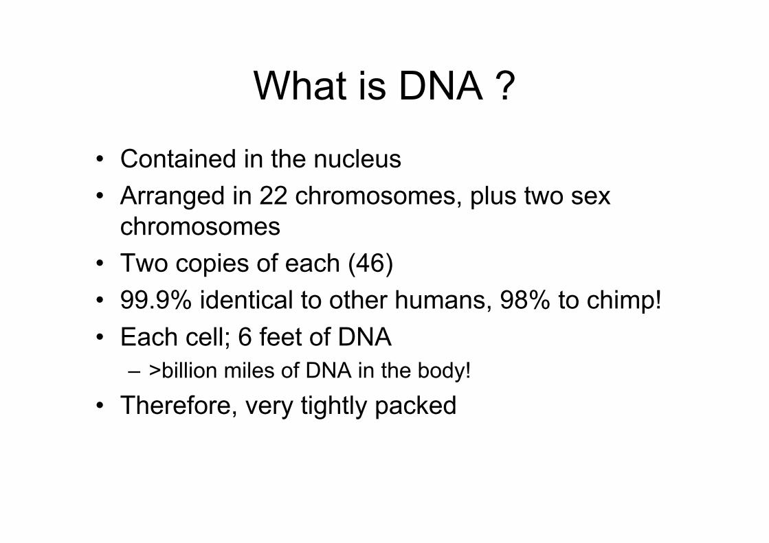
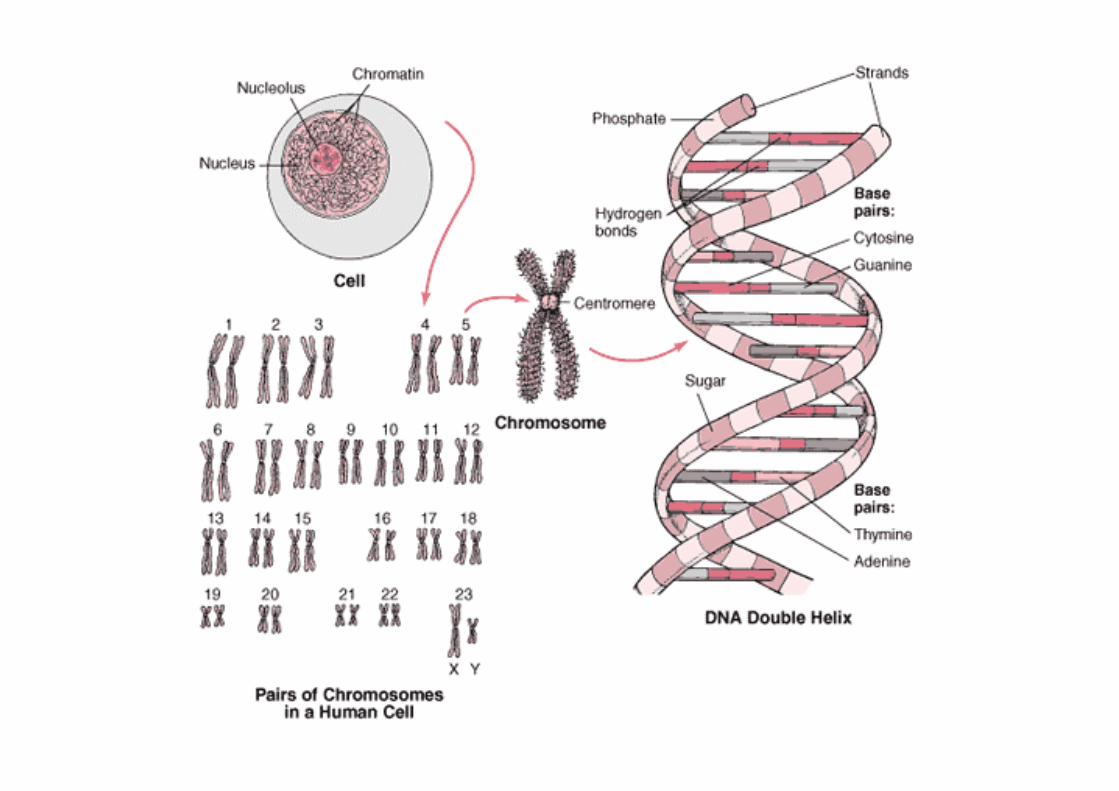
What is DNA ?
• Contained in the nucleus• Arranged in 22 chromosomes, plus two sex
chromosomes• Two copies of each (46)• 99.9% identical to other humans, 98% to chimp!• Each cell; 6 feet of DNA
– >billion miles of DNA in the body!• Therefore, very tightly packed
The Structure of DNA and RNA
• DNA (deoxyribonucleic acid)• RNA (ribonucleic acid)
– What’s the Difference ?• Both composed of two different
classes of nitrogen containingbases:– the purines and pyrimidines.
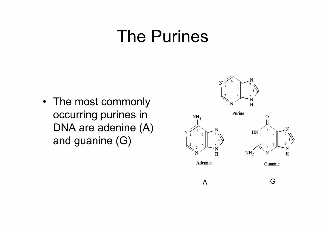
The Purines
• The most commonlyoccurring purines inDNA are adenine (A)and guanine (G)
A G
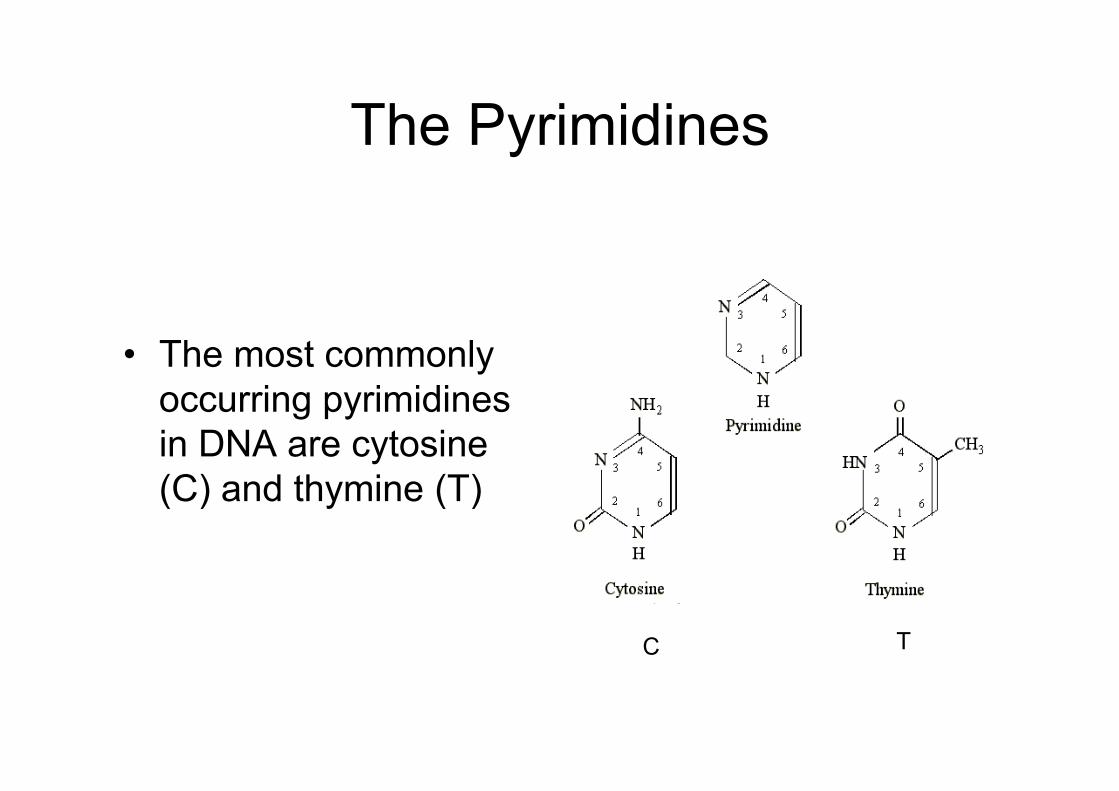
The Pyrimidines
• The most commonlyoccurring pyrimidinesin DNA are cytosine(C) and thymine (T)
C T
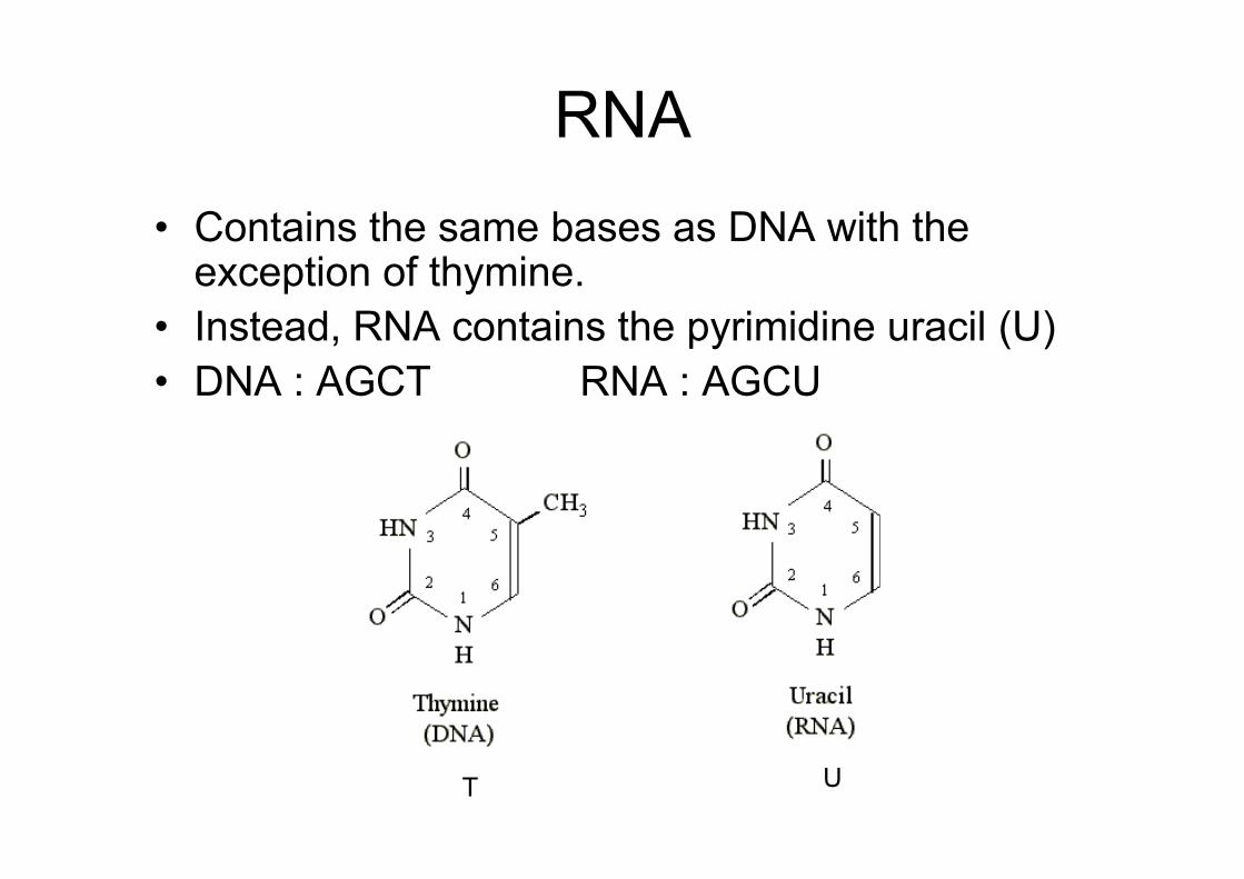
• Contains the same bases as DNA with theexception of thymine.
• Instead, RNA contains the pyrimidine uracil (U)• DNA : AGCT RNA : AGCU
RNA
T U
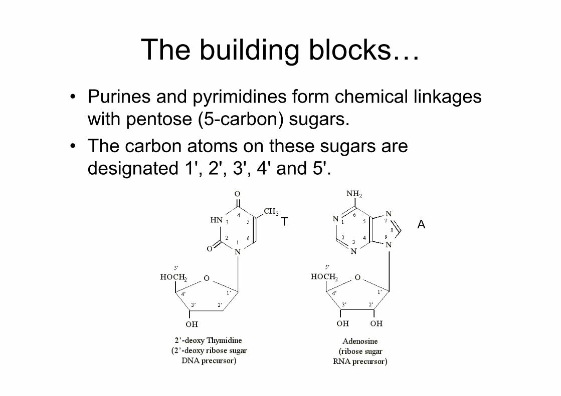
• Purines and pyrimidines form chemical linkageswith pentose (5-carbon) sugars.
• The carbon atoms on these sugars aredesignated 1', 2', 3', 4' and 5'.
The building blocks…
T A
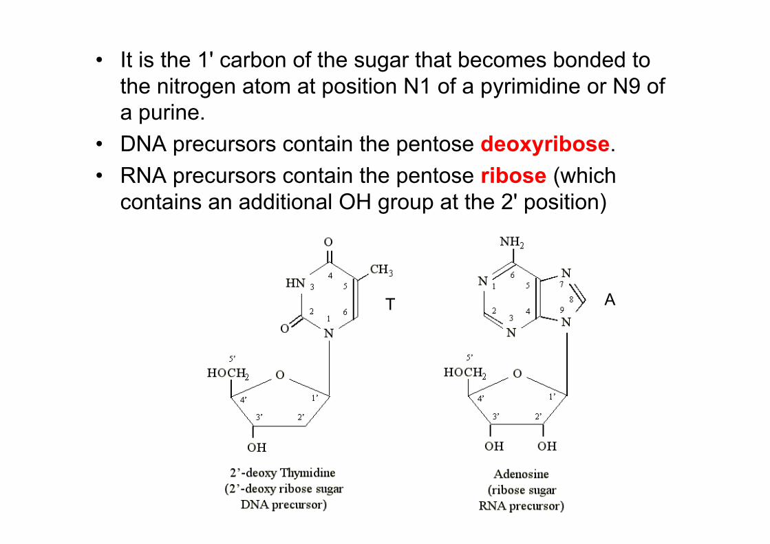
• It is the 1' carbon of the sugar that becomes bonded tothe nitrogen atom at position N1 of a pyrimidine or N9 ofa purine.
• DNA precursors contain the pentose deoxyribose.• RNA precursors contain the pentose ribose (which
contains an additional OH group at the 2' position)
T A
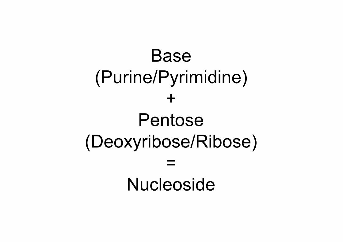
Base(Purine/Pyrimidine)
+Pentose
(Deoxyribose/Ribose)=
Nucleoside
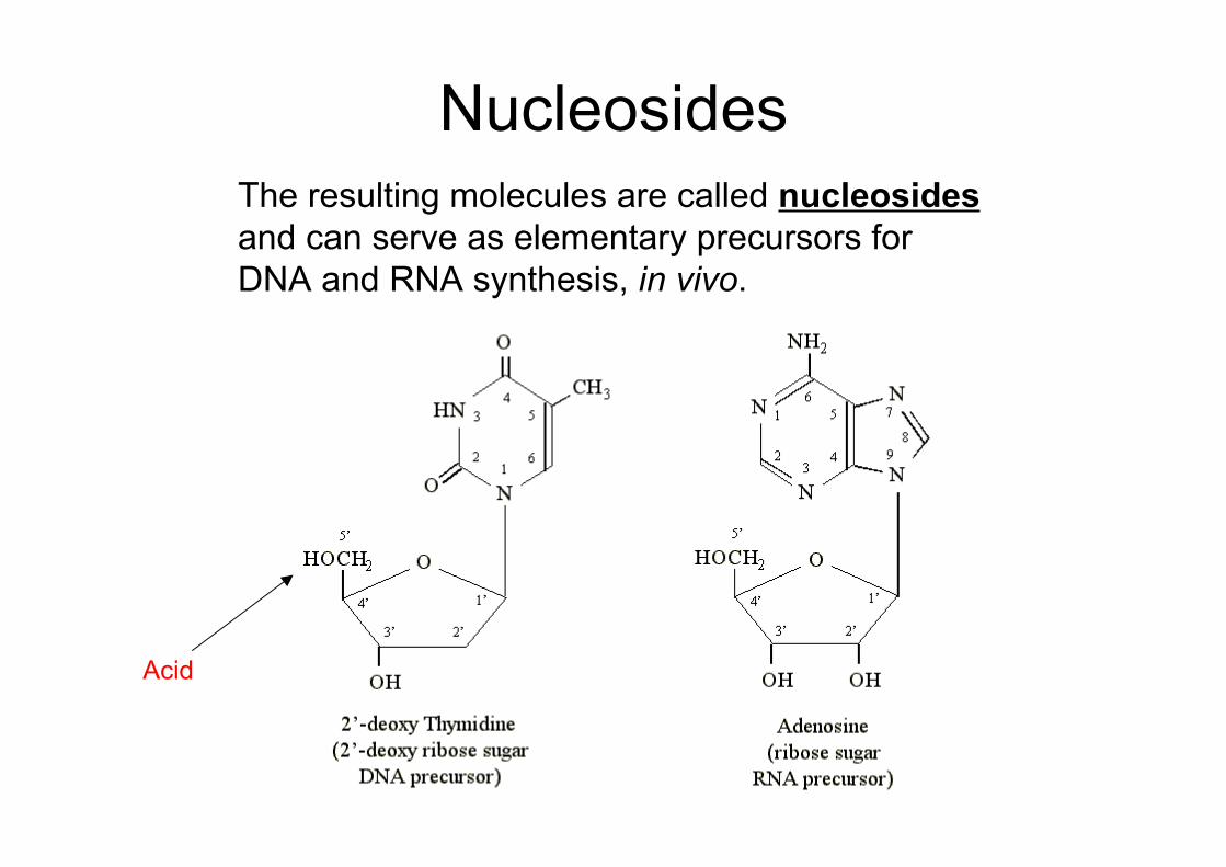
The resulting molecules are called nucleosidesand can serve as elementary precursors forDNA and RNA synthesis, in vivo.
Nucleosides
Acid
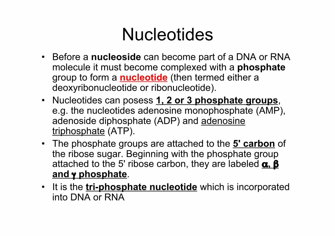
Nucleotides• Before a nucleoside can become part of a DNA or RNA
molecule it must become complexed with a phosphategroup to form a nucleotide (then termed either adeoxyribonucleotide or ribonucleotide).
• Nucleotides can posess 1, 2 or 3 phosphate groups,e.g. the nucleotides adenosine monophosphate (AMP),adenoside diphosphate (ADP) and adenosinetriphosphate (ATP).
• The phosphate groups are attached to the 5' carbon ofthe ribose sugar. Beginning with the phosphate groupattached to the 5' ribose carbon, they are labeled α, βand γ phosphate.
• It is the tri-phosphate nucleotide which is incorporatedinto DNA or RNA
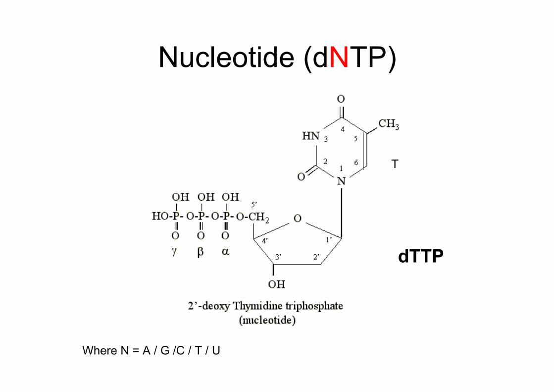
Nucleotide (dNTP)
dTTP
T
Where N = A / G /C / T / U
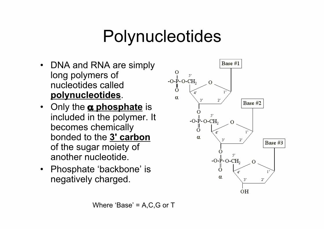
Polynucleotides• DNA and RNA are simply
long polymers ofnucleotides calledpolynucleotides.
• Only the α phosphate isincluded in the polymer. Itbecomes chemicallybonded to the 3' carbonof the sugar moiety ofanother nucleotide.
• Phosphate ‘backbone’ isnegatively charged.
Where ‘Base’ = A,C,G or T
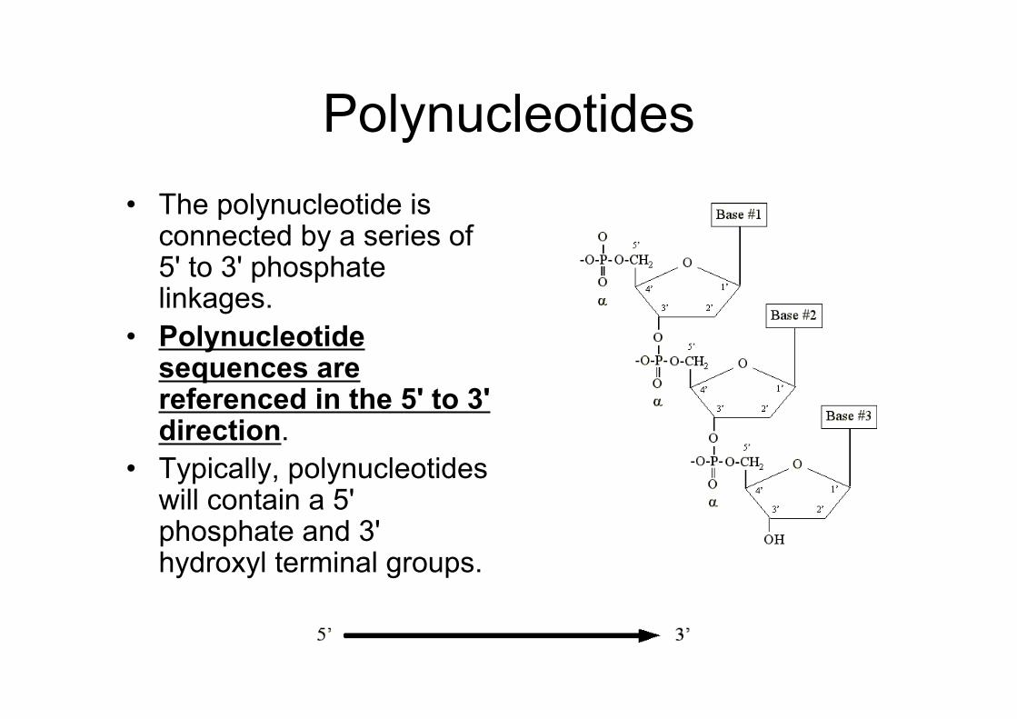
Polynucleotides• The polynucleotide is
connected by a series of5' to 3' phosphatelinkages.
• Polynucleotidesequences arereferenced in the 5' to 3'direction.
• Typically, polynucleotideswill contain a 5'phosphate and 3'hydroxyl terminal groups.
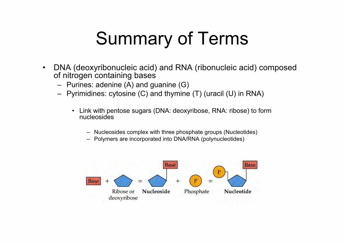
Summary of Terms• DNA (deoxyribonucleic acid) and RNA (ribonucleic acid) composed
of nitrogen containing bases– Purines: adenine (A) and guanine (G)– Pyrimidines: cytosine (C) and thymine (T) (uracil (U) in RNA)
• Link with pentose sugars (DNA: deoxyribose, RNA: ribose) to formnucleosides
– Nucleosides complex with three phosphate groups (Nucleotides)– Polymers are incorporated into DNA/RNA (polynucleotides)
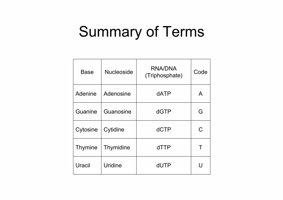
Summary of Terms
UdUTPUridineUracil
TdTTPThymidineThymine
CdCTPCytidineCytosine
GdGTPGuanosineGuanine
AdATPAdenosineAdenine
CodeRNA/DNA(Triphosphate)NucleosideBase
What is the structure of DNA?How is the structure related to
function?
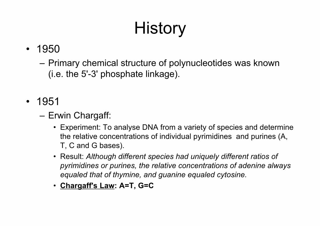
History• 1950
– Primary chemical structure of polynucleotides was known(i.e. the 5'-3' phosphate linkage).
• 1951– Erwin Chargaff:
• Experiment: To analyse DNA from a variety of species and determinethe relative concentrations of individual pyrimidines and purines (A,T, C and G bases).
• Result: Although different species had uniquely different ratios ofpyrimidines or purines, the relative concentrations of adenine alwaysequaled that of thymine, and guanine equaled cytosine.
• Chargaff's Law: A=T, G=C
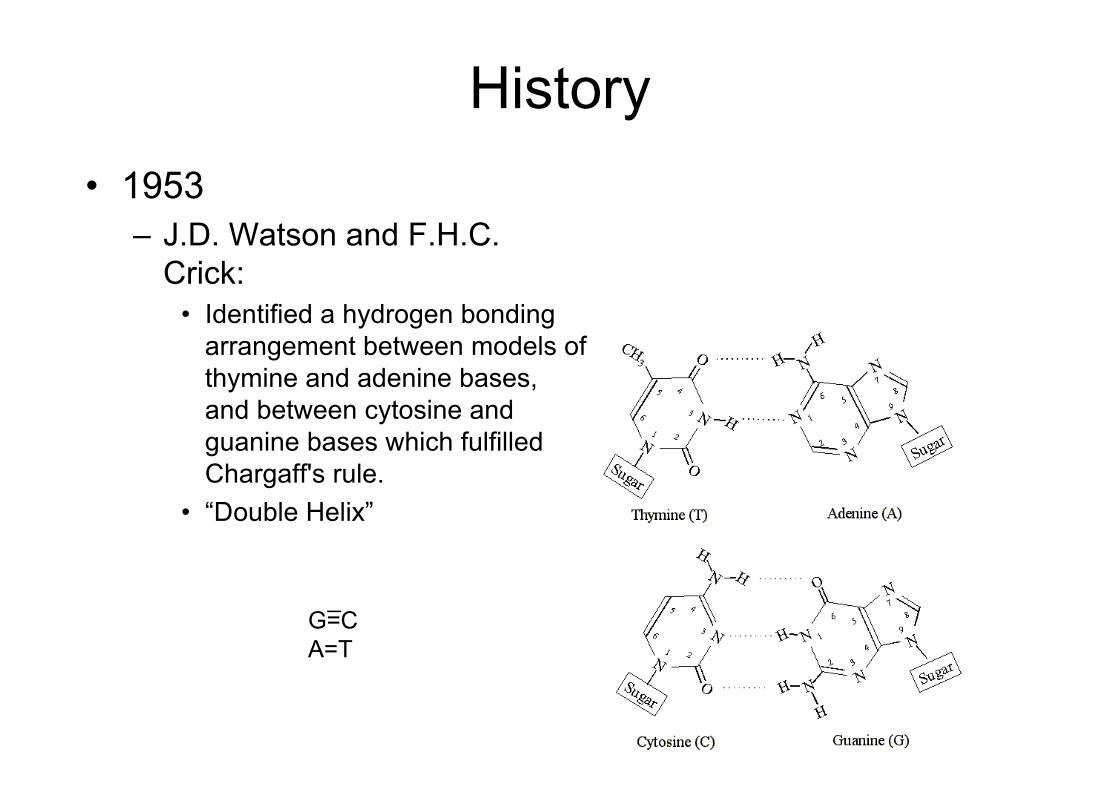
History• 1953
– J.D. Watson and F.H.C.Crick:
• Identified a hydrogen bondingarrangement between models ofthymine and adenine bases,and between cytosine andguanine bases which fulfilledChargaff's rule.
• “Double Helix”
G=CA=T
_
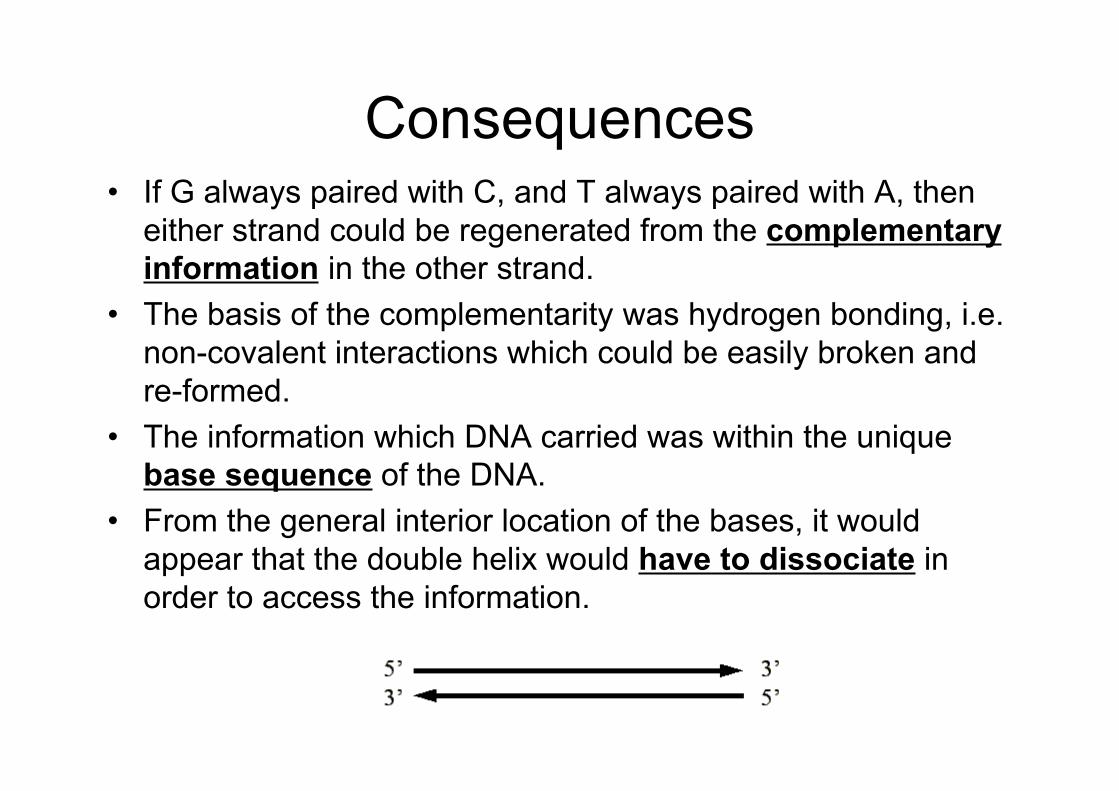
Consequences• If G always paired with C, and T always paired with A, then
either strand could be regenerated from the complementaryinformation in the other strand.
• The basis of the complementarity was hydrogen bonding, i.e.non-covalent interactions which could be easily broken andre-formed.
• The information which DNA carried was within the uniquebase sequence of the DNA.
• From the general interior location of the bases, it wouldappear that the double helix would have to dissociate inorder to access the information.
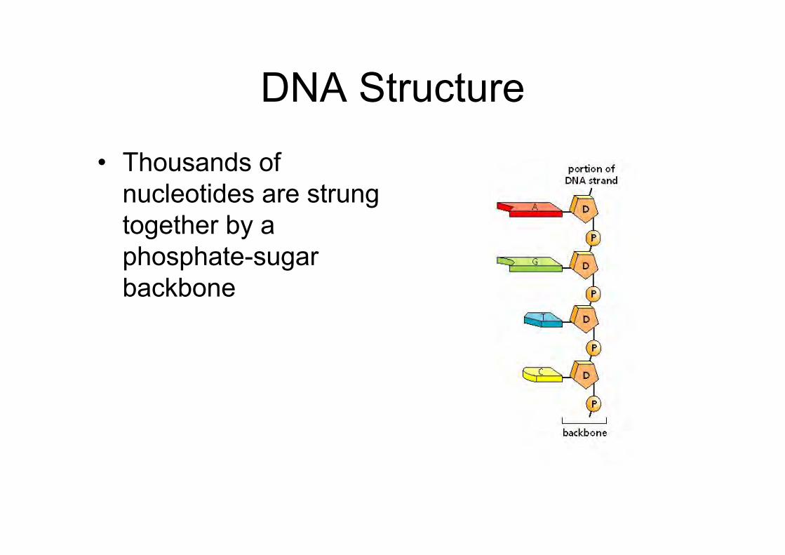
DNA Structure
• Thousands ofnucleotides are strungtogether by aphosphate-sugarbackbone
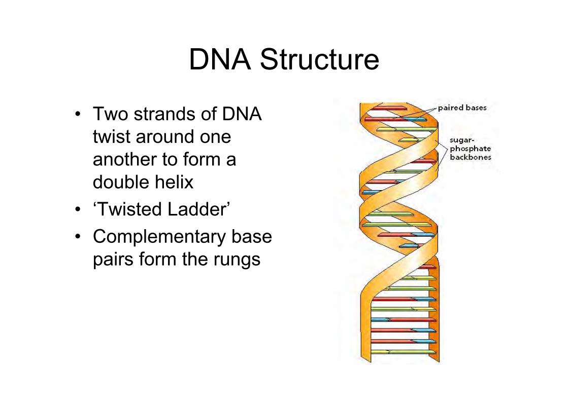
DNA Structure
• Two strands of DNAtwist around oneanother to form adouble helix
• ‘Twisted Ladder’• Complementary base
pairs form the rungs
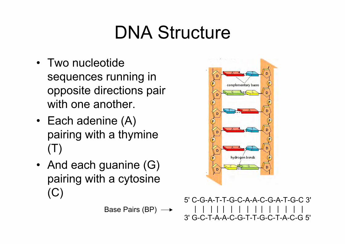
DNA Structure• Two nucleotide
sequences running inopposite directions pairwith one another.
• Each adenine (A)pairing with a thymine(T)
• And each guanine (G)pairing with a cytosine(C)
5' C-G-A-T-T-G-C-A-A-C-G-A-T-G-C 3' | | | | | | | | | | | | | | | 3' G-C-T-A-A-C-G-T-T-G-C-T-A-C-G 5'
Base Pairs (BP)
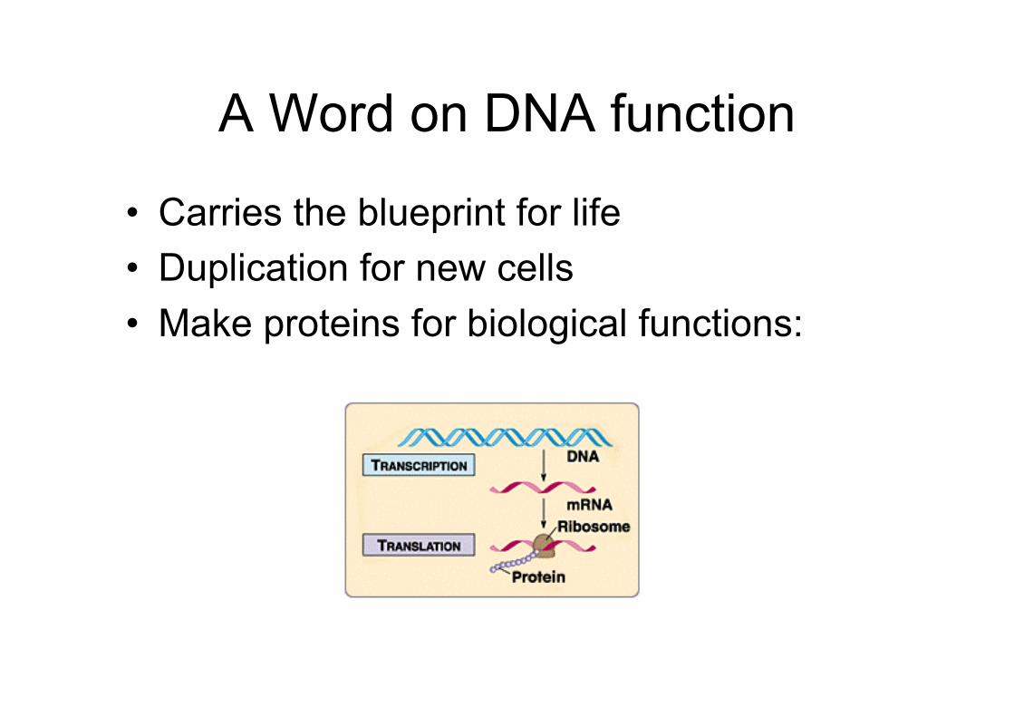
A Word on DNA function
• Carries the blueprint for life• Duplication for new cells• Make proteins for biological functions:
DNA• Prior to cell division, DNA is replicated
before being it is passed on to daughtercells
• The DNA within our cells contains theinformation for everything which occurswithin each cell,– every action– every substance made– every event– every response– everything!
The Genome
The Genome
• The complete set of information in an organism'sDNA is called its genome
• Carries the information for all the proteins theorganism will ever synthesize.
• Typical human cell– 6 feet of DNA– Written in the four-letter nucleotide alphabet that
spells out the linear sequence of amino acids in aprotein.
– Carries instructions for ~ 30,000 different proteins
Gene Expression
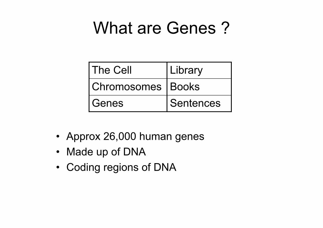
What are Genes ?
• Approx 26,000 human genes• Made up of DNA• Coding regions of DNA
GenesChromosomesThe Cell
SentencesBooksLibrary
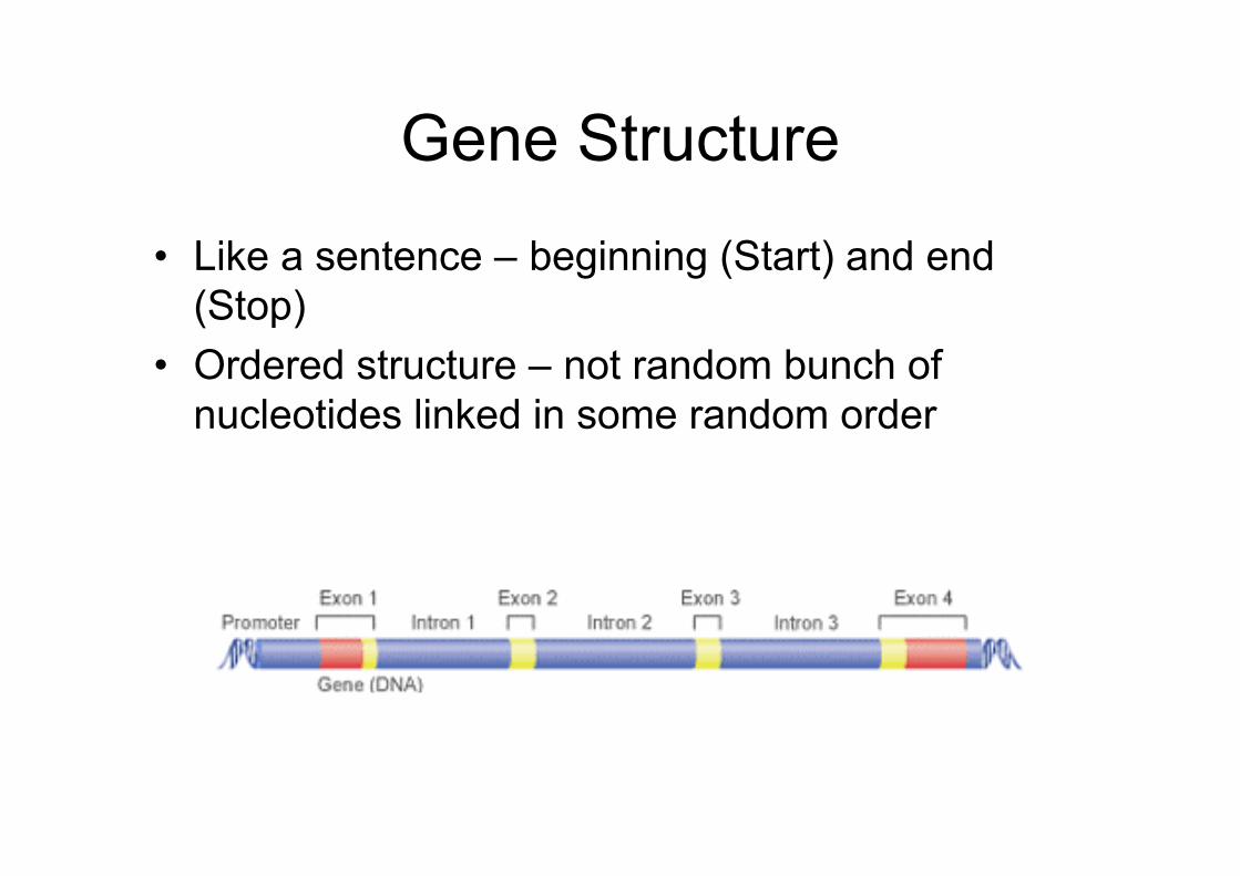
Gene Structure
• Like a sentence – beginning (Start) and end(Stop)
• Ordered structure – not random bunch ofnucleotides linked in some random order
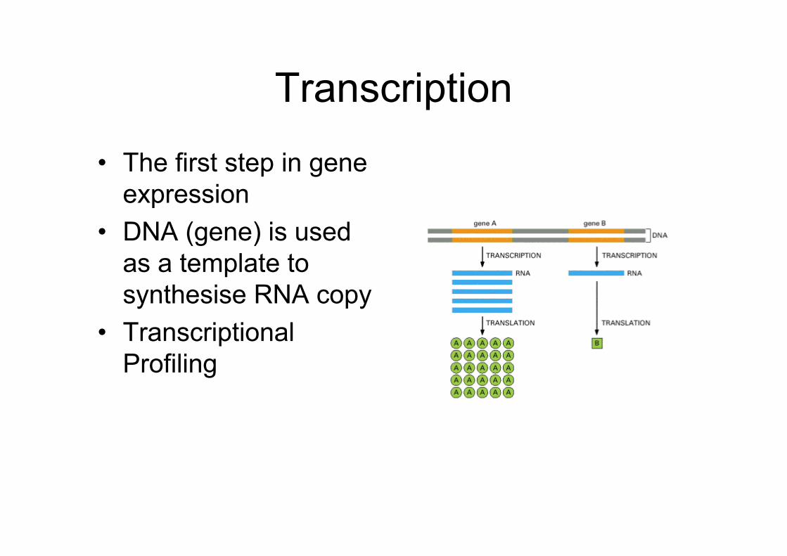
Transcription
• The first step in geneexpression
• DNA (gene) is usedas a template tosynthesise RNA copy
• TranscriptionalProfiling
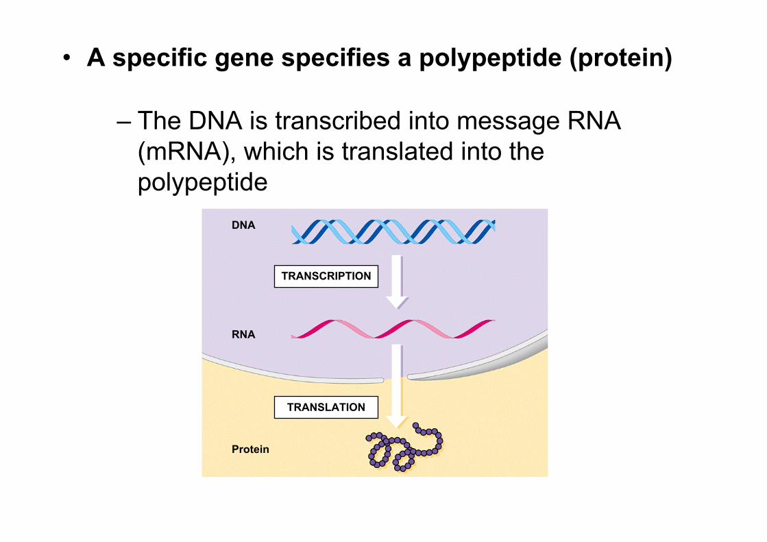
• A specific gene specifies a polypeptide (protein)
– The DNA is transcribed into message RNA(mRNA), which is translated into thepolypeptide
DNA
RNA
Protein
TRANSCRIPTION
TRANSLATION
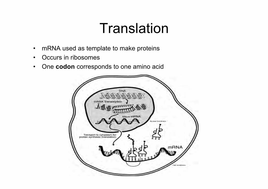
Translation• mRNA used as template to make proteins• Occurs in ribosomes• One codon corresponds to one amino acid
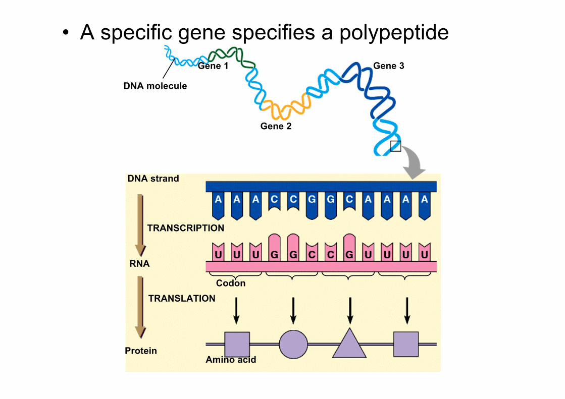
DNA molecule
Gene 1
Gene 2
Gene 3
DNA strand
TRANSCRIPTION
RNA
Protein
TRANSLATIONCodon
Amino acid
• A specific gene specifies a polypeptide
The Genetic Code
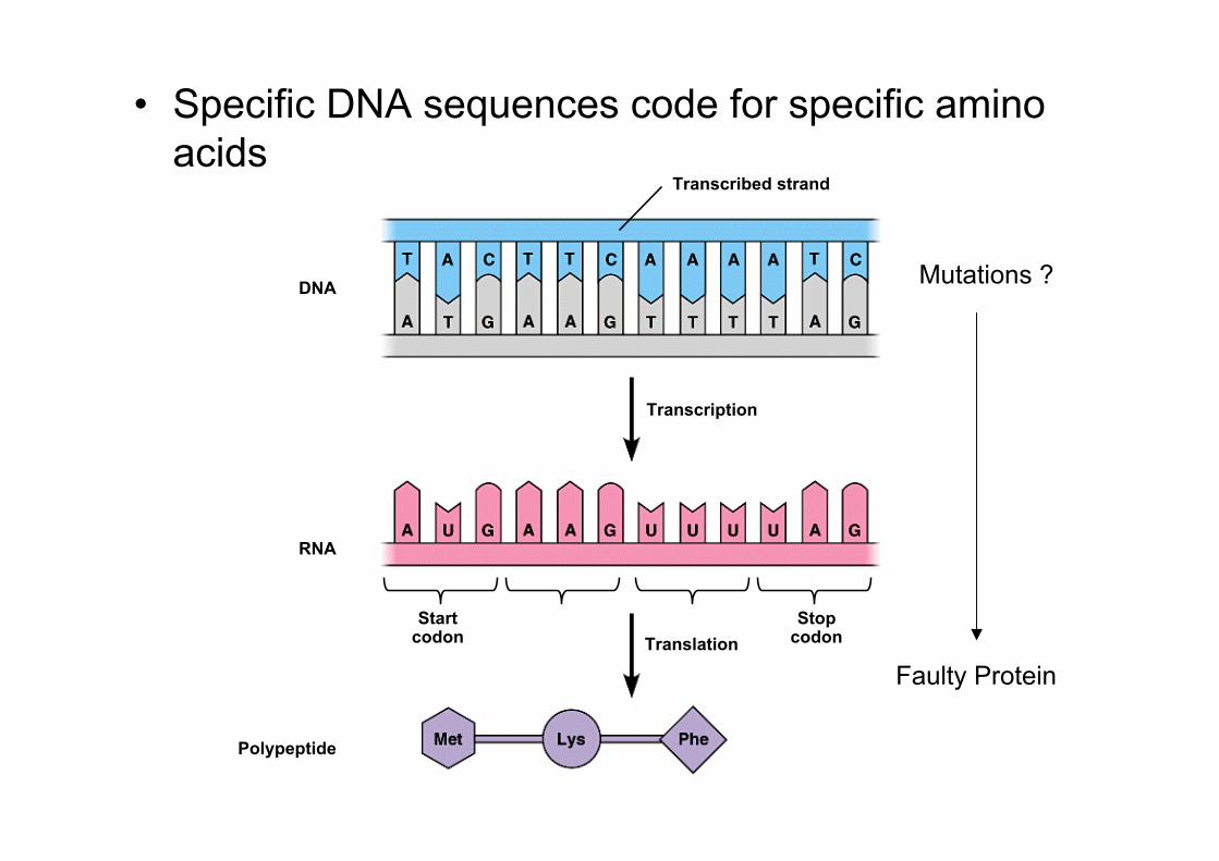
• Specific DNA sequences code for specific aminoacids
Startcodon
RNA
Transcribed strand
StopcodonTranslation
Transcription
DNA
Polypeptide
Mutations ?
Faulty Protein
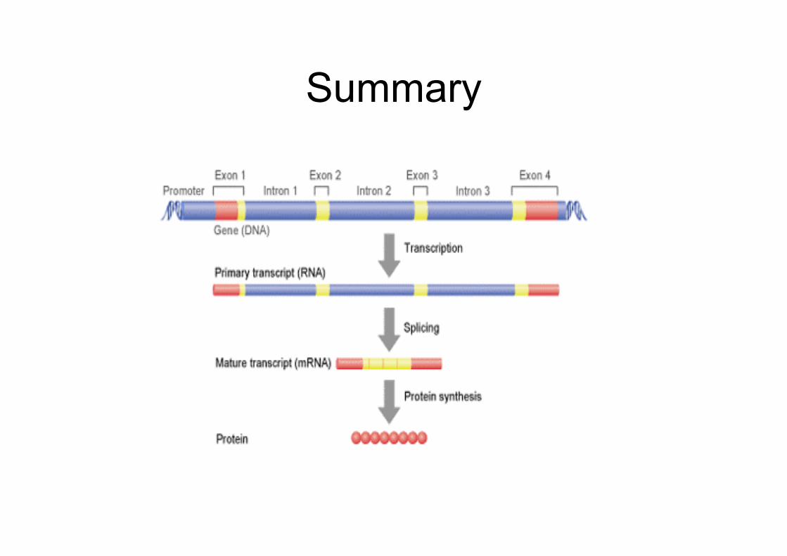
Summary
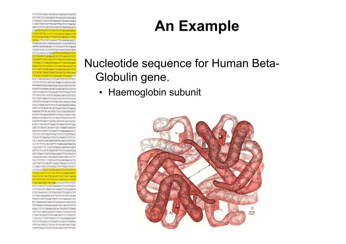
An Example
Nucleotide sequence for Human Beta-Globulin gene.• Haemoglobin subunit
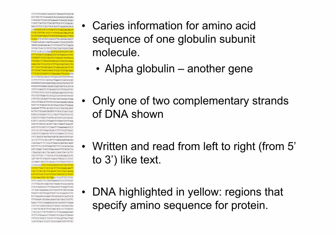
• Caries information for amino acidsequence of one globulin subunitmolecule.• Alpha globulin – another gene
• Only one of two complementary strandsof DNA shown
• Written and read from left to right (from 5’to 3’) like text.
• DNA highlighted in yellow: regions thatspecify amino sequence for protein.
Review
• DNA and RNA– What they are– Structure and Function
• Genes and Genomes• Transcription and Translation
• Any Questions ?
Nucleic Acids Nucleic Acids –– Analytical Techniques
David Murray PhD
UCD Clinical Research CentreUCD School of Medicine and Medical Sciences
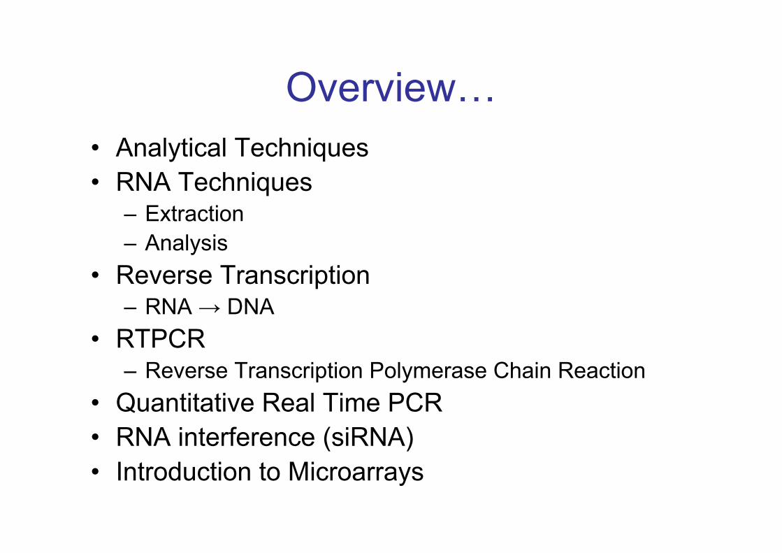
Overview…• Analytical Techniques• RNA Techniques
– Extraction– Analysis
• Reverse Transcription– RNA → DNA
• RTPCR– Reverse Transcription Polymerase Chain Reaction
• Quantitative Real Time PCR• RNA interference (siRNA)• Introduction to Microarrays
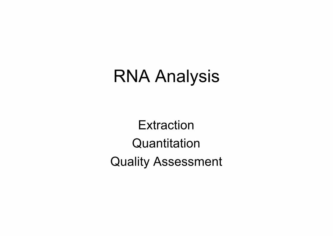
RNA Analysis
ExtractionQuantitation
Quality Assessment
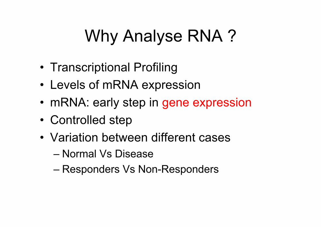
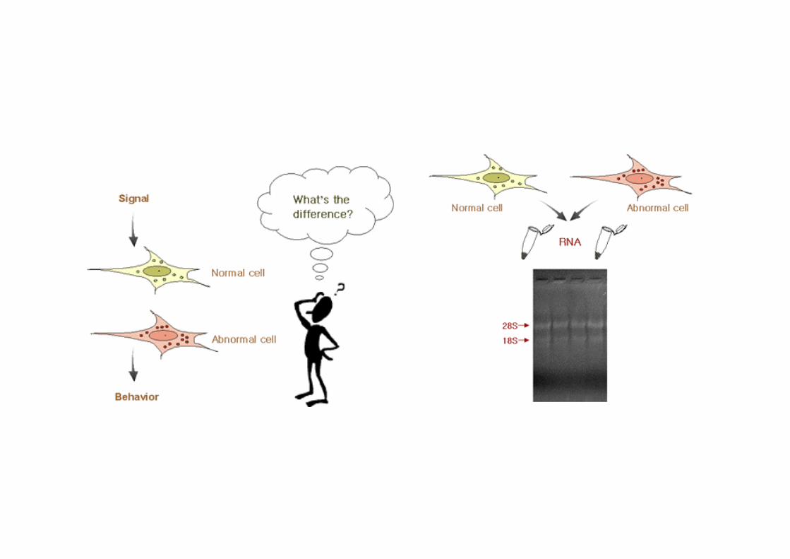
Why Analyse RNA ?
• Transcriptional Profiling• Levels of mRNA expression• mRNA: early step in gene expression• Controlled step• Variation between different cases
– Normal Vs Disease– Responders Vs Non-Responders
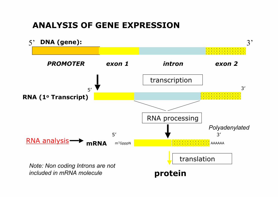
ANALYSIS OF GENE EXPRESSION
PROMOTER exon 1 exon 2intron
5’ 3’
transcription
RNA processing
protein
3’5’
RNA (1o Transcript)
m7GpppN AAAAAA
5’ 3’
mRNARNA analysis
DNA (gene):
Note: Non coding Introns are notincluded in mRNA molecule
translation
Polyadenylated
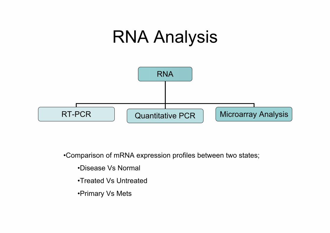
RNA Analysis
RNA
RT-PCR Quantitative PCR Microarray Analysis
•Comparison of mRNA expression profiles between two states;
•Disease Vs Normal
•Treated Vs Untreated
•Primary Vs Mets
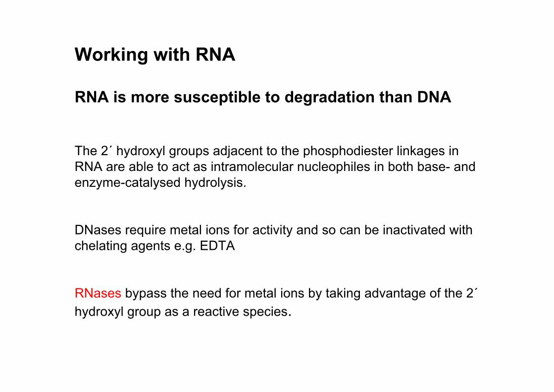
RNA is more susceptible to degradation than DNA
The 2´ hydroxyl groups adjacent to the phosphodiester linkages inRNA are able to act as intramolecular nucleophiles in both base- andenzyme-catalysed hydrolysis.
DNases require metal ions for activity and so can be inactivated withchelating agents e.g. EDTA
RNases bypass the need for metal ions by taking advantage of the 2´hydroxyl group as a reactive species.
Working with RNA
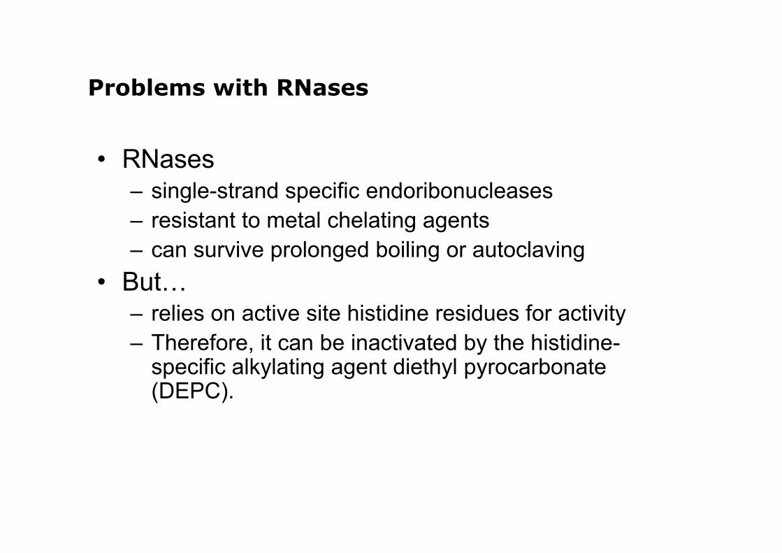
Problems with RNases
• RNases– single-strand specific endoribonucleases– resistant to metal chelating agents– can survive prolonged boiling or autoclaving
• But…– relies on active site histidine residues for activity– Therefore, it can be inactivated by the histidine-
specific alkylating agent diethyl pyrocarbonate(DEPC).
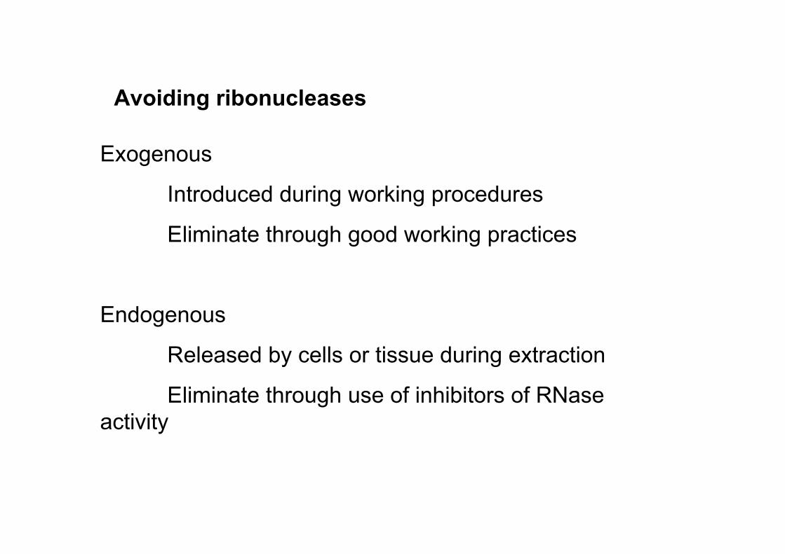
Avoiding ribonucleases
Exogenous
Introduced during working procedures
Eliminate through good working practices
Endogenous
Released by cells or tissue during extraction
Eliminate through use of inhibitors of RNase activity

Always wear gloves - Skin is an abundant source ofribonucleases.
Prepare solutions for RNA work using autoclaved glassware,then autoclave the solutions after they are prepared. Better stilluse disposable plastic ware if possible.
If possible use pre-sterilized water.
Use separate solutions for RNA work and only use them forRNA.
DEPC treatment of water isn’t always necessary. Autoclavingwater and solutions can sometimes be more effective inremoving RNases than chemical treatment.
Working with RNA – Dos and Don’ts
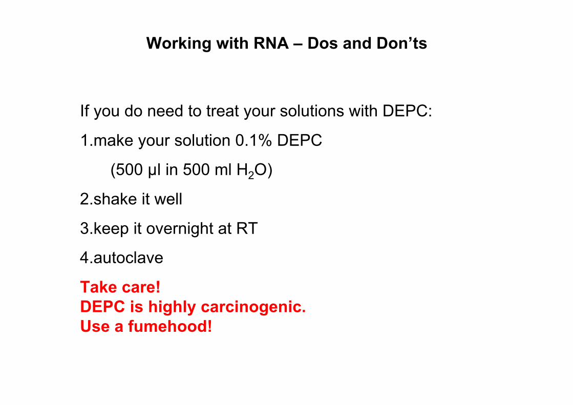
If you do need to treat your solutions with DEPC:
1.make your solution 0.1% DEPC
(500 µl in 500 ml H2O)
2.shake it well
3.keep it overnight at RT
4.autoclave
Take care!DEPC is highly carcinogenic.Use a fumehood!
Working with RNA – Dos and Don’ts
Maintain a separate area for RNA work that has its own setof pipettes.
This is especially important if your work requires the use ofRNase A (e.g. plasmid preps).
Sterile, disposable plasticware can safely be consideredRNase-free and should be used when possible
Use RNase away or RNase zap!!
Working with RNA – Dos and Don’ts
RNA Extraction
…an example
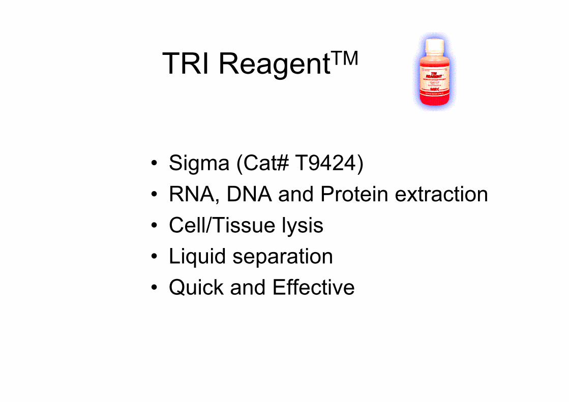
TRI ReagentTM
• Sigma (Cat# T9424)• RNA, DNA and Protein extraction• Cell/Tissue lysis• Liquid separation• Quick and Effective
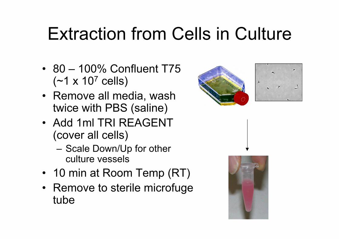
Extraction from Cells in Culture
• 80 – 100% Confluent T75(~1 x 10⁷ cells)
• Remove all media, washtwice with PBS (saline)
• Add 1ml TRI REAGENT(cover all cells)– Scale Down/Up for other
culture vessels• 10 min at Room Temp (RT)• Remove to sterile microfuge
tube
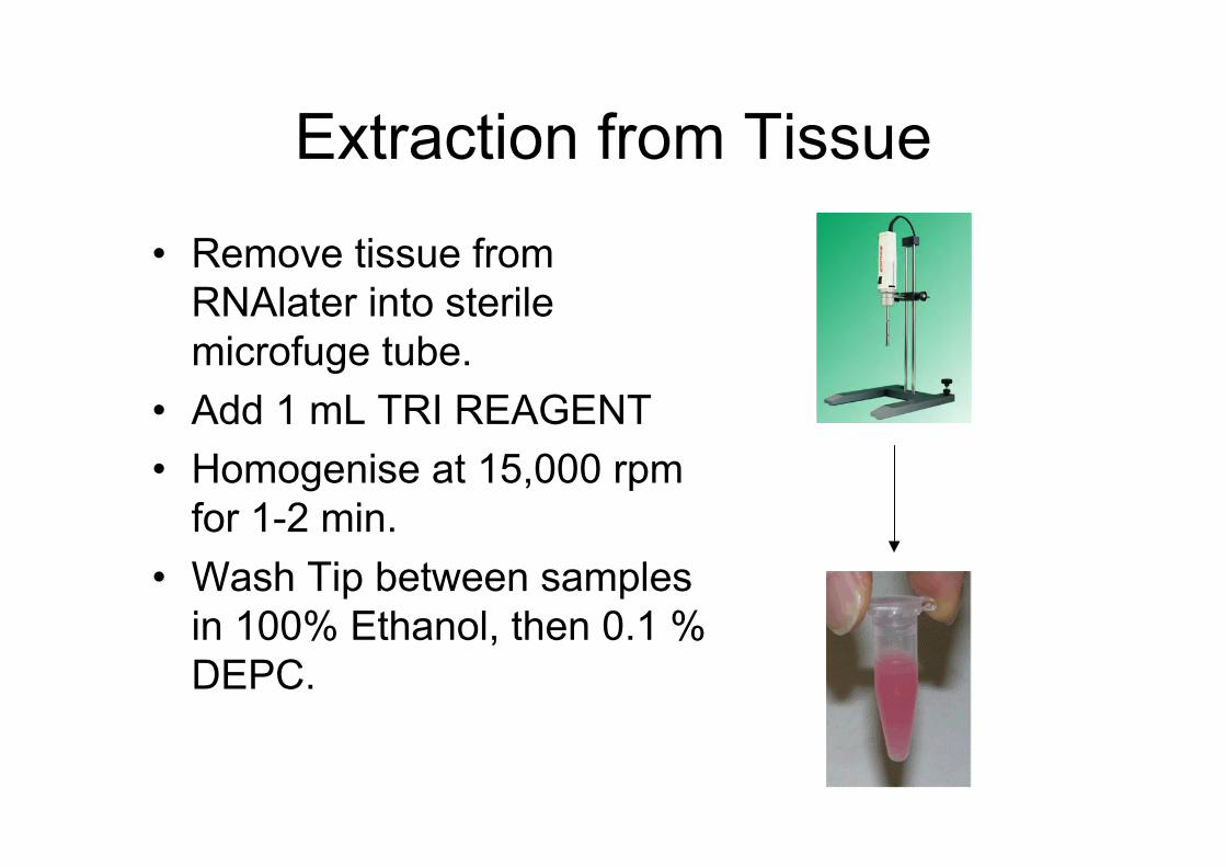
Extraction from Tissue
• Remove tissue fromRNAlater into sterilemicrofuge tube.
• Add 1 mL TRI REAGENT• Homogenise at 15,000 rpm
for 1-2 min.• Wash Tip between samples
in 100% Ethanol, then 0.1 %DEPC.
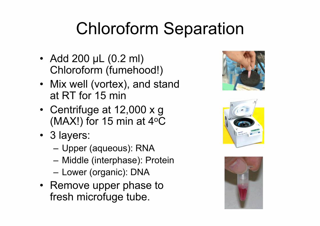
Chloroform Separation
• Add 200 µL (0.2 ml)Chloroform (fumehood!)
• Mix well (vortex), and standat RT for 15 min
• Centrifuge at 12,000 x g(MAX!) for 15 min at 4oC
• 3 layers:– Upper (aqueous): RNA– Middle (interphase): Protein– Lower (organic): DNA
• Remove upper phase tofresh microfuge tube.
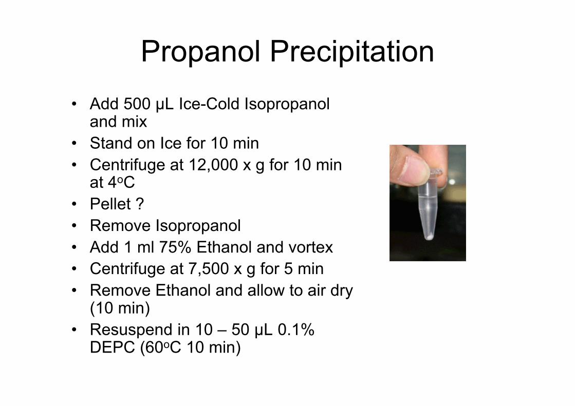
Propanol Precipitation• Add 500 µL Ice-Cold Isopropanol
and mix• Stand on Ice for 10 min• Centrifuge at 12,000 x g for 10 min
at 4oC• Pellet ?• Remove Isopropanol• Add 1 ml 75% Ethanol and vortex• Centrifuge at 7,500 x g for 5 min• Remove Ethanol and allow to air dry
(10 min)• Resuspend in 10 – 50 µL 0.1%
DEPC (60oC 10 min)
RNA Quantitation
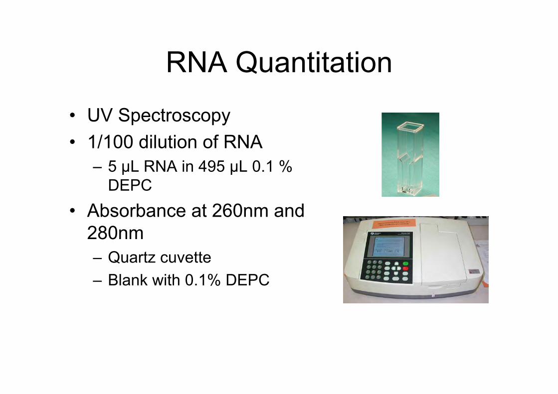
RNA Quantitation
• UV Spectroscopy• 1/100 dilution of RNA
– 5 µL RNA in 495 µL 0.1 %DEPC
• Absorbance at 260nm and280nm– Quartz cuvette– Blank with 0.1% DEPC
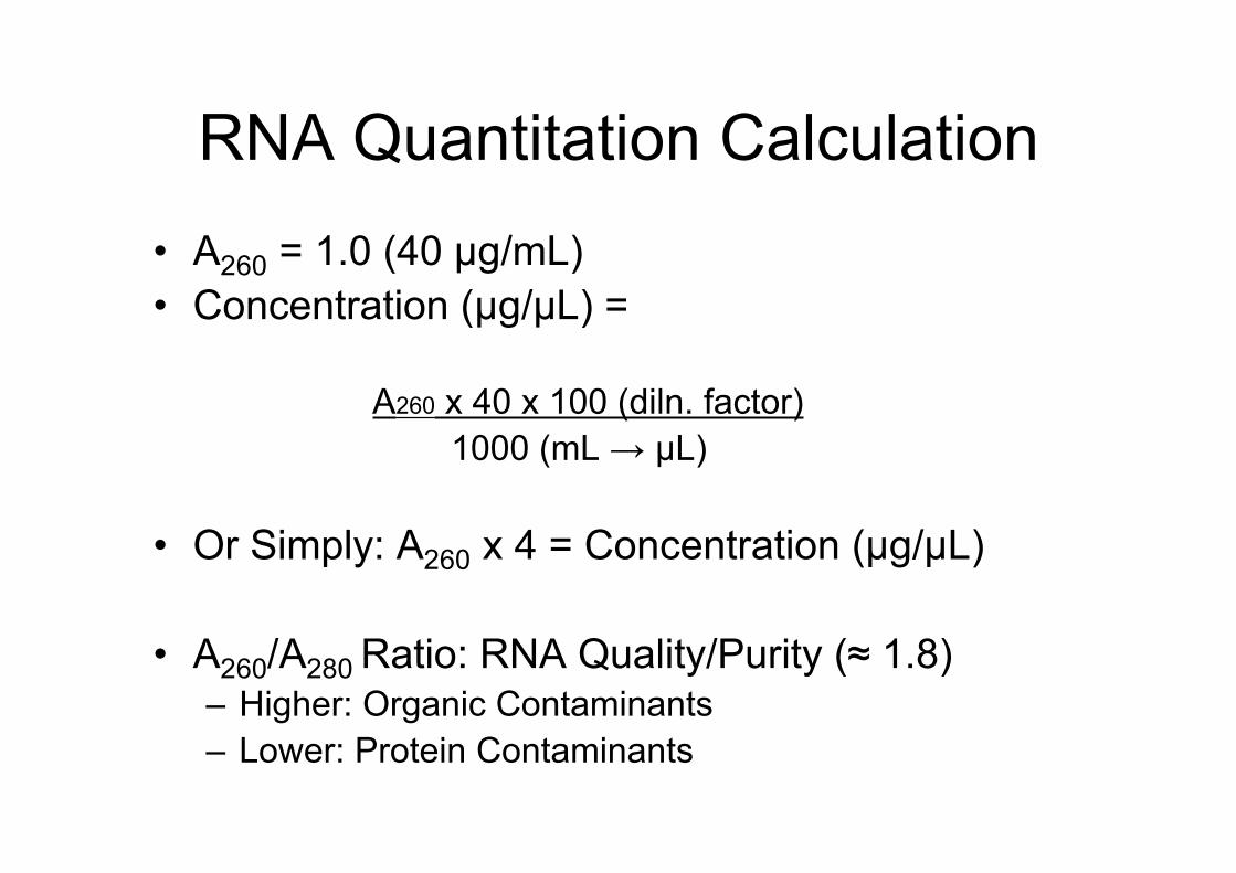
RNA Quantitation Calculation• A260 = 1.0 (40 µg/mL)• Concentration (µg/µL) =
A260 x 40 x 100 (diln. factor) 1000 (mL → µL)
• Or Simply: A260 x 4 = Concentration (µg/µL)
• A260/A280 Ratio: RNA Quality/Purity (≈ 1.8)– Higher: Organic Contaminants– Lower: Protein Contaminants
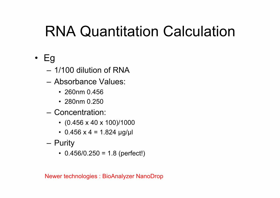
RNA Quantitation Calculation
• Eg– 1/100 dilution of RNA– Absorbance Values:
• 260nm 0.456• 280nm 0.250
– Concentration:• (0.456 x 40 x 100)/1000• 0.456 x 4 = 1.824 µg/µl
– Purity• 0.456/0.250 = 1.8 (perfect!)
Newer technologies : BioAnalyzer NanoDrop
Next?
Assessment of RNA Quality
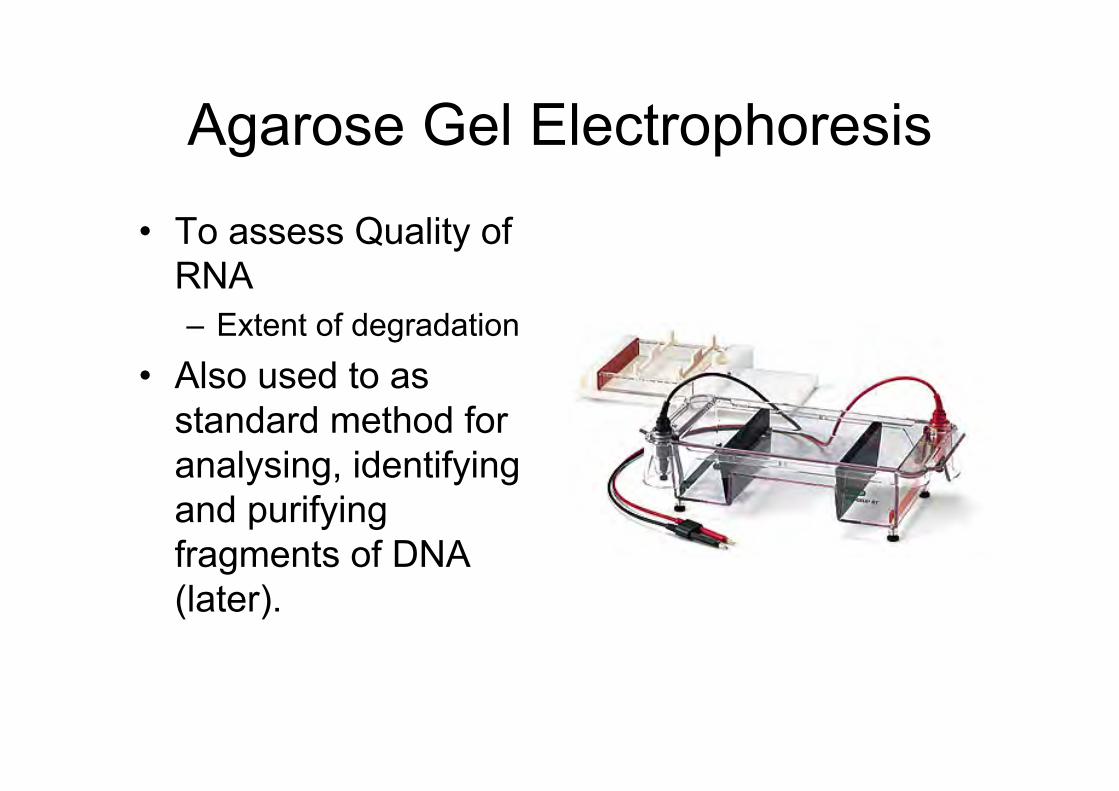
Agarose Gel Electrophoresis
• To assess Quality ofRNA– Extent of degradation
• Also used to asstandard method foranalysing, identifyingand purifyingfragments of DNA(later).
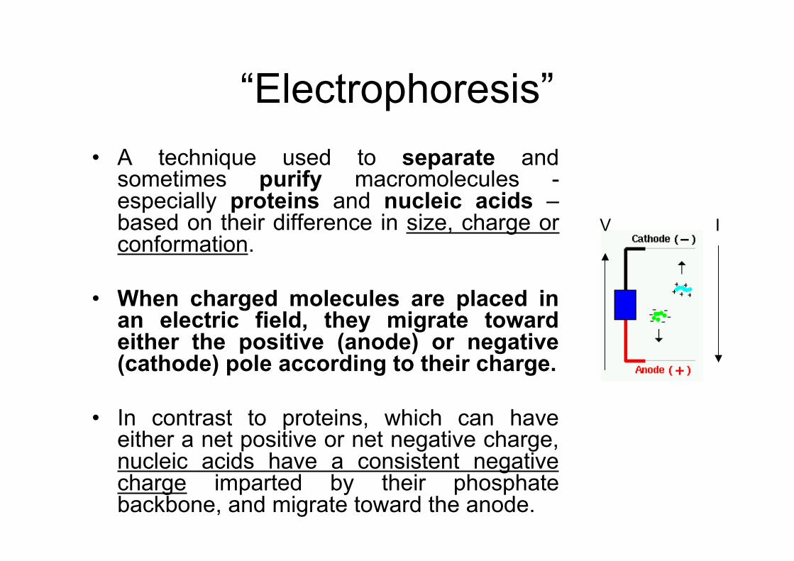
“Electrophoresis”• A technique used to separate and
sometimes purify macromolecules -especially proteins and nucleic acids –based on their difference in size, charge orconformation.
• When charged molecules are placed inan electric field, they migrate towardeither the positive (anode) or negative(cathode) pole according to their charge.
• In contrast to proteins, which can haveeither a net positive or net negative charge,nucleic acids have a consistent negativecharge imparted by their phosphatebackbone, and migrate toward the anode.
IV
“Electrophoresis”• Nucleic acids are electrophoresed within a matrix
or "gel".• The gel is cast in the shape of a thin slab, with wells for
loading the sample.• Agarose is typically used at concentrations of 0.5 to
2%.• The higher the agarose concentration the "stiffer" the
gel.• Agarose gels are extremely easy to prepare: simply
mix agarose powder with buffer solution (TAE/TBE),melt it by heating, and pour the gel. It is also non-toxic.
• The gel is immersed within an electrophoresis buffer(same as above) that provides ions to carry a currentand it also maintains the pH at a relatively constantvalue.
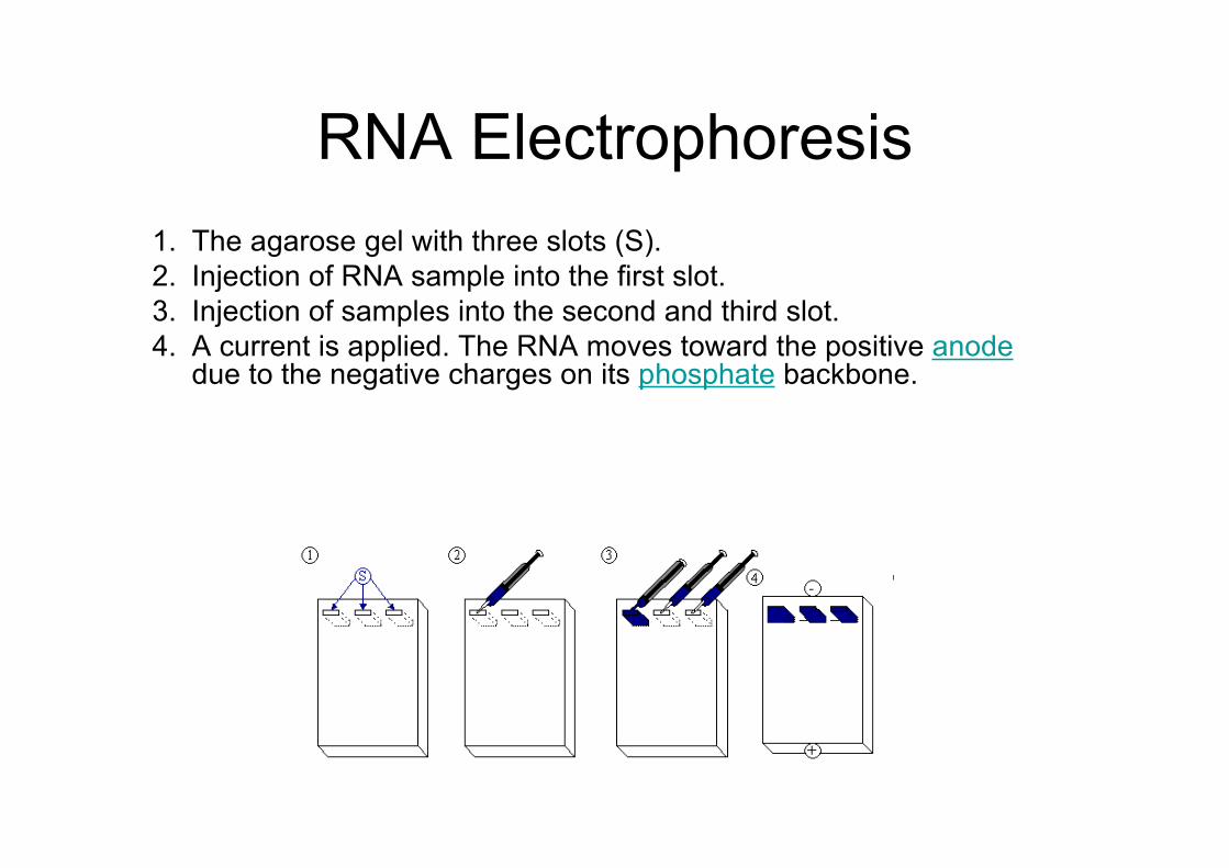
RNA Electrophoresis1. The agarose gel with three slots (S).2. Injection of RNA sample into the first slot.3. Injection of samples into the second and third slot.4. A current is applied. The RNA moves toward the positive anode
due to the negative charges on its phosphate backbone.
-
+
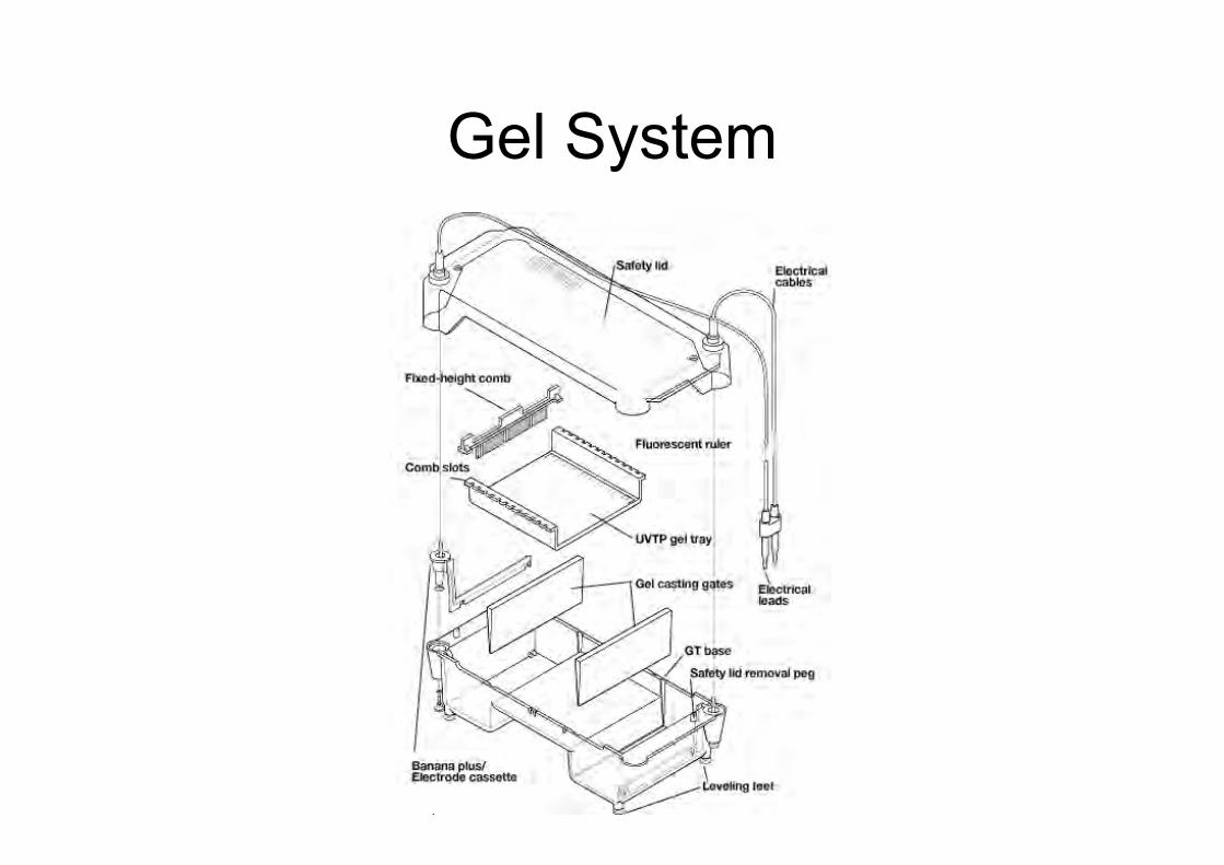
Gel System
Procedure
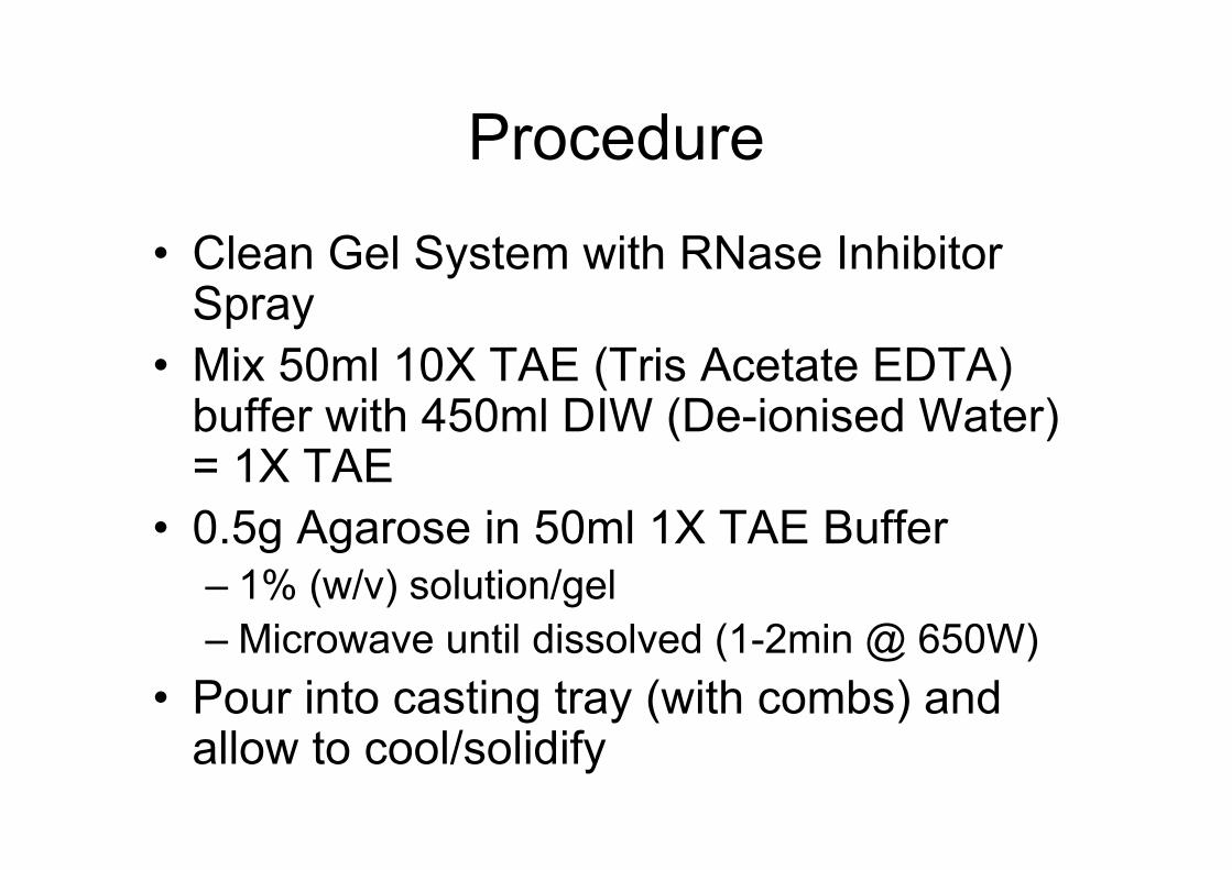
• Clean Gel System with RNase InhibitorSpray
• Mix 50ml 10X TAE (Tris Acetate EDTA)buffer with 450ml DIW (De-ionised Water)= 1X TAE
• 0.5g Agarose in 50ml 1X TAE Buffer– 1% (w/v) solution/gel– Microwave until dissolved (1-2min @ 650W)
• Pour into casting tray (with combs) andallow to cool/solidify
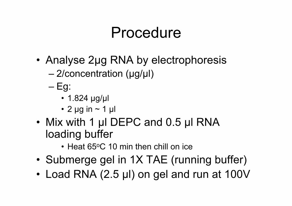
Procedure
• Analyse 2µg RNA by electrophoresis– 2/concentration (µg/µl)– Eg:
• 1.824 µg/µl• 2 µg in ~ 1 µl
• Mix with 1 µl DEPC and 0.5 µl RNAloading buffer
• Heat 65oC 10 min then chill on ice
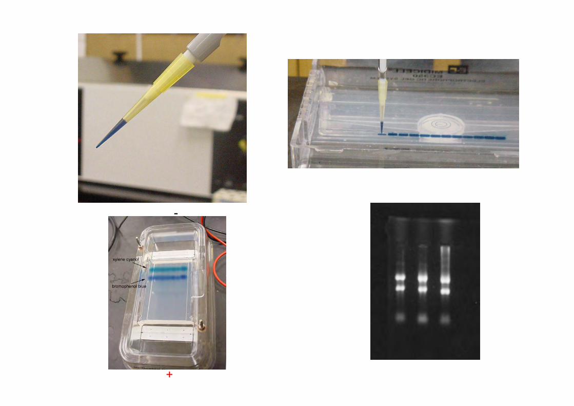
• Submerge gel in 1X TAE (running buffer)• Load RNA (2.5 µl) on gel and run at 100V
Procedure
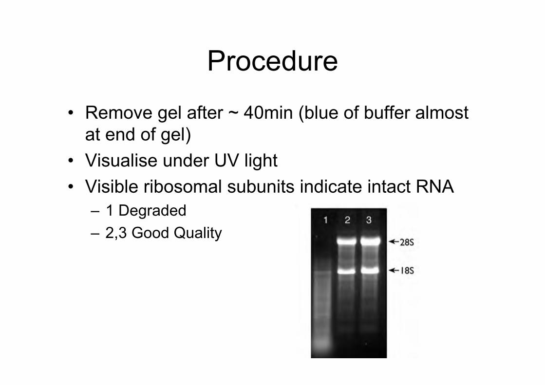
• Remove gel after ~ 40min (blue of buffer almostat end of gel)
• Visualise under UV light• Visible ribosomal subunits indicate intact RNA
– 1 Degraded– 2,3 Good Quality
RT-PCR
Reverse TranscriptasePolymerase Chain Reaction
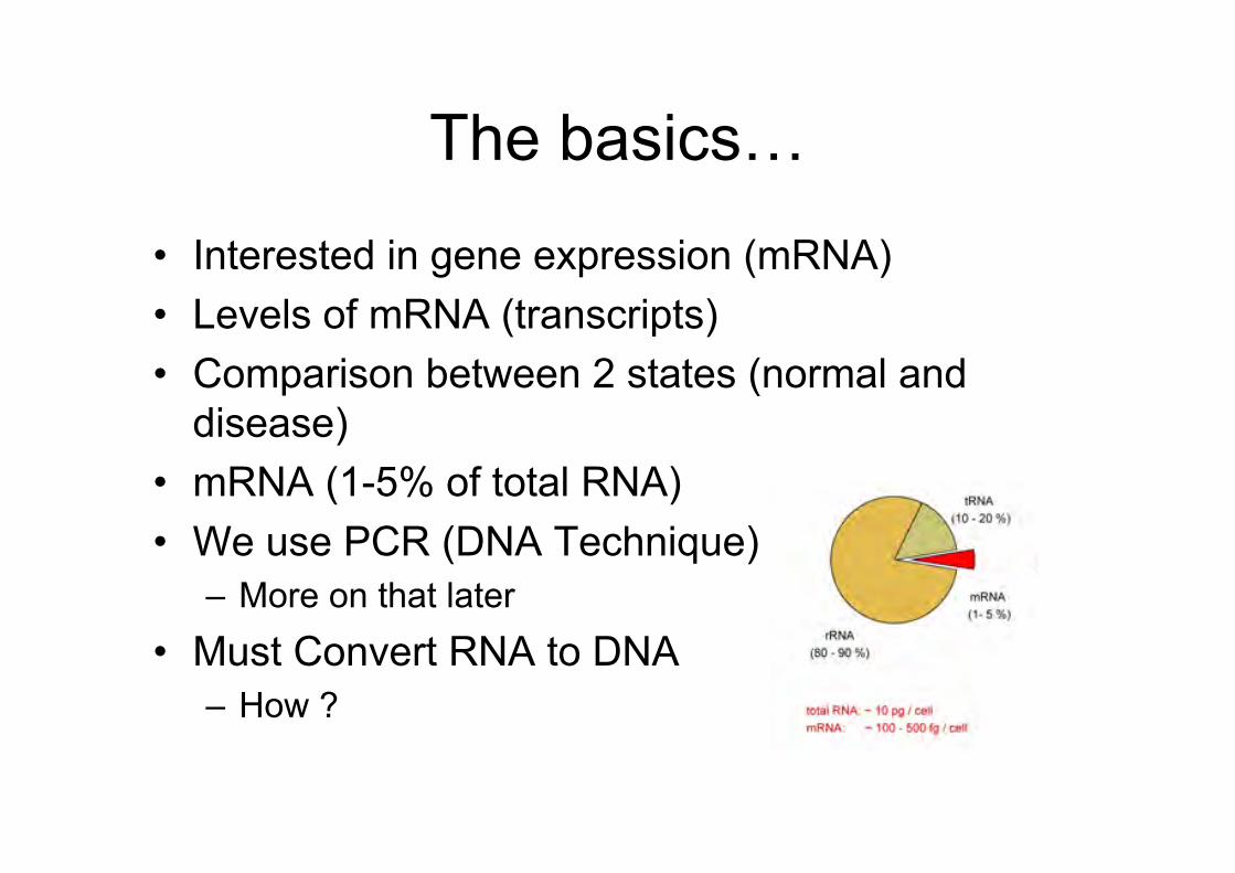
The basics…
• Interested in gene expression (mRNA)• Levels of mRNA (transcripts)• Comparison between 2 states (normal and
disease)• mRNA (1-5% of total RNA)• We use PCR (DNA Technique)
– More on that later• Must Convert RNA to DNA
– How ?
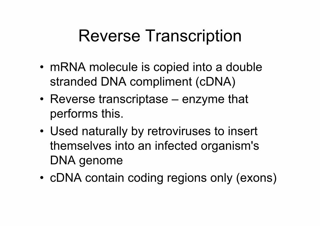
Reverse Transcription
• mRNA molecule is copied into a doublestranded DNA compliment (cDNA)
• Reverse transcriptase – enzyme thatperforms this.
• Used naturally by retroviruses to insertthemselves into an infected organism'sDNA genome
• cDNA contain coding regions only (exons)
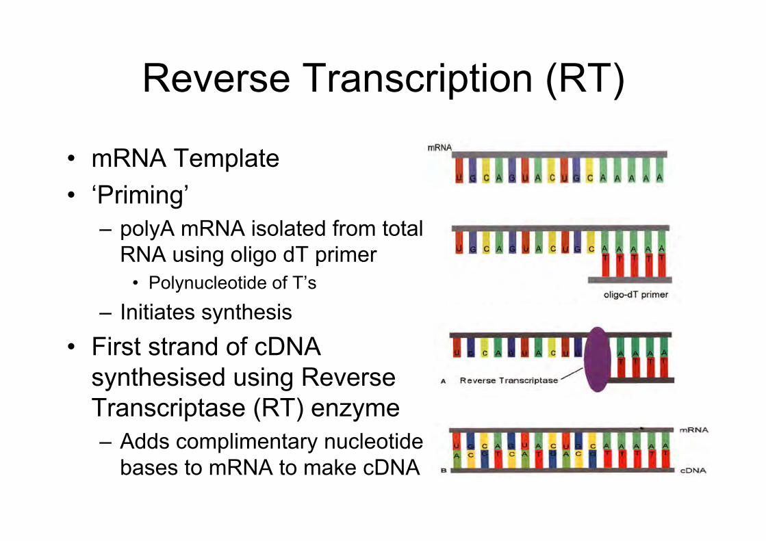
Reverse Transcription (RT)
• mRNA Template• ‘Priming’
– polyA mRNA isolated from totalRNA using oligo dT primer
• Polynucleotide of T’s
– Initiates synthesis• First strand of cDNA
synthesised using ReverseTranscriptase (RT) enzyme– Adds complimentary nucleotide
bases to mRNA to make cDNA
cDNA is then used as atemplate in PCR
PCR• Polymerase Chain Reaction
– Technique for Targeted DNA Amplification– Starting material ('target sequence’);
• A gene or segment of DNA (cDNA in our case)
– Target sequence can be amplified a billion fold in a matterof hours
• PCR allows one to take a specimen of geneticmaterial, even from just one cell, copy its geneticsequence over and over, and generate a testsample sufficient to detect the presence or absenceof a specific virus, bacterium or any particularsequence of genetic material
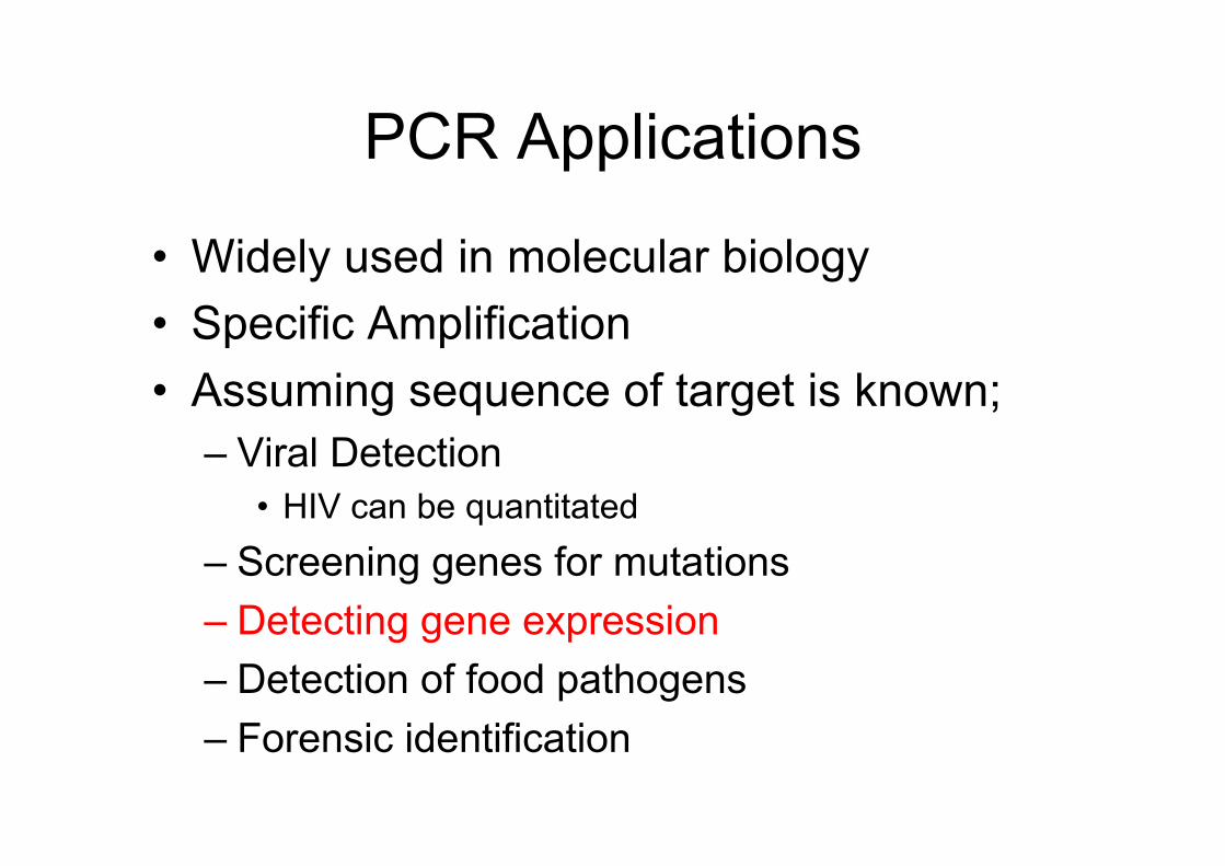
PCR Applications
• Widely used in molecular biology• Specific Amplification• Assuming sequence of target is known;
– Viral Detection• HIV can be quantitated
– Screening genes for mutations– Detecting gene expression– Detection of food pathogens– Forensic identification
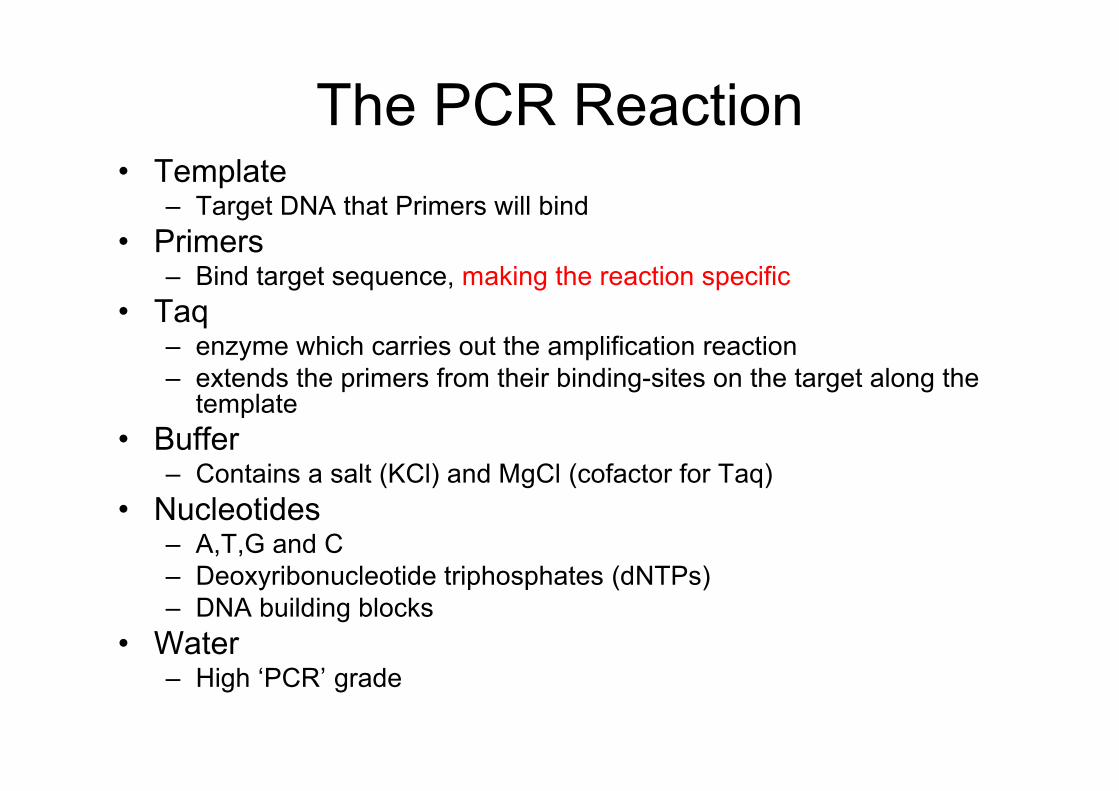
The PCR Reaction• Template
– Target DNA that Primers will bind• Primers
– Bind target sequence, making the reaction specific• Taq
– enzyme which carries out the amplification reaction– extends the primers from their binding-sites on the target along the
template• Buffer
– Contains a salt (KCl) and MgCl (cofactor for Taq)• Nucleotides
– A,T,G and C– Deoxyribonucleotide triphosphates (dNTPs)– DNA building blocks
• Water– High ‘PCR’ grade
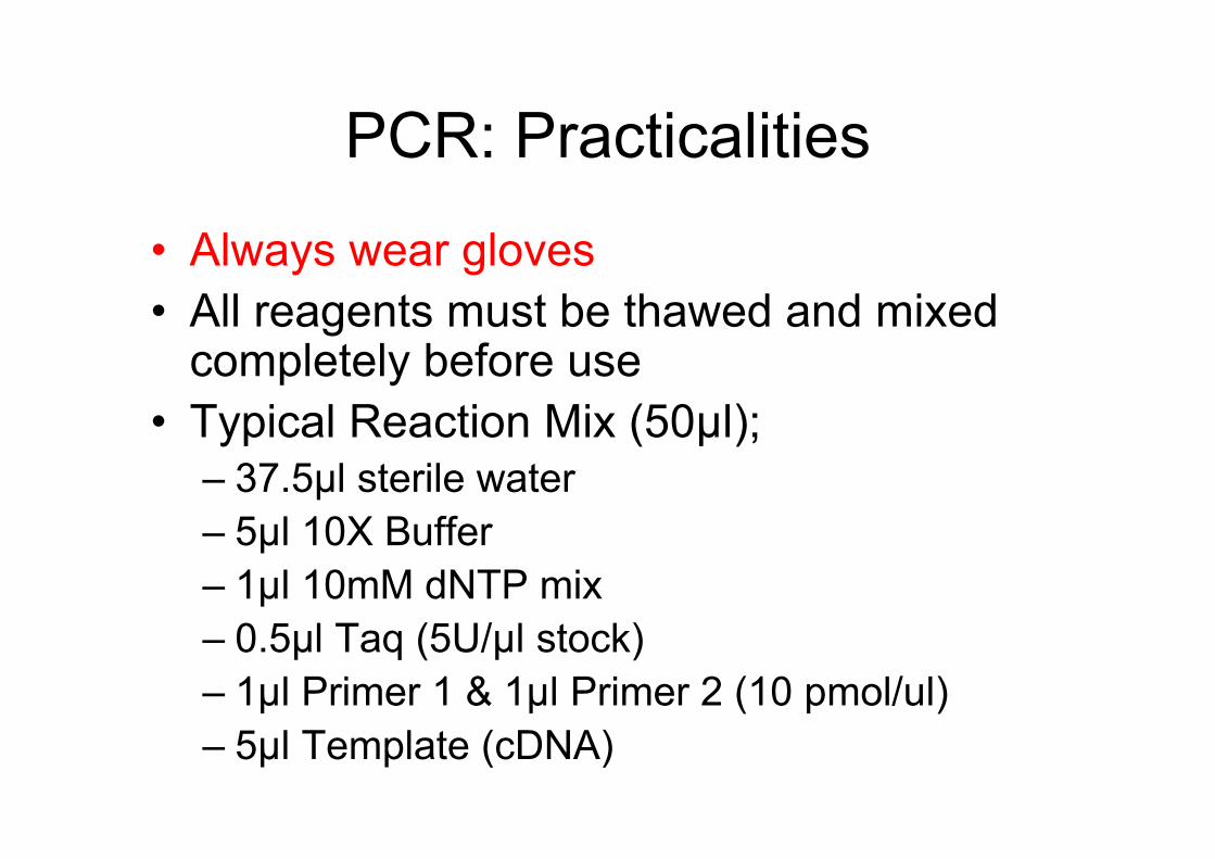
PCR: Practicalities
• Always wear gloves• All reagents must be thawed and mixed
completely before use• Typical Reaction Mix (50µl);
– 37.5µl sterile water– 5µl 10X Buffer– 1µl 10mM dNTP mix– 0.5µl Taq (5U/µl stock)– 1µl Primer 1 & 1µl Primer 2 (10 pmol/ul)– 5µl Template (cDNA)
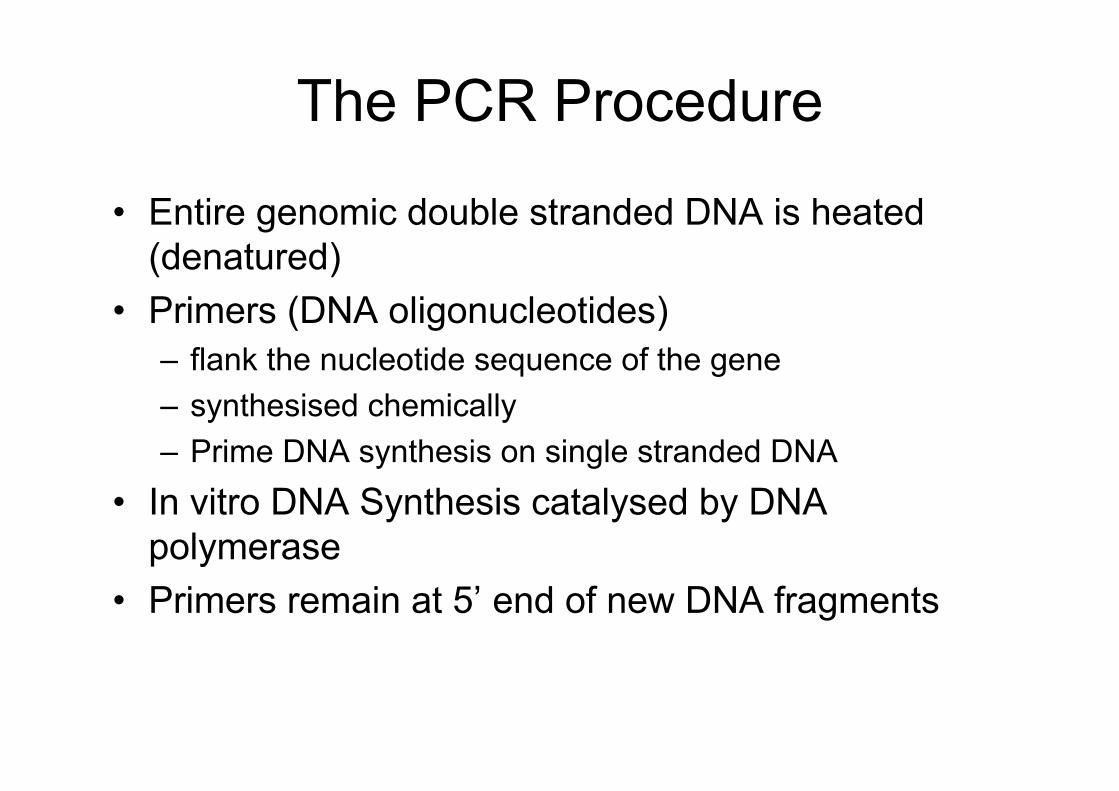
The PCR Procedure
• Entire genomic double stranded DNA is heated(denatured)
• Primers (DNA oligonucleotides)– flank the nucleotide sequence of the gene– synthesised chemically– Prime DNA synthesis on single stranded DNA
• In vitro DNA Synthesis catalysed by DNApolymerase
• Primers remain at 5’ end of new DNA fragments
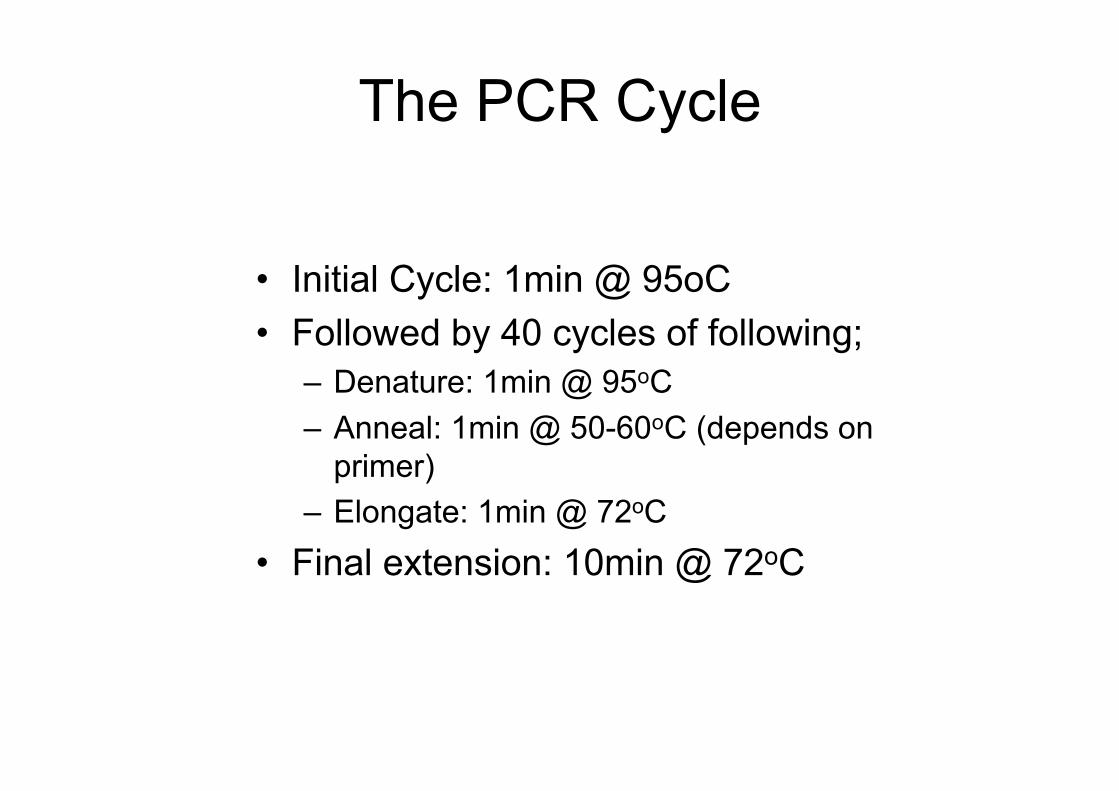
The PCR Cycle
• Initial Cycle: 1min @ 95oC• Followed by 40 cycles of following;
– Denature: 1min @ 95oC– Anneal: 1min @ 50-60oC (depends on
primer)– Elongate: 1min @ 72oC
• Final extension: 10min @ 72oC

PCR Links
• Calculate Annealing Temperature– http://www.bioinformatics.vg/bioinformatics_to
ols/oligo2002.shtml• Primer Design
– http://frodo.wi.mit.edu/cgi-bin/primer3/primer3_www.cgi
– http://www.basic.nwu.edu/biotools/oligocalc.html
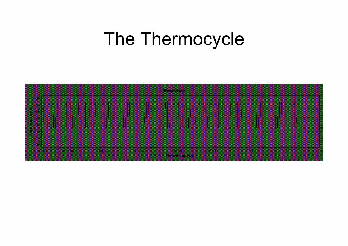
The Thermocycle
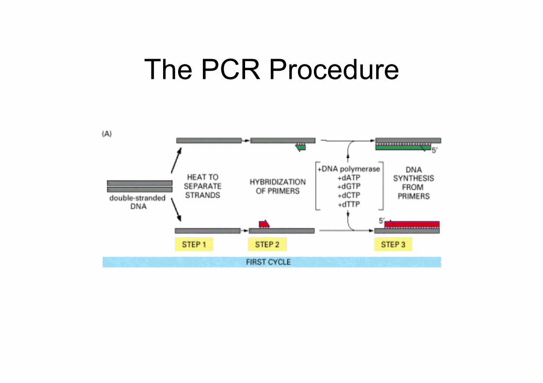
The PCR Procedure
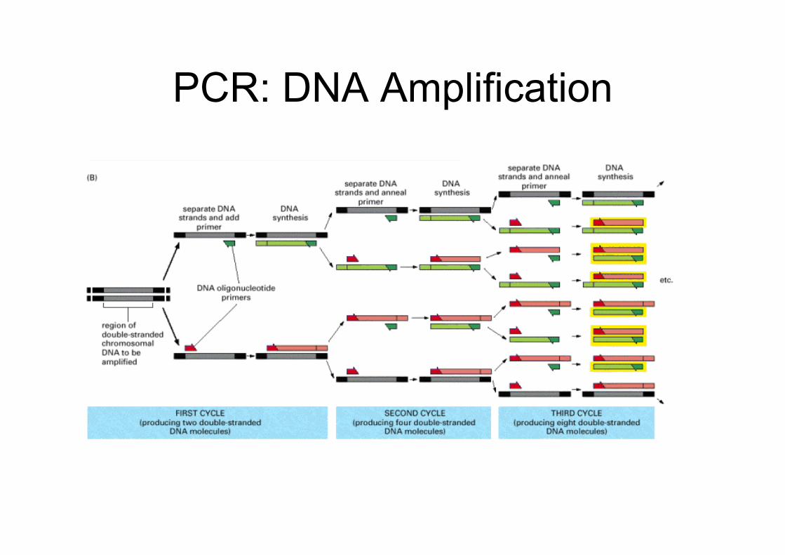
PCR: DNA Amplification
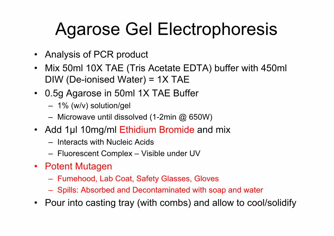
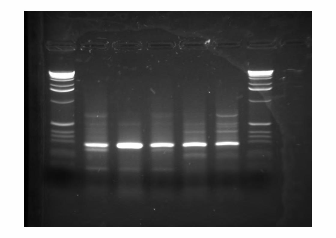
Agarose Gel Electrophoresis• Analysis of PCR product• Mix 50ml 10X TAE (Tris Acetate EDTA) buffer with 450ml
DIW (De-ionised Water) = 1X TAE• 0.5g Agarose in 50ml 1X TAE Buffer
– 1% (w/v) solution/gel– Microwave until dissolved (1-2min @ 650W)
• Add 1µl 10mg/ml Ethidium Bromide and mix– Interacts with Nucleic Acids– Fluorescent Complex – Visible under UV
• Potent Mutagen– Fumehood, Lab Coat, Safety Glasses, Gloves– Spills: Absorbed and Decontaminated with soap and water
• Pour into casting tray (with combs) and allow to cool/solidify
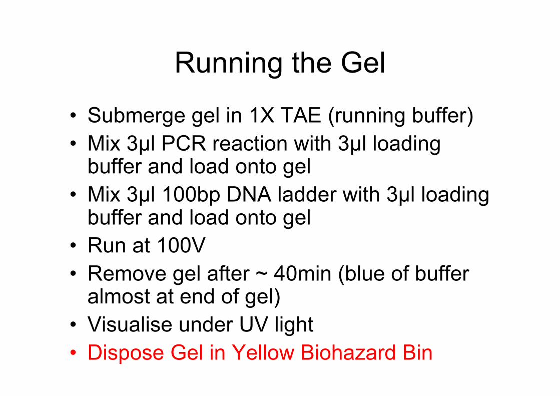
Running the Gel
• Submerge gel in 1X TAE (running buffer)• Mix 3µl PCR reaction with 3µl loading
buffer and load onto gel• Mix 3µl 100bp DNA ladder with 3µl loading
buffer and load onto gel• Run at 100V• Remove gel after ~ 40min (blue of buffer
almost at end of gel)• Visualise under UV light• Dispose Gel in Yellow Biohazard Bin
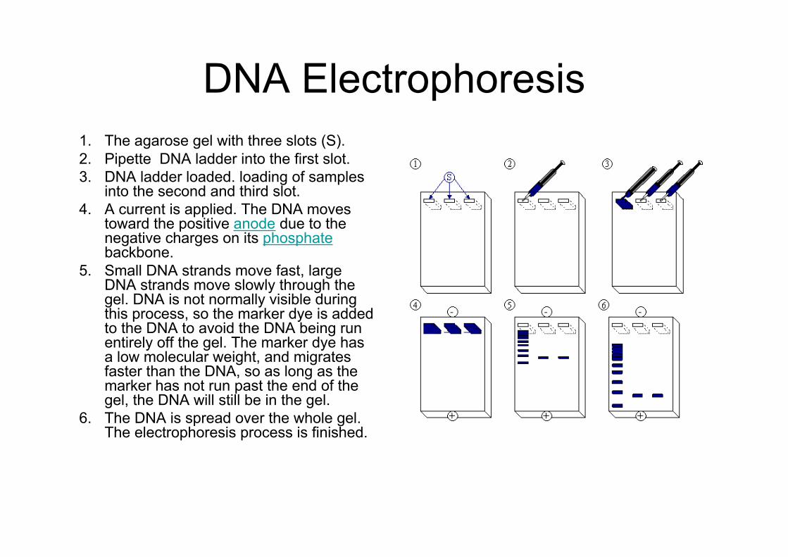
DNA Electrophoresis1. The agarose gel with three slots (S).2. Pipette DNA ladder into the first slot.3. DNA ladder loaded. loading of samples
into the second and third slot.4. A current is applied. The DNA moves
toward the positive anode due to thenegative charges on its phosphatebackbone.
5. Small DNA strands move fast, largeDNA strands move slowly through thegel. DNA is not normally visible duringthis process, so the marker dye is addedto the DNA to avoid the DNA being runentirely off the gel. The marker dye hasa low molecular weight, and migratesfaster than the DNA, so as long as themarker has not run past the end of thegel, the DNA will still be in the gel.
6. The DNA is spread over the whole gel.The electrophoresis process is finished.
Real Time PCR
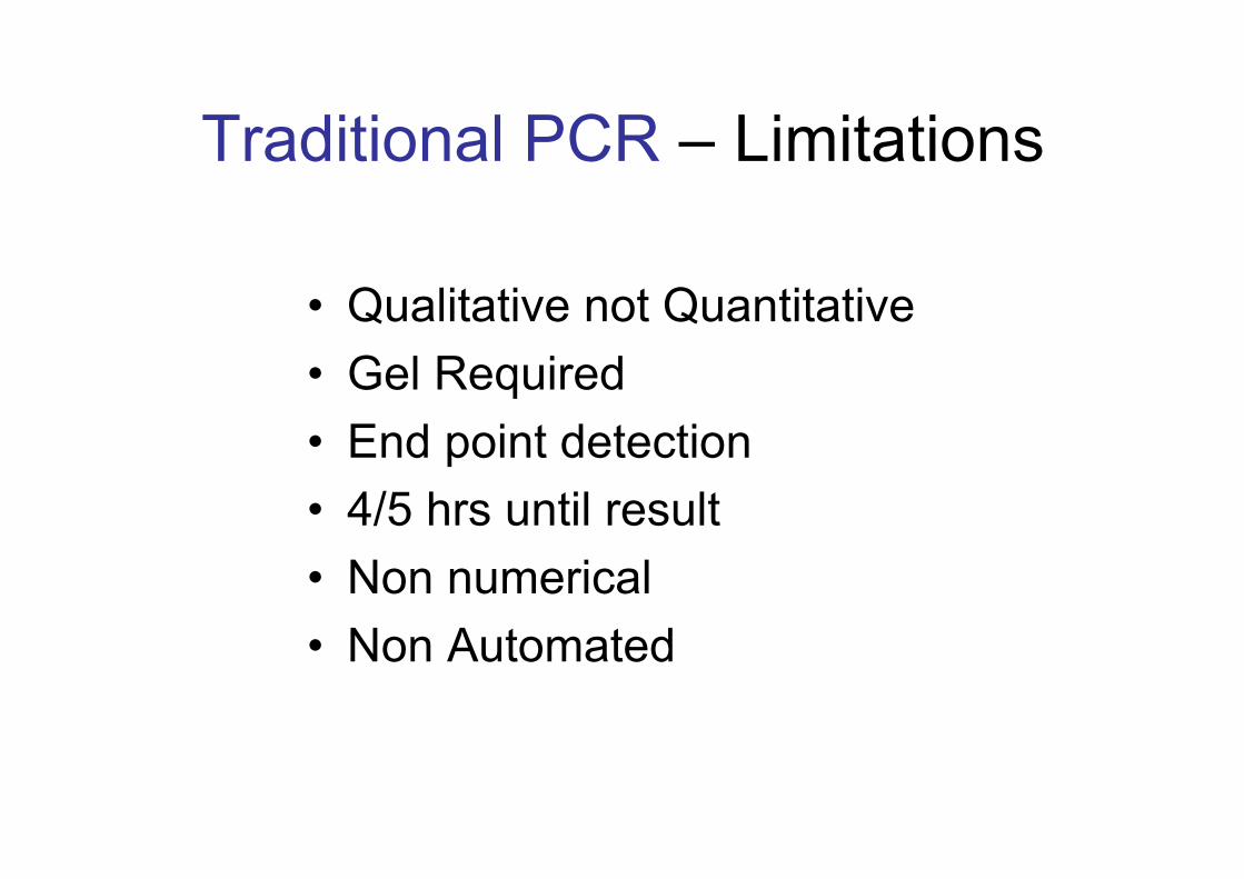
Traditional PCR – Limitations
• Qualitative not Quantitative• Gel Required• End point detection• 4/5 hrs until result• Non numerical• Non Automated
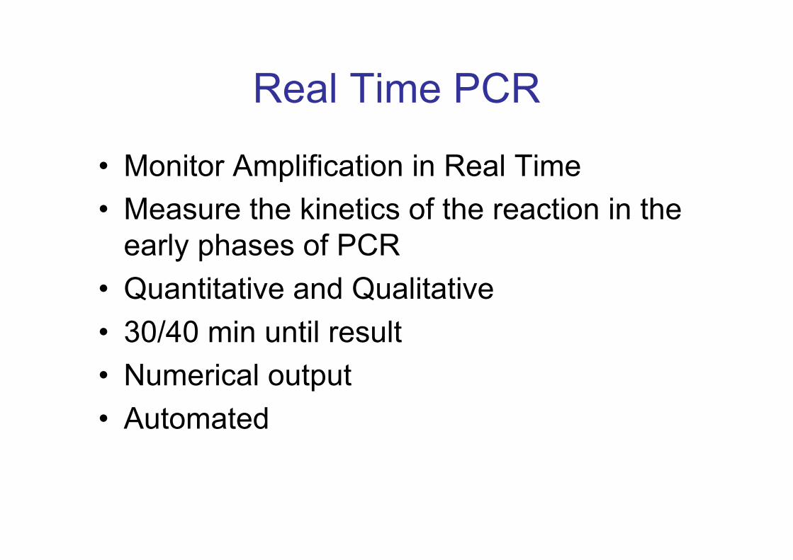
Real Time PCR
• Monitor Amplification in Real Time• Measure the kinetics of the reaction in the
early phases of PCR• Quantitative and Qualitative• 30/40 min until result• Numerical output• Automated
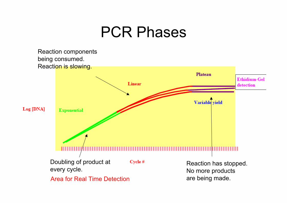
PCR Phases
Doubling of product atevery cycle.
Reaction componentsbeing consumed.Reaction is slowing.
Reaction has stopped.No more productsare being made.Area for Real Time Detection
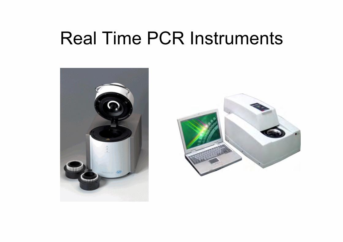
Real Time PCR Instruments
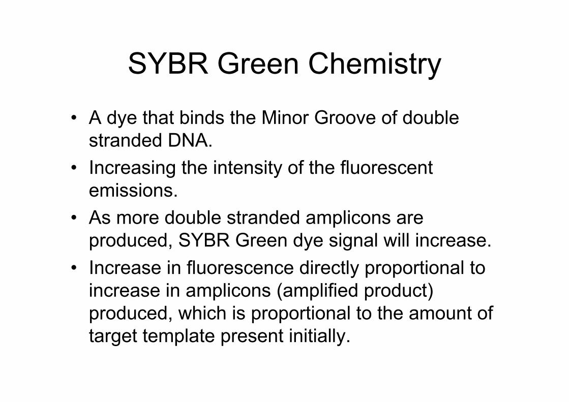
SYBR Green Chemistry
• A dye that binds the Minor Groove of doublestranded DNA.
• Increasing the intensity of the fluorescentemissions.
• As more double stranded amplicons areproduced, SYBR Green dye signal will increase.
• Increase in fluorescence directly proportional toincrease in amplicons (amplified product)produced, which is proportional to the amount oftarget template present initially.
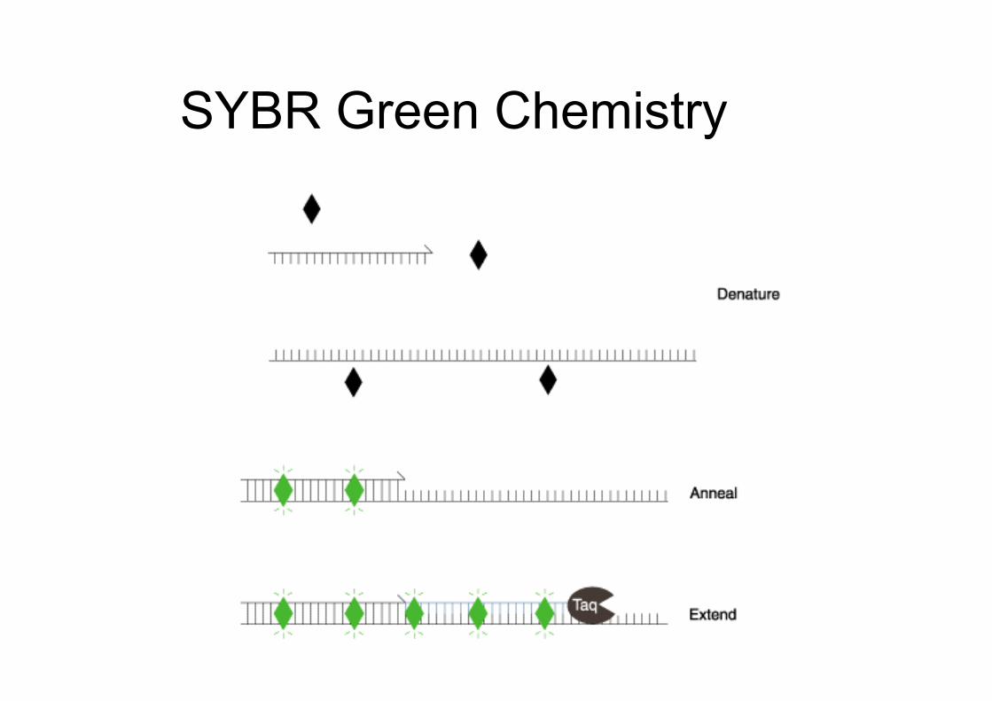
SYBR Green Chemistry
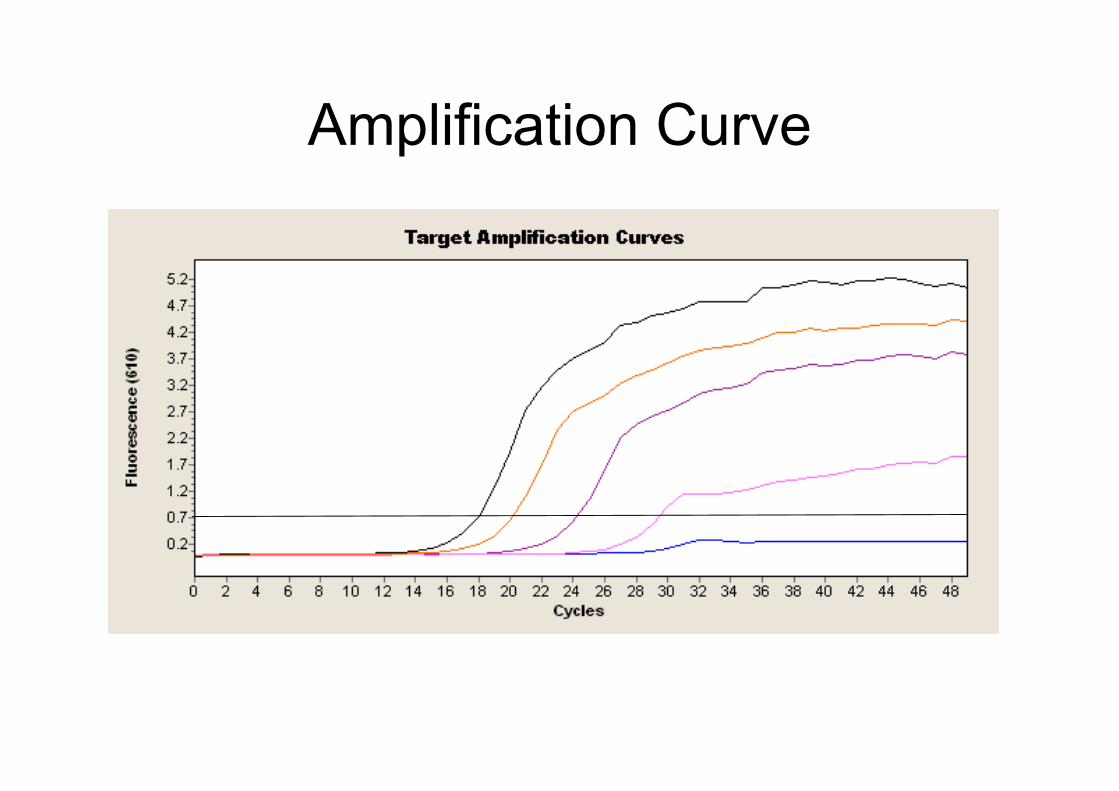
Amplification Curve
Real Time PCR
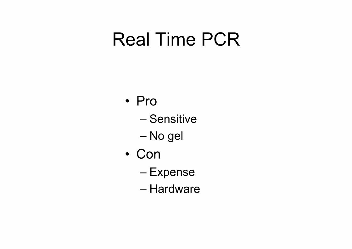
• Pro– Sensitive– No gel
• Con– Expense– Hardware
Real Time PCR Links• Good Article
– http://dorakmt.tripod.com/genetics/realtime.html• Troubleshooting
– http://www.eurogentec.com/module/FileLib/GRT-TSGCUST-0304-V4.pdf
Microarrays
Microarray Analysis
Monitor the activity of thousands of genes simultaneously
Compare activity of one gene in many samples.
Compare activity of many genes in one sample.
Take a photo of genes in action!
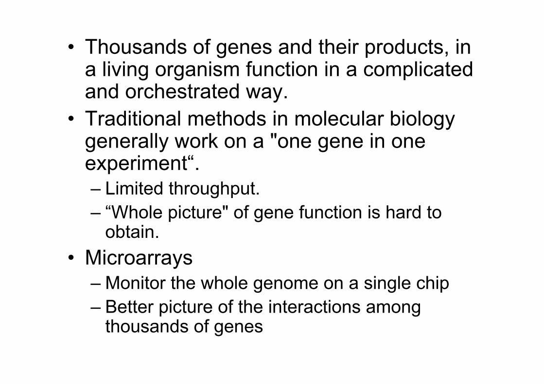
• Thousands of genes and their products, ina living organism function in a complicatedand orchestrated way.
• Traditional methods in molecular biologygenerally work on a "one gene in oneexperiment“.– Limited throughput.– “Whole picture" of gene function is hard to
obtain.• Microarrays
– Monitor the whole genome on a single chip– Better picture of the interactions among
thousands of genes
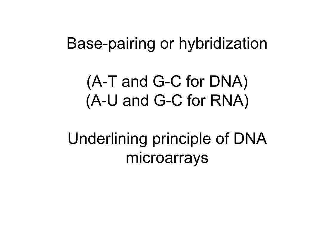
Base-pairing or hybridization
(A-T and G-C for DNA)(A-U and G-C for RNA)
Underlining principle of DNAmicroarrays
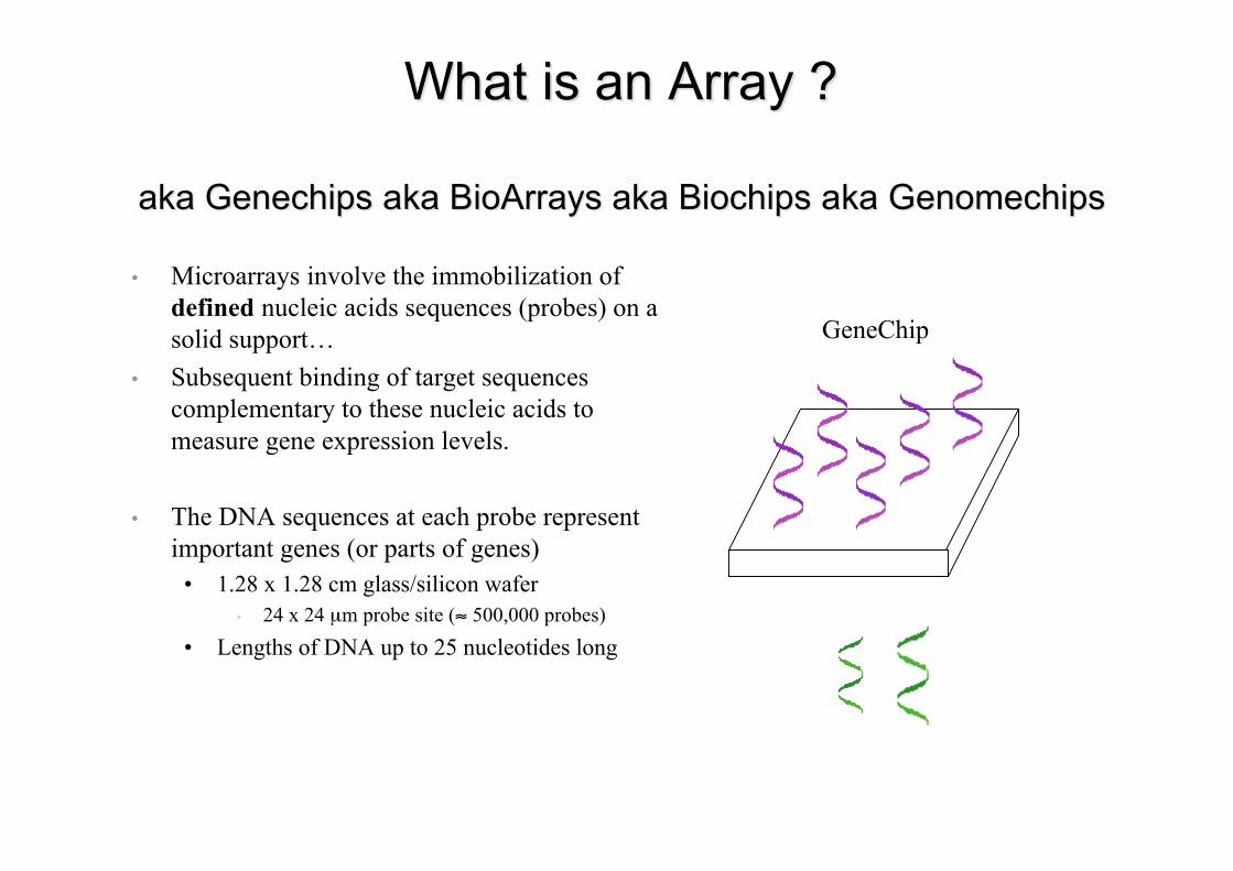
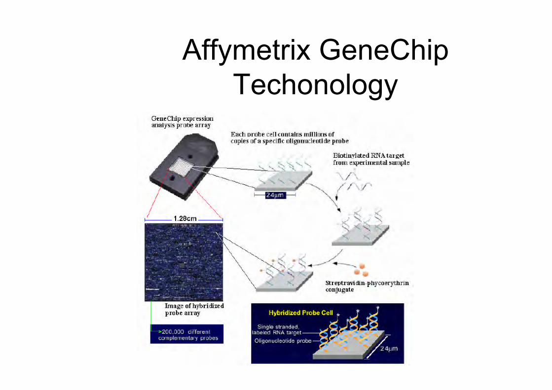
What is an Array ?What is an Array ?
akaaka GenechipsGenechips akaaka BioArraysBioArrays akaaka Biochips Biochips akaaka GenomechipsGenomechips
• Microarrays involve the immobilization ofdefined nucleic acids sequences (probes) on asolid support…
• Subsequent binding of target sequencescomplementary to these nucleic acids tomeasure gene expression levels.
• The DNA sequences at each probe representimportant genes (or parts of genes)
• 1.28 x 1.28 cm glass/silicon wafer• 24 x 24 µm probe site (≈ 500,000 probes)
• Lengths of DNA up to 25 nucleotides long
GeneChip
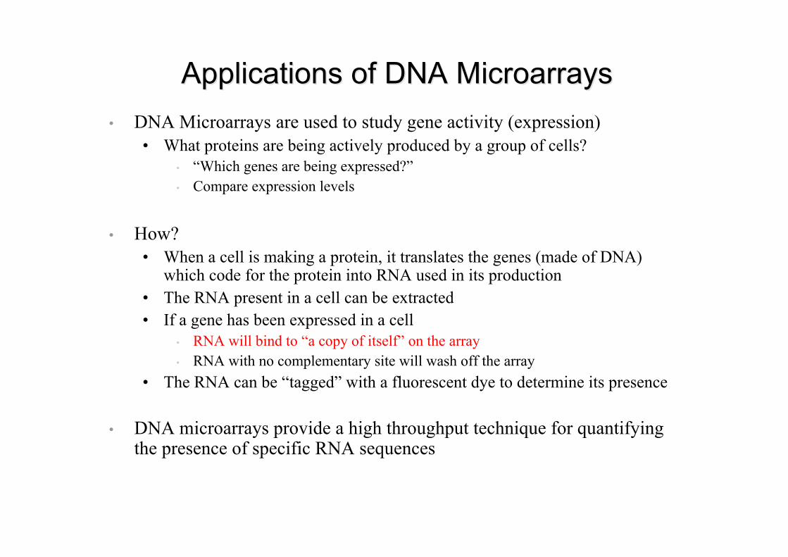
Applications of DNA Applications of DNA MicroarraysMicroarrays• DNA Microarrays are used to study gene activity (expression)
• What proteins are being actively produced by a group of cells?• “Which genes are being expressed?”• Compare expression levels
• How?• When a cell is making a protein, it translates the genes (made of DNA)
which code for the protein into RNA used in its production• The RNA present in a cell can be extracted• If a gene has been expressed in a cell
• RNA will bind to “a copy of itself” on the array• RNA with no complementary site will wash off the array
• The RNA can be “tagged” with a fluorescent dye to determine its presence
• DNA microarrays provide a high throughput technique for quantifyingthe presence of specific RNA sequences
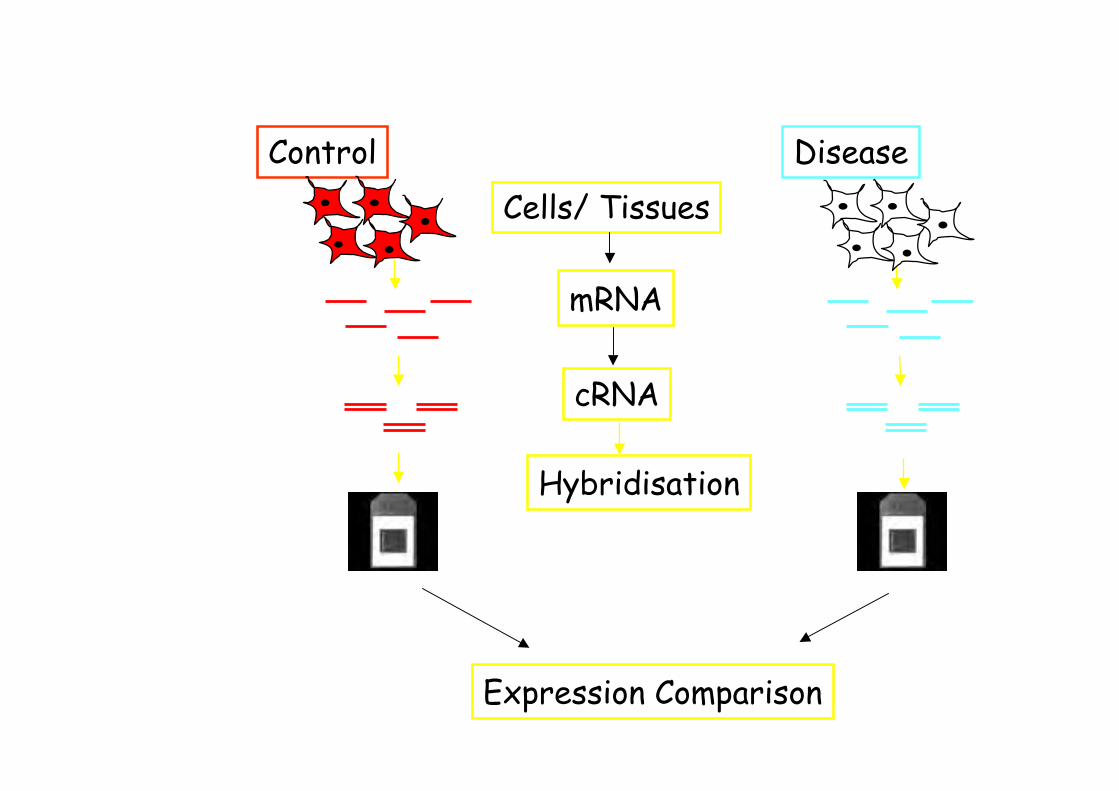
Control Disease
cRNA
Cells/ Tissues
mRNA
Hybridisation
Expression Comparison
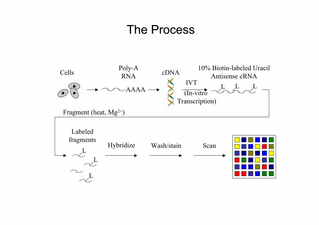
The ProcessThe Process
CellsPoly-ARNA
AAAA
cDNA
L L L
L
IVT
10% Biotin-labeled UracilAntisense cRNA
L
Fragment (heat, Mg2+)
Labeledfragments
Hybridize Wash/stain Scan
L
(In-vitro Transcription)
Affymetrix GeneChipTechonology
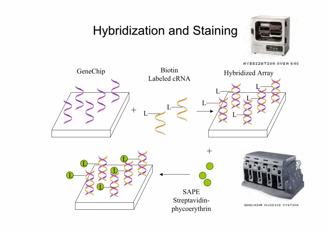
Hybridization and StainingHybridization and Staining
LL
GeneChip BiotinLabeled cRNA
+L
L
L
L
L
L
L
L
L
L+
SAPEStreptavidin-phycoerythrin
Hybridized Array
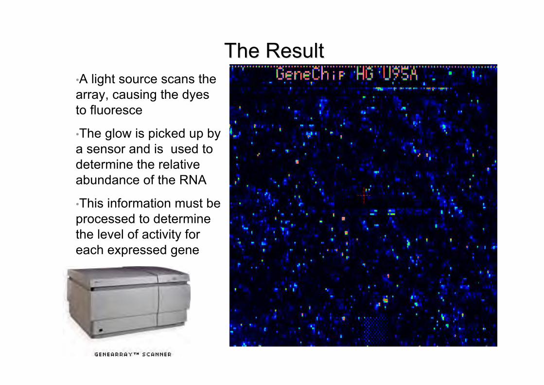
The ResultThe Result•A light source scans thearray, causing the dyesto fluoresce
•The glow is picked up bya sensor and is used todetermine the relativeabundance of the RNA
•This information must beprocessed to determinethe level of activity foreach expressed gene
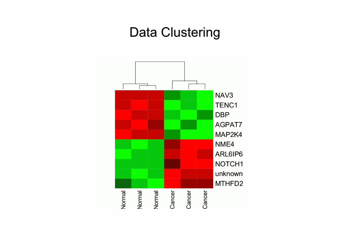
Data ClusteringData Clustering
Array ApplicationsArray Applications
• Basic Understanding• Arrays can take a snap shot of which subset of genes in a cell is
actively making proteins
• Medical diagnosis• Used to determine if a person’s genetic profile would make him or
her more or less susceptible to drug side effects• Used to distinguish between similar diseases or to define previously
unknown subsets within a disease.
• Drug design• Translate the human genome results into new products
• Must figure out what the genes do, how they interact, and howthey relate to diseases.

Microarray Links
•Affymetrix–http://www.affymetrix.com
RNA interference (siRNA)
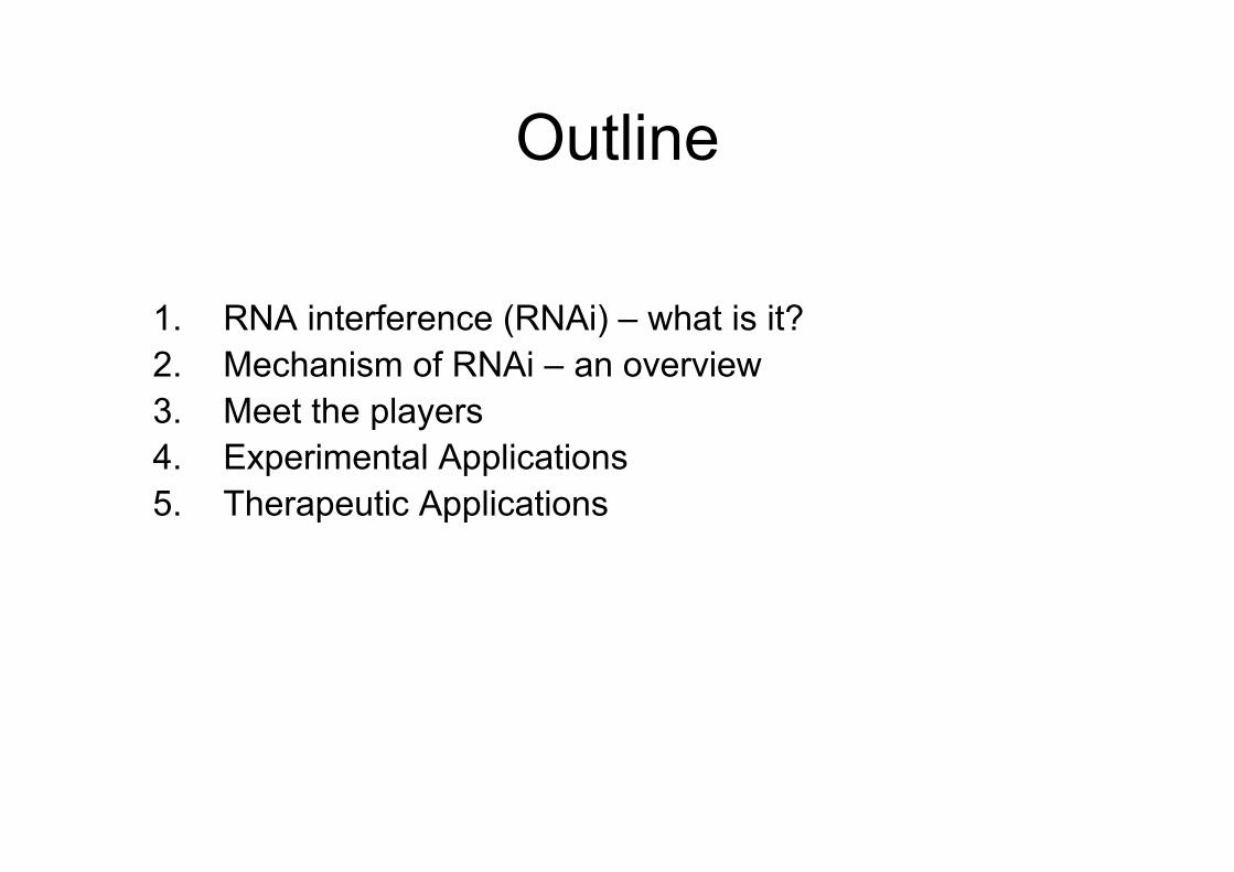
Outline
1. RNA interference (RNAi) – what is it?2. Mechanism of RNAi – an overview3. Meet the players4. Experimental Applications5. Therapeutic Applications
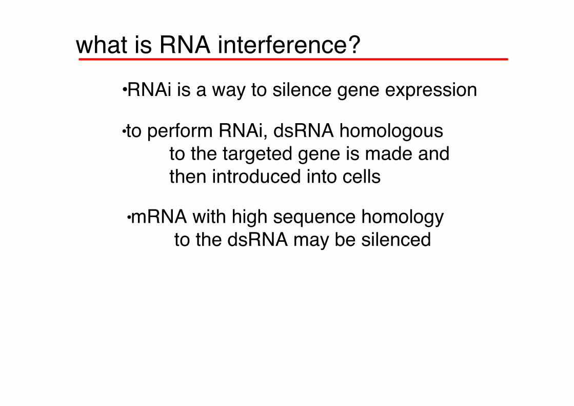
what is RNA interference?•RNAi is a way to silence gene expression
•to perform RNAi, dsRNA homologous to the targeted gene is made andthen introduced into cells
•mRNA with high sequence homology to the dsRNA may be silenced
RNA Interference
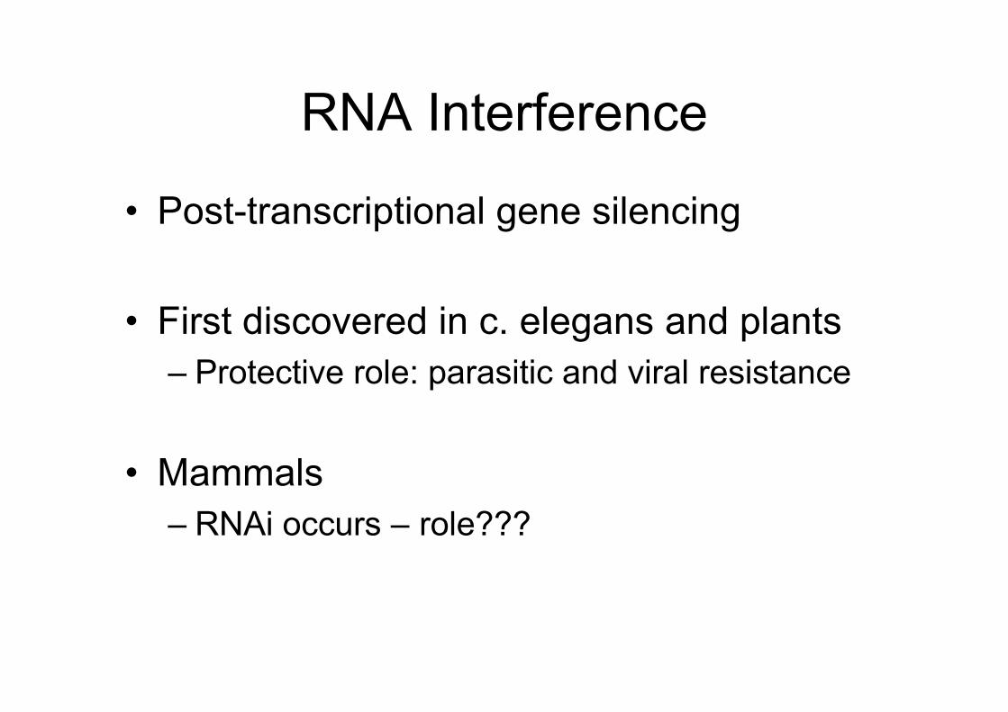
• Post-transcriptional gene silencing
• First discovered in c. elegans and plants– Protective role: parasitic and viral resistance
• Mammals– RNAi occurs – role???
How does RNAi work?
RNAi works postranscriptionally……..
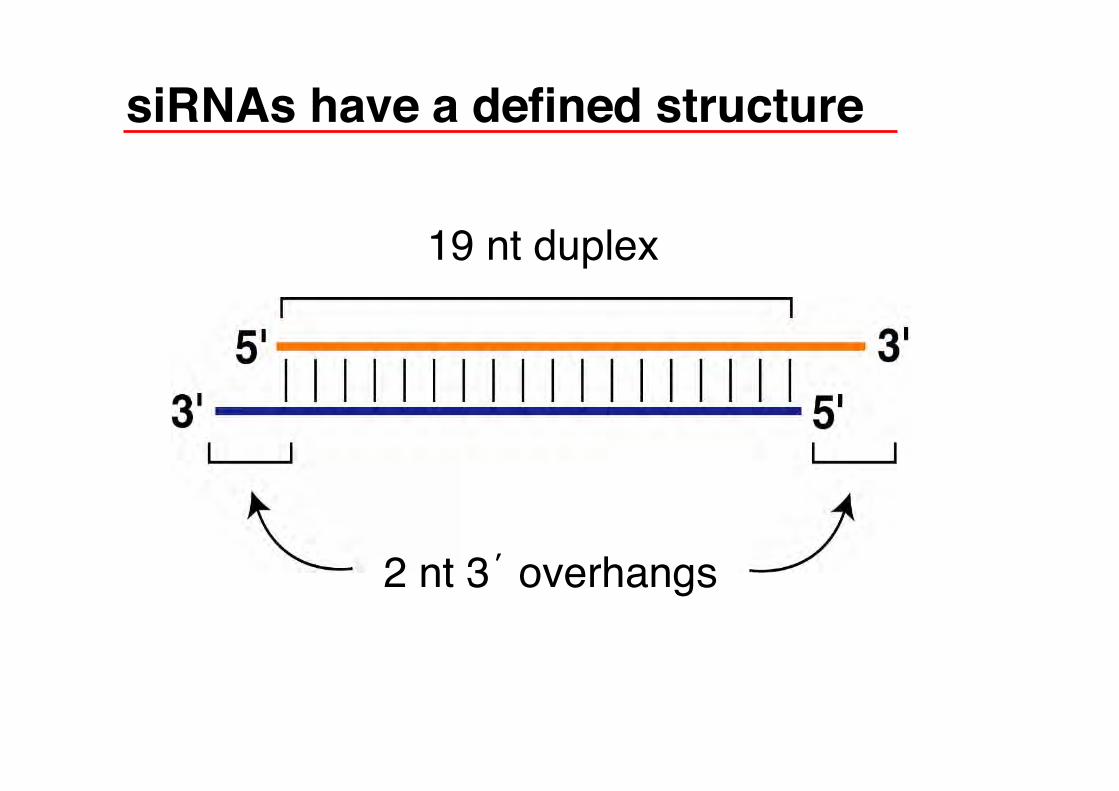
siRNAs have a defined structure
19 nt duplex
2 nt 3’ overhangs
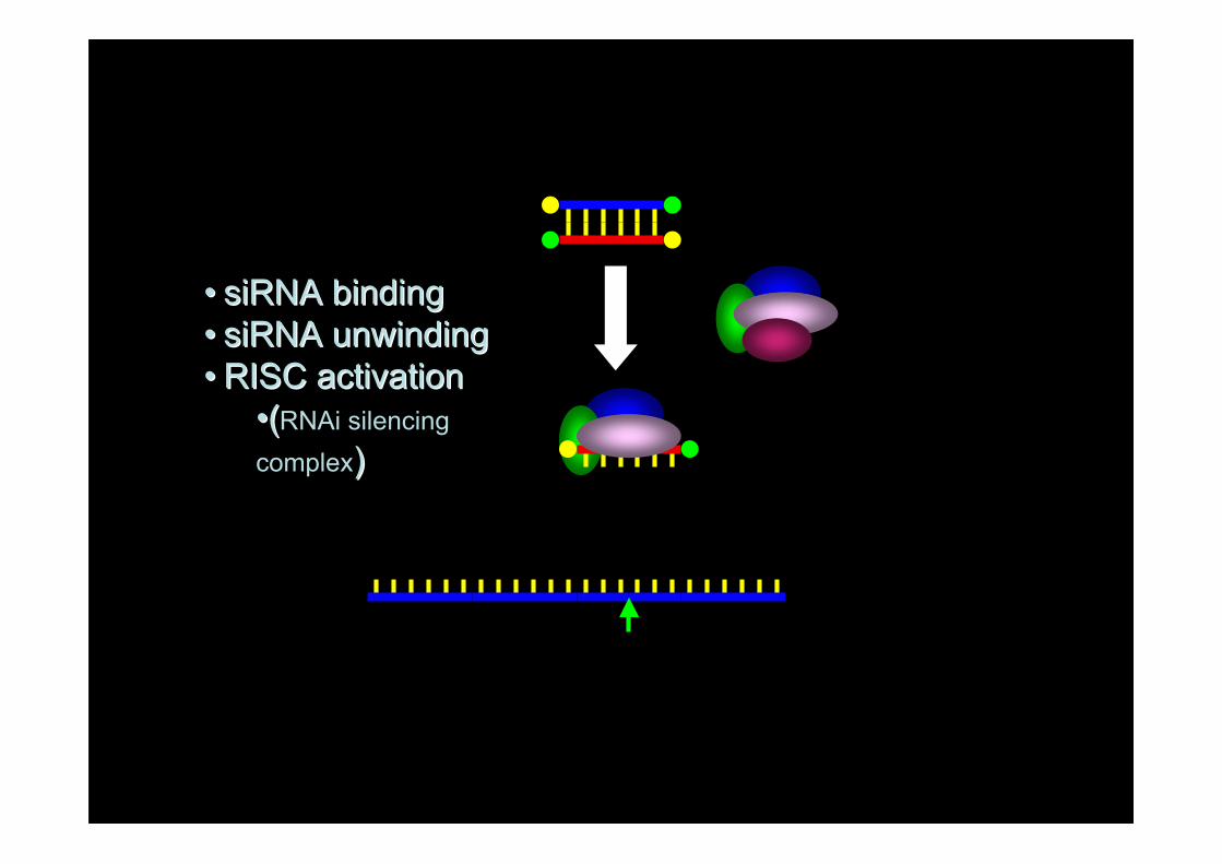
•• siRNA bindingsiRNA binding•• siRNA unwindingsiRNA unwinding•• RISC activationRISC activation
••((RNAi silencingcomplex))
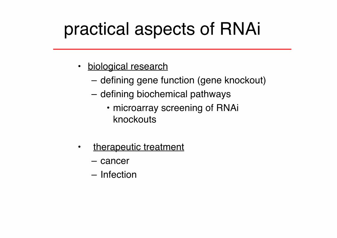
practical aspects of RNAi
• biological research– defining gene function (gene knockout)– defining biochemical pathways
• microarray screening of RNAiknockouts
• therapeutic treatment– cancer– Infection
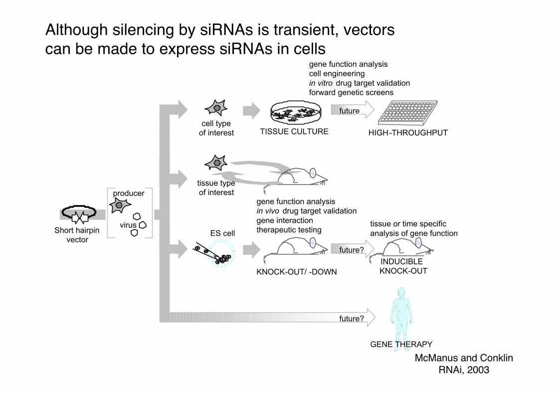
INDUCIBLE KNOCK-OUT
HIGH-THROUGHPUT
tissue or time specific analysis of gene function
KNOCK-OUT/ -DOWN
gene function analysis cell engineeringin vitro drug target validationforward genetic screens
ES cell
gene function analysisin vivo drug target validationgene interactiontherapeutic testing
cell typeof interest
GENE THERAPY
tissue typeof interest
Short hairpinvector
TISSUE CULTURE
future?
future
future?
producer
virus
McManus and ConklinRNAi, 2003
Although silencing by siRNAs is transient, vectorscan be made to express siRNAs in cells
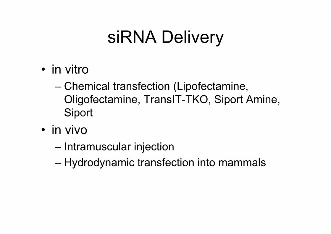
siRNA Delivery
• in vitro– Chemical transfection (Lipofectamine,
Oligofectamine, TransIT-TKO, Siport Amine,Siport
• in vivo– Intramuscular injection– Hydrodynamic transfection into mammals
siRNA Therapeutics
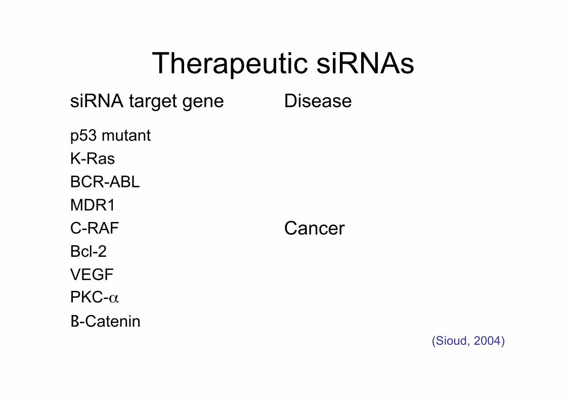
Therapeutic siRNAs
Cancer
p53 mutantK-RasBCR-ABLMDR1C-RAFBcl-2VEGFPKC-αΒ-Catenin
DiseasesiRNA target gene
(Sioud, 2004)
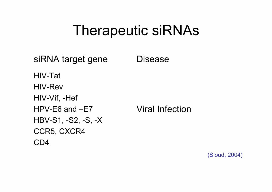
Therapeutic siRNAs
Viral Infection
HIV-TatHIV-RevHIV-Vif, -HefHPV-E6 and –E7HBV-S1, -S2, -S, -XCCR5, CXCR4CD4
DiseasesiRNA target gene
(Sioud, 2004)
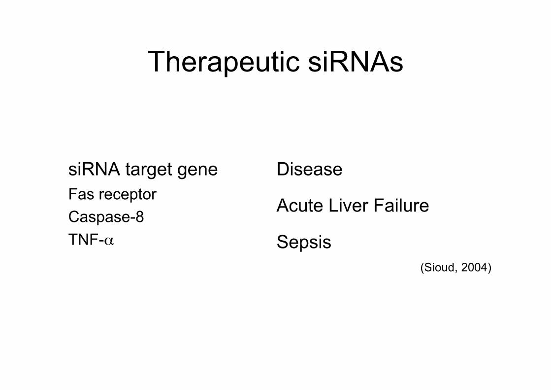
Therapeutic siRNAs
SepsisTNF-α
Acute Liver FailureFas receptorCaspase-8
DiseasesiRNA target gene
(Sioud, 2004)
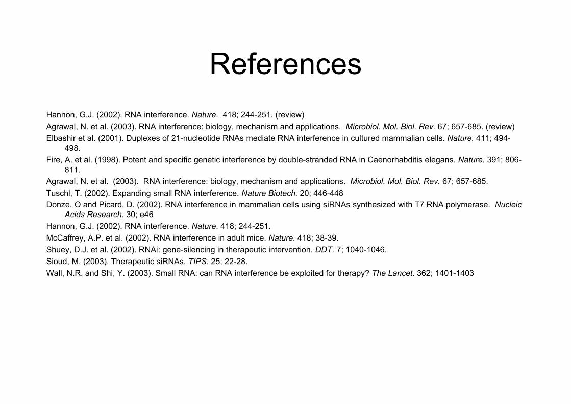
ReferencesHannon, G.J. (2002). RNA interference. Nature. 418; 244-251. (review)Agrawal, N. et al. (2003). RNA interference: biology, mechanism and applications. Microbiol. Mol. Biol. Rev. 67; 657-685. (review)Elbashir et al. (2001). Duplexes of 21-nucleotide RNAs mediate RNA interference in cultured mammalian cells. Nature. 411; 494-
498.Fire, A. et al. (1998). Potent and specific genetic interference by double-stranded RNA in Caenorhabditis elegans. Nature. 391; 806-
811.Agrawal, N. et al. (2003). RNA interference: biology, mechanism and applications. Microbiol. Mol. Biol. Rev. 67; 657-685.Tuschl, T. (2002). Expanding small RNA interference. Nature Biotech. 20; 446-448Donze, O and Picard, D. (2002). RNA interference in mammalian cells using siRNAs synthesized with T7 RNA polymerase. Nucleic
Acids Research. 30; e46Hannon, G.J. (2002). RNA interference. Nature. 418; 244-251.McCaffrey, A.P. et al. (2002). RNA interference in adult mice. Nature. 418; 38-39.Shuey, D.J. et al. (2002). RNAi: gene-silencing in therapeutic intervention. DDT. 7; 1040-1046.Sioud, M. (2003). Therapeutic siRNAs. TIPS. 25; 22-28.Wall, N.R. and Shi, Y. (2003). Small RNA: can RNA interference be exploited for therapy? The Lancet. 362; 1401-1403
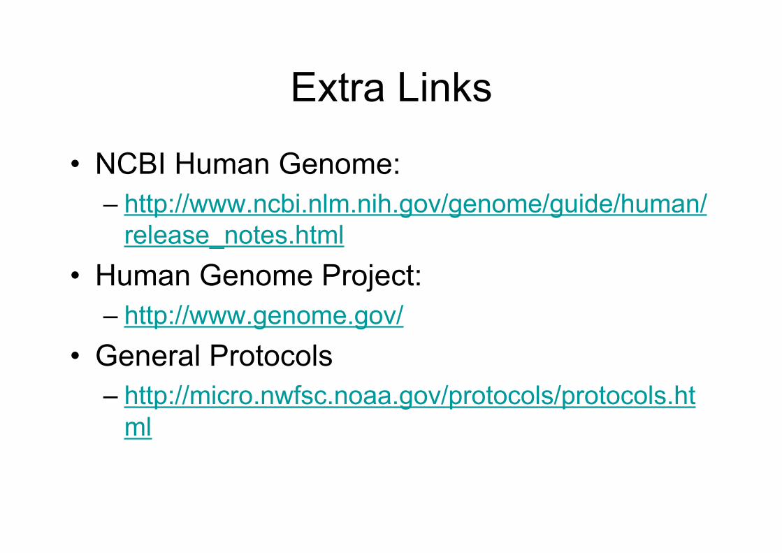
Extra Links
• NCBI Human Genome:– http://www.ncbi.nlm.nih.gov/genome/guide/human/
release_notes.html• Human Genome Project:
– http://www.genome.gov/• General Protocols
– http://micro.nwfsc.noaa.gov/protocols/protocols.html