Nucleic acid hybridisation and polymerase chain reaction in the
19
Rev. sci. tech. Off. int. Epiz., 1993,12 (2), 405-423 Nucleic acid hybridisation and polymerase chain reaction in the diagnosis of infectious animal diseases M. RODRÍGUEZ and A.A. SCHUDEL * Summary: The authors describe and summarise the use of nucleic acid hybridisation and polymerase chain reaction (PCR) technologies in the diagnosis of animal diseases. PCR is a powerful biological tool which enables exponential enzymatic amplification in vitro of a given deoxyribonucleic acid sequence. This technique is currently used to study the molecular pathogenesis of many infectious diseases and also for diagnosis. PCR is usually more sensitive than conventional methods and does not require complex facilities, and will therefore soon become the preferred technology of laboratory diagnosticians, field veterinarians and health officers. KEYWORDS: Animal health - Diagnosis - Polymerase chain reaction - Probes. INTRODUCTION In the past, the diagnosis of infectious animal diseases has been hampered by the complexity of the technology, the facilities required and the amount of time needed for detection and characterisation of most pathogens. With the advent of probe technology, new methods are available to field veterinarians, diagnosticians and animal health officers. These new methods offer specificity and sensitivity equal to those of conventional pathogen detection procedures, while possessing the advantage that they are often able to produce results in a single day, thus providing the opportunity of taking more effective control measures. The presence of infectious agents in fluid or tissue samples can be revealed by microscopic visualisation, or by detection of biological activity or structural components (antigenic proteins and nucleic acids). The nucleotide sequence of the nucleic acid of each pathogen is different to that of other micro-organisms and susceptible hosts. This uniqueness forms the basis of two recently-developed diagnostic tools: nucleic acid hybridisation and polymerase chain reaction (PCR). The characteristics and application of these techniques in the diagnosis of infectious diseases of domestic animals are considered below. Excellent earlier analyses of the technology should be consulted in order to obtain a broader view (15,20,24,27,52,56,57,61,67). * Institute of Virology, Centro de Investigación en Ciencias Veterinarias, Instituto Nacional de Tecnología Agropecuaria, CC 77, Moron 1708, Argentina.
Transcript of Nucleic acid hybridisation and polymerase chain reaction in the

Rev. sci. tech. Off. int. Epiz., 1993,12 (2), 405-423
Nucleic acid hybridisation and polymerase chain reaction in the diagnosis of infectious
animal diseases M. R O D R Í G U E Z and A . A . S C H U D E L *
Summary: The authors describe and summarise the use of nucleic acid hybridisation and polymerase chain reaction (PCR) technologies in the diagnosis of animal diseases.
PCR is a powerful biological tool which enables exponential enzymatic amplification in vitro of a given deoxyribonucleic acid sequence. This technique is currently used to study the molecular pathogenesis of many infectious diseases and also for diagnosis. PCR is usually more sensitive than conventional methods and does not require complex facilities, and will therefore soon become the preferred technology of laboratory diagnosticians, field veterinarians and health officers.
KEYWORDS: Animal health - Diagnosis - Polymerase chain reaction -Probes.
INTRODUCTION
In the past, the diagnosis of infectious animal diseases has been hampered by the complexity of the technology, the facilities required and the amount of time needed for detection and characterisation of most pathogens. With the advent of probe technology, new methods are available to field veter inarians, diagnosticians and animal heal th officers. These new methods offer specificity and sensitivity equal to those of conventional pathogen detection procedures, while possessing the advantage that they are often able to produce results in a single day, thus providing the opportunity of taking more effective control measures.
The presence of infectious agents in fluid or tissue samples can be revealed by microscopic visualisation, or by detection of biological activity or structural components (antigenic proteins and nucleic acids). The nucleotide sequence of the nucleic acid of each pathogen is different to that of other micro-organisms and susceptible hosts. This uniqueness forms the basis of two recently-developed diagnostic tools: nucleic acid hybridisation and polymerase chain reaction (PCR). The characteristics and application of these techniques in the diagnosis of infectious diseases of domestic animals are considered below. Excellent earlier analyses of the technology should be consulted in order to obtain a broader view (15,20,24,27,52,56,57,61,67) .
* Institute of Virology, Centro de Investigación en Ciencias Veterinarias, Instituto Nacional de Tecnología Agropecuaria, CC 77, Moron 1708, Argentina.
406
TECHNICAL CONSIDERATIONS
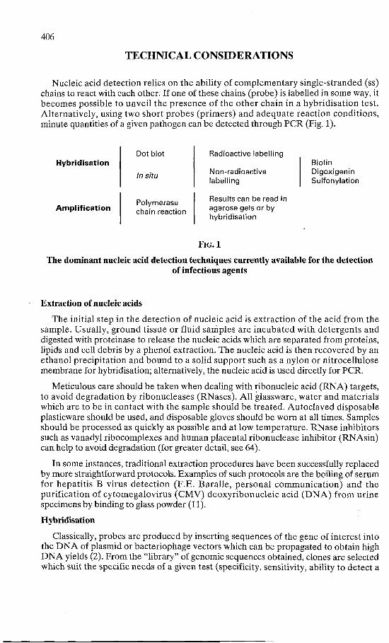
Nucleic acid detection relies on the ability of complementary single-stranded (ss) chains to react with each other. If one of these chains (probe) is labelled in some way, it becomes possible to unveil the presence of the o ther chain in a hybridisation test. Alternatively, using two short probes (primers) and adequate reaction conditions, minute quantities of a given pathogen can be detected through PCR (Fig. 1).
H y b r i d i s a t i o n
A m p l i f i c a t i o n
Dot blot
In situ
Polymerase chain reaction
Radioactive labelling
Non-radioactive labelling
Results can be read in agarose gels or by hybridisation
Biotin Digoxigenin Sulfonylation
F I G . l
The dominant nucleic acid detection techniques currently available for the detection of infectious agents
Extraction of nucleic acids
The initial step in the detection of nucleic acid is extraction of the acid from the sample. Usually, ground tissue or fluid samples are incubated with detergents and digested with proteinase to release the nucleic acids which are separated from proteins, lipids and cell debris by a phenol extraction. The nucleic acid is then recovered by an ethanol precipitation and bound to a solid support such as a nylon or nitrocellulose membrane for hybridisation; alternatively, the nucleic acid is used directly for PCR.
Meticulous care should be taken when dealing with ribonucleic acid (RNA) targets, to avoid degradation by ribonucleases (RNases). All glassware, water and materials which are to be in contact with the sample should be treated. Autoclaved disposable plasticware should be used, and disposable gloves should be worn at all times. Samples should be processed as quickly as possible and at low temperature. RNase inhibitors such as vanadyl ribocomplexes and human placental ribonuclease inhibitor (RNAsin) can help to avoid degradation (for greater detail, see 64).
In some instances, traditional extraction procedures have been successfully replaced by more straightforward protocols. Examples of such protocols are the boiling of serum for hepati t is B virus detect ion (F.E. Baral le , personal communicat ion) and the purification of cytomegalovirus (CMV) deoxyribonucleic acid ( D N A ) from urine specimens by binding to glass powder (11).
Hybridisation
Classically, probes are produced by inserting sequences of the gene of interest into the D N A of plasmid or bacteriophage vectors which can be propagated to obtain high D N A yields (2). From the "library" of genomic sequences obtained, clones are selected which suit the specific needs of a given test (specificity, sensitivity, ability to detect a

407
broad spectrum of field strains). For diagnostic purposes, the purity of any nucleic acid to be used as a probe is of prime importance, in order to avoid background signals which would complicate the interpretation of results. Background noise due to non-specific hybridisation to host or contaminant sequences is reduced by the excision of the cloned fragment.
More recent approaches for p robe product ion are the use of synthetic oligonucleotides, RNA probes and PCR products. The design of oligonucleotide probes is based on available sequence information. Base-pair mismatches might occur when such probes are used for the detection of field isolates, leading to reduced interaction between probe and target. Several reports indicate that when highly-conserved regions of the genome are selected and hybridisation conditions are optimised, oligonucleotide probes can be useful for viral detect ion (9, 10, 30). R N A probes , which are more difficult to handle because of the widespread presence of RNases, can be synthesised in vitro in large amounts using specially-constructed recombinant plasmids such as those containing the SP6 promoter. RNA probes are single-stranded (ss) and thus there is no competing reassociation during hybridisation; they are therefore more sensitive than double-stranded (ds) D N A probes (1). If the PCR has been optimised for a particular pathogen, probes for detect ion of the pa thogen can be p repared very easily using labelled primers or deoxyribonucleotides linked to a biotin or digoxigenin residue.
Probes can be labelled with isotopes or non-radioactive markers such as biotin or digoxigenin. Labelling involves replacement of a proportion of nucleotides in the probe with labelled nucleotides (nick translation and random priming) or end labelling. The nucleic acid can also be chemically modified (sulfonylated) in such a way that it becomes detectable by specific antibodies. Although 3 2 P has been the label of choice when sensitivity is of pr ime impor tance , many repor ts (and the experience of the present authors) indicate that 3 2 P can be replaced, with important operative advantages, by non-radioactive markers which do not constitute a health hazard to operators and have a long shelf life.
Biotin has a high affinity for avidin and, when coupled to peroxidase or alkaline phosphatase, serves to detect hybrids through a colorimetric reaction. Similarly, the hapten digoxigenin is used to label probes which are detectable by means of specific antibodies conjugated with an enzyme. F luorochromes conjugated with biot in or antibodies have also been used to label probes for in situ hybridisation, allowing the location of target sequences in tissue sections and cell monolayers.
Hybridisation is carried out by incubating the labelled probe, under appropr ia te conditions, with the membrane in which the nucleic acids extracted from the sample have been spotted. After several washes to remove unbound probe, the membrane is exposed to X-ray film (if the p robe is labelled with an isotope) or processed for colorimetric visualisation of results if non-radioactive radiation is used (27).
Polymerase chain reaction
This technique, first described by Saiki and colleagues (63), enables the amplification of a specific D N A sequence a million-fold or more. If the target is an RNA sequence, a copy D N A has to be synthesised by reverse transcription before the PCR is carried out. The reaction requires a cyclic, three-step process:
a) denaturation of D N A b) annealing of primers c) primer extension.

408
Denaturation by heating at 93-95°C separates the two D N A strands. In the annealing process, primers attach to complementary regions on either the left (5') or the right (3') side of the sequence of interest. During primer extension, new strands of D N A are produced by the addition of nucleotides (starting at the 3 ' end of the annealed primer), to the unpaired D N A strand. This process takes place by the catalytic effect of a thermostable D N A polymerase. The number of D N A strands doubles upon completion of each cycle, and after 30-45 cycles millions of copies of the target sequence (known as amplicons) are present in the test tube (Fig. 2).
(a)
(b)
(c)
2 DNA strands (original)
4 DNA strands (first cycle)
8 DNA strands (second cycle)
Millions of copies (30-40 cycles)
FIG. 2
The polymerase chain reaction process
The deoxyribonucleic acid (DNA) strands of the pathogen nucleic acid (a) are separated (denatured) at 94°C, and the primers hybridise (anneal) to the complementary sequences at 37-60°C (b).
In a third step (extension), a thermostable DNA polymerase produces complementary strands at 72°C (c). If this three-step process is repeated 30-40 times, an exponential accumulation of DNA copies occurs (d)
and the original target sequence is amplified millions of times
Selection of primers
Primers are synthetic ss oligonucleotides, the sequence of which is complementary either to the left side of one strand of the target sequence (upstream primer) or to the right side of the other strand (downstream pr imer) . In order to select pr imers for detection of a particular pathogen, sequence data relating to the genome of the primers must be available. Usually, the most suitable genomic portions for PCR tests intended for detection of field strains are those which are conserved among isolates, although some primer/template mismatches do not necessarily prevent amplification.
(d)

409
After a genomic por t ion is chosen, a search is conducted for pairs of 18-24 mer oligonucleotides with several characteristics:
- 40-60% G+C content
- absence of polypurines or polypirimidines
- absence of secondary structure
- absence of complementarity, particularly at the 3 ' end, which may promote the formation of an artifact called "primer dimer".
Computer programmes, available upon request, can help to perform this task (62). Since the efficiency of the PCR is lower when long genomic fragments are amplified, the selected primers should be less than 600 bases pairs (bp) apart.
Optimisation of PCR
The basic components of a P C R react ion are the pr imers , deoxynucleot ides (dNTPs), a buffer containing magnesium chloride, a thermostable D N A polymerase and target D N A . The concentrat ion of reaction components and t ime/ temperature parameters must be adjusted for efficient amplification of specific targets. A suitable starting point would be 200 µM of each dNTP, 0.2 µM of each pr imer , a C l 2 Mg concentration of 1.5 mM, 2.5 units of enzyme and 10,000 copies of semi-purified target DNA. The anneal ing t empera tu re is initially defined by the characteristics of the primers, while denaturat ion and extension are usually performed at 94°C and 72°C, respectively. Thirty-five cycles with 90 sec for each step usually allow the visualisation of a reaction product in agarose gels. The optimal concentration of Cl 2 Mg, in the range of 1-6 mM, should be subsequently de termined. If smears are de tec ted in the gel, in addition to the expected fragment size, it might be necessary to raise the annealing temperature, optimise the primer concentrat ion or use other primers. Similar tests should then be conducted with fewer target molecules and using artificial samples. In this process, positive and negative controls should be included in each experiment. Finally, to determine the specificity and sensitivity of the test, a significant number of field and control samples should be examined and the results correlated with those obtained using conventional techniques.
Amplicon detection
The PCR product (amplicon) is usually visualised as a distinct band in agarose gels stained with ethidium bromide. The size of the amplicon, compared with molecular weight s tandards, corresponds to the length of the target sequence defined by the primers. The identity of the amplified fragment is confirmed by hybridisation with an oligonucleotide p robe complementary to an internal area of the amplicon and the presence of restr ict ion enzyme sites. The hybridisat ion is performed using a PCR product spotted on membranes (dot blot), or after transfer of the nucleic acids present in the agarose gel to a solid suppor t (Southern blot) . Restr ic t ion enzymes cut the amplicons into fragments of defined size which are visible in agarose gels.
These procedures are relatively complex and are excessively time-consuming for the clinical laboratory. An interesting alternative which might allow the processing of many samples simultaneously, and even automat ion , is the specific capture of amplified material onto solid phases, followed by colorimetric assay. This approach uses 96-well microtitre plates to which a "capture p robe" is covalently attached (55). Amplicons

410
generated using biotin-labelled primers are t rapped, and the presence of hybrids detected by a streptavidin-peroxidase conjugate and a chromogenic substrate (35).
Laboratory procedure
False-positive reactions can arise due to contamination of a sample being examined by PCR products of previous tests. Since millions of copies of target D N A are produced in each positive react ion and only a few are needed to produce a false posit ive, precautions should be taken to avoid this "carryover". The incorporation of positive, negative and "no D N A " controls in each exper iment is essential to detect this phenomenon. Good general laboratory practice should be followed to avoid this, along with observance of the following guidelines (37):
- different laboratories and equipment should be used for pre- and pos t -PCR manipulations
- water, buffers, tips and tubes should be autoclaved, and work surfaces exposed to ultraviolet light
- aliquots of reagents should be p repared daily for use in a PCR product-free environment
- the D N A sample should be the last element added to each tube.
Critical experiments should be repeated and, whenever possible, more than one pair of primers should be used to amplify disparate genomic fragments.
DIAGNOSTIC APPLICATIONS
While hybridisation tests serve some specific purposes quite well, relatively low sensitivity and technical difficulties have limited the application of these tests in clinical laboratories. This issue has been extensively reviewed by Knowles and Gorham (36). Nevertheless, the high sensitivity of PCR, which is usually more sensitive than conventional methods, makes this technique a true candidate to replace many of the procedures currently in use for the rapid detection of pathogens. The fact that very few copies of the nucleic acid of a micro-organism are needed for rapid detect ion has produced a marked increase in the number of laborator ies implementing P C R for diagnostic purposes.
In contrast with conventional techniques, which demand a different procedure for each pathogen, the implementation of PCR requires similar procedures and reagents for all pathogens. After optimisation, P C R can be performed in low-complexity facilities using relatively low-cost equipment.
Detection of foot and mouth disease virus by PCR
The following provides an illustration of a strategy to develop and assess a PCR assay for the detection of foot and mouth disease virus (FMDV). The gene coding for the viral polymerase (located near the 3 ' end of the viral genome) was chosen for PCR because it is highly-conserved among serotypes and knowledge of the sequence is available (45). Oligonucleotides with suitable characteristics, including 50-60% G+C content and the absence of secondary structure, were selected using a computer programme (62). The reagent concentrat ions and t ime/ tempera ture parameters were optimised using as

411
template a plasmid in which the sequence of interest was inserted. The conditions for reverse transcription were defined using purified viral RNA. Nucleic acid extraction was performed following the method described by Meyer and colleagues (47). Results were read on agarose gels s tained with e thidium bromide and confirmed by hybridisation of the PCR products spotted on nylon membranes with a 30 mer internal oligonucleotide labelled with digoxigenin (M. Rodríguez and colleagues, unpublished findings).
In order to determine the sensitivity and specificity of the assay, experiments were carried out using dilutions of viral stocks of serotypes A, O and C, artificial samples, and bovine tissues taken at necropsy from experimentally-infected cattle (Tables I and II). In addition, further proof of the specificity of the test was obtained by examining fifty tissue samples (tongue epithelia, lung, soft palate, oesophagus, kidney and lymph node) from uninfected and experimental ly-infected cattle (P. Murphy and colleagues, personal communication).
TABLE I
Assessment of the sensitivity of the polymerase chain reaction for the detection of foot and mouth disease virus using dilutions of viral stocks of serotypes A, O and C
Viral stock Serotype Titre (log 1 0LD 5 0/ml)
Sensitivity in gels (LD 5 0 )
No. 644 No. 547 No. 622 No. 578
Oi Oi
A 8 7
6.90 7.04 7.04 6.80
1.90 0.27 0.27 0.19
LD50 :50% lethal dose for newborn mice Results were read directly on agarose gels stained with ethidium bromide and
confirmed by hybridisation
The experimental data indicates that the test is specific and that sensitivity is at least of the same order of magnitude as conventional methods when results are read directly on agarose gels. Increased sensitivity is observed after hybridisat ion of the P C R product. Many samples may be processed simultaneously in less than 24 h, and once the test is optimised complex laboratory facilities are not required.
OTHER APPLICATIONS OF POLYMERASE CHAIN REACTION FOR DETECTION AND CHARACTERISATION OF INFECTIOUS
ANIMAL DISEASE AGENTS
Viruses
African swine fever virus
Four PCR primers and one oligonucleotide probe were designed and synthesised to amplify a 740 bp fragment corresponding to the conserved region of the genome of African swine fever virus (ASFV). A specific 640 bp PCR product was amplified using
412
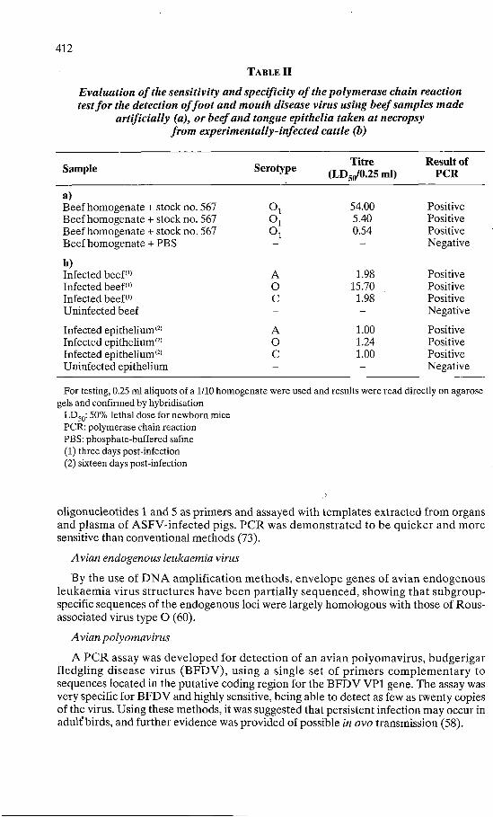
TABLE II
For testing, 0.25 ml aliquots of a 1/10 homogenate were used and results were read directly on agarose gels and confirmed by hybridisation
LD 5 0:50% lethal dose for newborn mice PCR: polymerase chain reaction PBS: phosphate-buffered saline (1) three days post-infection (2) sixteen days post-infection
oligonucleotides 1 and 5 as primers and assayed with templates extracted from organs and plasma of ASFV-infected pigs. PCR was demonstra ted to be quicker and more sensitive than conventional methods (73).
Avian endogenous leukaemia virus
By the use of D N A amplification methods , envelope genes of avian endogenous leukaemia virus structures have been partially sequenced, showing that subgroup-specific sequences of the endogenous loci were largely homologous with those of Rous-associated virus type O (60).
Avian Polyomavirus
A PCR assay was developed for detection of an avian Polyomavirus, budgerigar fledgling disease virus (BFDV) , using a single set of pr imers complementary to sequences located in the putative coding region for the BFDV VP1 gene. The assay was very specific for BFDV and highly sensitive, being able to detect as few as twenty copies of the virus. Using these methods, it was suggested that persistent infection may occur in adult'birds, and further evidence was provided of possible in ovo transmission (58).
Sample Serotype Titre (LD 5 0/0.25 ml)
Result of PCR
a) Beef homogenate + stock no. 567 Beef homogenate + stock no. 567 Beef homogenate + stock no. 567 Beef homogenate + PBS
Oi Oi Oi
54.00 5.40 0.54
Positive Positive Positive Negative
b) Infected beef(1)
Infected beef" Infected beef(" Uninfected beef
A O c
1.98 15.70 1.98
Positive Positive Positive Negative
Infected epithelium ( 2 )
Infected epithelium<2)
Infected epithelium<2)
Uninfected epithelium
A O C
1.00 1.24 1.00
Positive Positive Positive Negative
Evaluation of the sensitivity and specificity of the polymerase chain reaction test for the detection of foot and mouth disease virus using beef samples made
artificially (a), or beef and tongue epithelia taken at necropsy from experimentally-infected cattle (b)

413
Bluetongue virus
PCR was adopted for the identification of a globally-conserved, serogroup-specific nucleic acid sequence (210 bp) of bluetongue virus (BTV) R N A in BTV serotypes from the United States of America (USA). Preliminary results indicate detection of target RNA sequences at a level of less than 2 fg, equivalent to 7,500 viral particles (16). When segment 7 primers (from BTV-ISA) were used, it was possible to detect as few as six molecules of segment 7 ds R N A per sample and also to detect purified ds R N A from different isolates of other serotypes. Positive results were obtained on red blood cells from infected catt le containing as much as 16 T C I D 5 0 , but not those containing 1.6 T C I D 5 0 (81). Primers which encode virus reactive protein VP3, constructed using sequences based on R N A segment 3, were used to detect serogroups of B T V on platelet, buffy coat and packed red cells of BTV-infected sheep (43).
Bovine Coronavirus
PCR was used to synthesise ds and ss probes for detection of bovine Coronavirus (BCV) using recombinant plasmids as template molecules. After fragment-specific amplification by PCR, approximately ten viral genomes were detected by agarose gel analysis (78). Diagnosis of BCV in 134 clinical samples by hybridisat ion was more effective with ss than with either ds probes produced by PCR, or a combination of six recombinant plasmid probes containing non-overlapping sequences (79).
Bovine herpesvirus-4
PCR was used and compared with hybridisation and cell-culture isolation of samples from rabbits experimentally infected with bovine herpesvirus-4 (BHV-4). Virus was detected by PCR in several organs (including nervous tissues) which were found negative by other techniques (50).
Bovine immunodeficiency virus
Infection of embryonic bovine lung cells by bovine immunodeficiency virus was monitored both by syncytia formation and by reverse transcription followed by PCR, showing the advantages of the latter (33).
Bovine leukaemia virus
Proviral bovine leukaemia virus (BLV) D N A was detected in lymphocytic and tumour D N A at all stages of infection in cattle, by the use of a proviral BLV D N A probe and amplification of the D N A (51). P C R amplification (gag-p24, env-gp51, polymerase reverse transcriptase) was used to examine seropositive and seronegative cattle for the presence of BLV D N A in per iphera l blood mononuc lear cells. This protocol enabled the detect ion of one viral genome per 100,000 cells (49). When primers for the polymerase and px regions of BLV were used, as few as ten copies of BLV D N A were consistently amplified. This technique was successfully used in a blind study on blood mononuclear cells of eighteen cows (72).
Bovine papillomavirus
Using PCR, bovine papillomavirus (BPV) D N A was detected in 70% of commercial calf serum batches and 100% correlation was established with virus isolation (69). BPV DNA was detected by PCR using a set of primers located in the E5 open reading frame fitting BPV-1 and BPV-2, after extraction from formalin-fixed frozen sections of twenty equine sarcoids. These results support the general view that BPV plays an important role in equine sarcoids (74).

414
Bovine rotavirus
A full-length complementary D N A (cDNA) copy of the gene encoding the major neutralising antigen of bovine rotavirus was amplified, enabling the detection of this virus in infected faeces with 100,000 times greater sensitivity than the conventional electropherotype method and a 5,000-fold increase in sensitivity over hybridisation (89).
Bovine virus diarrhoea virus
By the amplification and sequencing of the p l 2 5 coding regions of a base pair homologous to the bovine virus diarrhoea virus (BVDV) biotype, cytopathogenic and non-cytopathogenic Pe515 strains were detected (18). Using a set of 20 bp primers located in the conserved 3' region of the B V D V genome, it was possible to amplify a 205 bp target sequence from B V D V D N A . The amplification assay was sensitive enough to detect one molecule of cloned BVDV cDNA, showing that the sensitivity of the test is higher than 1 T C I D 5 0 (42). PCR for BVDV was also shown to be ten times more sensitive than virus isolation, by Vaniddekinge and colleagues (77). On the basis of published sequence data on the N A D L (National Animal Disease Laboratory, USA) strain of BVDV, primers from nucleotide 6322 to 7475 were extracted from serum and white blood cells of persistently-infected heifers and used for P C R (8). Six 20 bp sequences from across the viral genome were selected by homology analysis of the published genomic sequence of the N A D L and Osloss isolates of BVDV. The probe originating nearest the 5 ' end of the viral RNA, ND001, detected 86% of the cytopathic and non-cytopathic viral isolates analysed (38). By using primers from the gp48 region of the cytopathic N A D L strain, it was possible to detect B V D V infection in serum samples of persistently-infected animals (6). Degenera te ol igonucleotide primers designed on the basis of the sequence of two strains of B V D V and one strain of hog cholera virus were used in the PCR to detect viral R N A in cells infected in vitro or in circulating lymphocytes (83).
Caprine arthritis-encephalitis
Caprine arthritis-encephalitis virus and maedi-visna virus were detected by PCR (90).
Distemper virus
Morbillivirus from Phoca vitulina was identified using PCR with cDNA derived from rinderpest morbillivirus (25).
Equine arteritis virus
Three different primer pairs were used for reverse transcriptase PCR genomic RNA amplification of clinical samples of equine arteritis virus, detecting as few as 600 plaque-forming units (PFU) per ml in seminal plasma (14).
Equine herpesvirus-1
PCR was performed on infected and uninfected cultured cells and on 63 specimens from 29 aborted equine foetuses. Results were evaluated by electrophoresis and dot-blot hybridisation using an oligonucleotide probe labelled with biotin. Results were obtained in 24 h and the close correlation with virus isolation results was established (3). Unpurified D N A derived from cultures of equine foetal kidney cells infected with equine herpesvirus ( E H V ) - l or E H V - 4 was amplified by PCR using one pair of oligonucleotide primers. Restriction endonuclease digestion of the amplified segments with PVuII , followed by electrophoresis , revealed restriction fragment length polymorphisms which enabled the two virus types to be differentiated (53).
415
Equine infectious anaemia virus
PCR was used to detect the presence of equine infectious anaemia virus (EIAV) genomes in peripheral mononuclear cells of horses experimentally infected with the CL 22 strain of E I A V (87). Proviral sequences in the peripheral blood mononuclear cells of three horses with acute E I A V infection were monitored using PCR. Provirus was detected in the initial viraemic episode in each horse and during each of three relapsing viraemic cycles. Following each viraemic episode, provirus levels in the peripheral monocytes decreased to less than one copy per 5 x 10 6 cells (54).
Feline leukaemia virus
PCR was used to study the pathogenesis of experimental co-infection of cats with feline influenza virus and feline leukaemia virus (FeLV) (76). PCR has also been used for identification of FeLV infection on feline olfactory neuroblastoma (68).
Foot and mouth disease virus
Rapid and sensitive detect ion of F M D V in bovine and porcine tissues was successfully achieved by enzymatic R N A amplification of the polymerase gene. This method was able to detect amounts smaller than 1 T C I D 5 0 when combined with hybridisation to a labelled probe. No cross-reaction with twelve other viruses (including enterovirus) was demonstrated (47).
Hog cholera virus
Total RNA isolated from hog cholera virus-infected tissue was reverse transcribed and the result ing complementary D N A was amplified with a sensitivity of 10,000 TCID 5 0 . Sensitivity increased 1,000-fold when the PCR product was reamplified with a set of nested primers (40).
Infectious avian laryngotracheitis virus
The infectious laryngotracheitis virus (IALV) homologue of the herpes simplex virus glycoprotein B (gB) gene was identified by PCR amplification of genomic IALV D N A (59).
Infectious bronchitis virus
A PCR technique was developed which permit ted the typing of avian infectious bronchitis virus isolates which caused nephritis (39).
Influenza virus
In order to determine the persistence of influenza virus in ducks after oral infection, PCR analysis of the haemagglutinine gene was used, showing no detectable R N A in spleen samples (82).
Maedi-visna virus
By using a multiple primer set which generates D N A segments with overlapping cohesive termini, maedi-visna virus can be amplified, retained, and detected in infected cells with greater sensitivity (by more than two orders of magnitude) than using existing methods (26). A maedi-visna-like virus (designated EV-1) isolated from sheep showing symptoms of arthritis and pneumonia was analysed by PCR in order to demonstrate the degree of homology with known maedi-visna virus strains (65).

416
Malignant catarrhal fever virus (Alcelaphine herpesvirus-1)
Two pairs of thirty nucleot ide pr imers were selected, corresponding to the nucleotide sequence of the genomic D N A of alcelaphine herpesvirus-1 (AHV-1) (the causative virus of bovine malignant catarrhal fever). The primers were synthesised and successfully used for PCR on crude cell lysates infected with AHV-1 (31). A fragment of AHV-1 D N A was subcloned into pVC18 and sequenced. The subclone hybridised strongly to AHV-1 DNA, weakly to AHV-2 D N A and not at all to D N A from bovine herpesvirus (BHV)- l , BHV-2 and BHV-4. A two-stage (nested) PCR diagnostic test was devised, using a portion of the subcloned AHV-1 D N A sequence. Five AHV-1 and AHV-2 isolates were detected identically and specifically by PCR, while the same procedure failed to detect BHV-1, BHV-2 and BHV-4 viruses. By this technique, levels of AHV-1 as low as 0.01 T C I D 5 0 were detected (34).
Pseudorabies virus (Aujeszky virus)
Genomic sequences of Aujeszky virus (pseudorabies virus: PRV) were amplified by PCR from cells of infected cultures, nasal cells and organs from acutely-diseased pigs, as well as from organs of latently-infected pigs (5). When used directly on disrupted cells with twenty-five cycles, results were obta ined in 1 h which were equal in specificity and sensitivity to conventional methods (32). Various aspects of the latency of PRV in swine were investigated using in vitro nucleic acid amplification methods based on PCR. Primers flanking the 156 bp region of the PRV gpII gene were annealed to purified P R V D N A , as well as D N A isolated from tr igeminal ganglia of swine latently infected with P R V . Following amplification, 100 fg of P R V D N A were visualised on stained gels and 1 fg (equivalent to six viral genome copies) was detected when amplification was combined with blot hybridisat ion (41). When blot hybridisation of PCR-amplified D N A (gX and gII genes) was used, latent virus was detected in seven of eight samples (44). A PCR protocol which specifically amplifies sequences coding for the glycoprotein gp50 (a 217 bp sequence between 433 and 651 of the gp50 gene) was able to consistently detect PRV in tissues of latently-infected swine. Results were obtained in 24 h by sampling tonsil tissues to investigate latent P R V infection (85). Latent viral D N A was detected by PCR in trigeminal ganglia of all of ten pigs which were necropsied 81 or more days after being infected intranasally with a thymidine kinase-negative vaccine strain of PRV (80).
Rabies virus
PCR was used to study the primary multiplication site of a recombinant vaccinia virus (VV) expressing the rabies virus G glycoprotein V V T G g R A B , in comparison with the paren te ra l VV Copenhagen strain, after oral adminis t ra t ion to foxes. V V T G g R A B was detected in the tonsils of foxes tested after 24 h and 48 h, in the buccal mucosa of foxes tested after 24 h and 48 h and in the soft palate of one of two foxes tested after 24 h and one of three foxes after 48 h. Foxes were inoculated with virus isolated from fox tonsils 24 h after oral administrat ion (with or without cell-culture amplification) to perform back passages. No virus could be subsequent ly isolated in either case. The innocuity of V V T G g R A B was also demonstra ted when foxes were inoculated with passaged virus (75). P C R was also used to study the pathogenesis of rabies virus in a mouse model by inoculation of the maseter muscle, showing that virus replication occurred on the trigeminal ganglia at early stages, while at later stages (96 h post-infection) positive sense RNA was present in large amounts in the maseter muscle (71).
417
Bacteria
Brucella abortus
A specific and sensitive PCR test was developed which was capable of detecting 0.1 pg of Brucella abortus D N A (less than 100 brucella cells) by the use of primers which allowed the amplification of a 635 bp fragment of a 43 kDa outer membrane protein gene from B. abortus strain 19 (21).
Escherichia coli
A PCR has been developed to detect Escherichia coli with sensitivity and specificity equal to conventional methods in the following substances: water (4), cheese (46), pig stools (29) and minced meat (84). Similarly sensitive and specific detection of E. coli has been achieved using ol igonucleotide pr imers containing inosine for enzymatic amplification of different alleles of the gene coding for heat-s table toxin type I of enterotoxigenic E. coli (13). Amplification of the mal B operon of E. coli A I I E strains yielded a specific D N A fragment. The minimum detection limit was ten bacteria and the PCR systems were val idated by testing twenty-seven E. coli strains of known enterotoxigenic properties (13).
Leptospira spp.
A PCR was developed to detect leptospira. Primers were used to amplify a 631 bp section of the 5 ' region of 16S ribosomal DNA. (28). When applied to the detection of leptospira in urine samples from cows, as few as 5-10 leptospira per ml of urine were detected (23).
Listeria monocytogenes
Five oligonucleotides were used as pr imers in the P C R for the amplification of specific sequences from D N A of Listeria monocytogenes. This technique was used for detection of listeria in food products (7). Amplification of the alpha and be ta haemolysin gene was used for detection of L. monocytogenes D N A in cooked sausages and milk, with a detection limit of ten bacteria per 10 ml of milk in 48 h (22). Variable results were observed when PCR was used on cheese samples (84). With the aid of PCR amplification, it was de termined that the absence of a gene coding for phosphatidylinositol is associated with the pathogenesis of L. monocytogenes (12).
Mycobacterium paratuberculosis
A 278 bp D N A probe (PCR 278) fragment was obtained by PCR amplification of the 5'region of IS900, an insertion element contained in the genome of M. paratuberculosis. When used in conjunction with PCR, this probe could detect as little as 10 fg (equivalent to two genomes) of M. paratuberculosis starting material (48).
Mycobacterium tuberculosis
Two oligonucleotides were synthesised and used as primers for PCR amplification. A 396 bp fragment was specifically amplified from the Mycobacterium tuberculosis genome. No amplification was observed from any of ten different Mycobacterium strains, including those belonging to the M. tuberculosis complex. The PCR product was detected by dot blot hybridisation, even when as little as 10 fg of purified M. tuberculosis DNA was used. A good correlation with isolation methods was established (17). Using a 123 bp segment of IS6110, which is repea ted in the M. tuberculosis chromosome, a sensitive and specific assay system was devised for detect ion within 48 h of
418
M. tuberculosis on sputum samples (19). PCR was also found to be the most sensitive test for the diagnosis of tuberculous meningitis (70).
Salmonella spp.
Specific primers and a magnetic immuno method (magnetic particles coated with monoclonal antibodies) were used to amplify a 163 bp genomic fragment of Salmonella typhimurium. By this method, a PCR amplification of 25 Salmonella strains but not of 19 other Enterobacteraceae was achieved. A sensitivity of 100 PFU was attained (88).
Other pathogens
Mycoplasma spp.
Primers designed on the basis of the sequences of the 16S ribosomal D N A genes of Mycoplasma pneumoniae and M. genitalis enabled the development of a specific and sensitive in vitro amplification assay system, which detected as few as 100 M. pneumoniae cells and 1,000 M. genitalis cells (66).
Toxoplasma spp.
Tissue samples from abor ted foetuses were examined for the presence of Toxoplasma by PCR amplification of the p30 gene of Toxoplasma. All samples were shown to be infected (86).
* * *
REFERENCES
1. ANDERSON M.L.M. & YOUNG B.D. (1987). - Quantitative filter hybridization. In Nucleic acid hybridization: a practical approach (B.D. Hames & S.J. Higgins, eds). IRL Press, Oxford & Washington D.C.
2. ARRAND J.E. (1987). - Preparation of nucleic acid probes. In Nucleic acid hybridization: a practical approach (B.D. Hames & S.J. Higgins, eds). IRL Press, Oxford & Washington D.C.
3. BALLAGI-PORDANY A., KLINGEBORN B., FLENSBURG J. & BELAK S. (1990). - Equine herpesvirus type 1: detection of viral DNA sequences in aborted foetuses with the polymerase chain reaction. Vet. Microbiol., 22 (4), 373-381.
4. BEJ A.K. , MCCARTY S.C. & ATLAS R.M. (1991). - Detection of coliform bacteria and Escherichia coli by multiplex polymerase chain reaction. Comparison with defined substrate and plating methods for water quality monitoring. Appl. environ. Microbiol., 57, 2429-2432.
5. BELAK S., BALLAGI-PORDANY A., FLENSBURG J. & VIRTANEN A. (1989). - Detection of Pseudorabies virus DNA sequences by the polymerase chain reaction. Arch. Virol., 108 (3-4), 279-286.
6. BELAK S. & BALLAGI-PORDANY A. (1991). - Bovine viral diarrhea virus infection. Rapid diagnosis by the polymerase chain reaction. Arch. Virol., (Suppl. 3), 181-190.
7. BORDER P.M., HOWARD J.J., PLASTOW G.S. & SIGGENS K. (1990). - Detection of Listeria species and Listeria monocytogenes using polymerase chain reaction. Letters appl. Microbiol., 11,158-162.

419
8. BROCK K.V. (1991). - Detection of persistent bovine viral diarrhea virus infections by DNA hybridization and polymerase chain reaction assay. Arch. Virol., (Suppl. 3), 199-208.
9. BRUCE C , AL-NAKIB W., FORSYTH M., STANWAY G . & ALMOND J. W. (1989). - Detection of enterovirus using cDNA and synthetic oligonucleotide probes. J. virol. Meth., 25, 233-240.
10. BRUCE C., AL-NAKIB W., ALMOND J.W. & TYRREL D.A.W. (1989). - Use of synthetic oligonucleotide probes to detect rhinovirus RNA. Arch. Virol., 105,179-187.
11. BUFFONE G . J . , DEMMLER G . J . , SCHIMBOR C.M. & G R E E R J . (1991). - Improved cytomegalovirus DNA amplification from urine after purification of DNA by using glass beads. Clin. Chem., 37,1945-1949.
12. CAMILLI A., GOLDFINE H . & PORTNOY D.A. (1991). - Listeria monocytogenes mutants lacking phosphatidylinositol-specific phospholipase C are avirulent. J. exp. Med., 173, 751-754.
13. CANDRIAN U., FURRER B., HÖFELEIN C. & LÜTHY J . (1991). - Use of inosine-containing oligonucleotide primers for enzymatic amplification of different alleles of the gene coding for heat-stable toxin type I of enterotoxigenic Escherichia coli. Appl. environ. Microbiol, 57,955-961.
14. CHIRNSIDE E . D . & SPAAN W.J.M. (1990). - Reverse transcription and cDNA amplification by the polymerase chain reaction of equine arteritis virus. J. virol. Meth., 30,133-140.
15. CLEWLEY J .P. (1989). - The polymerase chain reaction, a review of the practical limitations for human immunodeficiency virus diagnosis. J. virol. Meth., 25,179-188.
16. DANGLER C.A., MATTOS C.A. DE, MATTOS C.C. DE & OSBURN B.I. (1990). - Identifying bluetongue virus ribonucleic acid sequences by the polymerase chain reaction. J. virol. Meth., 28 (3), 281-292.
17. D E L PORTILLO P., MURILLO L . A . & PATARROYO M . E . (1991). - Amplification of a species-specific DNA fragment of Mycobacterium tuberculosis and its possible use in diagnosis. J. clin. Microbiol., 29,2163-2168.
18. DESPORT M. & BROWNLIE J . (1991). - Molecular characterisation of the coding region for the pl25 from homologous BVDV biotypes. Arch. Virol, (Suppl. 3), 261-265.
19. EISENACH K . D . , SIFFORD M.D., CAVE M.D., BATES J . H . & CRAWFORD J .T. (1991). -Detection of Mycobacterium tuberculosis in sputum samples using a polymerase chain reaction. Am. Rev. respir. Dis., 144, 1160-1163.
20. ERLICH H . A . (1991). - Recent advances in the polymerase chain reaction. Science, 252, 1643-1651.
21. FEKETE A., BANTLE J .A. , HALLING S.M. & SANBORN M.R. (1990). - Preliminary development of a diagnostic test for Brucella using polymerase chain reaction. J. appl. Bacteriol.,69,216-227.
22. FURRER B., CANDRIAN U., HÖFELEIN C . & LÜTHY J . (1991). - Detection and identification of Listeria monocytogenes in cooked sausage products and in milk by in vitro amplification of haemolysin gene fragments. J. appl. Bacteriol., 70, 372-379.
23. GERRITSEN M . J . , OLYHOEK T., SMITS M.A. & B O K H O U T B.A. (1991). - Sample preparation method for polymerase chain reaction-based semiquantitative detection of Leptospira interrogans serovar hardjo subtype hardjobovis in bovine urine. J. clin. Microbiol, 29, 2805-2808.
24. GINGERAS T.R., RICHMAN D.D., KWOH D.Y. & GUATELLI J . C . (1990). - Methodologies for in vitro nucleic acid amplification and their applications. Vet. Microbiol, 24, 235-251.

4 2 0
2 5 . H A A S L., BARRETT T., H A R D E R T. & BOSTOCK C.J. ( 1 9 9 0 ) . - Detection of phocine distemper virus using the polymerase chain reaction. Dt. tierärztl. Wschr., 97, 93 -95 .
26 . H A A S E A.T., R E T Z E L E . F . & STASKUS K . A . ( 1 9 9 0 ) . - Amplification and detection of lentiviral DNA inside cells. Proc. Natl Acad. Sci. USA, 87, 4971 -4975 .
27 . HAMES B.D. & HIGGINS S.J . (eds) ( 1 9 8 7 ) . - Nucleic acid hybridization: a practical approach. IRL Press, Oxford & Washington D.C.
2 8 . HOOKEY J.V. ( 1 9 9 2 ) . - Detection of Leptospiraceae by amplification of 1 6 S ribosomal DNA. FEMS Microbiol. Letters, 90 ,267-274.
29 . HORNES E., WASTESON Y. & OLSVIK O. ( 1 9 9 1 ) . - Detection of Escherichia coli heat-stable enterotoxin genes in pig stool specimens by an immobilized, colorimetric, nested polymerase chain reaction. J. clin. Microbiol., 29 ( 1 1 ) , 2375-2379 .
30 . HOWARD M.J., COELEN R.J. & M A C K E N Z I E J . S . ( 1 9 9 1 ) . - Detection of immobilized Murray Valley encephalitis virus using oligonucleotide probes with varying degrees of mismatch. J., virol. Meth., 34 ,333-341 .
3 1 . Hsu D., SHIH L.M., CASTRO A.E. & Z E E Y.C. ( 1 9 9 0 ) . - A diagnostic method to detect alcelaphine herpesvirus 1 of malignant catarrhal fever using the polymerase chain reaction. Arch. Virol., 114 ( 3 -4 ) , 2 5 9 - 2 6 3 .
32 . JESTIN A., FOULON T., PERTUISET B., BLANCHARD P. & LABOURDET M. ( 1 9 9 0 ) . - Rapid detection of Pseudorabies virus genomic sequences in biological samples from infected pigs using polymerase chain reaction DNA amplification. Vet. Microbiol., 23 (1 -4 ) , 317-328.
3 3 . KASHANCHI F. , LIU Z . Q . , ATKINSON B. & W O O D C. (1991) . - Comparative evaluation of bovine immunodeficiency-like virus infection by reverse transcriptase and polymerase chain reaction. J. virol. Meth., 31 ,197-209.
34 . KATZ J., SEAL B. & RIDPATH J. (1991) . - Molecular diagnosis of alcelaphine herpesvirus (malignant catarrhal fever) infections by nested amplification of viral DNA in bovine blood buffy coat specimens. J. vet. Diagn. Invest., 3 ( 3 ) , 193-198 .
3 5 . KELLER G.H. , HUANG D.P., SHIRAND J .W.K. & MANAK M.M. ( 1 9 9 0 ) . - Detection of hepatitis B virus DNA in serum by PCR and microtiter sandwich hybridization. J. clin. Microbiol, 28, 1411-1416 .
36. KNOWLES D.P. JR & GORHAM J.R. ( 1 9 9 0 ) . - Diagnosis of viral and bacterial diseases. In Biotechnology and veterinary science. Rev. sci. tech. Off. int. Epiz., 9 ( 3 ) , 733 -757 .
37 . KWOK S. & HIGUCHI R. ( 1 9 8 9 ) . - Avoiding false positives with PCR. Nature, 339, 237-238 .
3 8 . LEWIS T.L., RIDPATH J .F. , BOLIN S.R. & BERRY E.S. (1991) . - Detection of BVD viruses using synthetic oligonucleotides. Arch. Virol, 117, 269 -278 .
39 . LIN Z . , KATO A., KUDOU Y., UMEDA K. & U E D A S. (1991) . - Typing of recent infectious bronchitis virus isolates causing nephritis in chicken. Arch. Virol, 120, 145-149.
4 0 . Liu S.T., Li S.N., WANG D.C., CHANG S .F . , CHIANG S.C., H O W.C., CHANG Y.S. & LAI S.S. (1991) . - Rapid detection of hog cholera virus in tissues by PCR. J. virol. Meth., 35 (1 ) , 227-236 .
4 1 . LOKENSGARD J.R., THAWLEY D . G . & MOLITOR T.W. ( 1 9 9 1 ) . - Enzymatic amplification of latent Pseudorabies virus nucleic acid sequences. J. virol. Meth., 34 ,45-55.
42 . LOPEZ O., OSORIO F.A. & DONIS R.O. ( 1 9 9 1 ) . - Rapid detection of bovine diarrhea virus by PCR. J. clin. Microbiol, 29 ,578-582.
4 3 . MCCOLL K.A. & GOULD A.R. ( 1 9 9 1 ) . - Detection and characterisation of bluetongue virus using the polymerase chain reaction. Virus Res., 21, 19-34.

421
44. MAES R.K., BEISEL C.E., SPATZ S.J. & THACKER B J . (1990). - Polymerase chain reaction amplification of Pseudorabies virus DNA from acutely and latently infected cells. Vet. Microbiol., 24 (3-4), 273-280.
45. MARTINEZ-SALAS E., ORTIN J. & DOMINGO E. (1985). - Sequence of the viral replicase gene from FMDV C1-Santa Pau. Gene, 35,55-61.
46. MEYER R., LÜTHY J. & CANDRIAN U. (1991). - Direct detection by polymerase chain reaction (PCR) of Escherichia coli in water and soft cheese and identification of enterotoxigenic strains. Letters appl. Microbiol., 13, 268-271.
47. MEYER R.F., BROWN C.C., HOUSE C , HOUSE J.A. & MOLITOR T.W. (1991). - Rapid and sensitive detection of foot-and-mouth disease virus in tissues by enzymatic RNA amplification of the polymerase gene. J. virol. Meth., 34 (2), 161-172.
48. Moss M.T., G R E E N E.P., T IZARD M.L., MALIK Z.P. & HERMON-TAYLOR J. (1991). -Specific detection of Mycobacterium paratuberculosis by DNA hybridisation with a fragment of the insertion element IS900. Gut, 32,395-398.
49. MURTAUGH M.P., LIN G . F . , H A G G A R D D.L. , W E B E R A.F. & MEISKE J . C . (1991). -Detection of bovine leukaemia virus in cattle by the polymerase chain reaction. J. virol. Meth., 33,73-85.
50. NAEEM K . , M U R T A U G H M.P. & GOYAL S.M. (1991). - Tissue distribution of bovid herpesvirus-4 in inoculated rabbits and its detection by DNA hybridization and PCR. Arch. Virol., 119, 239-255.
51. NAIF H.M., BRANDON R.B., DANIEL R.C.W. & LAVIN M.F. (1990). - Bovine leukaemia proviral DNA detection in cattle using the polymerase chain reaction. Vet. Microbiol., 25 (2-3), 117-129.
52. NORVAL M. & BINGHAM R .W. (1987). - Advances in the use of nucleic acid probes in diagnosis of viral diseases of man. Arch. Virol., 97,151-165.
53. O 'KEEFE J.S., MURRAY A., WILKS CR. & MORIARTY K.M. (1991). - Amplification and differentiation of the DNA of an abortigenic (type 1) and a respiratory (type 4) strain of equine herpesvirus by the polymerase chain reaction. Res. vet. Sci., 50, 349-351.
54. O 'ROURKE K.I. , BESOLA M.L. & MCGUIRE T.C. (1991). - Proviral sequences detected by polymerase chain reaction in peripheral bloodcells of horses with equine infectious anemia lentivirus. Arch. Virol., 117,109-119.
55. PACCIARINI M.L., SAVIO M.L., DONINI G . & TAGLIABUE S. (1993). - The search for improved methods for diagnosing leptospirosis: the approach of a laboratory in Brescia, Italy. In Biotechnology applied to the diagnosis of animal diseases. Rev. sci. tech. Off. int. Epiz., 12 (2), 647-663.
56. PERSING D . H . (1991). - Polymerase chain reaction: trenches to benches. J. clin. Microbiol., 29,1281-1285.
57. PERSING D . H . & LANDRY M.L. (1989). - In vitro amplification techniques for the detection of nucleic acids. Yale J. biol. Med., 62,159-171.
58. PHALEN D.N., WILSON V.G. & GRAHAM D.L. (1991). - Polymerase chain reaction assay for avian Polyomavirus. J. clin. Microbiol., 29,1030-1037.
59. POULSEN D.J., BURTON C.R.A., O ' B R I A N J .A. , RABIN S.J. & KEELER C . L . JR (1991). -Identification of the infectious laryngotracheitis virus glycoprotein gB gene by the polymerase chain reaction. Virus Genes, 5 (4), 335-347.
60. RONFORT C , AFANASSIEFF M., CHEBLOUNE Y., DAMBRINE G . , NIGON V.M. & VERDIER G . (1991). - Identification and structure analysis of endogenous proviral sequences in a brown leghorn chicken strain. Poult. Sci., 70,2161-2175.
422
61. R O Y P. (1989). - Use of nucleic acid probes. In Biotechnology for livestock production. FAO Animal Production and Health Division. Plenum Press, New York, 301-310.
62. RYCHLIK W . & RHOADS E. (1989). - A computer program for choosing oligonucleotides for filter hybridization, sequencing and in vitro amplification of DNA. Nucleic Acids Res., 17,8543-8551.
63. SAIKI R.K. , SCHARF S., FALOONA F., MULLÍS K . B . , H O R N G.T., ERLICH H.A. & AMHEIM N. (1985). - Enzymatic amplification of beta-globin sequences and restriction site analysis for diagnosis of sickle cell anemia. Science, 230, 1350-1354.
64. SAMBROOK J. , FRITSCH E.F. & MANIATIS T. (1989). - Molecular cloning. A laboratory manual. Cold Spring Harbor Laboratory Press, New York.
65. SARGAN D.R., BENNET I.D., COUSENS C , ROY D.J., BLACKLAWS B.A., DALZIEL R.G. & WATT N.J. (1991). - Nucleotide sequence of EV1, a British isolate of maedi-visna virus. J. gen. Virol., 72,1893-1903.
66. SASAKI Y., SHINTANI T., SHIMADA T., WATANABE H. & SASAKI T. (1992). - Detection and discrimination of Mycoplasma pneumoniae and Mycoplasma genitalium by in vitro DNA amplification. Microbiol. Immunol, 36, 21-27.
67. SCHOCHETMAN G., Ou Y.C . & JONES W . (1988). - Polymerase chain reaction. J. infect. Dir., 158, 1154-1157.
68. SCHRENZEL M.D., HIGGINS R.J., HINRICHS S.H., SMITH M.O. & TORTEN M. (1990). -Type C retroviral expression in spontaneous feline olfactory neuroblastomas. Acta neuropathol.,80 (5), 547-553.
69. SCHUURMAN R., VAANSTENIS B., VANSTRIEN A., VANDERNOORDAA J . & SOL C. (1991). -Frequent detection of bovine Polyomavirus in commercial batches of calf serum by using PCR. J. gen. Virol, 72, 2739-2745.
70. SHANKAR P., MANJUNATH N., M O H A N K . K . , PRASAD K . , BEHARI M., SHRINIWAS & AHUJA G.K. (1991). - Rapid diagnosis of tuberculous meningitis by polymerase chain reaction. Lancet, 337 (8732), 5-7.
71. SHANKAR V., DIETZSCHOLD B. & KOPROWSKI H. (1991). - Direct entry of rabies virus into the central nervous system without prior local replication. J. Virol, 65, 2736-2738.
72. SHERMAN M.P., EHRLICH G.D., FERRER J.F., SNINSKY J.J . , ZANDOMENI R., DOCK N.L. & POIESZ B.J. (1992). - Amplification and analysis of specific DNA and RNA sequences of bovine leukaemia virus from infected cows by polymerase chain reaction. J. clin. Microbiol, 30, 185-191.
73. STEIGER Y., ACKERMANN M., METTRAUX C. & KIHM U. (1992). - Rapid and biologically safe diagnosis of African swine fever infection by using PCR. J. clin. Microbiol., 30, 1-8.
74. TEIFKE J.P. & WEISS E. (1991). - Detection of bovine papillomavirus DNA in equine sarcoids using the polymerase chain reaction. Berl. Münch, tierärztl. Wschr., 104, 185-187.
75. THOMAS I., BROCHIER B., LANGUET B . , BLANCOU J . , PEHARPRE D., KIENY M.P., DESMETTRE P., CHAPPUIS G. & PASTORET P.-P. (1990). - Primary multiplication site of the vaccinia-rabies glycoprotein recombinant virus administered to foxes by the oral route. J. gen. Virol, 71 (1), 37-42.
76. TORTEN M., SPARGER E.E., R I D E O U T B.A., PEDERSEN N.C. & Luciw P.A. (1990). -Coinfection of cats with FIV and FeLV affects both quantity and distribution of FIV DNA in various tissues. In Vaccines 90. Modern approaches to new vaccines including prevention of AIDS (F. Brown, R.M. Chanock, H.S. Ginsberg & R.A. Lerner, eds). Cold Spring Harbor Laboratory Press, New York, 375-378.
423
77. VANIDDEKINGE B.J.L.H., VANWAMEL J.L.B., VANGENNIP H . G . P . & MOORMANN R.J.M. (1992). - Application of the PCR to the detection of bovine viral diarrhea virus infections in cattle. Vet. Microbiol., 30,21-34.
78. VERBEEK A. & TIJSSEN P. (1990). - Polymerase chain reaction for probe synthesis and for direct amplification in detection of bovine Coronavirus. J. virol. Meth., 29, 243-256.
79. VERBEEK A., D E A S. & TIJSSEN P. (1991). - Genomic relationships between turkey and bovine enteric coronaviruses identified by hybridization with BCV or TCV specific cDNA probes. Arch. Virol., 121 (1-4), 199-211.
80. VOLTZ D.M., LAGER K.M. & MENGELING W.L. (1992). - Latency of a thymidine kinase-negative Pseudorabies vaccine virus detected by the PCR. Arch. Virol., 122, 341-348.
81. WADE-EVANS A.M., MERTENS P.P.C. & BOSTOCK C.J. (1990). - Development of the polymerase chain reaction for the detection of bluetongue virus in tissue samples. J. virol. Meth., 30 (1), 15-24.
82. WANG M. & WEBSTER R . G . (1990). - Lack of persistence of influenza virus genetic information in ducks. Arch. Virol., 111, 263-267.
83. WARD P. & MISRA V. (1991). - Detection of bovine viral diarrhea virus, using degenerate oligonucleotide primers and the polymerase chain reaction. Am. J. vet. Res., 52, 1231-1236.
84. WERNARS K . , D E L F G O U E., SOENTORO P.S. & NOTERMANS S. (1991). - Successful approach for detection of low numbers of enterotoxigenic Escherichia coli in minced meat by using the polymerase chain reaction. Appl. environ. Microbiol., 57,1914-1919.
85. WHEELER J . G . & OSORIO F.A. (1991). - Investigation of sites of Pseudorabies latency, using PCR. Am. J. vet. Res., 52, 1799-1803.
86. WHEELER R., WILMORE H . , SAVVA D. & TURNER C.B. (1990). - Diagnosis of ovine toxoplasmosis using PCR. Vet. Rec., 126,249.
87. WHETTER L., ARCHAMBAULT D., PERRY S., G A Z I T A., COGGINS L., YANIV A., CLABOUGH D., DAHLBERG J. , FULLER F. & TRONICK S. (1990). - Equine infectious anemia virus derived from a molecular clone persistently infects horses. J. Virol., 64 (12), 5750-5756.
88. WIDJOJOATMODJO M.N., FLUIT A.C., TORENSMA R., KELLER B .H. I . & V E R H O E F J . (1991). - Evaluation of the magnetic immuno PCR assay for rapid detection of Salmonella. Eur. J. clin. Micro. infect. Dis., 10 (11), 935-938.
89. Xu L., HARBOUR D. & M C C R A E M A . (1990). - The application of polymerase chain reaction to the detection of rotaviruses in faeces. J. virol. Meth., 27 (1), 29-37.
90. ZANONI R., PAULI U. & PETERHANS E. (1989). - Caprine arthritis-encephalitis (CAE)-and maedi-visna viruses detected by the polymerase chain reaction (PCR). In First Congress of the European Society for Veterinary Virology, Liege, Belgium, 6-7 April.