Nucleic Acid Aptamers as Potential Therapeutic and...
19
Journal of Cancer Therapy, 2013, 4, 872-890 http://dx.doi.org/10.4236/jct.2013.44099 Published Online June 2013 (http://www.scirp.org/journal/jct) Nucleic Acid Aptamers as Potential Therapeutic and Diagnostic Agents for Lymphoma Ka-To Shum 1* , Jiehua Zhou 1* , John J. Rossi 1,2# 1 Department of Molecular and Cellular Biology, Beckman Research Institute of City of Hope, Duarte, CA, USA; 2 Irell and Manella Graduate School of Biological Sciences, Beckman Research Institute of City of Hope, Duarte, CA, USA. Email: # [email protected] Received April 9 th , 2013; revised May 8 th , 2013; accepted May 16 th , 2013 Copyright © 2013 Ka-To Shum et al. This is an open access article distributed under the Creative Commons Attribution License, which permits unrestricted use, distribution, and reproduction in any medium, provided the original work is properly cited. ABSTRACT Lymphomas are cancers that arise from white blood cells and usually present as solid tumors. Treatment of lymphoma often involves chemotherapy, and can also include radiotherapy and/or bone marrow transplantation. There is an un- questioned need for more effective therapies and diagnostic tool for lymphoma. Aptamers are single stranded DNA or RNA oligonucleotides whose three-dimensional structures are dictated by their sequences. The immense diversity in function and structure of nucleic acids enable numerous aptamers to be generated through an iterative in vitro selection technique known as Systematic Evolution of Ligands by EXponential enrichment (SELEX). Aptamers have several biochemical properties that make them attractive tools for use as potential diagnostic and pharmacologic agents. Iso- lated aptamers may directly inhibit the function of target proteins, or they can also be formulated for use as delivery agents for other therapeutic or imaging cargoes. More complex aptamer identification methods, using whole cancer cells (Cell-SELEX), may identify novel targets and aptamers to affect them. This review focuses on recent advances in the use of nucleic acid aptamers as diagnostic and therapeutic agents and as targeted delivery carriers that are relevant to lymphoma. Some representative examples are also discussed. Keywords: Lymphoma; Nucleic Acid Aptamer; SELEX; Cell-Type Specific Drug Delivery; Biosensor; Nanotechnology 1. Introduction Lymphoma describes a group of blood cancers that de- velops in the lymphatic system and arise from affecting lymphocytes. In United States, more than 21,000 new cases of lymphoma are identified annually, affecting people of all ages [1,2]. Lymphoma is broadly used to describe at least 35 types of lymphomas, which are grouped based on cell histology, genetic mutations, ex- pression of certain genes and many other characteristics [3]. Although there are many types of lymphoma, Hodg- kin’s lymphoma and non-Hodgkin’s lymphoma are two major categories of lymphomas [4-6]. Lymphoma can originate from a variety of causes, but most lymphomas are curable if caught during the earliest stage of the dis- ease [1]. Untreated or advanced lymphoma leads to an uncontrolled proliferation of lymphocytes that spreads throughout the lymphatic system. Aptamer technology has emerged as a valuable tool for detecting, imaging, diagnosing and treating cancer [7-10]. Aptamers are short oligonucleotides that are se- lected from large combinatorial pools and can bind ex- traordinary tightly to and effectively inhibit protein tar- gets [11]. Aptamers can be evolved and manipulated with simplicity, but have versatility in structure and function similar to that of proteins. Since the inception of aptamer technology two decades ago, aptamers are now regularly considered along with small molecules and antibodies as potential diagnostic probes, pharmaceuticals and targeted delivery vehicles for cancer diagnosis and treatment [10, 12,13]. Regarded as “chemical antibodies”, aptamers have many attributes that are advantageous in compari- son with monoclonal antibodies and small molecules. These attributes include higher affinity and specificity, greater ease of manufacture and modification in vitro, and better tissue penetration [14,15]. United States Food and Drug Administration (FDA) approval of the first aptamer drug (Macugen) for the treatment of age-related macular degeneration has proven to be a milestone in the * The first two authors contributed equally to this work. # Corresponding author. Copyright © 2013 SciRes. JCT
Transcript of Nucleic Acid Aptamers as Potential Therapeutic and...
Journal of Cancer Therapy, 2013, 4, 872-890 http://dx.doi.org/10.4236/jct.2013.44099 Published Online June 2013 (http://www.scirp.org/journal/jct)
Nucleic Acid Aptamers as Potential Therapeutic and Diagnostic Agents for Lymphoma
Ka-To Shum1*, Jiehua Zhou1*, John J. Rossi1,2#
1Department of Molecular and Cellular Biology, Beckman Research Institute of City of Hope, Duarte, CA, USA; 2Irell and Manella Graduate School of Biological Sciences, Beckman Research Institute of City of Hope, Duarte, CA, USA. Email: #[email protected] Received April 9th, 2013; revised May 8th, 2013; accepted May 16th, 2013 Copyright © 2013 Ka-To Shum et al. This is an open access article distributed under the Creative Commons Attribution License, which permits unrestricted use, distribution, and reproduction in any medium, provided the original work is properly cited.
ABSTRACT
Lymphomas are cancers that arise from white blood cells and usually present as solid tumors. Treatment of lymphoma often involves chemotherapy, and can also include radiotherapy and/or bone marrow transplantation. There is an un- questioned need for more effective therapies and diagnostic tool for lymphoma. Aptamers are single stranded DNA or RNA oligonucleotides whose three-dimensional structures are dictated by their sequences. The immense diversity in function and structure of nucleic acids enable numerous aptamers to be generated through an iterative in vitro selection technique known as Systematic Evolution of Ligands by EXponential enrichment (SELEX). Aptamers have several biochemical properties that make them attractive tools for use as potential diagnostic and pharmacologic agents. Iso- lated aptamers may directly inhibit the function of target proteins, or they can also be formulated for use as delivery agents for other therapeutic or imaging cargoes. More complex aptamer identification methods, using whole cancer cells (Cell-SELEX), may identify novel targets and aptamers to affect them. This review focuses on recent advances in the use of nucleic acid aptamers as diagnostic and therapeutic agents and as targeted delivery carriers that are relevant to lymphoma. Some representative examples are also discussed. Keywords: Lymphoma; Nucleic Acid Aptamer; SELEX; Cell-Type Specific Drug Delivery; Biosensor;
Nanotechnology
1. Introduction
Lymphoma describes a group of blood cancers that de- velops in the lymphatic system and arise from affecting lymphocytes. In United States, more than 21,000 new cases of lymphoma are identified annually, affecting people of all ages [1,2]. Lymphoma is broadly used to describe at least 35 types of lymphomas, which are grouped based on cell histology, genetic mutations, ex- pression of certain genes and many other characteristics [3]. Although there are many types of lymphoma, Hodg- kin’s lymphoma and non-Hodgkin’s lymphoma are two major categories of lymphomas [4-6]. Lymphoma can originate from a variety of causes, but most lymphomas are curable if caught during the earliest stage of the dis-ease [1]. Untreated or advanced lymphoma leads to an uncontrolled proliferation of lymphocytes that spreads throughout the lymphatic system.
Aptamer technology has emerged as a valuable tool
for detecting, imaging, diagnosing and treating cancer [7-10]. Aptamers are short oligonucleotides that are se- lected from large combinatorial pools and can bind ex- traordinary tightly to and effectively inhibit protein tar- gets [11]. Aptamers can be evolved and manipulated with simplicity, but have versatility in structure and function similar to that of proteins. Since the inception of aptamer technology two decades ago, aptamers are now regularly considered along with small molecules and antibodies as potential diagnostic probes, pharmaceuticals and targeted delivery vehicles for cancer diagnosis and treatment [10, 12,13]. Regarded as “chemical antibodies”, aptamers have many attributes that are advantageous in compari- son with monoclonal antibodies and small molecules. These attributes include higher affinity and specificity, greater ease of manufacture and modification in vitro, and better tissue penetration [14,15]. United States Food and Drug Administration (FDA) approval of the first aptamer drug (Macugen) for the treatment of age-related macular degeneration has proven to be a milestone in the
*The first two authors contributed equally to this work. #Corresponding author.
Copyright © 2013 SciRes. JCT
Nucleic Acid Aptamers as Potential Therapeutic and Diagnostic Agents for Lymphoma 873
applications of aptamer technology [16,17]. Many ap- tamers against cancer-related proteins or even intact cancer cells have been tested in clinical trials [18,19]. In addition, use of aptamers to target membrane receptors and their adoption as delivery carriers has progressed rapidly [20,21]. In this review, we highlight the impor- tance of aptamers in lymphoma research. We discuss strategies to identify potent cancer-targeting aptamers and focus on recent advances in the use of nucleic acid aptamers as diagnostics, therapeutics and targeted deliv- ery vehicles that are relevant to lymphoma. Some repre- sentative examples are also discussed.
2. The Overview of Systematic Evolution of Ligands by Exponential Enrichment (SELEX)
Aptamer sequences that have defined and unique proper- ties are identified from a large pool comprising a ran- domized combinatorial library through an iterative pro-
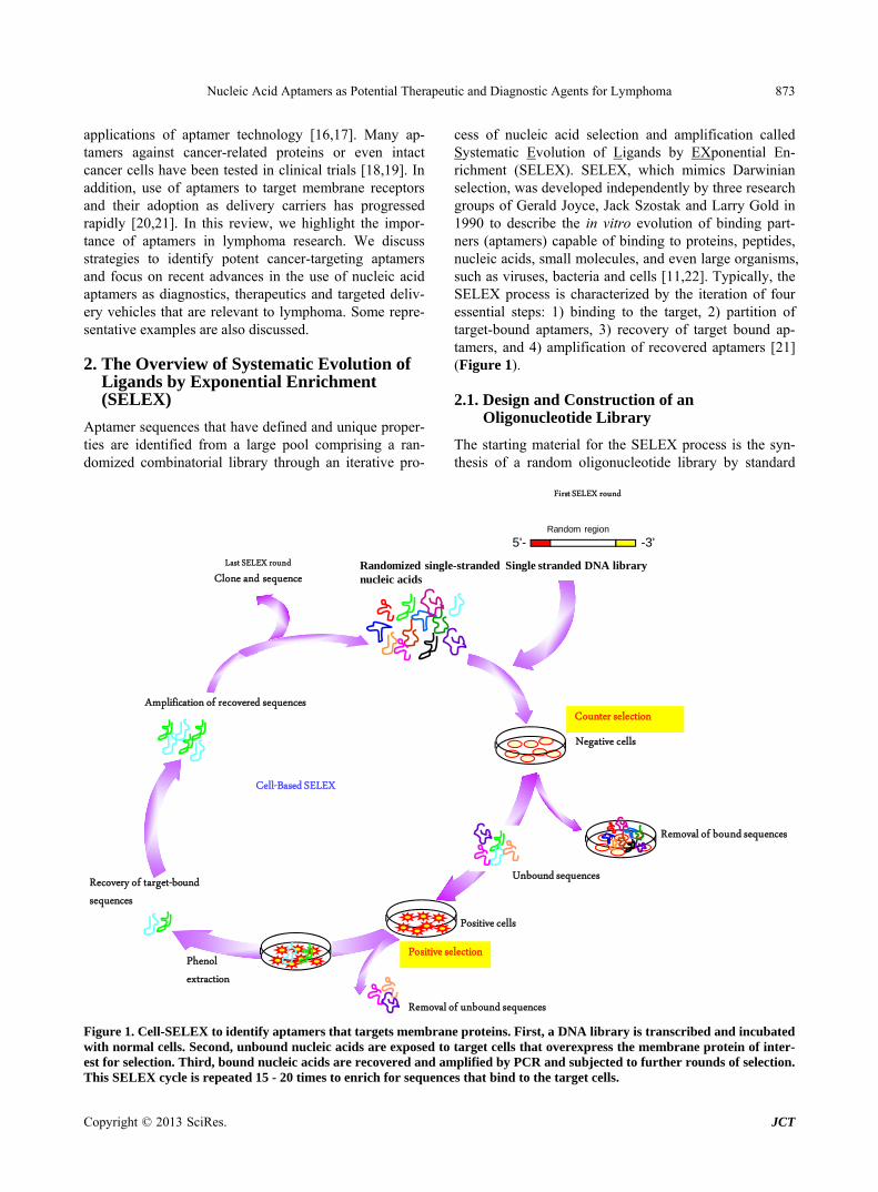
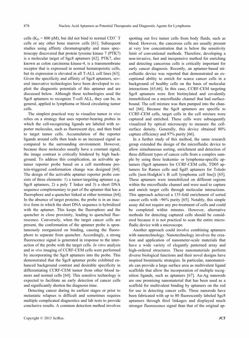
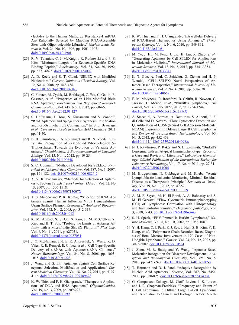
cess of nucleic acid selection and amplification called Systematic Evolution of Ligands by EXponential En- richment (SELEX). SELEX, which mimics Darwinian selection, was developed independently by three research groups of Gerald Joyce, Jack Szostak and Larry Gold in 1990 to describe the in vitro evolution of binding part- ners (aptamers) capable of binding to proteins, peptides, nucleic acids, small molecules, and even large organisms, such as viruses, bacteria and cells [11,22]. Typically, the SELEX process is characterized by the iteration of four essential steps: 1) binding to the target, 2) partition of target-bound aptamers, 3) recovery of target bound ap- tamers, and 4) amplification of recovered aptamers [21] (Figure 1).
2.1. Design and Construction of an Oligonucleotide Library
The starting material for the SELEX process is the syn- thesis of a random oligonucleotide library by standard
Negative cells
Cell-Based SELEX
Removal of bound sequences
Unbound sequences
Positive cells
Removal of unbound sequences
Recovery of target-bound sequences
Positive selection
Last SELEX roundClone and sequence
Amplification of recovered sequences
5'- -3'
Single stranded DNA library
Random region
Randomized single-stranded nucleic acids
Counter selection
Phenolextraction
First SELEX round
Figure 1. Cell-SELEX to identify aptamers that targets membrane proteins. First, a DNA library is transcribed and incubated with normal cells. Second, unbound nucleic acids are exposed to target cells that overexpress the membrane protein of inter- est for selection. Third, bound nucleic acids are recovered and amplified by PCR and subjected to further rounds of selection. This SELEX cycle is repeated 15 - 20 times to enrich for sequences that bind to the target cells.
Copyright © 2013 SciRes. JCT
Nucleic Acid Aptamers as Potential Therapeutic and Diagnostic Agents for Lymphoma 874
solid phase methodology. The random single-stranded DNA consists of 20 - 80 nucleotides, flanked by a region of known sequence that can be recognized by primers in a PCR reaction. If RNA selection is to take place, a T7 RNA polymerase promoter is incorporated into the for- ward primer to allow in vitro transcription. In principle, it is thought that RNA selection provides more structural diversity than DNA selection because of the presence of 2' OH and non-Watson and Crick base pairing in RNA, but there is no clear pattern of specificity and affinity related to the chemistry of aptamers observed today [23]. Random base incorporation is achieved by using an al- most equimolar mix of the four phosphoramidite precur- sors during the random sequence of the synthesis. Be- cause of the varied coupling efficiencies of different nu- cleobases, the concentrations of phosphoramidite pre- cursors may need to be adjusted accordingly (e.g. a ratio of A:C:G:U/T = 1.5:1.25:1.15:1.0) so there is an equal chance of each nucleotide being at any precision and no bias is introduced during this step [24].
Another issue is the length of the randomized se- quence that determines the complexity of the library and its molecular diversity. If the random region is short (~10 nucleotides), then every permutation can be synthesized and the entire sequence space explored. However, such a short sequence is not sufficient for many single-stranded nucleic acid structures. The length of the oligonucleotide is also limited by the difficulty of producing longer oli- gonucleotide sequences by standard DNA synthesis methods. Because many natural nucleic acid-protein re- cognition sites comprise 15 - 25 nucleotides, a library containing 25 random nucleotides is generally considered appropriate [25]. For a randomized single-stranded nu- cleic acids consisting of four bases, the number of possi- ble sequences scale at 4n, where n is the number of ran- domized base position. Typically the starting number of individual molecules used is 1013 - 1015. For a 25-mer library, there are 4n = 1015 individual sequences in the pool, which reach the practically possible limit of se- quence diversity.
After the initial DNA strand is synthesized, a primer is annealed onto the known region and the complementary strand is synthesized using the Klenow fragment of DNA polymerase I. In an RNA selection experiment, RNA is transcribed by T7 RNA polymerase using the promoter that was incorporated during synthesis of oligonucleo- tides. Modified nucleotides are often used instead of the natural ones to confer extra stability to the RNA mole- cules produced and to maintain the defined functions in the absence of toxicity [26]. For example, some popular modifications of aptamers are derivatives of the 2' ribose, such as 2' fluoro, 2' amino-methyl and 2' O-methyl de- rivatives [26]. Recently, two classes of modified nucleo- tides, Locked Nucleic Acids and spiegelmers (mirror
image of aptamers) have been adapted into the PCR am- plification and T7 transcription [27,28]. Thus, aptamers can be tailored to achieve certain functions through site- specific chemical modifications that are especially im- portant for in vivo uses [29].
2.2. Partitioning and the Iterative Cycle of SELEX
The critical experimental challenge of SELEX is to de- sign selection processes that can distinguish those mole- cules that are able to perform the required task, such as binding, from those that cannot, a process known as peti- tioning. Many powerful tools have been developed to fractionate target bound from unbound species, including centrifugation, gel shift assay, affinity chromatography, co-immunoprecipitation, surface plasmon resonance, flow cytometry, capillary electrophoresis, microfluidic devices and nitrocellulose filter binding [30-33]. As an example, in the filter binding assay, the target could be immobilized on a filter and affinity chromatography could be used; aptamers that bind to the target would be retained on the filter, while those that do not would pass through. Partitioning is the most important variable for a SELEX experiment because it can greatly simplify the processes, thereby reducing the number of selection cy- cles and accelerating identification of potent aptamers [22]. In general, approximate 12 iterative cycles of SE- LEX are sufficient to achieve significant enrichment within the library of an aptamer that has high binding affinity with the target. The inherent properties of se- lected oligonucleotides can be further fine-tuned for dif- ferent purposes by adjusting the stringency parameters, such as salt concentration and number of washes.
2.3. Generation of Cell-Specific Aptamers
To date, a considerable number of aptamers have been evolved to target a specific cell type or subpopulation of cancer cells through either protein-based or cell-based SELEX [18,19]. The traditional approach requires a puri- fied soluble form of the target proteins in desirable con- centrations. However, this method has inherent limita- tions because the purified recombinant protein may not fold into a similar conformation as under physiological conditions. Moreover, the native protein may be present in a modified form or exist as a protein complex that may be masked and therefore inaccessible to the aptamers [34]. This problem is especially common when the trans- membrane domain of a receptor embedded in the plasma membrane is not exposed to aptamer binding. To over- come this, research groups have modified the traditional SELEX protocol to enable use of whole living cells as targets (Cell-SELEX).
The advantages of Cell-SELEX include being able to
Copyright © 2013 SciRes. JCT
Nucleic Acid Aptamers as Potential Therapeutic and Diagnostic Agents for Lymphoma 875
select native aptamers without going through the proc- esses of protein expression and purification [35]. In addi- tion, Cell-SELEX can be performed when unknown tar- gets are expressed on the cell surface (Blind Cell-SELEX) as this strategy relies on the differences between two distinct populations of cells (target diseased cells versus control healthy cells) that have particular defined features, such as protein expression levels and phenotypes [36-38]. In theory, aptamers isolated from cell-SELEX will only recognize the target cells, but not control cells. Akin to traditional protein-based SELEX, Cell-SELEX is also characterized by the iterative cycles of the four main steps described above, albeit using living cells instead of purified proteins (Figure 1). Because intact living cells contain many native receptor proteins, a counter-selec- tion step is required to avoid non-specific binders. For example, if the selection step involves a cell line that overexpresses a cell surface receptor of interest, the counter-selection step could involve a related cell line that lacks or has a very low expression of the target pro- tein. Generally, Cell-SELEX requires approximately 15 - 20 cycles of selection, which is more than that of protein- SELEX [7]. Because many surface complexes are pre- sent on cells and these complexes consist of multiple pro- teins, multiple families of aptamers are expected to be identified, and the selected aptamers must be carefully characterized [39]. Moreover, Cell-SELEX does not dis- criminate between living and dead cells. Because dead cells have reduced membrane integrity and can non-spe- cifically internalize nucleic acids in a sequence-indepen- dent manner, cells should be kept healthy during selec- tion as any damages to the cells may incur risk of selec- tion failure [7,21]. Because Cell-SELEX facilitates the development of and simplifies the identification of mo- lecular probes for recognizing cancer cells, this technol- ogy opens the way to an improved understanding of can- cer development and provides a platform for developing
new tools for the diagnosis and treatment of cancer (Ta- ble 1).
3. Aptamers as a Potential “Next-Generation” of Diagnostic Tools
Lymphoma is fatal if left untreated or diagnosed at a late stage. Some classes of lymphoma, such as Burkitt’s lymphoma, are particularly acute and aggressive and therefore require rapid and accurate diagnosis and prog- nosis to aid in prevention, therapy and detection of mini- mal residual diseases [40]. There are a battery of options to diagnose lymphoma in the clinic, involving a combi- nation of technologies, such as immunophenotypic analy- ses using flow cytometry or microarray, karyotyping analysis of peripheral blood and bone marrow, and de- tection of malignant cell mutations through PCR [41-43]. These methods require relatively simple technical skills and have a short turnaround time, but impose certain limitations. For example, immunophenotyping by flow cytometry requires the use of multiple monoclonal anti- bodies to reliably detect lymphoma, thereby complicating the diagnosis and increasing the cost of the method [44, 45]. Although PCR can sensitively examine the RNA expression of certain genes to detect cancerous cells, occult or early-stage tumors cells have very low signal levels that could result in a false-negative misdiagnosis [46]. Therefore, development of new diagnostic tests for sensitive, accurate and economical identification of lymphoma cells remains an unmet medical need.
Nucleic acid aptamers have several parallels with anti- body technology, but also have many advantages over antibodies as tools for molecular recognition [47,48]. The most significant advantage is that aptamers are more tol- erant of chemical and temperature changes and have longer shelf lives than antibodies, because they are stabi- lized nucleic acids. Other advantages include ease and
Table 1. Recent advances in the use of nucleic acid aptamers in lymphoma research.
Aptamers Targets Selection strategy Applications/Status
AS1411 DNA aptamers
Nucleolin N.A.
Therapeutic agent: Phase IIb clinical trials for treatment of AML (Completed: NCT00512083 and NCT01034410).
Targeted delivery for photodynamic drugs, miRNAs and splice-switching oligonucleotides.
NOX-A12 L-RNA aptamers
CXCL12/SDF-1 Spiegelmer technology Therapeutic agent: Phase IIa clinical trials for the treatment of CLL (on-going: NCT01486797).
Sgc8 DNA aptamers PTK7 Cell-based SELEX (CCRF-CEM cells)
Diagnostic probes for in vivo cancer imaging, microfluidic device and flow cytometry.
Targeted delivery for chemotherapy agents and photodynamic drugs using various nanocarriers (e.g. aptamer-drug conjugates, liposomes, hydrogels and viral capsids).
TD05 DNA aptamers IGHM Cell-based SELEX (Ramos cells)
Diagnostic probes for biosensor strip and fluorescence imaging. Targeted delivery for photosensitizer agents and micelles.
CD30 RNA aptamers CD30 receptor Protein-based SELEX (Recombinant RANK protein)
Diagnostic probes for flow cytometry and immunostaining. Targeted delivery for siRNA-loaded nano-polymers.
Copyright © 2013 SciRes. JCT

Nucleic Acid Aptamers as Potential Therapeutic and Diagnostic Agents for Lymphoma 876
reproducibility of synthesis, reducing both manufacturing costs and batch to batch variability, which are intrinsic problems with monoclonal antibody-based diagnostic kits. As small, nonimmunogenic probes, aptamers may have better tissue penetration and cellular uptake and a higher ratio of target accumulation, thereby affording great potential for in vivo use. Several aptamers are being used to lay the groundwork for new point-of-care diag- nostic approaches for leukemia and lymphoma.
3.1. Anti-CD30 Aptamers
CD30 antigen is a transmembrane protein receptor of the tumor necrosis factor (TNF) family that is normally ex- pressed in activated T and B cells, but is overexpressed in tumor cells of anaplastic large cell lymphoma (ALCL) and Hodgkin’s lymphoma [49,50]. Thus, expression of CD30 on tumor cells is an important and unique bio- marker for diagnosis of these types of lymphomas and for differentiating them from other tumors. Anti-CD30 antibodies have been routinely used in the clinic to detect CD30 expression [51]. Anti-CD30 RNA aptamers have also been explored for diagnosis of these lymphomas, and were identified using the extracellular domain of the receptor activator of nuclear factor-ĸB (RANK) as an evolution target. Interestingly, these aptamers demon- strated strong and specific binding to CD30 proteins, with an affinity (KD) more than 1000 times higher than that to other proteins in the TNF family [52].
The use of these anti-CD30 aptamers as probes to de- tect lymphoma cells was explored using flow cytometry and immunostaining [53,54]. In the flow cytometry ap- proach, the anti-CD30 aptamers were fluorescently la- beled and tested with cultured ALCL and Hodgkin’s lymphoma [53]. Both flow cytometry and fluorescence microscopy experiments demonstrated specific binding of the aptamers to these lymphoma cells at a sub-nano- molar scale and CD30 aptamer and the CD30 antibody recognized the same sets of cells among various cultured tumor or healthy cell lines and had identical specificity and sensitivity [53]. Molecular studies also showed that both anti-CD30 aptamers and antibodies independently bound to the CD30 proteins, suggesting that the CD30 aptamer probe could serve as a replacement and/or sup- plement for the CD30 antibodies in multicolor cell im- munophenotyping analysis [53]. Similarly, the same anti- CD30 aptamers were used as probes for immunostaining of paraffin-embedded and formalin-fixed lymphoma tis- sues. Expression of CD30 on the lymphoma cells was recognized by biotinylated anti-CD30 aptamers, which were subsequently visualized by horseradish-peroxidase color development [54]. In this assay, the aptamer ap- proach exhibited superior diagnostic values to the anti- body approach in that the anti-CD30 aptamers could spe-
cifically stain the lymphoma cells and required a shorter incubation time and lower temperature for antigen re- trieval [54].
3.2. TD05 Aptamers
Burkitt’s lymphoma is an acute blood cell cancer and one of the most aggressive of all human cancers. Because Burkitt’s lymphoma spreads so quickly, early and accu- rate diagnosis is essential for treating its victims. Burkitt’s lymphoma is typically diagnosed through mor- phological inspection of the blood cells and biopsied tissue samples, but this approach is rather time-consum- ing and ineffective [55]. Because aptamers represent great options in molecular recognition and understanding of diseases at the molecular level, TD05 aptamers have been designed to diagnose Burkitt’s lymphoma. TD05 aptamers were evolved by applying Cell-SELEX against a Burkitt’s lymphoma cell line, Ramos cells [56]. Ini- tially, a panel of DNA aptamers was selected for Ramos cells. These aptamers had strong binding affinity and some exhibited distinguished specificity. The TD05 ap- tamers were one of these aptamers, but they only recog- nized Ramos cells and did not bind to normal CD19+ B cells or other bone marrow cells [56]. Subsequent pro- teomics experiments determined that an immunoglobulin heavy mu chain, which is a B-cell antigen receptor ex- pressed on the surface of Burkitt’s lymphoma cells, was a binding target for the TD05 aptamers [57].
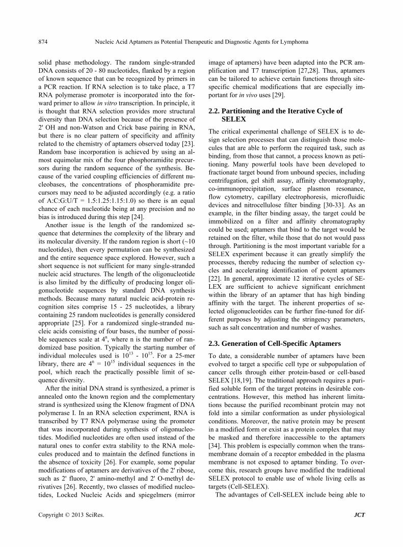
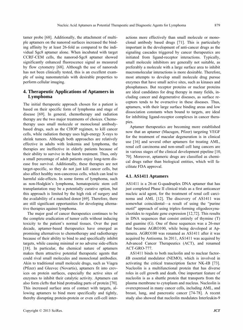
Because of their specificity and affinity to Ramos cells, TD05 aptamers have been widely reported for use as a diagnostic [58,59]. One significant step forward enabled by aptamers has been the development of a simple, sensi- tive dipstick test for lateral flow-based detection of Burkitt’s lymphoma cells, using an aptamer-nanoparticle strip biosensor (Figure 2) [60]. This approach uses tech- nology similar to that in commercially available preg- nancy test kits, and provides a litmus-test type of assay. In the original report of this approach, gold nanoparticle aggregates that were responsive to Ramos cells were designed in which the thiolated TD05 aptamers were immobilized on the gold nanoparticles, while another Ramos cell selective aptamer (TE02), which was identi- fied along with the TD05 aptamers in Cell-SELEX, was immobilized on the test zone of the strip [60]. The prin- ciple of the assay was that the sample solution or human blood sample would migrate by capillary action along the strip. In the presence of Ramos cells, the TD05 aptamers immobilized on the gold nanoparticles would interact with the cells to form the gold nanoparticles aptamer-cell complex and continued to migrate along the strip. The gold nanoparticle-aptamer-cell complexes were captured at the test zone by a second aptamer, TE02, immobilized on the strip. Because of the optical features of gold
Copyright © 2013 SciRes. JCT

Nucleic Acid Aptamers as Potential Therapeutic and Diagnostic Agents for Lymphoma 877
In the Presence of Ramos cell In the Absence of Ramos cell
Test ZoneTest Zone Control ZoneControl Zone
Sample Sample
Ramos Cell +Aptamer‐Au‐NP
Capture the Ramoscell-aptamer-Au-NP on the Test zone
Capture the excess of aptamer-Au-NP on the control zone
Capture the excess of aptamer-Au –NP on the control zone
Aptamer‐Au‐NP
Figure 2. A TD05 aptamer-nanoparticle strip biosensor for detecting a Burkitt’s lymphoma cell line (Ramos cells). A sample solution that contains Ramos cells is recognized by TD05 aptamer gold nanoparticles (TD05-Au-NP). The TD05 Au-NP-cell complexes are captured on the test zone by a second binding event between Ramos cells and immobilized Ramos cells that recognize aptamers (TE02). The accumulation of Au-NP results in a red band. The excess Au-NP continues to migrate and is captured on the control zone by a hybridization reaction between TD05-Au-NP and an oligonucleotide complementary to the TD05 aptamers, leading to a second red band. In the absence of Ramos cells, only the red band in the control zone is observed, showing that the assay is working properly. nanoparticles, the accumulation of gold nanoparticle- aptamer-cell complexes at the test zone produced a red line. Excessive amounts of gold nanoparticle-aptamer conjugates continued migrating to the control zone, in which the gold nanoparticle-aptamer complex was cap- tured by a DNA sequence complementary to the TD05 aptamers, thereby forming a second red line to show the biosensor was working properly. Whereas, if the sample did not contain Ramos cells, only one red line was ex- pected in the control zone and no red band would be seen in the test zone. Optimally, this biosensor could detecting a minimum of 4000 Ramos cells, thus providing a rapid, sensitive and economical tool for the detecting Burkitt’s lymphoma cells.
Alternatively, the TD05 aptamers were used as mo- lecular probes for real time in vivo tumor-target imaging inside live animals [58]. The TD05 aptamers were la- beled with fluorescent dyes, such as Cy5, and then in- jected into mice that were engrafted with Ramos cells. In vivo fluorescence imaging experiments showed that only the Cy-5-labeled TD05 aptamers accumulated in the tu- mor tissues, while non-specific control probes were rap- idly cleared from the body. Notably, the probe produced very high signal-to-background ratios during imaging and could distinguish Ramos cells from other circulating tumor cells, such as T-cell acute lymphoblastic leukemia (ALL) cells and CCRF-CEM in vivo. However, because nucleic acid aptamers have serum degradation issues in biological fluids, a number of chemical modifications have been introduced to various parts of aptamer struc-
tures to increase the feasibility of using aptamers in vivo (e.g. nuclease resistance and conformational stability). One notable modification for in vivo cancer imaging is the design of polyethylenimine (PEI)-aptamer probes [59]. When TD05 aptamers are immobilized onto PEI- polymers, the degradation rate of the aptamers is greatly reduced. A whole animal fluorescence imaging study demonstrated that the fluorescence signal from tumor tissues that were imaged by fluorescently labeled PEI/ TD05 aptamer nanoparticles remained elevated for five hours post-injection, while the signal when naked TD05 aptamers was used was only stayed for 45 minutes. This interesting strategy can significantly enhance the phar- macokinetics and half-life of aptamers, which is impor- tant for offering a feasible and versatile method to image tumors in vivo with high specificity and sensitivity.
3.3. Sgc8 Aptamers
Sgc8 aptamers are DNA aptamers that were generated through a cell-based selection strategy for the specific recognition of leukemia cells [61]. In the selection proc- ess, two tumor cell lines were selected as ligands. An ALL cell line, CCRF-CEM, was chosen as the target for selection, while a Ramos cell line was used as a counter selection to minimize the DNA sequences that could bind to common ligands on the surface of both hematopoietic cell lines [61]. The Sgc8 aptamers were one of the en- riched sequences that could specifically recognize cancer cells from T cell ALL patients and cultured CCRF-CEM
Copyright © 2013 SciRes. JCT
Nucleic Acid Aptamers as Potential Therapeutic and Diagnostic Agents for Lymphoma 878
cells (KD = 800 pM), but did not bind to normal CD3+ T cells or any other bone marrow cells [61]. Subsequent studies using affinity chromatography and mass spec-troscopy discovered that protein tyrosine kinase 7 (PTK7) is a molecular target of Sgc8 aptamers [62]. PTK7, also known as colon carcinoma kinase-4, is a transmembrane receptor that is expressed in normal hematopoietic cells, but its expression is elevated in all T-ALL cell lines [63]. Given the specificity and affinity of Sgc8 aptamers, sev- eral innovative technologies have been developed to ex- ploit the diagnostic potentials of this aptamer and are discussed below. Although these technologies used the Sgc8 aptamers to recognize T-cell ALL, they can be, in general, applied to lymphoma or blood circulating tumor cells.
The simplest practical way to visualize tumor in vivo relies on a strategy that uses reporter-bearing probes in which the cell-recognizing ligands are labeled with re- porter molecules, such as fluorescent dye, and then bind to target tumor cells. Accumulation of the reporter ligands around cells then leads to an increased signal as compared to the surrounding environment. However, because these molecules usually have a constant signal, the image contrast is critically hindered by high back- ground. To address this complication, an activable ap- tamer reporter probe based on a cell membrane pro- tein-triggered conformation change was designed [64]. The design of the activable aptamer reporter probe con- sists of three elements: 1) a tumor-targeting aptamer e.g. Sgc8 aptamers, 2) a poly T linker and 3) a short DNA sequence complementary to part of the aptamer that has a fluorophore and a quencher linked at either terminus [64]. In the absence of target proteins, the probe is in an inac- tive form in which the short DNA sequence is hybridized with the aptamer. This keeps the fluorophore and the quencher in close proximity, leading to quenched fluo- rescence. Conversely, when the target cancer cells are present, the conformation of the aptamer probe is spon- taneously reorganized on binding, causing the fluoro- phore to separate from quencher. Accordingly, a strong fluorescence signal is generated in response to the inter- action of the probe with the target cells. In vitro analysis and in vivo imaging of CCRF-CEM cells were performed by incorporating the Sgc8 aptamers into the probe. This demonstrated that the Sgc8 aptamer probe exhibited en- hanced background contrast and desirable specificity in differentiating CCRF-CEM tumor from other blood tu- mors and normal cells [64]. This sensitive technology is expected to facilitate an early detection of cancer cells and significantly shorten the diagnosis time.
Detecting cancer during its earliest stages or prior to metastatic relapses is difficult and sometimes requires multiple complicated diagnostics and lab tests to provide conclusive results. A common detection method involves
spotting out live tumor cells from body fluids, such as blood. However, the cancerous cells are usually present at very low concentration that is below the sensitivity limit of conventional methods. Therefore, developing a non-invasive, fast and inexpensive method for enriching and detecting cancerous cells is critically important for early cancer diagnosis. Recently, an aptamer-based mi- cofluidic device was reported that demonstrated an ex- ceptional ability to enrich for scarce cancer cells in a background of healthy cells on the basis of molecular interactions [65,66]. In this case, CCRF-CEM targeting Sgc8 aptamers were first biotinylated and covalently immobilized on a microfluidic channel that had surface- bound. The cell mixture was then pumped into the chan- nel [66]. Because the Sgc8 aptamers are specific to CCRF-CEM cells, target cells in the cell mixture were captured and enriched. These cells were subsequently visualized by optical microscopy to measure the cell- surface density. Generally, this device obtained 80% capture efficiency and 97% purity [66].
In a further study of this method, the same research group extended the design of the microfluidic device to allow simultaneous sorting, enrichment and detection of three different types of cancer cells from a complex sam- ple by using three leukemia- or lymphoma-specific ap- tamers (Sgc8 aptamers for CCRF-CEM cells, TD05 ap- tamers for Ramos cells and Sgd5 aptamers for Toledo cells [non-Hodgkin’s B cell lymphoma cell line]) [65]. These aptamers were immobilized on different regions within the microfluidic channel and were used to capture and enrich target cells through molecular interactions. This approach achieved a 135-fold enrichment of sorted cancer cells with ~96% purity [65]. Notably, this simple assay did not require any pre-treatment of cells and could be completed within minutes. However, alternative methods for detecting captured cells should be consid- ered because it is not practical to scan the entire micro- fludic device with a microscope.
Another approach could involve combining aptamers with nanotechnology. Nanotechnology involves the crea- tion and application of nanometer-scale materials that have a wide variety of elegantly patterned array and high-ordered structures. These nanomaterials perform diverse biological functions and their novel designs have inspired biomimetic strategies. In particular, nanomateri- als can provide a large surface area as multivalent ligand scaffolds that allow the incorporation of multiple recog- nition ligands, such as aptamers [67]. Au-Ag nanorods are one promising nanomaterial that has been used as a scaffold for multivalent binding by aptamers on the rod for use in detecting cancer cells. These nanorods have been fabricated with up to 80 fluorescently labeled Sgc8 aptamers through thiol linkages and displayed much stronger fluorescence signal than that of the original ap-
Copyright © 2013 SciRes. JCT
Nucleic Acid Aptamers as Potential Therapeutic and Diagnostic Agents for Lymphoma 879
tamer probe [68]. Additionally, the attachment of multi- ple aptamers on the nanorod surfaces increased the bind- ing affinity by at least 26-fold as compared to the indi- vidual Sgc8 aptamer alone. When incubated with target CCRF-CEM cells, the nanorod-Sgc8 aptamer showed significantly enhanced fluorescence signal as measured by flow cytometry [68]. Although the use of nanorods has not been clinically tested, this is an excellent exam- ple of using nanomaterials with desirable properties to perform cellular imaging.
4. Therapeutic Applications of Aptamers in Lymphoma
The initial therapeutic approach chosen for a patient is based on their specific form of lymphoma and stage of disease [69]. In general, chemotherapy and radiation therapy are the two major treatments of choices. Chemo- therapy uses small molecule or monoclonal antibody- based drugs, such as the CHOP regimen, to kill cancer cells, while radiation therapy uses high-energy X-rays to shrink tumors. Although both approaches are relatively effective in adults with leukemia and lymphoma, the therapies are ineffective in elderly patients because of their ability to survive in the harsh treatment. Thus, only a small percentage of adult patients enjoy long-term dis- ease free survival. Additionally, these therapies are not target-specific, so they do not just kill cancer cells, but also affect healthy non-cancerous cells, which can lead to harmful side-effects. In some forms of lymphoma, such as non-Hodgkin’s lymphoma, hematopoietic stem cell transplantation may be a potentially curative option, but this approach is limited by the high risk of surgery and the availability of a matched donor [69]. Therefore, there are still significant opportunities for developing alterna- tive therapies against lymphoma.
The major goal of cancer therapeutics continues to be the complete eradication of tumor cells without inducing toxicity to the patient’s normal tissues. Over the past decade, aptamer-based therapeutics have emerged as promising alternatives to chemotherapy and radiotherapy because of their ability to bind to and specifically inhibit targets, while causing minimal or no adverse side-effects [18]. In particular, the chemical nature of aptamers makes them attractive potential therapeutic agents that could rival small molecules and monoclonal antibodies. Akin to traditional small molecule drugs, such as Viagra (Pfizer) and Gleevec (Novartis), aptamers fit into crev- ices on protein surfaces, especially the active sites of enzymes to inhibit their catalytic activity. Aptamers can also form clefts that bind protruding parts of protein [70]. This increased surface area of contact with targets, al- lowing aptamers to bind more specifically and tightly, thereby disrupting protein-protein or even cell-cell inter-
actions more effectively than small molecule or mono- clonal antibody based drugs [71]. This is particularly important in the development of anti-cancer drugs as the signaling cascades triggered by cancer therapeutics are initiated from ligand-receptor interactions. Typically, small molecule inhibitors are generally not suitable, as preferably a molecule with a large surface area to inhibit macromolecular interactions is more desirable. Therefore, most attempts to develop small molecule drug pursue enzymes that have small active sites, such as kinases and phosphatases. But receptor proteins or nuclear proteins are ideal candidates for drug therapy in many fields, in- cluding cancer and degenerative diseases, as surface re- ceptors tends to be overactive in these diseases. Thus, aptamers, with their large surface binding areas and low dissociation constants when bound to targets, are ideal for inhibiting ligand-receptor complexes in cancer thera- pies.
Aptamer therapeutics are becoming more established now that an aptamer (Macugen, Pfizer) targeting VEGF for the treatment of macular degeneration is in clinical use [16] and several other aptamers for treating AML, renal cell carcinoma and non-small cell lung cancers are in various stages of the clinical development pipeline [10, 70]. Moreover, aptameric drugs are classified as chemi- cal drugs rather than biological entities, which will fa- cilitate FDA approval.
4.1. AS1411 Aptamers
AS1411 is a 26-nt G-quadruplex DNA aptamer that has just completed Phase II clinical trials as a first anticancer nucleic acid agent, for the treatment of renal cell carci- noma and AML [12]. The discovery of AS1411 was somewhat coincidental—a result of using the “purine motif” approach of using triplex-forming oligodeoxynu- cleotides to regulate gene expression [12,72]. This results in DNA sequences that consist entirely of thymine (T) and guanine (G). One of these sequences was a 26-mer that became AGRO100, while being developed at Ap- tamera. AGRO100 was renamed as AS1411 after it was acquired by Antisoma. In 2011, AS1411 was acquired by Advanced Cancer Therapeutics (ACT), and renamed ACT-GRO-777.
AS1411 binds to both nucleolin and to nuclear factor- kB essential modulator (NEMO), which is involved in activating the critical transcription factor NK-kB [73]. Nucleolin is a multifunctional protein that has diverse roles in cell growth and death. One important feature of nucleolin is as a shuttle protein that transports from the plasma membrane to cytoplasm and nucleus. Nucleolin is overexpressed in many cancer cells, including AML, and breast, lung, and pancreatic cancer [74-78]. A recent study also showed that nucleolin modulates Interleukin-9
Copyright © 2013 SciRes. JCT
Nucleic Acid Aptamers as Potential Therapeutic and Diagnostic Agents for Lymphoma 880
(Il9) expression in T-cell lymphoma cells [79]. Another major role of nucleolin is to bind to an AU-rich element in the 3'UTR of Bcl2 mRNA [80]. This binding protects the anti-apoptotic Bcl-2 from degradation, resulting in stabilization of Bcl2 mRNA, which subsequently pro- motes the cancer cells to overproduce Bcl2 and avoid apoptosis [80]. Early preclinical studies demonstrated that micromolar concentrations of AS1411 could inhibit proliferation of a wide variety of tumor cell lines, and suppressed tumor growth in a xenograft animal model [78]. Phase I clinical trials (involving 30 patients) were completed in 2006 and indicated that AS1411 was safe and well-tolerated [72]. Phase II trials for AML are showing positive interim results (ClinicalTrials.gov Iden- tifier: NCT00512083 and NCT01034410). Recent re- search has explored the use of AS1411 as a carrier for the delivery of cancer drugs specifically into cancer cells by nucleolin mediated internalization [81].
4.2. NOX-A12 Spiegelmers
NOX-A12 is a 45-nucleotide long spiegelmer aptamer that was developed by a proprietary spiegelmer technol- ogy® in Noxxon Pharma (Germany). Spiegelmers are mirror image (L-steroisomer) RNA aptamers. An impor- tant feature of spiegelmers is that they have tremendous stability in the body because nucleases recognize natural D-nucleic acids, but not L-nucleic acids (Spiegelmers) [82]. Additionally, spiegelmers possess all other charac- teristics of canonical aptamers, such as high binding af- finity and selectivity to the target, passive immunogenic- ity and ease of manufacture [82].
The identification of NOX-A12 spiegelmers combined screening of large combinatorial RNA libraries with a chemical mirror technology in which NOX-A12 was evolved from the natural D-RNA library by using a mir- ror image target (D-amino acid), and then was synthe- sized as L-RNA to bind the natural target (L-amino acid) [82,83]. NOX-A12 spiegelmers specifically antagonize CXC chemokine ligand 12/stromal cell derived factor-1 (CXCL12/SDF-1), which is a key regulatory chemokine for migration of leukemia stem cells to the bone marrow [84]. CXCL12 also plays an important role in tumor growth and metastasis by binding with high affinity to two chemokine receptors, CXCR5 and CXCR7 [84]. Both of these receptors appear to contribute to metastasis of cancer cells to regions with elevated CXCL12 expres- sion, such as lymph nodes and bone marrow. Moreover, CXCL12 signaling has been shown to play a major role in the pathophysiology of chronic lymphocytic leukemia (CLL), especially in the interaction of leukemic cells with the tissue microenvironment [85]. Thus, inhibition of CXCL12 binding to its receptors by NOX-A12 spie- gelmers has the potential to interfere with the CXCL12
anchor and prompt leukemia stems cells to re-enter the cell cycle and become available for chemotherapeutic attack.
NOX-A12 spiegelmers have also shown promising pre-clinical anti-tumor activity in various solid and he- matological tumor models. A Phase 1 clinical trial in- volving 48 healthy subjects was launched in 2009 and showed that NOX-A12 spiegelmers were safe and well- tolerated. Notably, the pharmacokinetic profile showed that NOX-A12 spiegelmers have a very long half-life of 37 hours. Recently, two Phase IIa clinical studies of NOX-A12 have begun for hematological oncology indi- cations, including non-Hodgkin’s lymphoma, chronic lymphocytic leukemia and multiple myeloma (Clinical- Trials.gov Identifier: NCT01486797 and NCT-01486797). This study will involve 33 relapsed CLL patients who will receive NOX-A12 spiegelmers in combination with a background therapy of chemotherapeutic agents. In-terim results are expected to be available in early 2013.
5. Aptamer “Smart Bomb” Mediates Delivery of Drugs to Cancer Cells
The emphasis on cancer therapeutics has now shifted to the development of tumor-specific, targeted therapies in the hope that only the cancer cells will be targeted and killed by the drugs, while non-cancerous normal cells will not be affected. Antibody drug conjugates, which couple the cell-recognizing advantage of antibodies with the cytotoxic potential of chemotherapy, herald the promise of targeted cancer therapy, and several antibody drug conjugates are currently undergoing clinical evalua- tion [86]. Indeed, the development of antibody drug con- jugates are one of the major interests in pharmaceutical and biotechnology companies nowadays [87]. Currently, only two antibody-drug conjugates have been approved by FDA for the treatment of hematological cancers, and only one of these is still in clinical use. Gemtuzumab ozogamicin (Mylotag, Pfizer) was the first FDA-ap- proved antibody-drug conjugates designed for patients with relapsed AML, but was discontinued because of sig- nificant side effects in a post-approval trial [87,88]. Gemtuzumab targets CD33 antigen expressed on both leukemic blast cells and normal hematopoietic cells [88]. Brentuximab vedotin (Adcetric, Seattle Genetics Inc.) was the second FDA approved antibody drug-conjugates for the treatment of Hodgkin’s Lymphoma [89]. Unlike gemtuzumab ozogamicin, which also targets normal cells, Brentuximab vedotin only targets CD30, which is an established marker for classic Hodgkin’s lymphoma, anaplastic large cell lymphoma and embryonal carcino- mas [90-92]. In brentuximab vedotin, the antibody is conjugated to monomethyl auristatin E, which inhibits cell division by preventing polymerization of tubulin [90,
Copyright © 2013 SciRes. JCT
Nucleic Acid Aptamers as Potential Therapeutic and Diagnostic Agents for Lymphoma 881
93]. Despite the seemingly promising clinical results on the
use of antibodies for targeted cancer therapy, these drugs face many challenges that limit their successful clinical translation. Some of these challenges might be due to the inherent properties of monoclonal antibodies as specific therapeutic deliverables [87]. For example, in gemtuzu- mab ozogamicin, only 50% of the anti-CD33 gemtuzu- mab antibody was attached to the chemotherapeutics (4 - 6 drug molecules per antibody), while the remaining an-tibody was unconjugated, suggesting that the potency of the antibody-drug conjugates was undermined [87,94]. Arguably, aptamer technology is an attractive alternative for use as drug carriers during targeted drug delivery. Aptamers have several advantages over antibodies used in targeted delivery, particularly for cancer therapy [15, 18]. First, manufacture of aptamers relies on chemical synthesis and therefore is much easier to scale-up with low batch-to-batch variation. Second, aptamers are much smaller than antibodies, which can lead to better tissue penetration and cell internalization. Third, using aptam- ers as anti-receptors will yield superior specificity and the lowest antibody-inducing activity as compared to protein anti-receptors, thus providing a means for re- peated administration and treatment of chronic diseases, such as cancers. Last, attachment of chemotherapeutic drugs, imaging agents or any functional group can be readily computed during aptamer synthesis. Many ap- tamers have been identified for hematological cells and examples of aptamer-mediated delivery are discussed below.
5.1. TD05 Aptamer-Mediated Delivery of a Photosensitizer
The TD05 aptamers, which target the immunoglobulin heavy mu chain in a Burkitt’s lymphoma cell line, were engineered with a photosensitizer for selective photody- namic therapy of targeted tumor cells [95]. Photody- namic therapy exploits the ability of nontoxic light-sensi- tive compounds called photosensitizers to produce cyto- toxic reactive oxygen species when illuminated and it is widely recognized as a minimally toxic and minimally invasive approach [96]. Recently, the photodynamic therapy-based approach has been used clinically to treat several medical conditions, but it is limited to locally exposed diseases, such as skin cancers and severe acne. The largest challenge of using this technology for dis- eases inside the body is the effective localization of the cytotoxic photosensitizers at the diseased cells. Because the TD05 aptamers can specifically recognize Ramos cells, an aptamer-photosensitizer conjugate was engi- neered. This conjugate contained the cytotoxic porphy- rin-based photosensitizer reagent Chlorin 6 [81,95]. In vitro studies revealed that these aptamer-photosensitizer
conjugates were highly selective to Ramos cells and ef- fectively generated free-radicals that triggered Ramos cell upon illumination. This aptamer-photosensitizer con- jugate was highly selective to Ramos cells, but did not affect acute lymphoblastic leukemia or myeloid leukemia cell lines [95]. To increase their binding affinity to the target and their half-life in the body, the TD05 aptamers were designed to attach a lipid tail at the end of the ap- tamers to form a self-assembled aptamer-micelle nanos- tructure [97]. Alternatively, a construct containing multi- ple TD05 aptamers also successfully improved the speci- ficity and binding kinetics of the aptamers.
5.2. AS1411 Nucleolin Aptamer-Mediated Drug Delivery
The aptamer-mediated delivery approach is not limited to delivery of therapeutic cargos to cell cytoplasm, but can also extend to nuclear delivery. Nucleolin acts as a shut- tle to transfer molecules between the cell surface and the nucleus [98], and several hematopoietic cancer cell lines, including AML and chronic lymphocytic leukemia/small lymphocytic lymphoma, expressed higher levels of nu- cleolin than normal cells [74,78]. Moreover, molecules that bind to nucleolin are then internalized by the cells. These data suggested nucleolin as an excellent candidate for targeted delivery of anti-cancer agents as a means for internalization of nucleolin-binding AS1411 aptamers.
Shieh et al. reported a novel strategy of using AS1411 aptamers as drug carriers to target cancer cells for photo- dynamic therapy [81]. The AS1411 aptamers were com- plexed with 6 photodynamic molecules, TMPyP4, to form multivalent AS1411 apt-TMPyP4 conjugates through electrostatic interactions. This approach is similar to cocktail therapy, in which a mixture of drugs is used to produce a synergistic effect. The AS1411 apt-TMPyP4 conjugates accumulated in nucleolin-expressing cells lines, likely because of nucleolin-mediated internaliza- tion via AS1411, and damaged these cells upon exposure of light [81]. Using a similar strategy, Aravind et al. re- ported the use of lipid-polymer hybrid nanoparticles loaded with chemotherapeutic agents and decorated with AS1411 aptamers for targeted drug delivery [99]. These lipid-polymer hybrid nanoparticles were composed of 6 elements 1) a hydrophobic polymeric core made of poly D, L lactic-co-glycolic acid (PLGA), 2) a chemothera- peutic drug, paclitaxel, 3) a fluorescent dye, 4) a lipid layer made of lecithin, 5) a PEGylated phospholipid, DSPE-PEG2000-COOH and 6) AS1411 aptamers on the surface [99]. Physical analyses confirmed that the nano- particles were spherical and ranged in a size from 60 to 110 nm. Subsequent drug-loading studies showed that this nanoparticle had an enhanced cytotoxic effect, higher encapsulation efficiency and superior sustained drug re- lease than corresponding non-target nanoparticles [99].
Copyright © 2013 SciRes. JCT
Nucleic Acid Aptamers as Potential Therapeutic and Diagnostic Agents for Lymphoma
Copyright © 2013 SciRes. JCT
882
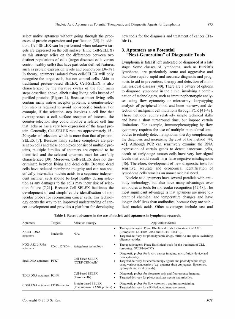
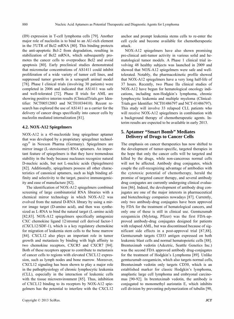
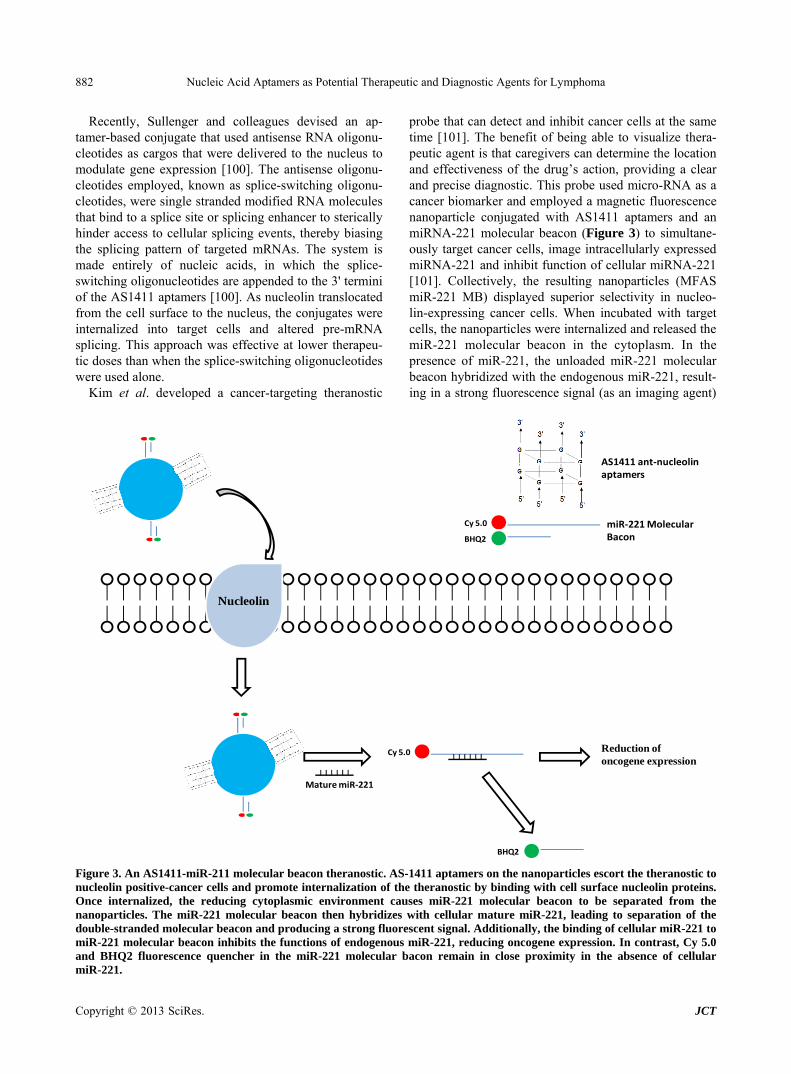
probe that can detect and inhibit cancer cells at the same time [101]. The benefit of being able to visualize thera- peutic agent is that caregivers can determine the location and effectiveness of the drug’s action, providing a clear and precise diagnostic. This probe used micro-RNA as a cancer biomarker and employed a magnetic fluorescence nanoparticle conjugated with AS1411 aptamers and an miRNA-221 molecular beacon (Figure 3) to simultane- ously target cancer cells, image intracellularly expressed miRNA-221 and inhibit function of cellular miRNA-221 [101]. Collectively, the resulting nanoparticles (MFAS miR-221 MB) displayed superior selectivity in nucleo- lin-expressing cancer cells. When incubated with target cells, the nanoparticles were internalized and released the miR-221 molecular beacon in the cytoplasm. In the presence of miR-221, the unloaded miR-221 molecular beacon hybridized with the endogenous miR-221, result- ing in a strong fluorescence signal (as an imaging agent)
Recently, Sullenger and colleagues devised an ap- tamer-based conjugate that used antisense RNA oligonu- cleotides as cargos that were delivered to the nucleus to modulate gene expression [100]. The antisense oligonu- cleotides employed, known as splice-switching oligonu- cleotides, were single stranded modified RNA molecules that bind to a splice site or splicing enhancer to sterically hinder access to cellular splicing events, thereby biasing the splicing pattern of targeted mRNAs. The system is made entirely of nucleic acids, in which the splice- switching oligonucleotides are appended to the 3' termini of the AS1411 aptamers [100]. As nucleolin translocated from the cell surface to the nucleus, the conjugates were internalized into target cells and altered pre-mRNA splicing. This approach was effective at lower therapeu- tic doses than when the splice-switching oligonucleotides were used alone.
Kim et al. developed a cancer-targeting theranostic
Nucleolin
Cy 5.0
BHQ2
AS1411 ant‐nucleolinaptamers
miR‐221 Molecular Bacon
Cy 5.0
BHQ2
Mature miR‐221
Reduction of oncogene expression
Figure 3. An AS1411-miR-211 molecular beacon theranostic. AS-1411 aptamers on the nanoparticles escort the theranostic to nucleolin positive-cancer cells and promote internalization of the theranostic by binding with cell surface nucleolin proteins. Once internalized, the reducing cytoplasmic environment causes miR-221 molecular beacon to be separated from the nanoparticles. The miR-221 molecular beacon then hybridizes with cellular mature miR-221, leading to separation of the double-stranded molecular beacon and producing a strong fluorescent signal. Additionally, the binding of cellular miR-221 to miR-221 molecular beacon inhibits the functions of endogenous miR-221, reducing oncogene expression. In contrast, Cy 5.0 and BHQ2 fluorescence quencher in the miR-221 molecular bacon remain in close proximity in the absence of cellular miR-221.
Nucleic Acid Aptamers as Potential Therapeutic and Diagnostic Agents for Lymphoma 883
and simultaneously miRNA-221 inhibition (as a thera- peutic agent), thereby reducing expression of oncogenes [101].
5.3. Sgc8 Aptamer-Mediated Drug Delivery
In addition to their ability to differentiate PTK7+ T-cell ALL cells from healthy cells, the Sgc8 aptamers can also be engineered as targeting ligands, thereby enabling the selective delivery of therapeutic cargos to target cells. One example is to use the cell targeting Sgc8 aptamers for delivery of anti-tumor chemotherapeutic agents, such as Doxorubicin (Dox) and Daunorubicin, with the aim of reducing off-target toxicity and lowering the concentra- tions of drug administered, thus reducing potential side effects [102,103]. Sgc8 aptamer-Dox conjugates were therefore designed. In one study, Dox was covalently cross-linked with the 5'-SH group of the Sgc8 aptamers through an acid-liable hydrozone linker [102]. When the Sgc8-Dox conjugate was incubated with a cell mixture, the conjugates recognized target CCRF-CEM cells and efficiently mediated the internalization of Dox through PTK7 receptors. Once inside the cells, Dox was subse- quently released into the cytoplasm from the conjugates because of the cleavage of the linker in the acidic en- dosomal environment. Although the Sgc8 aptamers were attached to Dox, molecular analyses demonstrated that Sgc8-Dox conjugates displayed many of the intrinsic properties of the Sgc8 aptamers, including strong binding strength and high specificity. Moreover, cell viability tests showed that the Sgc8-Dox conjugates were as po- tent as unconjugated Dox, suggesting that the function of Dox was not affected by aptamer conjugation. Impor- tantly, the Sgc8-Dox conjugates were not internalized by non-target cells; therefore their cellular toxicity was minimal. When combined with different aptamers that specifically target a wide array of cancer cells, this ap- tamer-drug assembly design lays the foundation for con- tinued development of targeted delivery of a variety of cancers [102].
Another common strategy for drug delivery uses lipo- somes. Because the cell plasma membrane is made of a dynamic lipid bi-layer, the liposome nanostructure can increase cell permeability and efficiency of drug delivery, akin to the natural process of cell-cell adhesion. One ad- vantage of a liposome-based approach is that the lipo- somes can be purposefully decorated. To deliver drugs to specific cells, a cell targeting aptamer can be anchored on the liposome to make a cell permeable Sgc8-PEG- liposome nanoparticles [104]. A design in which ~250 copies of Sgc8 aptamers were tethered to a liposome, facilitated multiple aptamer-receptor interactions, pro- vided stronger binding to the target cells and enhanced cellular internalization across the cell membrane [104]. In an initial study, fluorescein-isothiocyanate-dextran
was selected as a model molecule and was engineered into a Sgc8 aptamer-coated liposome. Aptamer-conju- gated liposomes involving fluorescein-isothiocyanate- dextran were selectively immobilized on the surface membrane of CCRF-CEM cells, but not on non-target NB4 cells [104]. Several studies using specialized lipids, such as diacyl phospholipid and perfluoropentane, to form the liposome nanostructures have also been re- ported [105].
Hydrogels are water-retainable materials generated from cross-linked polymers, and offer another possibility for aptamer-mediated delivery [106]. Because of their low toxicity and biocompatibility, hydrogel systems have long been recognized as valuable carriers for drug trans- port. In particular, stimuli-responsive hydrogels that are sensitive to a variety of chemical and physical alterna- tions fulfill many of the critical criteria of targeted drug delivery [106]. For example, an Sgc8 aptamer-incor- porated photoresponsive hydrogel system was developed as the recognition unit. The sgc8 aptamers were conju- gated to a DNA polyacrylamide chain in the hydrogel systems, which encapsulated Dox and the release of Dox from this system was triggered by exposure to near-in- frared light. Flow cytometry indicated that the hy- drogels bound to CCRF-CEM cells through the Sgc8 aptamers, then released Dox to kill the cancer cells after exposure to laser light. Collectively, treatment with the Dox-sgc8 aptamer-nanogel system increased death of CCRF-CEM cells from 3% ± 2% to 67% ± 5%, while only 3% ± 2% and 10% ± 3% cell death was observed after incubation with Dox-loaded nanogel conjugated to a DNA library sequence [107].
Finally, akin to liposomes and hydrogels, viral capsids are another nano-material that can serve as a core scaf- fold for targeted delivery. The protein shell of bacterio- phage MS2 consists of 180 amino acids that are arranged in a 27-nm hollow spherical structure. To functionalize the outer surface of the capsid with targeting capabilities, Sgc8 aptamers were chemically installed through a chemoselective oxidative coupling strategy [108]. Typi- cally, 20 - 40 copies of Sgc8 aptamers were attached on the surface, resulting in a multivalent aptamer-capsid system for targeted delivery. In proof-of-concept studies, acid-liable prodrugs and photodynamic therapy agents have been assembled in the Sgc8 aptamer-conjugated viral capsid core. These agents were able to selectively kill the target cells, while capsids modified with a control DNA strand did not have any cytotoxic effect [109]. This system can be extended to diagnostic imaging by adding radionuclides and contrast agents [109].
5.4. Anti-CD30 Aptamer-Mediated siRNA Delivery
CD30 aptamers have been described previously as diag-
Copyright © 2013 SciRes. JCT
Nucleic Acid Aptamers as Potential Therapeutic and Diagnostic Agents for Lymphoma 884
nostic probes for detection of CD30-expressing lym- phoma cells. Because of their selectivity and specificity to CD30 receptors, these aptamers were also used as agents to deliver siRNAs to Anaplastic lymphoma kinase (ALK) positive ALCL, which is an aggressive T-cell lymphoma that overexpresses ALK oncogenes and CD30 surface proteins. Through electrostatic interactions, ALK siRNAs and CD30 RNA aptamers were incorporated onto the cationic PEI-citrate carriers to form nanoparti- cles [110]. Notably, the assembled aptamer-siRNA- nanocomplexes were ~140 nm in diameter such that they are not too small to be rapidly excreted and not too large to be unavailable for cellular uptake. In fact, the nano- particles were stable for more than one day in culture medium and the CD30 aptamers directed the cellular uptake of the siRNA-loaded nanoparticles. Inside the cell, the ALK siRNAs were released from the nanoparticles to specifically silence ALK gene expression, resulting in growth arrest and apoptosis in CD30-expressing ALCL cells [110].
6. Conclusions and Perspectives
Aptamers are single stranded non-coding nucleic acids that can be evolved in vitro to carry out a specific func- tion by forming a functional three dimensional structure to their targets. Since the first demonstration of SELEX over two decades ago, the use of aptamer technology has shown promise in molecular diagnostics, medical im- aging, targeted delivery and pharmaceuticals [111]. Ap- tamers inhibit enzyme in an extraordinarily strong and specific manner that is comparable and often superior to therapeutic antibodies and small molecules, and yet avoid the toxicity and immunogenicity concerns of these traditional agents [70]. With current advances in cell- based SELEX methodology and versatile chemical modi- fication techniques, aptamers now have remarkable po- tential to be used as novel delivery vehicles that target a particular cell population, thereby providing enhanced therapeutic efficacy and reduced cellular toxicity [21]. Furthermore, several examples discussed in this review (e.g. Sgc8 aptamer hydrogels) provide complementary approaches for combining the strength of aptamer tech- nology with nanotechnology, offering a versatile plat- form for disease treatment and diagnosis. However, de- spite the advances described above, only a few examples have reached extensive clinical development for lym- phoma therapy [70,111-115]. Further research in suc- cessful translation of aptamer technology should be aimed at the following three main challenges.
First, although the technology for nucleic acid synthe-sis has improved greatly over the last decade, the cost of industrial-scale production of long, high quality cGMP- grade nucleic acids remains very high and significantly limits clinical development and pre-clinical testing in
vivo [7,37,116]. Aptamer sequences are usually more than 30 nucleotides long and are extensively modified to protect against nuclease and abrogate type 1 interferon activation, further increasing the cost and difficulties of standard chemical synthesis. Potential solutions to this problem include truncating aptamers to the minimal functional sequence or conjugating multiple aptamers on a polymer [117].
Second, for most uses of aptamer-mediated delivery to date, the aptamer-drug conjugates appear to internalize into the cell through endocytosis pathway [118-120]. Upon internalization, the conjugates encapsulate in en- dosomal vesicles, then fuse with early endosomes and finally enter into lysosomes, which are the last compart- ment of the endocytic pathway [121]. Lysosomes are highly acidic (pH ~4.5) and contain various nucleases, which could break down the cargo rapidly [37]. There- fore, efficient escape of therapeutic cargos from the en- dosome is required and is the most critical challenge in delivery. In this regard, various nanocarriers have been developed to improve the endosomal escape of cargos. Some amine-based materials that have strong buffering capacities, such as PEI, fusogenic lipids, peptides and β-amino ester, have been conjugated to siRNA therapeu- tics to facilitate endosome escape through the proton sponge effect, which induces rupture of the endosome to release siRNA cargos [122-125]. However, none of these reagents has yet been evaluated in the clinic as a way to promote endosome escape.
Third, the relative paucity of known internalization cell surface receptors limits the wide-applicability of this technique. It is possible that aptamers selected from Cell-SELEX only bind to the cell surface, but do not ef- ficiently internalize into the cytoplasm. In this case, therapeutic cargos escorted by aptamers cannot reach their site of action. Modified cell-internalization SELEX has recently been proposed by Giangrande and col- leagues to isolate aptamers that have high affinity and internalization capability. This approach would have a clear advantage compared to selections performed using canonical Cell-SELEX and purified recombinant proteins [126]. In-depth knowledge of cell-surface protein trans- port and trafficking will also help in identifying new tar- gets for aptamer-mediated delivery.
Continued effort in the development of SELEX tech- nology will enable aptamer-based systems to be devel- oped into their own niches for the treatment and diagno- sis of various diseases. In particular, nucleic acids have intrinsic features that can serve as building blocks for bottom-up fabrication of a drug conjugate architecture [127-129]. Their phenomenal diversity in function and versatility in structure make aptamers particularly attrac- tive for many biological applications, and the use of ap- tamers to deliver cancer treatments, including for lym-
Copyright © 2013 SciRes. JCT
Nucleic Acid Aptamers as Potential Therapeutic and Diagnostic Agents for Lymphoma 885
phoma, is a rapidly growing technique. Aptamers and other therapeutics cargos, including chemotherapeutics and RNA drugs, can assemble into one nanovector that has polyvalent functionalities. Using a similar rationale, we anticipate that novel aptamer-based nanoparticles, such as aptamer-aptamer chimeras or even polyvalent aptamer-siRNA-microRNA nanoparticles, will likely be developed soon to further improve drug delivery and imaging systems. In next few years, aptamer technology is sure to play a critical role in accelerating the transla- tion and development of lymphoma diagnosis and treat- ment.
7. Acknowledgements
We thank Keely Walker for reading this manuscript. This work was funded by NIH grants AI42552, HL07470 awarded to JJR and NCI. CA151648 awarded to Peixuan Guo (University of Kentucky) and subcontracted to JJR.
REFERENCES [1] N. Bakshi and I. Maghfoor, “The Current Lymphoma
Classification: New Concepts and Practical Applications Triumphs and Woes,” Annals of Saudi Medicine, Vol. 32. No. 3, 2012, pp. 296-305.
[2] J. W. Sweetenham, “Lymphoblastic Lymphoma in Adults,” Current Hematologic Malignancy Reports, Vol. 1. No. 4, 2006, pp. 241-247. doi:10.1007/s11899-006-0005-8
[3] S. Ogino, C. S. Fuchs and E. Giovannucci, “How Many Molecular Subtypes? Implications of the Unique Tumor Principle in Personalized Medicine,” Expert Review of Molecular Diagnostics, Vol. 12. No. 6, 2012, pp. 621- 628. doi:10.1586/erm.12.46
[4] K. R. Shankland, J. O. Armitage and B. W. Hancock, “Non-Hodgkin Lymphoma,” Lancet, Vol. 380. No. 9844, 2012, pp. 848-857. doi:10.1016/S0140-6736(12)60605-9
[5] R. Kuppers, “New Insights in the Biology of Hodgkin Lymphoma,” Hematology/The Education Program of the American Society of Hematology American Society of He- matology Education Program, Vol. 2012, 2012, pp. 328- 334.
[6] R. Kuppers, A. Engert and M. L. Hansmann, “Hodgkin Lymphoma,” The Journal of Clinical Investigation, Vol. 122. No. 10, 2012, pp. 3439-3447. doi:10.1172/JCI61245
[7] J. Zhou and J. J. Rossi, “Aptamer-Targeted Cell-Specific RNA Interference,” Silence, Vol. 1, No. 1, 2010, p. 4. doi:10.1186/1758-907X-1-4
[8] J. Zhou and J. J. Rossi, “The Therapeutic Potential of Cell-Internalizing Aptamers,” Current topics in Medicinal Chemistry, Vol. 9. No. 12, 2009, pp. 1144-1157. doi:10.2174/156802609789630893
[9] J. Zhou and J. J. Rossi, “Bivalent Aptamers Deliver the Punch,” Chemistry & Biology, Vol. 15. No. 7, 2008, pp. 644-645. doi:10.1016/j.chembiol.2008.07.004
[10] X. Ni, M. Castanares, A. Mukherjee and S. E. Lupold, “Nucleic Acid Aptamers: Clinical Applications and Prom-
ising New Horizons,” Current Medicinal Chemistry, Vol. 18, No. 27, 2011, pp. 4206-4214. doi:10.2174/092986711797189600
[11] A. D. Ellington and J. W. Szostak, “In Vitro Selection of RNA Molecules that Bind Specific Ligands,” Nature, Vol. 346, No. 6287, 1990, pp. 818-822. doi:10.1038/346818a0
[12] W. O. Tucker, K. T. Shum and J. A. Tanner, “G-Quadru- plex DNA Aptamers and Their Ligands: Structure, Func- tion and Application,” Current Pharmaceutical Design, Vol. 18, No. 14, 2012, pp. 2014-2026. doi:10.2174/138161212799958477
[13] J. Liu, M. You, Y. Pu, H. Liu, M. Ye and W. Tan, “Re- cent Developments in Protein and Cell-Targeted Aptamer Selection and Applications,” Current Medicinal Chemis- try, Vol. 18, No. 27, 2011, pp. 4117-4125. doi:10.2174/092986711797189619
[14] Y. Nakamura, A. Ishiguro and S. Miyakawa, “RNA Plas- ticity and Selectivity Applicable to Therapeutics and Novel Biosensor Development,” Genes to Cells: Devoted to Molecular & Cellular Mechanisms, Vol. 17, No. 5, 2012, pp. 344-364. doi:10.1111/j.1365-2443.2012.01596.x
[15] S. D. Jayasena, “Aptamers: An Emerging Class of Mole- cules that Rival Antibodies in Diagnostics,” Clinical Che- mistry, Vol. 45, No. 9, 1999, pp. 1628-1650.
[16] E. W. Ng, D. T. Shima, P. Calias, E. T. Cunningham, Jr., D. R. Guyer and A. P. Adamis, “Pegaptanib, a Targeted anti-VEGF Aptamer for Ocular Vascular Disease,” Na-ture Reviews Drug Discovery, Vol. 5, No. 2, 2006, pp. 123-132. doi:10.1038/nrd1955
[17] E. W. Ng and A. P. Adamis, “Anti-VEGF Aptamer (Pegap- tanib) Therapy for Ocular Vascular Diseases,” Annals of the New York Academy of Sciences, Vol. 1082, 2006, pp. 151-171. doi:10.1196/annals.1348.062
[18] G. Zhu, M. Ye, M. J. Donovan, E. Song, Z. Zhao and W. Tan, “Nucleic Acid Aptamers: An Emerging Frontier in Cancer Therapy,” Chemical Communications (Camb), Vol. 48, No. 85, 2012, pp. 10472-10480. doi:10.1039/c2cc35042d
[19] Y. Zhang, H. Hong and W. Cai, “Tumor-Targeted Drug Delivery with Aptamers,” Current Medicinal Chemistry, Vol. 18, No. 27, 2011, pp. 4185-4194. doi:10.2174/092986711797189547
[20] A. S. Barbas, J. Mi, B. M. Clary and R. R. White, “Ap- tamer Applications for Targeted Cancer Therapy,” Future Oncology, Vol. 6, No. 7, 2010, pp. 1117-1126. doi:10.2217/fon.10.67
[21] J. Zhou and J. J. Rossi, “Cell-Specific Aptamer-Mediated Targeted Drug Delivery,” Oligonucleotides, Vol. 21, No. 1, 2011, pp. 1-10. doi:10.1089/oli.2010.0264
[22] C. Tuerk and L. Gold, “Systematic Evolution of Ligands by Exponential Enrichment: RNA Ligands to Bacterio- phage T4 DNA Polymerase,” Science, Vol. 249, No. 4968, 1990, pp. 505-510. doi:10.1126/science.2200121
[23] P. Dua, S. Kim and D. K. Lee, “Nucleic Acid Aptamers Targeting Cell-Surface Proteins,” Methods, Vol. 54, No. 2, 2011, pp. 215-225. doi:10.1016/j.ymeth.2011.02.002
[24] S. P. Ho, D. H. Britton, B. A. Stone, D. L. Behrens, L. M. Leffet, F. W. Hobbs, et al., “Potent Antisense Oligonu-
Copyright © 2013 SciRes. JCT
Nucleic Acid Aptamers as Potential Therapeutic and Diagnostic Agents for Lymphoma 886
cleotides to the Human Multidrug Resistance-1 mRNA Are Rationally Selected by Mapping RNA-Accessible Sites with Oligonucleotide Libraries,” Nucleic Acids Re- search, Vol. 24. No. 10, 1996, pp. 1901-1907. doi:10.1093/nar/24.10.1901
[25] R. V. Talanian, C. J. McKnight, R. Rutkowski and P. S. Kim, “Minimum Length of a Sequence-Specific DNA Binding Peptide,” Biochemistry, Vol. 31, No. 30, 1992, pp. 6871-6875. doi:10.1021/bi00145a002
[26] A. D. Keefe and S. T. Cload, “SELEX with Modified Nucleotides,” Current Opinion in Chemical Biology, Vol. 12, No. 4, 2008, pp. 448-456. doi:10.1016/j.cbpa.2008.06.028
[27] C. Forster, M. Zydek, M. Rothkegel, Z. Wu, C. Gallin, R. Gessner, et al., “Properties of an LNA-Modified Ricin RNA Aptamer,” Biochemical and Biophysical Research Communications, Vol. 419. No. 1, 2012, pp. 60-65. doi:10.1016/j.bbrc.2012.01.127
[28] S. Hoffmann, J. Hoos, S. Klussmann and S. Vonhoff, “RNA Aptamers and Spiegelmers: Synthesis, Purification, and Post-Synthetic PEG Conjugation,” In: S. L. Beaucage, et al., Current Protocols in Nucleic Acid Chemistry, 2011, pp. 41-30.
[29] L. H. Lauridsen, J. A. Rothnagel and R. N. Veedu, “En- zymatic Recognition of 2'-Modified Ribonucleoside 5'- Triphosphates: Towards the Evolution of Versatile Ap- tamers,” Chembiochem: A European Journal of Chemical Biology, Vol. 13, No. 1, 2012, pp. 19-25. doi:10.1002/cbic.201100648
[30] S. C. Gopinath, “Methods Developed for SELEX,” Ana- lytical and Bioanalytical Chemistry, Vol. 387, No. 1, 2007, pp. 171-182. doi:10.1007/s00216-006-0826-2
[31] A. V. Kulbachinskiy, “Methods for Selection of Aptam- ers to Protein Targets,” Biochemistry (Mosc), Vol. 72, No. 13, 2007, pp. 1505-1518. doi:10.1134/S000629790713007X
[32] T. S. Misono and P. K. Kumar, “Selection of RNA Ap- tamers against Human Influenza Virus Hemagglutinin Using Surface Plasmon Resonance,” Analytical Biochem- istry, Vol. 342, No. 2, 2005, pp. 312-317. doi:10.1016/j.ab.2005.04.013
[33] K. M. Ahmad, S. S. Oh, S. Kim, F. M. McClellen, Y. Xiao and H. T. Soh, “Probing the Limits of Aptamer Af- finity with a Microfluidic SELEX Platform,” PloS One, Vol. 6, No. 11, 2011, p. e27051. doi:10.1371/journal.pone.0027051
[34] J. O. McNamara, 2nd, E. R. Andrechek, Y. Wang, K. D. Viles, R. E. Rempel, E. Gilboa, et al., “Cell Type-Specific Delivery of siRNAs with Aptamer-siRNA Chimeras,” Nature Biotechnology, Vol. 24, No. 8, 2006, pp. 1005- 1015. doi:10.1038/nbt1223
[35] J. Wang and G. Li, “Aptamers against Cell Surface Re- ceptors: Selection, Modification and Application,” Cur- rent Medicinal Chemistry, Vol. 18. No. 27, 2011, pp. 4107- 4116. doi:10.2174/092986711797189628
[36] K. W. Thiel and P. H. Giangrande, “Therapeutic Applica- tions of DNA and RNA Aptamers,” Oligonucleotides, Vol. 19, No. 3, 2009, pp. 209-222. doi:10.1089/oli.2009.0199
[37] K. W. Thiel and P. H. Giangrande, “Intracellular Delivery of RNA-Based Therapeutics Using Aptamers,” Thera- peutic Delivery, Vol. 1, No. 6, 2010, pp. 849-861. doi:10.4155/tde.10.61
[38] M. Ye, J. Hu, M. Peng, J. Liu, H. Liu, X. Zhao, et al., “Generating Aptamers by Cell-SELEX for Applications in Molecular Medicine,” International Journal of Mo- lecular Sciences, Vol. 13, No. 3, 2012, pp. 3341-3353. doi:10.3390/ijms13033341
[39] K. T. Guo, A. Paul, C. Schichor, G. Ziemer and H. P. Wendel, “CELL-SELEX: Novel Perspectives of Ap- tamer-Based Therapeutics,” International Journal of Mo- lecular Sciences, Vol. 9, No. 4, 2008, pp. 668-678. doi:10.3390/ijms9040668
[40] E. M. Molyneux, R. Rochford, B. Griffin, R. Newton, G. Jackson, G. Menon, et al., “Burkitt’s Lymphoma,” The Lancet, Vol. 379, No. 9822, 2012, pp. 1234-1244. doi:10.1016/S0140-6736(11)61177-X
[41] A. Stacchini, A. Barreca, A. Demurtas, S. Aliberti, P. F. di Celle and D. Novero, “Flow Cytometric Detection and Quantification of CD56 (Neural Cell Adhesion Molecule, NCAM) Expression in Diffuse Large B Cell Lymphomas and Review of the Literature,” Histopathology, Vol. 60, No. 3, 2012, pp. 452-459. doi:10.1111/j.1365-2559.2011.04098.x
[42] N. J. Rawlinson, P. Baker and S. B. Kahwash, “Burkitt’s Leukemia with an Atypical Immunophenotype: Report of a Case and Review of Literature,” Laboratory Hematol- ogy: Official Publication of the International Society for Laboratory Hematology, Vol. 17, No. 4, 2011, pp. 27-31. doi:10.1532/LH96.11004
[43] M. Bruggemann, N. Gokbuget and M. Kneba, “Acute Lymphoblastic Leukemia: Monitoring Minimal Residual Disease as a Therapeutic Principle,” Seminars in Oncol- ogy, Vol. 39, No. 1, 2012, pp. 47-57. doi:10.1053/j.seminoncol.2011.11.009
[44] A. M. El-Sayed, M. H. El-Borai, A. A. Bahnassy and S. M. El-Gerzawi, “Flow Cytometric Immunophenotyping (FCI) of Lymphoma: Correlation with Histopathology and Immunohistochemistry,” Diagnostic pathology, Vol. 3, 2008, p. 43. doi:10.1186/1746-1596-3-43
[45] S. H. Speck, “EBV Framed in Burkitt Lymphoma,” Na- ture Medicine, Vol. 8, No. 10, 2002, pp. 1086-1087.
[46] Y. H. Kang, C. J. Park, E. J. Seo, J. Huh, S. B. Kim, Y. K. Kang, et al., “Polymerase Chain Reaction-Based Diagno- sis of Bone Marrow Involvement in 170 Cases of Non- Hodgkin Lymphoma,” Cancer, Vol. 94, No. 12, 2002, pp. 3073-3082. doi:10.1002/cncr.10584
[47] J. Zhou, M. R. Battig and Y. Wang, “Aptamer-Based Molecular Recognition for Biosensor Development,” Ana- lytical and Bioanalytical Chemistry, Vol. 398, No. 6, 2010, pp. 2471-2480. doi:10.1007/s00216-010-3987-y
[48] T. Hermann and D. J. Patel, “Adaptive Recognition by Nucleic Acid Aptamers,” Science, Vol. 287, No. 5454, 2000, pp. 820-825. doi:10.1126/science.287.5454.820
[49] G. Campuzano-Zuluaga, M. Cioffi-Lavina, I. S. Lossos and J. R. Chapman-Fredricks, “Frequency and Extent of CD30 Expression in Diffuse Large B-Cell Lymphoma and Its Relation to Clinical and Biologic Factors: A Ret-
Copyright © 2013 SciRes. JCT
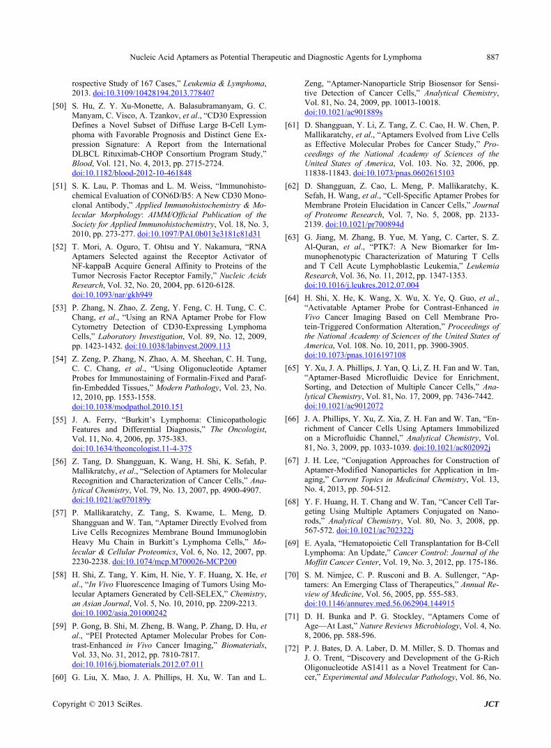
Nucleic Acid Aptamers as Potential Therapeutic and Diagnostic Agents for Lymphoma 887
rospective Study of 167 Cases,” Leukemia & Lymphoma, 2013. doi:10.3109/10428194.2013.778407
[50] S. Hu, Z. Y. Xu-Monette, A. Balasubramanyam, G. C. Manyam, C. Visco, A. Tzankov, et al., “CD30 Expression Defines a Novel Subset of Diffuse Large B-Cell Lym- phoma with Favorable Prognosis and Distinct Gene Ex- pression Signature: A Report from the International DLBCL Rituximab-CHOP Consortium Program Study,” Blood, Vol. 121, No. 4, 2013, pp. 2715-2724. doi:10.1182/blood-2012-10-461848
[51] S. K. Lau, P. Thomas and L. M. Weiss, “Immunohisto- chemical Evaluation of CON6D/B5: A New CD30 Mono- clonal Antibody,” Applied Immunohistochemistry & Mo- lecular Morphology: AIMM/Official Publication of the Society for Applied Immunohistochemistry, Vol. 18, No. 3, 2010, pp. 273-277. doi:10.1097/PAI.0b013e3181c81d31
[52] T. Mori, A. Oguro, T. Ohtsu and Y. Nakamura, “RNA Aptamers Selected against the Receptor Activator of NF-kappaB Acquire General Affinity to Proteins of the Tumor Necrosis Factor Receptor Family,” Nucleic Acids Research, Vol. 32, No. 20, 2004, pp. 6120-6128. doi:10.1093/nar/gkh949
[53] P. Zhang, N. Zhao, Z. Zeng, Y. Feng, C. H. Tung, C. C. Chang, et al., “Using an RNA Aptamer Probe for Flow Cytometry Detection of CD30-Expressing Lymphoma Cells,” Laboratory Investigation, Vol. 89, No. 12, 2009, pp. 1423-1432. doi:10.1038/labinvest.2009.113
[54] Z. Zeng, P. Zhang, N. Zhao, A. M. Sheehan, C. H. Tung, C. C. Chang, et al., “Using Oligonucleotide Aptamer Probes for Immunostaining of Formalin-Fixed and Paraf- fin-Embedded Tissues,” Modern Pathology, Vol. 23, No. 12, 2010, pp. 1553-1558. doi:10.1038/modpathol.2010.151
[55] J. A. Ferry, “Burkitt’s Lymphoma: Clinicopathologic Features and Differential Diagnosis,” The Oncologist, Vol. 11, No. 4, 2006, pp. 375-383. doi:10.1634/theoncologist.11-4-375
[56] Z. Tang, D. Shangguan, K. Wang, H. Shi, K. Sefah, P. Mallikratchy, et al., “Selection of Aptamers for Molecular Recognition and Characterization of Cancer Cells,” Ana- lytical Chemistry, Vol. 79, No. 13, 2007, pp. 4900-4907. doi:10.1021/ac070189y
[57] P. Mallikaratchy, Z. Tang, S. Kwame, L. Meng, D. Shangguan and W. Tan, “Aptamer Directly Evolved from Live Cells Recognizes Membrane Bound Immunoglobin Heavy Mu Chain in Burkitt’s Lymphoma Cells,” Mo- lecular & Cellular Proteomics, Vol. 6, No. 12, 2007, pp. 2230-2238. doi:10.1074/mcp.M700026-MCP200
[58] H. Shi, Z. Tang, Y. Kim, H. Nie, Y. F. Huang, X. He, et al., “In Vivo Fluorescence Imaging of Tumors Using Mo- lecular Aptamers Generated by Cell-SELEX,” Chemistry, an Asian Journal, Vol. 5, No. 10, 2010, pp. 2209-2213. doi:10.1002/asia.201000242
[59] P. Gong, B. Shi, M. Zheng, B. Wang, P. Zhang, D. Hu, et al., “PEI Protected Aptamer Molecular Probes for Con- trast-Enhanced in Vivo Cancer Imaging,” Biomaterials, Vol. 33, No. 31, 2012, pp. 7810-7817. doi:10.1016/j.biomaterials.2012.07.011
[60] G. Liu, X. Mao, J. A. Phillips, H. Xu, W. Tan and L.
Zeng, “Aptamer-Nanoparticle Strip Biosensor for Sensi- tive Detection of Cancer Cells,” Analytical Chemistry, Vol. 81, No. 24, 2009, pp. 10013-10018. doi:10.1021/ac901889s
[61] D. Shangguan, Y. Li, Z. Tang, Z. C. Cao, H. W. Chen, P. Mallikaratchy, et al., “Aptamers Evolved from Live Cells as Effective Molecular Probes for Cancer Study,” Pro- ceedings of the National Academy of Sciences of the United States of America, Vol. 103. No. 32, 2006, pp. 11838-11843. doi:10.1073/pnas.0602615103
[62] D. Shangguan, Z. Cao, L. Meng, P. Mallikaratchy, K. Sefah, H. Wang, et al., “Cell-Specific Aptamer Probes for Membrane Protein Elucidation in Cancer Cells,” Journal of Proteome Research, Vol. 7, No. 5, 2008, pp. 2133- 2139. doi:10.1021/pr700894d
[63] G. Jiang, M. Zhang, B. Yue, M. Yang, C. Carter, S. Z. Al-Quran, et al., “PTK7: A New Biomarker for Im- munophenotypic Characterization of Maturing T Cells and T Cell Acute Lymphoblastic Leukemia,” Leukemia Research, Vol. 36, No. 11, 2012, pp. 1347-1353. doi:10.1016/j.leukres.2012.07.004
[64] H. Shi, X. He, K. Wang, X. Wu, X. Ye, Q. Guo, et al., “Activatable Aptamer Probe for Contrast-Enhanced in Vivo Cancer Imaging Based on Cell Membrane Pro- tein-Triggered Conformation Alteration,” Proceedings of the National Academy of Sciences of the United States of America, Vol. 108. No. 10, 2011, pp. 3900-3905. doi:10.1073/pnas.1016197108
[65] Y. Xu, J. A. Phillips, J. Yan, Q. Li, Z. H. Fan and W. Tan, “Aptamer-Based Microfluidic Device for Enrichment, Sorting, and Detection of Multiple Cancer Cells,” Ana- lytical Chemistry, Vol. 81, No. 17, 2009, pp. 7436-7442. doi:10.1021/ac9012072
[66] J. A. Phillips, Y. Xu, Z. Xia, Z. H. Fan and W. Tan, “En- richment of Cancer Cells Using Aptamers Immobilized on a Microfluidic Channel,” Analytical Chemistry, Vol. 81, No. 3, 2009, pp. 1033-1039. doi:10.1021/ac802092j
[67] J. H. Lee, “Conjugation Approaches for Construction of Aptamer-Modified Nanoparticles for Application in Im- aging,” Current Topics in Medicinal Chemistry, Vol. 13, No. 4, 2013, pp. 504-512.
[68] Y. F. Huang, H. T. Chang and W. Tan, “Cancer Cell Tar- geting Using Multiple Aptamers Conjugated on Nano- rods,” Analytical Chemistry, Vol. 80, No. 3, 2008, pp. 567-572. doi:10.1021/ac702322j
[69] E. Ayala, “Hematopoietic Cell Transplantation for B-Cell Lymphoma: An Update,” Cancer Control: Journal of the Moffitt Cancer Center, Vol. 19, No. 3, 2012, pp. 175-186.
[70] S. M. Nimjee, C. P. Rusconi and B. A. Sullenger, “Ap- tamers: An Emerging Class of Therapeutics,” Annual Re- view of Medicine, Vol. 56, 2005, pp. 555-583. doi:10.1146/annurev.med.56.062904.144915
[71] D. H. Bunka and P. G. Stockley, “Aptamers Come of Age—At Last,” Nature Reviews Microbiology, Vol. 4, No. 8, 2006, pp. 588-596.
[72] P. J. Bates, D. A. Laber, D. M. Miller, S. D. Thomas and J. O. Trent, “Discovery and Development of the G-Rich Oligonucleotide AS1411 as a Novel Treatment for Can- cer,” Experimental and Molecular Pathology, Vol. 86, No.
Copyright © 2013 SciRes. JCT

Nucleic Acid Aptamers as Potential Therapeutic and Diagnostic Agents for Lymphoma 888
3, 2009, pp. 151-164. doi:10.1016/j.yexmp.2009.01.004
[73] A. C. Girvan, Y. Teng, L. K. Casson, S. D. Thomas, S. Juliger, M. W. Ball, et al., “AGRO100 Inhibits Activation of Nuclear Factor-kappaB (NF-kappaB) by Forming a Complex with NF-kappaB Essential Modulator (NEMO) and Nucleolin,” Molecular Cancer Therapeutics, Vol. 5, No. 7, 2006, pp. 1790-1799.
[74] S. Gattoni-Celli, C. L. Buckner, J. Lazarchick, R. K. Stu- art and D. J. Fernandes, “Overexpression of Nucleolin in Engrafted Acute Myelogenous Leukemia Cells,” Ameri- can Journal of Hematology, Vol. 84, No. 8, 2009, pp. 535-538. doi:10.1002/ajh.21461
[75] N. Tulchin, M. Chambon, G. Juan, S. Dikman, J. Strau- chen, L. Ornstein, et al., “BRCA1 Protein and Nucleolin Colocalize in Breast Carcinoma Tissue and Cancer Cell Lines,” The American Journal of Pathology, Vol. 176, No. 3, 2010, pp. 1203-1214. doi:10.2353/ajpath.2010.081063
[76] L. Peng, J. Liang, H. Wang, X. Song, A. Rashid, H. F. Gomez, et al., “High Levels of Nucleolar Expression of Nucleolin Are Associated with Better Prognosis in Pa- tients with Stage II Pancreatic Ductal Adenocarcinoma,” Clinical Cancer Research, Vol. 16, No. 14, 2010, pp. 3734-3742. doi:10.1158/1078-0432.CCR-09-3411
[77] H. Zhao, Y. Huang, C. Xue, Y. Chen, X. Hou, Y. Guo, et al., “Prognostic Significance of the Combined Score of Endothelial Expression of Nucleolin and CD31 in Surgi- cally Resected Non-Small Cell Lung Cancer,” PloS One, Vol. 8, No. 1, 2013, p. e54674. doi:10.1371/journal.pone.0054674
[78] F. Mongelard and P. Bouvet, “AS-1411, a Guanosine- Rich Oligonucleotide Aptamer Targeting Nucleolin for the Potential Treatment of Cancer, Including Acute Mye- loid Leukemia,” Current Opinion in Molecular Thera- peutics, Vol. 12, No. 1, 2010, pp. 107-114.
[79] Y. Shang, S. Kakinuma, M. Nishimura, Y. Kobayashi, K. Nagata and Y. Shimada, “Interleukin-9 Receptor Gene Is Transcriptionally Regulated by Nucleolin in T-Cell Lym- phoma Cells,” Molecular Carcinogenesis, Vol. 51, No. 8, 2012, pp. 619-627. doi:10.1002/mc.20834
[80] Y. Otake, S. Soundararajan, T. K. Sengupta, E. A. Kio, J. C. Smith, M. Pineda-Roman, et al., “Overexpression of Nucleolin in Chronic Lymphocytic Leukemia Cells In- duces Stabilization of bcl2 mRNA,” Blood, Vol. 109, No. 7, 2007, pp. 3069-3075. doi:10.1182/blood-2006-08-043257
[81] Y. A. Shieh, S. J. Yang, M. F. Wei and M. J. Shieh, “Ap- tamer-Based Tumor-Targeted Drug Delivery for Photo- dynamic Therapy,” ACS Nano, Vol. 4, No. 3, 2010, pp. 1433-1442. doi:10.1021/nn901374b
[82] A. Vater and S. Klussmann, “Toward Third-Generation Aptamers: Spiegelmers and Their Therapeutic Prospects,” Current Opinion in Drug Discovery & Development, Vol. 6, No. 2, 2003, pp. 253-261.
[83] D. Eulberg and S. Klussmann, “Spiegelmers: Biostable Aptamers,” Chembiochem: A European Journal of Che- mical Biology, Vol. 4, No. 10, 2003, pp. 979-983. doi:10.1002/cbic.200300663
[84] D. G. Duda, S. V. Kozin, N. D. Kirkpatrick, L. Xu, D.
Fukumura and R. K. Jain, “CXCL12 (SDF1alpha)- CXCR4/CXCR7 Pathway Inhibition: An Emerging Sen- sitizer for Anticancer Therapies?” Clinical Cancer Re- search, Vol. 17, No. 8, 2011, pp. 2074-2080. doi:10.1158/1078-0432.CCR-10-2636
[85] M. Borge, P. R. Nannini, P. E. Morande, C. Jancic, A. Bistmans, R. F. Bezares, et al., “CXCL12 Is a Costimu- lator for CD4+ T Cell Activation and Proliferation in Chronic Lymphocytic Leukemia Patients,” Cancer Im- munology, Immunotherapy, Vol. 62, No. 1, 2013, pp. 113-124. doi:10.1007/s00262-012-1320-7
[86] J. R. Adair, P. W. Howard, J. A. Hartley, D. G. Williams and K. A. Chester, “Antibody-Drug Conjugates—A Per- fect Synergy,” Expert Opinion on Biological Therapy, Vol. 12, No. 9, 2012, pp. 1191-1206. doi:10.1517/14712598.2012.693473
[87] M. A. Firer and G. Gellerman, “Targeted Drug Delivery for Cancer Therapy: The Other Side of Antibodies,” Jour- nal of Hematology & Oncology, Vol. 5, 2012, pp. 70.
[88] R. Stasi, “Gemtuzumab Ozogamicin: An Anti-CD33 Im- munoconjugate for the Treatment of Acute Myeloid Leu- kaemia,” Expert Opinion on Biological Therapy, Vol. 8, No. 4, 2008, pp. 527-540. doi:10.1517/14712598.8.4.527
[89] B. Pro and G. F. Perini, “Brentuximab Vedotin in Hodg- kin’s Lymphoma,” Expert Opinion on Biological Therapy, Vol. 12, No. 10, 2012, pp. 1415-1421. doi:10.1517/14712598.2012.718328
[90] K. Haddley, “Brentuximab Vedotin: Its Role in the Treatment of Anaplastic Large Cell and Hodgkin’s Lym- phoma,” Drugs of Today, Vol. 48, No. 4, 2012, pp. 259- 270.
[91] M. Furtado and S. Rule, “Emerging Pharmacotherapy for Relapsed or Refractory Hodgkin’s Lymphoma: Focus on Brentuximab Vedotin,” Clinical Medicine Insights On- cology, Vol. 6, 2012, pp. 31-39.
[92] G. P. Canellos, “Brentuximab Vedotin and Panobinostat: New Drugs for Hodgkin’s Lymphoma—Can They Make One of Medical Oncology’s Chemotherapy Success Sto- ries More Successful?” Journal of Clinical Oncology, Vol. 30, No. 18, 2012, pp. 2171-2172. doi:10.1200/JCO.2011.39.6416
[93] A. Younes, A. K. Gopal, S. E. Smith, S. M. Ansell, J. D. Rosenblatt, K. J. Savage, et al., “Results of a Pivotal Phase II Study of Brentuximab Vedotin for Patients with Relapsed or Refractory Hodgkin’s Lymphoma,” Journal of Clinical Oncology, Vol. 30, No. 18, 2012, pp. 2183- 2189. doi:10.1200/JCO.2011.38.0410
[94] P. F. Bross, J. Beitz, G. Chen, X. H. Chen, E. Duffy, L. Kieffer, et al., “Approval Summary: Gemtuzumab Ozo- gamicin in Relapsed Acute Myeloid Leukemia,” Clinical Cancer Research, Vol. 7, No. 6, 2001, pp. 1490-1496.
[95] P. Mallikaratchy, Z. Tang and W. Tan, “Cell Specific Aptamer-Photosensitizer Conjugates as a Molecular Tool in Photodynamic Therapy,” ChemMedChem, Vol. 3, No. 3, 2008, pp. 425-428. doi:10.1002/cmdc.200700260
[96] F. Schmitt and L. Juillerat-Jeanneret, “Drug Targeting Strategies for Photodynamic Therapy,” Anti-Cancer Agents in Medicinal Chemistry, Vol. 12, No. 5, 2012, pp. 500- 525. doi:10.2174/187152012800617830
Copyright © 2013 SciRes. JCT
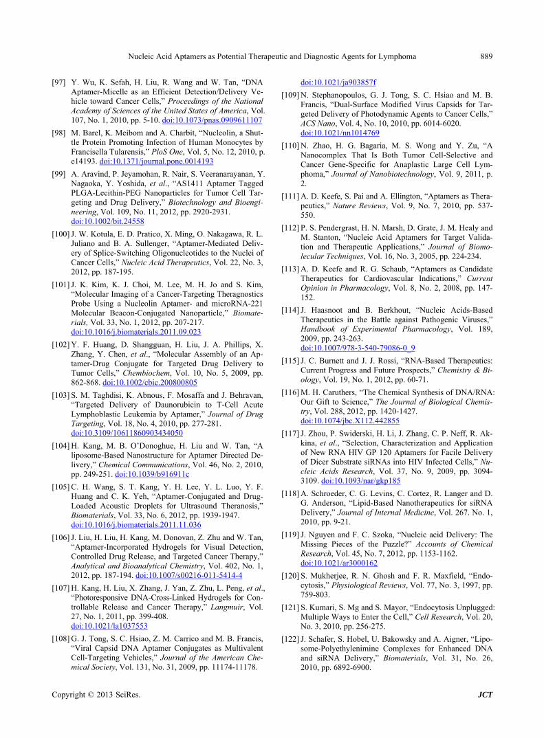
Nucleic Acid Aptamers as Potential Therapeutic and Diagnostic Agents for Lymphoma 889
[97] Y. Wu, K. Sefah, H. Liu, R. Wang and W. Tan, “DNA Aptamer-Micelle as an Efficient Detection/Delivery Ve- hicle toward Cancer Cells,” Proceedings of the National Academy of Sciences of the United States of America, Vol. 107, No. 1, 2010, pp. 5-10. doi:10.1073/pnas.0909611107
[98] M. Barel, K. Meibom and A. Charbit, “Nucleolin, a Shut- tle Protein Promoting Infection of Human Monocytes by Francisella Tularensis,” PloS One, Vol. 5, No. 12, 2010, p. e14193. doi:10.1371/journal.pone.0014193
[99] A. Aravind, P. Jeyamohan, R. Nair, S. Veeranarayanan, Y. Nagaoka, Y. Yoshida, et al., “AS1411 Aptamer Tagged PLGA-Lecithin-PEG Nanoparticles for Tumor Cell Tar- geting and Drug Delivery,” Biotechnology and Bioengi- neering, Vol. 109, No. 11, 2012, pp. 2920-2931. doi:10.1002/bit.24558
[100] J. W. Kotula, E. D. Pratico, X. Ming, O. Nakagawa, R. L. Juliano and B. A. Sullenger, “Aptamer-Mediated Deliv- ery of Splice-Switching Oligonucleotides to the Nuclei of Cancer Cells,” Nucleic Acid Therapeutics, Vol. 22, No. 3, 2012, pp. 187-195.
[101] J. K. Kim, K. J. Choi, M. Lee, M. H. Jo and S. Kim, “Molecular Imaging of a Cancer-Targeting Theragnostics Probe Using a Nucleolin Aptamer- and microRNA-221 Molecular Beacon-Conjugated Nanoparticle,” Biomate- rials, Vol. 33, No. 1, 2012, pp. 207-217. doi:10.1016/j.biomaterials.2011.09.023
[102] Y. F. Huang, D. Shangguan, H. Liu, J. A. Phillips, X. Zhang, Y. Chen, et al., “Molecular Assembly of an Ap- tamer-Drug Conjugate for Targeted Drug Delivery to Tumor Cells,” Chembiochem, Vol. 10, No. 5, 2009, pp. 862-868. doi:10.1002/cbic.200800805
[103] S. M. Taghdisi, K. Abnous, F. Mosaffa and J. Behravan, “Targeted Delivery of Daunorubicin to T-Cell Acute Lymphoblastic Leukemia by Aptamer,” Journal of Drug Targeting, Vol. 18, No. 4, 2010, pp. 277-281. doi:10.3109/10611860903434050
[104] H. Kang, M. B. O’Donoghue, H. Liu and W. Tan, “A liposome-Based Nanostructure for Aptamer Directed De- livery,” Chemical Communications, Vol. 46, No. 2, 2010, pp. 249-251. doi:10.1039/b916911c
[105] C. H. Wang, S. T. Kang, Y. H. Lee, Y. L. Luo, Y. F. Huang and C. K. Yeh, “Aptamer-Conjugated and Drug- Loaded Acoustic Droplets for Ultrasound Theranosis,” Biomaterials, Vol. 33, No. 6, 2012, pp. 1939-1947. doi:10.1016/j.biomaterials.2011.11.036
[106] J. Liu, H. Liu, H. Kang, M. Donovan, Z. Zhu and W. Tan, “Aptamer-Incorporated Hydrogels for Visual Detection, Controlled Drug Release, and Targeted Cancer Therapy,” Analytical and Bioanalytical Chemistry, Vol. 402, No. 1, 2012, pp. 187-194. doi:10.1007/s00216-011-5414-4
[107] H. Kang, H. Liu, X. Zhang, J. Yan, Z. Zhu, L. Peng, et al., “Photoresponsive DNA-Cross-Linked Hydrogels for Con- trollable Release and Cancer Therapy,” Langmuir, Vol. 27, No. 1, 2011, pp. 399-408. doi:10.1021/la1037553
[108] G. J. Tong, S. C. Hsiao, Z. M. Carrico and M. B. Francis, “Viral Capsid DNA Aptamer Conjugates as Multivalent Cell-Targeting Vehicles,” Journal of the American Che- mical Society, Vol. 131, No. 31, 2009, pp. 11174-11178.
doi:10.1021/ja903857f
[109] N. Stephanopoulos, G. J. Tong, S. C. Hsiao and M. B. Francis, “Dual-Surface Modified Virus Capsids for Tar- geted Delivery of Photodynamic Agents to Cancer Cells,” ACS Nano, Vol. 4, No. 10, 2010, pp. 6014-6020. doi:10.1021/nn1014769
[110] N. Zhao, H. G. Bagaria, M. S. Wong and Y. Zu, “A Nanocomplex That Is Both Tumor Cell-Selective and Cancer Gene-Specific for Anaplastic Large Cell Lym- phoma,” Journal of Nanobiotechnology, Vol. 9, 2011, p. 2.
[111] A. D. Keefe, S. Pai and A. Ellington, “Aptamers as Thera- peutics,” Nature Reviews, Vol. 9, No. 7, 2010, pp. 537- 550.
[112] P. S. Pendergrast, H. N. Marsh, D. Grate, J. M. Healy and M. Stanton, “Nucleic Acid Aptamers for Target Valida- tion and Therapeutic Applications,” Journal of Biomo- lecular Techniques, Vol. 16, No. 3, 2005, pp. 224-234.
[113] A. D. Keefe and R. G. Schaub, “Aptamers as Candidate Therapeutics for Cardiovascular Indications,” Current Opinion in Pharmacology, Vol. 8, No. 2, 2008, pp. 147- 152.
[114] J. Haasnoot and B. Berkhout, “Nucleic Acids-Based Therapeutics in the Battle against Pathogenic Viruses,” Handbook of Experimental Pharmacology, Vol. 189, 2009, pp. 243-263. doi:10.1007/978-3-540-79086-0_9
[115] J. C. Burnett and J. J. Rossi, “RNA-Based Therapeutics: Current Progress and Future Prospects,” Chemistry & Bi- ology, Vol. 19, No. 1, 2012, pp. 60-71.
[116] M. H. Caruthers, “The Chemical Synthesis of DNA/RNA: Our Gift to Science,” The Journal of Biological Chemis- try, Vol. 288, 2012, pp. 1420-1427. doi:10.1074/jbc.X112.442855
[117] J. Zhou, P. Swiderski, H. Li, J. Zhang, C. P. Neff, R. Ak- kina, et al., “Selection, Characterization and Application of New RNA HIV GP 120 Aptamers for Facile Delivery of Dicer Substrate siRNAs into HIV Infected Cells,” Nu- cleic Acids Research, Vol. 37, No. 9, 2009, pp. 3094- 3109. doi:10.1093/nar/gkp185
[118] A. Schroeder, C. G. Levins, C. Cortez, R. Langer and D. G. Anderson, “Lipid-Based Nanotherapeutics for siRNA Delivery,” Journal of Internal Medicine, Vol. 267. No. 1, 2010, pp. 9-21.
[119] J. Nguyen and F. C. Szoka, “Nucleic acid Delivery: The Missing Pieces of the Puzzle?” Accounts of Chemical Research, Vol. 45, No. 7, 2012, pp. 1153-1162. doi:10.1021/ar3000162
[120] S. Mukherjee, R. N. Ghosh and F. R. Maxfield, “Endo- cytosis,” Physiological Reviews, Vol. 77, No. 3, 1997, pp. 759-803.
[121] S. Kumari, S. Mg and S. Mayor, “Endocytosis Unplugged: Multiple Ways to Enter the Cell,” Cell Research, Vol. 20, No. 3, 2010, pp. 256-275.
[122] J. Schafer, S. Hobel, U. Bakowsky and A. Aigner, “Lipo- some-Polyethylenimine Complexes for Enhanced DNA and siRNA Delivery,” Biomaterials, Vol. 31, No. 26, 2010, pp. 6892-6900.
Copyright © 2013 SciRes. JCT

Nucleic Acid Aptamers as Potential Therapeutic and Diagnostic Agents for Lymphoma
Copyright © 2013 SciRes. JCT
890
[123] M. Gunther, J. Lipka, A. Malek, D. Gutsch, W. Kreyling and A. Aigner, “Polyethylenimines for RNAi-Mediated Gene Targeting in Vivo and siRNA Delivery to the Lung,” European Journal of Pharmaceutics and Biopharmaceu- tics, Vol. 77, No. 3, 2011, pp. 438-449.
[124] S. Y. Wu and N. A. McMillan, “Lipidic Systems for in Vivo siRNA Delivery,” AAPS Journal, Vol. 11, No. 4, 2009, pp. 639-652. doi:10.1208/s12248-009-9140-1
[125] Y. Sakurai, H. Hatakeyama, Y. Sato, H. Akita, K. Taka- yama, S. Kobayashi, et al., “Endosomal Escape and the Knockdown Efficiency of Liposomal-siRNA by the Fu- sogenic Peptide shGALA,” Biomaterials, Vol. 32, No. 24, 2011, pp. 5733-5742.
[126] K. W. Thiel, L. I. Hernandez, J. P. Dassie, W. H. Thiel, X.
Liu, K. R. Stockdale, et al., “Delivery of Chemo-Sensi- tizing siRNAs to HER2+-Breast Cancer Cells Using RNA Aptamers,” Nucleic Acids Research, Vol. 40, No. 13, 2012, pp. 6319-6337
[127] P. Guo, “The Emerging Field of RNA Nanotechnology,” Nature Nanotechnology, Vol. 5, No. 12, 2010, pp. 833- 842.
[128] G. C. Shukla, F. Haque, Y. Tor, L. M. Wilhelmsson, J. J. Toulme, H. Isambert, et al., “A Boost for the Emerging Field of RNA Nanotechnology,” ACS Nano, Vol. 5, No. 5, 2011, pp. 3405-3418. doi:10.1021/nn200989r
[129] J. J. Rossi, “RNA Nanoparticles Come of Age,” Acta Biochimica et Biophysica Sinica, Vol. 43, No. 4, 2011, pp. 245-247. doi:10.1093/abbs/gmr018