
Protein delivery: DNA nanostructures and cell-surface targeting Harvard iGEM August 27, 2006.

Nuclear Factor Erythroid-derived 2 (Nfe2) Regulates JunDDNA-binding Activity via AcetylationA NOVEL MECHANISM REGULATING TROPHOBLAST DIFFERENTIATION*□S
Received for publication, August 5, 2011, and in revised form, December 2, 2011 Published, JBC Papers in Press, December 15, 2011, DOI 10.1074/jbc.M111.289801
Muhammed Kashif‡, Andrea Hellwig§, Said Hashemolhosseini¶, Varun Kumar‡, Fabian Bock‡, Hongjie Wang�**,Khurrum Shahzad**, Satish Ranjan**, Juliane Wolter**, Thati Madhusudhan**, Angelika Bierhaus‡, Peter Nawroth‡,and Berend Isermann**1
From the ‡Department of Internal Medicine I and Clinical Chemistry, University of Heidelberg, INF 410, 69120 Heidelberg, Germany,the §Department of Neurobiology, Interdisciplinary Center for Neurosciences (IZN) University of Heidelberg, INF 364, 69120Heidelberg, Germany, the ¶Institute for Biochemistry, Emil-Fischer-Center, University of Erlangen-Nürnberg, Fahrstrasse 17, 91054Erlangen, Germany, the �Department of Cardiology, Tongji Hospital, Tongji Medical College, Huazhong University of Science andTechnology, 430030 Wuhan, China, and the **Department of Clinical Chemistry and Pathobiochemistry, Otto-von-Guericke-University Magdeburg, Leipziger Strasse 44, 39120 Magdeburg, Germany
Background: Nfe2 restricts Gcm1 expression, placental vascularization, and embryonic growth.Results: Nfe2 induces hypoacetylation of JunD, thus limiting JunD binding to the Gcm1 promoter (at �1441).Conclusion: In trophoblast cells Nfe2 negatively controls Gcm1 expression and syncytiotrophoblast formation by repressingJunD-binding activity.Significance: This identifies a novel, acetylation dependent interaction of bZip transcription factors regulating placental andembryonic development.
We recently demonstrated that the bZip transcription fac-tor nuclear factor erythroid-derived 2 (Nfe2) represses pro-tein acetylation and expression of the transcription factorglial cell missing 1 (Gcm1) in trophoblast cells, preventingexcess syncytiotrophoblast formation and permitting normalplacental vascularization and embryonic growth. However,theGcm1promoter lacks aNfe2-binding site andhence themech-anisms linking Nfe2 and Gcm1 expression remained unknown.Here we show that Nfe2 represses JunD DNA-binding activity tothe Gcm1 promoter during syncytiotrophoblast differentiation.Interventional studies using knockdown and knockin approachesshow that enhanced JunD DNA-binding activity is required forincreased expression of Gcm1 and syncytiotrophoblast formationaswellas impairedplacentalvascularizationandreducedgrowthofNfe2�/� embryos. Induction ofGcm1 expression requires bindingof JunD to the �1441 site within the Gcm1 promoter, which isdistinct from the �1314 site previously shown to induce Gcm1expression by other bZip transcription factors. Nfe2 modulatesJunD binding to the Gcm1 promoter via acetylation, as reducingJunD acetylation using the histone acetyltransferase inhibitor cur-cumin reverses the increased JunDDNA-binding activity observedin the absence of Nfe2. This identifies a novelmechanism throughwhich bZip transcription factors interact.Within the placenta thisinteraction regulates Gcm1 expression, syncytiotrophoblast for-mation, placental vascularization, and embryonic growth.
We recently identified a novel function of the bZip transcrip-tion factor Nfe2 in trophoblast cells (1). In trophoblast cellsNfe2 prevents excess syncytiotrophoblast formation during thelater pregnancy stages in the mouse (after day 14.5 p.c.,2 corre-sponding to the 3rd trimester), thus ensuring sufficient vascu-larization of the placenta and normal embryonic growth (1).This new function of Nfe2 may provide novel insight intomechanisms underlying placental dysfunction and intrauterinegrowth restriction. These are frequent but incompletely under-stood pregnancy complications associated not only withincreased peripartal morbidity and mortality of the newborn,but also with diseases such as diabetes mellitus or cardiovascu-lar diseases later on in adult life (2, 3). Hence, deciphering themechanism through which Nfe2 modulates trophoblast differ-entiation and placental development and function may revealnew diagnostic or therapeutic targets for placental dysfunctionand intrauterine growth restriction.Nfe2 is the larger (45 kDa) subunit of the heterodimeric tran-
scription factor NFE2. Until recently expression of Nfe2 wasthought to be restricted to hematopoietic cells (cells of themegakaryocyte, erythrocyte, or mast cell lineage) (4). We iden-tified a function of Nfe2 in nonhematopoietic cells (1). In tro-phoblast cells Nfe2 represses Gcm1 (also referred to as GCMa)activity (1). Gcm1 belongs to a unique family of transcriptionfactors hallmarked by a highly conserved zinc-coordinatedDNA-binding domain (5). Gcm1 is required for branching andsyncytiotrophoblast fusion during placental development, andboth loss and gain of Gcm1 function disturb syncytiotropho-
* This work was supported by Deutsche Forschungsgemeinschaft Grants IS67/4-1 and IS 67/4-2 (to B. I.) and a grant of the German Israeli Foundation(to B. I. and P. P. N.).
□S This article contains supplemental Figs. S1 and S2.1 To whom correspondence should be addressed. Tel.: 49-0-391-13900; Fax:
49-0-391-13902; E-mail: [email protected] or [email protected].
2 The abbreviations used are: p.c., post coitus; Nfe2, nuclear factor erythroid-derived 2; CREB, cAMP-response element-binding protein; TSA, trichosta-tin A; HDAC, histone deacetylase; TS, trophoblast stem; HAT, histoneacetyltransferase; kd, knockdown.
THE JOURNAL OF BIOLOGICAL CHEMISTRY VOL. 287, NO. 8, pp. 5400 –5411, February 17, 2012© 2012 by The American Society for Biochemistry and Molecular Biology, Inc. Published in the U.S.A.
5400 JOURNAL OF BIOLOGICAL CHEMISTRY VOLUME 287 • NUMBER 8 • FEBRUARY 17, 2012
by guest on May 11, 2018
http://ww
w.jbc.org/
Dow
nloaded from

blast formation and placental development (1, 6). Loss of Nfe2function increases acetylation of histone H4 within the Gcm1promoter and of Gcm1 itself, enhancing Gcm1 expression inmurine placenta and trophoblast cells (1). Although these stud-ies identified a novel function of Nfe2 as a repressor duringsyncytiotrophoblast differentiation, the positive gene regula-tors repressed byNfe2 during normal trophoblast and placentaldifferentiation remain unknown. Based on in silico analyses ofthe Gcm1 promoter the 6000-bp 5� of the ATG lack potentialNfe2-binding sites, indicating that Nfe2 must regulate Gcm1expression through an indirectmechanism. Interaction of Nfe2with CBP or other bZip transcription factors has been reportedand may be mechanistically linked to the developmental func-tion of Nfe2 (1, 7, 8).Indeed, bZip transcription factors have established functions
for Gcm1 expression and placental development. Thus, thebZip transcription factors CREB and OASIS stimulate Gcm1expression in vitro and enhance expression of syncytiotropho-blast markers in vivo (9). The relevant cAMP response element(CRE)-binding site was located at �1337 within the mouseGcm1 promoter. In vivo, expression of CREB was replaced byexpression ofOASIS around latemidgestation (E13.5), at whichtime the hemochorial placenta is fully established. This demon-strates a dynamic regulation of bZip transcription factors dur-ing placental development.Other bZip transcription factorswith known function during
placentation belong to the AP-1 proteins. Knockout studies inmice revealed embryonic lethal placental defects in JunB andFra1-deficient embryos (10, 11), whereas placental develop-mentwas normal inmice lacking otherAP-1 proteins. PlacentalJunB or Fra1 deficiency impairs vascularization of the labyrin-thine layer, causing early embryonic lethality between days 8.5and 10.5 p.c. (10, 11).Whether Nfe2 regulates syncytiotrophoblast formation and
vascularization of the placental labyrinthine layer through aninteraction with TORC1, OASIS, JunB, Fra1, or yet to be iden-tified transcription factors remained hitherto unknown.Within the current study we explored the interaction partnersof Nfe2 during placental development, identifying a so farunknown interaction of Nfe2 with JunD. During placentaldevelopment Nfe2 regulates through an acetylation dependentmechanism JunD-binding activity to the �1441 site of theGcm1 promoter, regulating syncytiotrophoblast formation,placental vascularization, and embryonic growth.
EXPERIMENTAL PROCEDURES
Materials—Heparin sodium salt from porcine intestinalmucosa, human acidic fibroblast growth factor 4 (aFGF4), tri-chostatin A (TSA), Polybrene, and puromycin dihydrochloridefrom Streptomyces albonigerwere obtained fromSigma. All cellculture media, FBS, glutamine, penicillin/streptomycin mix-ture, neuromycin, nonessential amino acids, and sodium pyru-vate were from PAA, Cölbe, Germany. P24 tag antigen ELISAand the CHIP assay kit were from BioCat, Heidelberg, Ger-many. ECL reagent was from Amersham Biosciences. MurineNfe2 and JunD shRNA constructs were fromOpen Biosystems,Huntsville, AL. GoTaqDNApolymerase, AP-1, andCREB con-sensus oligonucleotides for EMSA were obtained from Pro-
mega. Rabbit polyclonal IgG antibodies against Nfe2, c-Jun,JunD, JunB, FosB, and Fra1 were purchased from Santa CruzBiotechnology. Rabbit polyclonal IgG antibody against acety-lated lysine was purchased from Cell Signaling Technology,Boston, MA. CellMask Deep Red stain was bought from Invit-rogen. QuikChange XL Site-directed Mutagenesis kit was pur-chased from Stratagene. All primers were synthesized byThermo Scientific, Langenselbold, Germany.Plasmids—Murine JunDexpression (pCMV-JunD-SPORT6)
and JunD knockdown (pLKO1-JunD) constructs were pur-chased from Open Biosystems. The JunD expression plasmidpLV-JunD was generated by replacing the EGFP fragment(EcoRI/XhoI) of the plasmid pLV-EGFP (kindly provided byMasahito Ikawa, Osaka, Japan) (12) with the EcoRI/XhoI frag-ment of pCMV-JunD-SPORT6. To demonstrate specificity ofthe knockdown experiment six nucleotides within the JunDshRNAhybridization sequence (5�-ACTCGAGTTGAGCAGCCC AAG ATC CGG CTT TTT G-3�) were replaced in pLV-JunD (5�-ACT CGA GTT GAG CAA CCG AAA ATC CGCCTG TTC G-3�) by site-directed mutagenesis. This resulted insilent mutations yielding a JunD resistant to the shRNA used(pLV-JunD-shRes).Reporter constructs consisting of various fragments of the
murine Gcm1 promoter (starting at �4041, �2892, �1637,and �396 relative to ATG) linked to the luciferase codingsequence (Promega, Mannheim, Germany) have been previ-ously described (9). Gcm1 promoter constructs containingmutated AP-1-binding sites were generated by site-directedmutagenesis at positions �1572/�1571 (AC to TG), �1434/�1433 (TC to AG), and �1310/�1309 (AC to TG). The Gcm1�1637 PGL2 luciferase reporter construct was used as the tem-plate. The following primers were used: gcm-1 �1572, AC toTG (5�-GGTTGTCTGTGATGCTTGTGTGTACAGTTTTAT TTC TCA-3� and 5�-TGA GAA ATA AAA CTG TACACA CAA GCA TCA CAG ACA ACC-3�); gcm-1 �1434, TCto AG (5�-CCC AGC GTG GCC TCT GAT AGA TTG TAAGAA TTC AGT CAA-3� and 5�-TTG ACT GAA TTC TTACAATCTATCAGAGGCCACGCTGGG-3�); gcm-1�1310,AC toTG (5�-GCCTGGTCTATAGGAGGTCTGTCCCATCTC AAA AAA CTA-3� and 5�-TAG TTT TTT GAG ATGGGACAGACCTCCTATAGACCAGGC-3�). All mutationswere generated using the QuikChange� II Site-directedMutagenesis kit (Stratagene) and identity of all mutations wasconfirmed by sequencing.Mice—Nfe2-deficient mice were kindly provided by R.
Shivdasani (Harvard Medical School, Cambridge, MA). Allanimals were housed and the experiments were performed atthe Interdisciplinary Biomedical Research institution (IBF)of the University of Heidelberg, Germany. For in vivo inter-vention studies we used TSA (15 �g/mouse (13)) or valproicacid (sodium salt, 155 mM, 25 ml/kg of body weight (14)) asHDAC (histone deacetylase) inhibitors and epigallocat-echin-3-gallate (50 mg/kg in saline (15)) or curcumin (0.5mg/kg (16)) as HAT inhibitors. Animal experiments wereconducted following standards and procedures approved bythe local Animal Care and Use Committee (Regierungsprä-sidium Karlsruhe, Germany).
Nfe2 Regulates JunD Binding Activity via Acetylation
FEBRUARY 17, 2012 • VOLUME 287 • NUMBER 8 JOURNAL OF BIOLOGICAL CHEMISTRY 5401
by guest on May 11, 2018
http://ww
w.jbc.org/
Dow
nloaded from

Cell Culture—HEK293T and HEK293 cells were kindly pro-vided by Dr. Peter Seeburg (Max-Planck-Institute for MedicalResearch, Heidelberg, Germany). They were used to generatelentiviral particles and perform the luciferase assay. These cellsweremaintained in DMEM supplemented with 4.5 g/ml of glu-cose, 10% FBS, 2 mM L-glutamine, 50 �g/ml of penicillin/strep-tomycin, and 4 �g/ml of neomycin at 37 °C in a humidifiedincubator with 5% CO2. For transfection experiments mediumwithout antibiotics was used.Mouse trophoblast stem (TS) cells were obtained from J.
Rossant (Hospital for SickChildren, Toronto,Ontario, Canada)and were induced to differentiate in plain TS cell medium(RPMI 1640 supplemented with 25 mM HEPES, 2.0 g/liter ofNaHCO3, 20% FBS, 1 mM sodium pyruvate, 100 �M �-mercap-toethanol, 2 mM L-glutamine, and 50 �g/ml of penicillin/strep-tomycin) or maintained as stem cells in complete TS cellmedium (30% plain TS cell medium plus 70% fibroblast-condi-tioned TS cell medium, 25 ng/ml of FGF4, and 1 �g/ml of hep-arin) as described previously (1, 17–19). In some experimentsTS cells were treatedwith 100 nMTSAor 2.5mM sodiumbutyr-ate for HDAC inhibition (20) or 30 nM curcumin or 5 �M epi-gallocatechin-3-gallate for HAT inhibition (21–23).In Vitro Syncytium Formation—Syncytiotrophoblast forma-
tion was monitored in nearly confluent TS cells differentiatedfor 6 days, washed with PBS, incubated with 2.5 �M CellMaskDeep Red for 5 min at 37 °C, and then fixed for 3 min in 3.75%formaldehyde at RT. Cells were counterstained with 0.5 �g/mlof DAPI in PBS for 10 min at RT in the dark. For quantificationof syncytiotrophoblasts at least 30 random images per slidewere captured (University of Texas Health Science Center SanAntonio ImageTool software) and a blinded investigator deter-mined the frequency of syncytial cells in these random images(1).Production of Lentiviral Particles—Highly purified plasmid
DNA prepared by alkaline lysis preparation was used for lenti-viral particle production. Vesicular stomatitis virus-G pseu-dotyped lentiviral particles were generated as described (1, 24).Briefly, HEK 293T cells were transfected with pLKO1-JunD,pLV-JunD, or pLV-JunD-shRes plasmids together with thepackaging plasmid (pCMV-dR8.91) and vesicular stomatitisvirus-G-expressing plasmid using the standard calcium phos-phate method. Lentiviral particles were harvested 2 and 3 daysafter transfection and concentrated by ultracentrifugation(50,000� g, 2 h, 2 times). After resuspension inHBSS buffer theviral particle concentration was determined by measuring thep24 gag antigen by ELISA according to the manufacturer’sinstruction (BioCat,Heidelberg,Germany). Viral particleswerestored at �80 °C.Luciferase Assay—The HEK293 cells were transfected in
60-mmdishes with 1�g of luciferase reporter construct and 0.1�g of CMV driven expression vectors using a standard calciumphosphate method. At 48 h post-transfection, cells were har-vested for luciferase assays using the luciferase assay systemaccording to the manufacturer’s instruction.Electrophoretic Mobility Shift Assay (EMSA)—Nuclear pro-
teins were isolated and binding activity of CREB and AP-1 wasdetermined as described previously (1, 25). Briefly, for isolationof nuclear proteins cells were washed in PBS and resuspended
in 200 �l of ice-cold buffer A (10 mM HEPES-KOH, pH 7.9, 1.5mMMgCl2, 10mMKCl, 0.5 mMDTT, 0.2 mM PMSF). For tissueextracts the tissue samples were homogenized in 400 �l of ice-cold buffer B (10 mM HEPES-KOH, pH 7.9, 1.5 mM MgCl2, 10mMKCl, 0.5mMDTT0.2mMPMSF, 1mMEDTA, 0.6%NonidetP-40), vortexed for 10 s, and incubated on ice for 10 min. Sam-ples were centrifuged at 12,000 � g for 2 min and the superna-tant was discarded. The pellet was either resuspended in 100 �lof buffer C (cell lysates: 20 mMHEPES-KOH, pH 7.9, 25% glyc-erol, 420mMNaCl, 1.5mMMgCl2, 0.2mMEDTA, 0.5mMDTT,0.2 mM PMSF) or 200 �l of buffer D (tissue lysates: 20 mM
HEPES-KOH, pH 7.9, 25% glycerol, 420 mM NaCl, 1.5 mM
MgCl2, 0.2 mM EDTA, 0.5 mM DTT, 0.2 mM PMSF, 0.2mM benzamidine, 5 �g/ml of leupeptin) and incubated on icefor 20 min. After centrifugation (12,000 � g, 5 min, 4 °C) thesupernatant was transferred to a new tube, protein concentra-tion was measured by colorimetric assay (BSA), and sampleswere quick-frozen at�80 °C. Binding of transcription factors to1 ng of radiolabeled oligonucleotides (AP-1 consensus: 5�-CGCTTG ATG ACT CAG CCG GAA-3�, JunD-binding site at�1441 derived from Gcm1: 5�-G CGT GGC CTC TGA TTCATT GTA AG-3�, JunD mutated (TC to AG), derived fromGcm1 �1441: 5�-G CGT GGC CTC TGA TAG ATT GTAAG-3�, CREB consensus: 5�-AGA GAT TGC CTG ACG TCAGAG AGC TAG-3�) was performed for 20 min at RT in 10 mM
HEPES, pH 7.5, 0.5 mM EDTA, 100 mM KCl, 2 mM DTT, 2%glycerol, 4% Ficoll, 0.25% Nonidet P-40, 1 mg/ml of BSA, and0.1 mg/ml of poly(dI/dC). Protein-DNA complexes were sepa-rated on 5% native polyacrylamide gels containing 3.25% gly-cerol and 0.5� Tris/boric acid/EDTA buffer. Identity of AP-1subunits was determined by supershift experiments. To thisend 4 �g of antibody against AP-1 subunits (Fra1, JunB, c-Jun,JunD, and FosB) were preincubated with 10 �g of proteinextract for 20min at RTbefore adding radiolabeled nucleotides.Transduction of Preimplantation Embryos—FemaleNfe2�/�
or wild type (WT) mice were superovulated by intraperitonealinjection of pregnant mare’s serum gonadotropin (5 units) fol-lowed by human chorionic gonadotropin (5 units) 48 h laterand thenmated with Nfe2�/� orWTmales, respectively. Two-cell stage embryos were collected from the females at day1.5 p.c. and incubated for 2 days to obtain blastocysts. The zonapellucida was removed with acidic Tyrode solution. Zona pel-lucida-free embryos were incubated individually in 50 �l ofKSOM medium containing JunD with either shRNA or JunDexpressing lentiviral particles (1 � 103 ng of p24/ml) for 4 h (1,12). Transduced blastocysts were transferred into pseudo-pregnant females. The transfer day was counted as day 3.5 p.c.and analyses were performed at day 14.5 or 16.5 p.c. Embryosgenerated from blastocysts transduced with lentiviral particlesgenerated from nonspecific shRNA or empty vector served ascontrols.Morphological Analyses of Placenta—Placental tissues were
obtained from Nfe2�/� or WT females after lentiviral trans-duction at day 14.5 p.c. Half of the placental tissue was fixed in4% buffered formalin, whereas the other half was furtherdivided and stored either in RNAlater or quick frozen in liquidnitrogen for protein isolation. The fixed tissuewas embedded inparaffin, sectioned, and stained with hematoxylin and eosin.
Nfe2 Regulates JunD Binding Activity via Acetylation
5402 JOURNAL OF BIOLOGICAL CHEMISTRY VOLUME 287 • NUMBER 8 • FEBRUARY 17, 2012
by guest on May 11, 2018
http://ww
w.jbc.org/
Dow
nloaded from

For vascular space quantification randomly selected micro-scopic fields (magnification �10) of the labyrinthine regionfrom 3 nonconsecutive placental sections (each section �10images) were acquired at 1280 � 960 resolution using a digitalcamera (Nikon digital sight DS-U1) connected to a light micro-scope (Nikon Eclipse TE2000). We implemented an imageanalysis routine using NIS Elements AR2.30 imaging software.Briefly, after acquisition, the images underwent an automatedanalysis procedure and were given a color threshold to coverthe area corresponding to the lumen of blood spaces. The cov-erage percentage was calculated as the ratio between the num-ber of pixels covered by the area defined by the threshold andthe overall number of pixels in the image (1). A blinded inves-tigator performed analyses.Transmission Electron Microscopic Analyses of Placenta—
Placenta tissues obtained at day 16.5 p.c. were fixed with 2.5%glutaraldehyde, 2.5% polyvinylidone 25, 0.1 M sodium cacody-late, pH 7.4. After washing with 0.1 M sodium cacodylate buffer,pH 7.4, samples were post-fixed with 2% osmium tetroxide,1.5% potassium ferrocyanide in 0.1 M sodium cacodylate buffer,pH 7.4, for 1 h, washed, contrasted en bloc with uranyl acetate,dehydrated with an ascending series of ethanol, and embeddedin glycid ether 100-based resin. Ultrathin sectionswere cutwitha Reichert ULTRACUT S ultramicrotome (Leica Microsys-tems, Wetzlar, Germany), contrasted with uranyl acetate andlead citrate, and viewed with an EM 10 CR electronmicroscope(Carl Zeiss NTS, Oberkochen, Germany).Gene Knockdown and Knockin inMouse Trophoblast Cells—
Generation of stably transfected Nfe2 knockdown mouse tro-phoblast cells (Nfe2kd) has been previously described (1). Tran-sient knockdown or knockin of JunD in control, Nfe2kd, orNfe2kd JunDkd TS cells was performed by lentiviral transduc-tion. Briefly, pLKO1-JunD, pLV-JunD-shRes, or pLV-JunDviral particles (5 � 105 TU/ml) were generated as describedabove and used to infect Nfe2kd (yielding Nfe2kdJunDkd cells),Nfe2kd JunDkd (yielding Nfe2kdJunDkdRes cells), or control(yielding Nfe2cJunD� cells) TS cells, respectively, in the pres-ence of 8 �g/ml of Polybrene in TS cell medium. The efficiencyof the JunD knockdown or knockin was determined after 3 daysof differentiation by RT-PCR and EMSA. In all experimentscells transducedwith viral particles generated fromnonspecificshRNA or empty pLV-CAG1.1 plasmid served as controls.Quantitative Real Time PCR (qRT-PCR)—RNA was isolated
from mouse placental tissue or TS cells. Placental tissues werestored initially in RNAlater and placed into TRIzol for RNAisolation, whereas TRIzol was added directly to TS cells. Fol-lowing incubating with TRIzol (5 min at RT), 0.2 ml of chloro-form/1 ml of TRIzol was added, mixed, and centrifuged(12,000 � g, 15 min, 4 °C). The aqueous phase containing theRNA was transferred to a fresh tube and RNA was precipitatedby adding 0.5 ml of 2-propanol/1 ml of TRIzol reagent initiallyused. Samples were incubated at RT (10 min) and centrifuged(12,000 � g, 10 min, 4 °C). Supernatant was removed and RNAwas washed by adding 1 ml of 75% ethanol/1 ml of TRIzol.Samples were vortexed, centrifuged (7,500� g, 5min, 4 °C), theRNA pellet was air dried (5 min), dissolved in 20 �l of diethylpyrocarbonate/water, and incubated for 10 min at 55 °C. TheRNA concentration was measured in a photometer, and RNA
purity and integrity were verified on a 1.8% agarose gel. cDNAwas synthesized according to the manufacturer’s protocol(SuperScript First-Strand Synthesis System). All primers werecustom synthesized by Thermo Fisher Scientific and quantita-tive RT-PCR was performed as described previously (1, 26).The following primer sequences were used for amplification:
actin, 5�-CTA GAC TTC GAG CAG GAG ATG G-3� and5�-GCTAGGAGCCAGAGCAGTAATC-3�; Cebpa, 5�-CGCTGG TGA TCA AAC AAG AG-3� and 5�-GTC ACT GGTCAA CTC CAG CA-3�; Gcm1, 5�-GAC TGC TCC ACA GAGGAA GG-3� and 5�-GGA GAG CCA TAG GTG AGC AG-3�;Synb, 5�-TGA CCT TGA AGT GGG TAG GG-3� and 5�-TGACCT TGA AGT GGG TAG GG-3�; Syna, 5�-GGT TCG TCCTTG GGT TTT CT-3� and 5�-GGA GTG GGG AGT CAATGG T-3�; and Cstq, 5�-TTC CTG TGA CTG GTG CCATA-3� and 5�-CCA GAC CTC CAT TGT GCA AA-3�).Chromatin Immunoprecipitation—Chromatin immunopre-
cipitation (ChIP) was performed on placental tissue extractsusing the Epiquik ChIP assay kit for tissue following the manu-facturer’s instructions. Briefly, tissue samples wereminced intosmall fragments using a scalpel and exposed to 1% formalde-hyde in TS cell medium for cross-linking (20 min at RT on arocking platform). Tissue extracts were washed in 125 mM gly-cine in TS cell medium, centrifuged, and the pellet was disag-gregated using a Dounce homogenizer in 1 ml of homogeniza-tion buffer/200mg of tissue. Following centrifugation the pelletwas resuspended in lysis buffer CP3 containing protease inhib-itors and incubated on ice for 10 min. DNA was sheared bysonication (5 intervals, each 7 s with 5-s breaks). Following cen-trifugation (8,000� g, 10min, 4 °C) the supernatantwas dilutedwith buffer CP4, an aliquot was removed as “input” control, andfrom the remaining solution proteins were precipitated usingrabbit polyclonal anti-JunD IgG antibody. Following incuba-tion (60min at RT) supernatants were removed and the precip-itates were washed 6 times (buffer CP1). DNA was releasedusing proteinase K in buffer CP5 (15 min at 65 °C) and thencaptured, washed, and finally eluted using spin columns. Puri-fied DNA was stored at �20 °C until final analyses. RecoveredDNA was analyzed by PCR using the following primers for thevarious mouse Gcm1 promoter regions: �3150 to �2950 (5�-GAA GAA ACC ACC TAA AGC TA-3� and 5�-GGG AAGACT CAC CCA ACT CA-3�), �2930 to �2720 (5�-AGG ATGCTG TAT AAG CCC TTC-3� and 5�-CCT CAG GCA TGCTAT GGG TGA-3�), �2140 to �1930 (5�-TGG AAC ACTAGT TGA AAT AG-3� and 5�-GCA TTC ATA ACC CAGGCA TC-3�), �1910 to �1720 (5�-CAG AAT GTA ATA TCTAGG GA-3� and 5�-GGA GTA GCT GAC CTT ACC TG-3�),�1630 to �1460 (5�-ACG GAG GCG ACA AAG AC-3� and5�-CTA AAT TAC GCT TGA TGC GC-3�), �1450 to �1300(5�-CAG CGT GGC CTC TGA TTC AT-3� and 5�-GAG ATGGGA GTG ACC TCC TA-3�), and �1100 to �900 (5�-AAAAACCCTTCGACTCAGAG-3� and 5�-TTATCATTGTGGCTA ATC CA-3�). Cycling conditions were 35 cycles with anannealing temperature of 58 °C.Statistical Analysis—All in vitro experiments were per-
formed at least three times in duplicates. All data wereexpressed as mean � S.E. Statistical analyses were performedusing Student’s t test or analysis of variance as appropriate. Post
Nfe2 Regulates JunD Binding Activity via Acetylation
FEBRUARY 17, 2012 • VOLUME 287 • NUMBER 8 JOURNAL OF BIOLOGICAL CHEMISTRY 5403
by guest on May 11, 2018
http://ww
w.jbc.org/
Dow
nloaded from

hoc comparisons of analysis of variance were corrected usingthe method of Tukey. Excel and StatistiXL software were usedfor all statistical analyses. Statistical significance was acceptedat p � 0.05.
RESULTS
Nfe2Modulates AP-1 Binding inMurine Placenta—We haverecently shown thatNfe2 suppressesGcm1 expression and syn-cytiotrophoblast formation in trophoblast cells. Consideringthe negative gene regulatory function of Nfe2 in regard toGcm1 and the absence of a Nfe2-binding site within the first6000 bp of the Gcm1 promoter (both in humans and mice) wehypothesized that bZip transcription factor Nfe2 regulatesGcm1 indirectly, potentially by interacting with other bZiptranscription factors. To this end we analyzed binding activityto two bZip transcription factor-binding sites, which have beenpreviously shown to regulate placental development and/orGcm1 expression (CREB and AP-1) (9–11, 27). We observed amarked induction of AP-1 DNA-binding activity (251 versus100% in Nfe2�/� embryos, p � 0.001), but not of CREB DNA-binding activity (121 versus 100% in Nfe2�/� embryos, p � 0.8,Fig. 1, A and B), in placental tissue nuclear extracts obtainedfrom day 14.5 p.c. Nfe2�/� murine placenta. Next, we per-formed EMSA using cellular nuclear extracts obtained fromcontrol (Nfe2c) andNfe2 knockdown (Nfe2kd) trophoblast cellsdifferentiated for 3 days. Again, AP-1 DNA-binding activity(206 versus 100% in Nfe2c, p � 0.01), but not CREB DNA-binding activity (117 versus 100% in Nfe2c, p � 0.12, Fig. 1, Cand D), was markedly enhanced in Nfe2kd samples. Theseresults suggest that in the absence of Nfe2 expression of Gcm1is induced through an AP-1-dependent mechanism.
Nfe2 Modulates JunD Binding in Murine Placenta and Tro-phoblast Cells—Given the strong induction of AP-1-bindingactivity in the absence of Nfe2 and the established role of someAP-1 family members (JunB and Fra1) during placental devel-opment (10, 11, 27) we next performed supershift analyses.These revealed enhanced binding activity particularly of theJunD subunit in the absence of Nfe2 both ex vivo (Nfe2�/�, Fig.1E) and in vitro (Nfe2kd, Fig. 1F). No relevant contribution ofc-Jun, JunB, Fra1, or FosB to the enhanced AP-1-binding activ-ity could be detected. Thus, in the absence of Nfe2, bindingactivity of JunD is markedly increased. This indicates that Nfe2may reduce Gcm1 expression and trophoblast syncytium for-mation by repressing JunD, an AP-1 protein not previouslyknown to be relevant for placental development.JunD Is Required for Nfe2-dependent Syncytiotrophoblast
Formation in Vitro—To explore whether enhanced syncy-tiotrophoblast formation in the absence of Nfe2 depends onJunD we reduced expression of JunD in trophoblast cells invitro. Consistent with our previous observation, syncytiotro-phoblast formation and expression of Gcm1, Cebpa, and Synbwere increased in Nfe2-deficient trophoblast cells (Fig. 2,A–C)(1). Following inhibition of JunD using shRNA (supplementalFig. S1) the frequency of syncytial trophoblast cells in 6-daydifferentiated TS cells was normalized (2.7 syncytial tropho-blast cells/random image in Nfe2kd versus 0.8 in Nfe2kdJunDkd,p � 0.01, Fig. 2, A and B). Likewise, expression of Gcm1 (109versus 183%, p � 0.001), Cebpa (103 versus 138%, p � 0.001),and Synb (108 versus 162%, p� 0.001, Fig. 2C) were normalizedin Nfe2 and JunD double knockdown trophoblast cells as com-pared with Nfe2kd cells after 3 days of differentiation. In agree-
FIGURE 1. Nfe2 inhibits JunD-binding activity in murine placenta. A–D, enhanced AP-1-binding activity in the absence of Nfe2. Representative EMSA imagesof protein lysates obtained from Nfe2�/� and Nfe2�/� placenta (A) and cell lysates obtained from 3-day differentiated control (Nfe2c) or Nfe2-deficient (Nfe2kd)TS cells (C) using radiolabeled oligonucleotides for the detection of AP-1 or CREB. Bar graph summarizing results of placental (B, n � 10) and trophoblast cell (D,5 independent repeat experiments) analyses. E and F, binding of the JunD subunit is predominately increased in the absence of Nfe2. Analyses of AP-1 subunits(JunD, c-Jun, JunB, Fra1, or FosB) by supershift using protein lysates obtained from Nfe2�/� (�) or Nfe2�/� (�) placenta (E) or from 3-day differentiated Nfe2control (c) or Nfe2-deficient (kd) TS cells (F). Mean value � S.E., **, p � 0.01 (t test).
Nfe2 Regulates JunD Binding Activity via Acetylation
5404 JOURNAL OF BIOLOGICAL CHEMISTRY VOLUME 287 • NUMBER 8 • FEBRUARY 17, 2012
by guest on May 11, 2018
http://ww
w.jbc.org/
Dow
nloaded from

ment with our previous observation, other differentiationmarkers, e.g. Syna or Cstq, were not dependent onNfe2 or JunDlevels in trophoblast cells (Fig. 2C)(1).Additional experiments were conducted to confirm the role
of JunD in regulating syncytiotrophoblast formation. First,transient overexpression of a shRNA-resistant JunD mutant inNfe2kdJunDkd trophoblast cells (Nfe2kd JunDkdRes) reversed thephenotype to that observed in single Nfe2kd trophoblast cells.Thus, syncytiotrophoblast formation increased to 2.6 per ran-dom image (Fig. 2, A and C) and the expression levels of Gcm1(173%), Cebpa (131%), and Synb (158%) increased to levels notdifferent from those in single Nfe2kd trophoblast cells (Fig. 2C).Second, isolated overexpression of JunD in trophoblast cells(Nfe2c JunD�) increased syncytiotrophoblast formation (to1.93 syncytial trophoblast cells per random image versus 0.56 inNfe2c, p � 0.01, Fig. 2, A and B) and induced expression ofGcm1 (149%, p � 0.001), Cebpa (120%, p � 0.001), and Synb(138%, p � 0.001, Fig. 2C). These experiments establish thatgain of JunD function is sufficient for increased syncytiotropho-blast formation, confirming the mechanistic relevance ofincreased JunD activity for the enhanced syncytiotrophoblastformation in the absence of Nfe2. Furthermore, these in vitrostudies demonstrate that syncytiotrophoblast formationdepends on a cell autonomous cross-talk between Nfe2 andJunD.Nfe2 and JunD Coordinately Control Placental Vasculariza-
tion and Syncytiotrophoblast Formation in Vivo—We haverecently shown that Nfe2 restricts syncytiotrophoblast forma-tion, thus regulating vascularization of the labyrinthine layer ofthe placenta and embryonic growth (1). Given the before men-
tioned results we next exploredwhether cross-coupling ofNfe2with JunD in trophoblast cells represses Gcm1 in vivo. Wereduced JunD expression in cells of the trophectodermal line-age by transducing Nfe2�/� blastocysts with JunD shRNAexpressing lentiviral particles. This resulted in a marked reduc-tion of JunD expression and of JunD DNA-binding activitywithin the placenta of Nfe2�/�JunDkd embryos as comparedwithNfe2�/� embryos (see supplemental Fig. S2). Reduction ofJunD expression normalizes placental vascularization withinthe labyrinthine layer of day 14.5 p.c. placental tissues (93% inNfe2�/� JunDkd versus 75% inNfe2�/� embryos,p� 0.001, Fig.3, A and B). Restoration of placental vascularization was asso-ciated with a normalization of growth in Nfe2�/� JunDkd
embryos (weight 97% and length 95% of that of control and Fig.3C). Furthermore, electron microscopic analyses revealed anormalization of the total width of the feto-maternal placentalbarrier (108% in Nfe2�/� JunDkd versus 122% in Nfe2�/�
embryos, p � 0.001, Fig. 3, D and E) and syncytiotrophoblastlayer II (110 versus 138%, p � 0.001, Fig. 3, D and E). Usingquantitative RT-PCR we observed a normalization of Gcm1(112 versus 192%, p � 0.001), Cepba (107 versus 142%, p �0.001), and Synb (110 versus 172%, p � 0.001, Fig. 3F) expres-sion in Nfe2�/� placenta following JunD knockdown in cells ofthe trophectodermal lineage. In agreement with our previousobservations we did not observe any morphological change insyncytiotrophoblast layers I and III, in endothelial cells, or inthe expression of Syna or Cstq (Fig. 3F and data not shown) (1).
We next explored whether gain of JunD function is sufficientfor the changes observed in Nfe2�/� placenta. To this end wetransduced wild type blastocysts with JunD expressing lentivi-
FIGURE 2. The interaction of JunD and Nfe2 regulates syncytiotrophoblast formation in vitro. A–C, reduction of JunD expression in Nfe2-deficienttrophoblast cells (Nfe2kdJunDkd, gray bars) reduces syncytiotrophoblast formation and expression of syncytiotrophoblast markers, which are characteristicallyincreased in Nfe2-deficient (Nfe2kdc, white bars) trophoblast cells, to levels observed in control trophoblast cells (control, black bars). Overexpression of ashRNA-resistant JunD in Nfe2kdJunDkd trophoblast cells reverses the phenotype to that observed in Nfe2kdc cells (Nfe2kd JunDkdRes cells; increased syncytiotro-phoblast formation and increased expression of Gcm1, Cebpa, and Synb, striped bars) and isolated gain of JunD function (Nfe2c JunD�) is sufficient to increasesyncytiotrophoblast formation and expression of Gcm1, Cebpa, and Synb (dotted bars). Representative images of 6-day differentiated trophoblast cells stainedwith CellMask (red) and counterstained with DAPI (blue) (A, scale bar: 20 �m) and the bar graph summarizing results (B). C, bar graph summarizing results ofquantitative RT-PCR analyses of the syncytiotrophoblast markers Gcm1, Cepba, Synb, Syna, and Cstq. Mean value � S.E., **, p � 0.01 (analysis of variance).
Nfe2 Regulates JunD Binding Activity via Acetylation
FEBRUARY 17, 2012 • VOLUME 287 • NUMBER 8 JOURNAL OF BIOLOGICAL CHEMISTRY 5405
by guest on May 11, 2018
http://ww
w.jbc.org/
Dow
nloaded from
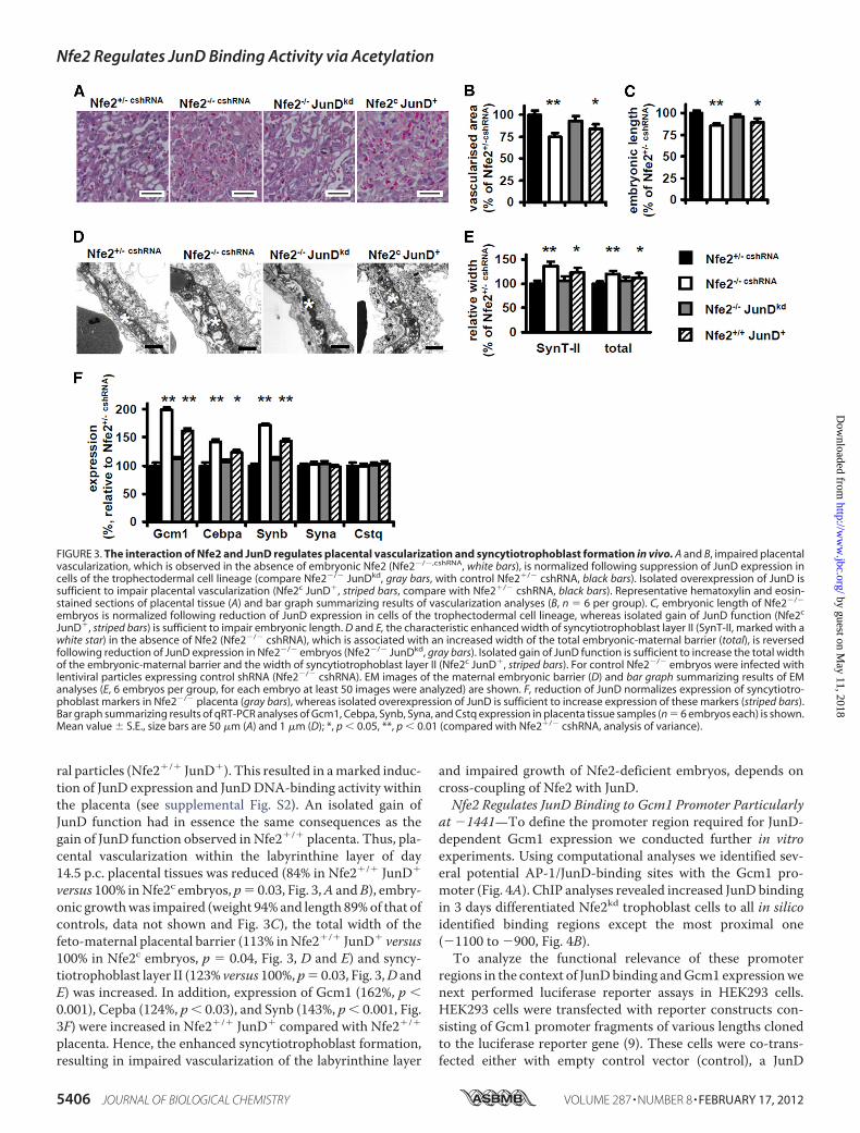
ral particles (Nfe2�/� JunD�). This resulted in amarked induc-tion of JunD expression and JunDDNA-binding activity withinthe placenta (see supplemental Fig. S2). An isolated gain ofJunD function had in essence the same consequences as thegain of JunD function observed in Nfe2�/� placenta. Thus, pla-cental vascularization within the labyrinthine layer of day14.5 p.c. placental tissues was reduced (84% in Nfe2�/� JunD�
versus 100% inNfe2c embryos, p� 0.03, Fig. 3,A andB), embry-onic growthwas impaired (weight 94% and length 89%of that ofcontrols, data not shown and Fig. 3C), the total width of thefeto-maternal placental barrier (113% inNfe2�/� JunD� versus100% in Nfe2c embryos, p � 0.04, Fig. 3, D and E) and syncy-tiotrophoblast layer II (123% versus 100%, p� 0.03, Fig. 3,D andE) was increased. In addition, expression of Gcm1 (162%, p �0.001), Cepba (124%, p� 0.03), and Synb (143%, p� 0.001, Fig.3F) were increased in Nfe2�/� JunD� compared with Nfe2�/�
placenta. Hence, the enhanced syncytiotrophoblast formation,resulting in impaired vascularization of the labyrinthine layer
and impaired growth of Nfe2-deficient embryos, depends oncross-coupling of Nfe2 with JunD.Nfe2 Regulates JunD Binding to Gcm1 Promoter Particularly
at �1441—To define the promoter region required for JunD-dependent Gcm1 expression we conducted further in vitroexperiments. Using computational analyses we identified sev-eral potential AP-1/JunD-binding sites with the Gcm1 pro-moter (Fig. 4A). ChIP analyses revealed increased JunD bindingin 3 days differentiated Nfe2kd trophoblast cells to all in silicoidentified binding regions except the most proximal one(�1100 to �900, Fig. 4B).To analyze the functional relevance of these promoter
regions in the context of JunDbinding andGcm1 expressionwenext performed luciferase reporter assays in HEK293 cells.HEK293 cells were transfected with reporter constructs con-sisting of Gcm1 promoter fragments of various lengths clonedto the luciferase reporter gene (9). These cells were co-trans-fected either with empty control vector (control), a JunD
FIGURE 3. The interaction of Nfe2 and JunD regulates placental vascularization and syncytiotrophoblast formation in vivo. A and B, impaired placentalvascularization, which is observed in the absence of embryonic Nfe2 (Nfe2�/�,cshRNA, white bars), is normalized following suppression of JunD expression incells of the trophectodermal cell lineage (compare Nfe2�/� JunDkd, gray bars, with control Nfe2�/� cshRNA, black bars). Isolated overexpression of JunD issufficient to impair placental vascularization (Nfe2c JunD�, striped bars, compare with Nfe2�/� cshRNA, black bars). Representative hematoxylin and eosin-stained sections of placental tissue (A) and bar graph summarizing results of vascularization analyses (B, n � 6 per group). C, embryonic length of Nfe2�/�
embryos is normalized following reduction of JunD expression in cells of the trophectodermal cell lineage, whereas isolated gain of JunD function (Nfe2c
JunD�, striped bars) is sufficient to impair embryonic length. D and E, the characteristic enhanced width of syncytiotrophoblast layer II (SynT-II, marked with awhite star) in the absence of Nfe2 (Nfe2�/� cshRNA), which is associated with an increased width of the total embryonic-maternal barrier (total), is reversedfollowing reduction of JunD expression in Nfe2�/� embryos (Nfe2�/� JunDkd, gray bars). Isolated gain of JunD function is sufficient to increase the total widthof the embryonic-maternal barrier and the width of syncytiotrophoblast layer II (Nfe2c JunD�, striped bars). For control Nfe2�/� embryos were infected withlentiviral particles expressing control shRNA (Nfe2�/� cshRNA). EM images of the maternal embryonic barrier (D) and bar graph summarizing results of EManalyses (E, 6 embryos per group, for each embryo at least 50 images were analyzed) are shown. F, reduction of JunD normalizes expression of syncytiotro-phoblast markers in Nfe2�/� placenta (gray bars), whereas isolated overexpression of JunD is sufficient to increase expression of these markers (striped bars).Bar graph summarizing results of qRT-PCR analyses of Gcm1, Cebpa, Synb, Syna, and Cstq expression in placenta tissue samples (n � 6 embryos each) is shown.Mean value � S.E., size bars are 50 �m (A) and 1 �m (D); *, p � 0.05, **, p � 0.01 (compared with Nfe2�/� cshRNA, analysis of variance).
Nfe2 Regulates JunD Binding Activity via Acetylation
5406 JOURNAL OF BIOLOGICAL CHEMISTRY VOLUME 287 • NUMBER 8 • FEBRUARY 17, 2012
by guest on May 11, 2018
http://ww
w.jbc.org/
Dow
nloaded from

expression construct (JunD), or both JunD andNfe2 expressionconstructs (JunD � Nfe2). The �4041, �2982, and �1637reporter constructs, but not the �396 reporter construct, con-tained inducible JunD-binding site(s). Hence, the functionalrelevant JunD-binding site must be within the �1637 to �396promoter fragment. Of note, co-transfection with Nfe2 sup-pressed JunD-binding activity, directly demonstrating therepressive effect of Nfe2 in regard to JunD driven Gcm1 pro-moter activation.Considering that the �1637 to �396 region contained
inducible JunD-binding site(s) and that, based on the aboveChIP analyses, JunD binds to the �1630 to �1460 and the�1450 to �1300 promoter regions, but not to the �1100 to�900 promoter fragment (see above, Fig. 4B), we hypothesizedthat the functional relevant JunD-binding site(s) must residewithin the�1637 to�1300 region of the Gcm1 promoter. Thisregion contains three potential JunD-binding sites (at �1576,
�1441 and �1314). To determine their functional relevancefor Gcm1 expression these sites were inactivated by targetedpointmutations. JunD-mediated induction of the reporter con-struct containing the mutated JunD-binding site at �1441 wasreduced to 45% (p � 0.02, Fig. 4D), whereas induction of thereporter construct containing themutated JunD-binding site at�1576 or �1314 was not significantly reduced (94 and 88%comparedwith the control construct, p� 0.48 and 0.26, respec-tively, Fig. 4D).To establish whether JunD binds to the endogenous Gcm1
promoter sequence we next performed EMSA using an oligo-nucleotide derived from the Gcm1 sequence (�1426 to�1448). Binding of nuclear extracts to this oligonucleotide wasenhanced in the absence of Nfe2 (Nfe2kd, Fig. 4E). Using amutated oligonucleotide (CT3 AG) no binding activity couldbe detected (Fig. 4E). Supershift analyses confirmed binding ofJunD to the Gcm1-derived oligonucleotide (Fig. 4E). Taken
FIGURE 4. Nfe2 regulates JunD binding to the �1441 site within the mGcm1 promoter. A, graphical illustration of potential JunD bindings sites within themGcm1 promoter, identified by in silico analyses (top), and of the promoter deletion constructs used (bottom). B, binding of JunD to the mGcm1 promoter isincreased in the absence of Nfe2. Potential JunD-binding sites identified in silico were analyzed by ChIP assay. In the absence of Nfe2 enhanced binding of JunDcan be detected in all regions except �1100 to �900. Representative images of semiquantitative RT-PCR using Nfe2 expressing control (c) or Nfe2-deficient(kd) trophoblast cells; input DNA serves as positive control. C, the �1637 to �396 Gcm1 promoter region contains a functional relevant JunD-binding site,which is negatively controlled by Nfe2. Analyses of promoter activity using mGcm1 promoter deletion constructs fused to a luciferase reporter gene andtransfected into HEK293 cells. Cells were co-transfected with empty vector (control), JunD alone (JunD), or JunD and Nfe2 (JunD� Nfe2) expression constructs.D, the functional JunD-binding site within the mGcm1 promoter is at position �1441. The three potential AP-1/JunD-binding sites within the �1637 to �1430promoter fragment were inactivated by targeted mutagenesis. Only inactivation of the �1441 JunD-binding site results in impaired promoter activity. HEK293cells were transfected with the Gcm1 �1637 promoter/luciferase reporter constructs (nonmutated control; �1576 AC/TG mutant; �1441 TC/AG mutant; and�1314 AC/TG mutant) and a JunD expression construct. E, JunD-binding activity determined using a probe corresponding to the mGcm1 promoter region�1426 to �1448 with the JunD-binding site at �1441 intact or mutated (TC3 AG). Binding to the intact but not to the mutated Gcm1 probe is enhanced inNfe2kd trophoblast cells (kd) compared with control (c) cells (left image). Supershift analyses demonstrating binding of JunD to the Gcm1-derived oligonucleo-tide probe (right image) are shown. Mean value � S.E., **, p � 0.01 (C and D, analysis of variance).
Nfe2 Regulates JunD Binding Activity via Acetylation
FEBRUARY 17, 2012 • VOLUME 287 • NUMBER 8 JOURNAL OF BIOLOGICAL CHEMISTRY 5407
by guest on May 11, 2018
http://ww
w.jbc.org/
Dow
nloaded from

together, JunD binds to the Gcm1 promoter at�1441, which isrequired for JunD-mediated Gcm1 promoter activation.Regulation of JunD-binding Activity by Nfe2 Is Acetylation
Dependent—We have previously shown that the regulation ofGcm1 by Nfe2 is acetylation dependent (1). Given the currentfinding that JunD is crucially involved in the regulation ofGcm1we next evaluated the role of acetylation for the Nfe2-depen-dent regulation of JunD.Using immunoprecipitation we detected a marked enhance-
ment of JunD acetylation in Nfe2-deficient placenta (ex vivo:Nfe2�/�, c, Fig. 5,A andB) or trophoblast cells (in vitro: Nfe2kd,c, Fig. 5, A and B). Inhibition of acetylation using the HATinhibitors curcumin or epigallocatechin-3-gallate reducedacetylation of JunD (103 versus 175% in Nfe2�/� placenta, p �0.001, 98 versus 165% inNfe2kd trophoblast cells, p� 0.001, Fig.5, A and B, and data not shown) and JunD-binding activity inNfe2-deficient placenta (Nfe2�/�) and trophoblast (Nfe2kd)cells (Fig. 5C). Conversely, enhancement of acetylation by inhi-bition of HDAC activity using trichostatin A (TSA, TS), valp-roic acid (in vivo, data not shown), or sodium butyrate (in vitro,data not shown) increased acetylation of JunD (182 versus 100%in Nfe2�/� placenta, p � 0.001, 158 versus 100% in Nfe2c tro-phoblast cells, p � 0.001, Fig. 5, b and C) and JunD-bindingactivity in Nfe2 expressing placenta (Nfe2�/�) and trophoblastcells (Nfe2�) (Fig. 5C). Modulation of JunD acetylation wasassociated with corresponding changes of JunD binding to the
Gcm1 promoter. Thus, inhibition of acetylation using curcu-min reduced JunD binding to the �1450 to �1300 Gcm1 pro-moter region in Nfe2-deficient placenta (Nfe2�/�, ex vivo) andin Nfe2kd trophoblast cells (in vitro), whereas enhancing acety-lation in Nfe2 expressing trophoblast cells using TSA was suf-ficient to enhance JunD binding to the �1450 to �1300 Gcm1promoter region both inNfe2 expressing placenta (Nfe2�/�, exvivo) and trophoblast cells (Nfe2c, in vitro, Fig. 5D). These dataprovide experimental evidence that Nfe2 regulates JunDDNA-binding activity to the Gcm1 promoter via acetylation.
DISCUSSION
Previously we identified a novel function of the bZip tran-scription factor Nfe2 during trophoblast differentiation. Wedemonstrated that Nfe2 represses Gcm1 expression, a key reg-ulator of syncytiotrophoblast differentiation and placental vas-cularization, in trophoblast cells through an unknown modusoperandi. In the present study we establish that the AP-1 pro-tein JunD is a positive gene regulator during syncytiotropho-blast differentiation. Nfe2 prevents excess syncytiotrophoblastformation, which would impair placental vascularization andembryonic growth (1) by limiting JunD DNA-binding activity.The Nfe2-dependent repression of JunD DNA-binding activityis mediated via acetylation.The acetylation dependent cross-coupling ofNfe2with JunD
during trophoblast differentiation provides a novel mechanism
FIGURE 5. Modulation of JunD-binding activity by Nfe2 is acetylation dependent. A and B, acetylation of JunD is enhanced in the absence of Nfe2 (ex vivo,�/�; in vitro, kd). Enhanced acetylation is reversed following intervention with the HAT inhibitor curcumin (Cu), both in placental samples (ex vivo) and introphoblast cells (in vitro). Treatment with the HDAC inhibitor TSA (TS) is sufficient to increase JunD acetylation both ex vivo and in vitro. Immunoprecipitationusing an antibody against acetylated lysine and immunoblotting for JunD (JunDac, A, top). JunD immunoblot without prior immunoprecipitation serves asinput control (JunDinput, A, bottom). B, bar graph summarizing results. C and D, enhanced JunD DNA-binding activity (supershift analyses, C) and JunD bindingto the mGcm1 promoter (analyses of the �1450 to �1300 mGcm1 promoter region by ChIP assay, D) in the absence of Nfe2 is acetylation dependent.Enhanced JunD DNA-binding activity and binding to the mGcm1 promoter in the absence of Nfe2 (ex vivo, �/�; in vitro, kd) is reversed following treatmentwith the HAT inhibitor curcumin in placental samples (C and D, ex vivo) and trophoblast cells (C and D, in vitro). Conversely, increasing acetylation using theHDAC inhibitor TSA (TS) is sufficient to increase JunD DNA-binding activity and JunD binding to the mGcm1 promoter both ex vivo (C and D) and in vitro (C andD). Representative images of JunD supershifts (C) and ChIP assay using an anti-JunD antibody for DNA precipitation (D) are shown.
Nfe2 Regulates JunD Binding Activity via Acetylation
5408 JOURNAL OF BIOLOGICAL CHEMISTRY VOLUME 287 • NUMBER 8 • FEBRUARY 17, 2012
by guest on May 11, 2018
http://ww
w.jbc.org/
Dow
nloaded from

controlling Gcm1 expression, syncytiotrophoblast formation,placental vascularization, and embryonic growth. A function ofother members of the AP-1 protein family, namely JunB andFra1, for placental development has been previously described(10, 11). The function of JunD must be different from JunB orFra1. Thus, loss of JunB or Fra1 function results in embryoni-cally lethal defects, whereas in mice with a gain of JunB or Fra1function no embryonic phenotype has been reported (28). Con-trary, loss of JunD function has no impact on embryonic devel-opment (29), whereas gain of function impairs placental vascu-larization and embryonic growth (this study).In contrast to the nonlethal consequences of increased JunD-
binding activity in Nfe2�/� embryos after day 14.5 p.c. theabsence of JunB or Fra1 results in a lethal placental defect asso-ciated with defective yolk sac vascularization during establish-ment of the hemochorial placenta (day 8.5 p.c. in JunB�/� and10.5 p.c. in Fra1�/� embryos) (10, 11). Thus, whereas loss offunction of JunB and Fra1 results in early fetal loss, bZip tran-scription factors Nfe2 and JunD coordinately regulate placentaldevelopment and embryonic growth at later developmentalstages. Such diverse functions of distinct Jun family proteinslikely reflect their ability to form various heterodimers withother bZiP family proteins resulting in either positive or nega-tive regulation of target genes (28, 30). The newly identifiedinteraction of JunD andNfe2 expands our knowledge about therole of bZip transcription factors during placental developmentand demonstrates that this expanding network conveys crucialroles for placentation at various developmental stages.Transcriptional activation of AP-1 proteins is regulated by
MAPKs, in particular by the c-JunNH2-terminal kinases (JNKs)and the extracellular signal-regulated kinases (ERKs), whichphosphorylate AP-1 proteins (31, 32). The molecular mecha-nism of transcriptional activation of AP-1 proteins by MAPKsremained unknown until recently. Aguilera and colleagues (27)demonstrated that transcriptional activation of the AP-1 pro-tein c-Jun is linked to increased histone acetylation withinAP-1-dependent promoters. Nonphosphorylated c-Junrecruits the Mbd3/nucleosome remodeling and histonedeacetylation (NuRD) repressor complex through an interac-tion with its transactivation domain, resulting in gene repres-sion of AP-1 target genes. This repression is relieved by JNK-mediated c-Jun NH2-terminal phosphorylation. In the currentstudy we demonstrate that DNA-binding and transcriptionalactivity of JunD is likewisemodulated by acetylation, indicatingthat the mechanism identified by Aguilera and colleagues (27)is not specific for c-Jun. We previously demonstrated thatacetylation of histone H4 within the Gcm1 promoter is modu-lated by Nfe2 in trophoblast cells and placenta (1), which isentirely consistent with the observationmade by Aguilera et al.(27). In addition, we demonstrate now that the AP-1 proteinJunD itself is modulated by acetylation. Whether other AP-1proteins are directly modulated by acetylation and the relativeimportance of either histone or AP-1 protein acetylation fortranscriptional activation needs to be explored in futurestudies.Whether Nfe2-dependent acetylation of JunD depends on
MAPK activity remains currently unknown.Unlike c-Jun, JunDlacks a functional JNK docking site within the NH2-terminal
region (28). However, following heterodimerizationwith c-Jun,the serine residues of JunD can be phosphorylated (33), indicat-ing that MAPK-mediated JunD phosphorylation may beinvolved in the regulation of acetylation. However, whereasregulation of Nfe2 by JNK has been demonstrated (34), thereverse, e.g. regulation ofMAPK activity byNfe2, has to the bestof our knowledge not been reported. Hence the mechanismunderlying Nfe2-dependent regulation of acetylation remainspresently unknown. Based on previous and the current findingswe propose a MAPK independent mechanism through whichNfe2 may inhibit acetylation. Interaction of Nfe2 with CBP inhematopoietic and trophoblast cells is established (1, 7, 35).CBP/p300 are coactivators generally influencing gene expres-sion by functioning as scaffolding proteins facilitating protein-protein interactions and/or through their intrinsic acetyltrans-ferase activity toward histone proteins and other nearbynuclear factors (36). Considering that Nfe2 acts as a repressorduring trophoblast differentiation we speculate that Nfe2 mayinhibit the intrinsic acetylation activity of CBP in trophoblastcells, thus inducing hypoacetylation of JunD and histoneswithin the Gcm1 promoter (1) (and current results). The Nfe2-dependent repression of acetylation may result from a compe-tition of bZip transcription factors for a limited CBP pool (37).Indeed, a competitive interaction between CBP and bZip pro-teins, including Nfe2 and c-Jun, has been previously reported(38). In addition, we observed a markedly increased interactionbetween CBP and Gcm1 in the absence of Nfe2 (1), which is inagreement with a competitive interaction of CBP with varioustranscription factors during trophoblast differentiation. Suchcompetitive interaction with CBP may be the mechanismunderlying the functional antagonism between bZip transcrip-tion factors like Nfe2 and JunD during trophoblast differentia-tion and may constitute an alternative, MAPK independentpathway through which AP-1 proteins regulate acetylation andgene expression. The existence of such a competitive mecha-nism, as proposed, needs to be explored in future studies.Given the current and previous findings Nfe2 acts as an epi-
genetic and post-translational repressor during placental devel-opment, providing a mechanism limiting syncytiotrophoblastformation. It appears that all mechanisms through which Nfe2modulates placental vascularization converge at the regulatorylevel of Gcm1. Thus, the current finding underscores a crucialfunction of Gcm1 in regulating placental labyrinthine forma-tion (6). This novel, Nfe2-dependent pathway controllingGcm1 activity is important to ensure sufficient placental vascu-larization and embryonic growth during later pregnancy stages(1) (and current findings).Future studies are required to determine whether the same
pathway is of functional relevance in the human placenta. Weobserved expression of Nfe2 in human placenta, specifically inhuman syncytiotrophoblast cells3 and expression of Nfe2within human syncytiotrophoblast is depicted on the humanprotein atlas webpage. Interestingly, JunD, but not other AP-1proteins, is specifically expressed in human syncytiotropho-blast (39). Hence it appears possible, but remains to be shown,
3 M. Kashif and B. Isermann, unpublished observation.
Nfe2 Regulates JunD Binding Activity via Acetylation
FEBRUARY 17, 2012 • VOLUME 287 • NUMBER 8 JOURNAL OF BIOLOGICAL CHEMISTRY 5409
by guest on May 11, 2018
http://ww
w.jbc.org/
Dow
nloaded from

that cross-coupling of Nfe2 and JunD via acetylation consti-tutes a novel mechanism regulating placental development inhumans as well. Providing that the relevance of this pathwaycan be confirmed in humans this may identify new diagnosticand therapeutic targets for placental diseases associated withaltered expression of Gcm1, such as pre-eclampsia.
Acknowledgments—Nfe2-deficient mice were kindly provided by R.Shivdasani (Harvard Medical School, Cambridge, MA). We thank S.Schmidt and F. Zimmermann for technical support. We are gratefulto Hilmar Bading, Department of Neurobiology, InterdisciplinaryCenter for Neurosciences, University of Heidelberg, for providing theopportunity to carry out the electron microscopy work in hislaboratory.
REFERENCES1. Kashif, M., Hellwig, A., Kolleker, A., Shahzad, K., Wang, H., Lang, S.,
Wolter, J., Thati, M., Vinnikov, I., Bierhaus, A., Nawroth, P. P., and Iser-mann, B. (2011) p45NF-E2 represses Gcm1 in trophoblast cells to regulatesyncytium formation, placental vascularization, and embryonic growth.Development 138, 2235–2247
2. McIntire, D. D., Bloom, S. L., Casey, B. M., and Leveno, K. J. (1999) Birthweight in relationship tomorbidity andmortality among newborn infants.N. Engl. J. Med. 340, 1234–1238
3. Gluckman, P. D., Hanson, M. A., Cooper, C., and Thornburg, K. L. (2008)Effect of in utero and early-life conditions on adult health and disease.N. Engl. J. Med. 359, 61–73
4. Chen, Z., Hu, M., and Shivdasani, R. A. (2007) Expression analysis ofprimary mouse megakaryocyte differentiation and its application in iden-tifying stage-specific molecular markers and a novel transcriptional targetof NF-E2. Blood 109, 1451–1459
5. Hashemolhosseini, S., Schmidt, K., Kilian, K., Rodriguez, E., and Wegner,M. (2004) Conservation and variation of structure and function in a newlyidentified GCM homolog from chicken. J. Mol. Biol. 336, 441–451
6. Anson-Cartwright, L., Dawson, K., Holmyard, D., Fisher, S. J., Lazzarini,R. A., and Cross, J. C. (2000) The glial cells missing-1 protein is essentialfor branching morphogenesis in the chorioallantoic placenta. Nat. Genet.25, 311–314
7. Hung, H. L., Kim, A. Y., Hong, W., Rakowski, C., and Blobel, G. A. (2001)Stimulation of NF-E2 DNA binding by CREB-binding protein (CBP)-me-diated acetylation. J. Biol. Chem. 276, 10715–10721
8. Johnson, K. D., Grass, J. A., Boyer, M. E., Kiekhaefer, C. M., Blobel, G. A.,Weiss, M. J., and Bresnick, E. H. (2002) Cooperative activities of hemato-poietic regulators recruit RNA polymerase II to a tissue-specific chroma-tin domain. Proc. Natl. Acad. Sci. U.S.A. 99, 11760–11765
9. Schubert, S. W., Abendroth, A., Kilian, K., Vogler, T., Mayr, B., Knerr, I.,and Hashemolhosseini, S. (2008) bZIP-type transcription factors CREBandOASIS bind and stimulate the promoter of themammalian transcrip-tion factor GCMa/Gcm1 in trophoblast cells. Nucleic Acids Res. 36,3834–3846
10. Schorpp-Kistner, M., Wang, Z. Q., Angel, P., and Wagner, E. F. (1999)JunB is essential for mammalian placentation. EMBO J. 18, 934–948
11. Schreiber, M., Wang, Z. Q., Jochum,W., Fetka, I., Elliott, C., andWagner,E. F. (2000) Placental vascularization requires the AP-1 component fra1.Development 127, 4937–4948
12. Okada, Y., Ueshin, Y., Isotani, A., Saito-Fujita, T., Nakashima, H., Kimura,K., Mizoguchi, A., Oh-Hora, M., Mori, Y., Ogata, M., Oshima, R. G., Ok-abe, M., and Ikawa, M. (2007) Complementation of placental defects andembryonic lethality by trophoblast-specific lentiviral gene transfer. Nat.Biotechnol. 25, 233–237
13. Nervi, C., Borello, U., Fazi, F., Buffa, V., Pelicci, P. G., and Cossu, G. (2001)Inhibition of histone deacetylase activity by trichostatin Amodulates geneexpression during mouse embryogenesis without apparent toxicity. Can-cer Res. 61, 1247–1249
14. Göttlicher, M., Minucci, S., Zhu, P., Krämer, O. H., Schimpf, A., Giavara,
S., Sleeman, J. P., Lo Coco, F., Nervi, C., Pelicci, P. G., and Heinzel, T.(2001) Valproic acid defines a novel class of HDAC inhibitors inducingdifferentiation of transformed cells. EMBO J. 20, 6969–6978
15. Giunta, B., Obregon, D., Hou, H., Zeng, J., Sun, N., Nikolic, V., Ehrhart, J.,Shytle, D., Fernandez, F., and Tan, J. (2006) EGCGmitigates neurotoxicitymediated by HIV-1 proteins gp120 and Tat in the presence of IFN-�. Roleof JAK/STAT1 signaling and implications for HIV-associated dementia.Brain Res. 1123, 216–225
16. Pan, Y., Chen, C., Shen, Y., Zhu, C.H.,Wang, G.,Wang, X. C., Chen,H.Q.,and Zhu,M. S. (2008) Curcumin alleviates dystrophicmuscle pathology inmdx mice.Mol. Cells 25, 531–537
17. Tanaka, S., Kunath, T., Hadjantonakis, A. K., Nagy, A., and Rossant, J.(1998) Promotion of trophoblast stem cell proliferation by FGF4. Science282, 2072–2075
18. Isermann, B., Sood, R., Pawlinski, R., Zogg, M., Kalloway, S., Degen, J. L.,Mackman, N., and Weiler, H. (2003) The thrombomodulin-protein Csystem is essential for the maintenance of pregnancy. Nat. Med. 9,331–337
19. Himeno, E., Tanaka, S., and Kunath, T. (2008) Curr. Protoc. Stem Cell.Biol., Chapter 1, Unit 1E 4
20. Maltepe, E., Krampitz, G. W., Okazaki, K. M., Red-Horse, K., Mak, W.,Simon,M. C., and Fisher, S. J. (2005) Hypoxia-inducible factor-dependenthistone deacetylase activity determines stem cell fate in the placenta. De-velopment 132, 3393–3403
21. Balasubramanyam, K., Varier, R. A., Altaf,M., Swaminathan, V., Siddappa,N. B., Ranga, U., and Kundu, T. K. (2004) Curcumin, a novel p300/CREB-binding protein-specific inhibitor of acetyltransferase, represses the acety-lation of histone/nonhistone proteins and histone acetyltransferase-de-pendent chromatin transcription. J. Biol. Chem. 279, 51163–51171
22. Tachibana, H., Fujimura, Y., and Yamada, K. (2004) Tea polyphenol epi-gallocatechin-3-gallate associates with plasmamembrane lipid rafts. Lipidrafts mediate anti-allergic action of the catechin. Biofactors 21, 383–385
23. Tachibana, H., Koga, K., Fujimura, Y., and Yamada, K. (2004) A receptorfor green tea polyphenol EGCG. Nat. Struct. Mol. Biol. 11, 380–381
24. Follenzi, A., Bakovic, S., Gual, P., Stella, M. C., Longati, P., and Comoglio,P. M. (2000) Cross-talk between the proto-oncogenes Met and Ron. On-cogene 19, 3041–3049
25. Bierhaus, A., Schiekofer, S., Schwaninger, M., Andrassy, M., Humpert,P. M., Chen, J., Hong, M., Luther, T., Henle, T., Klöting, I., Morcos, M.,Hofmann, M., Tritschler, H., Weigle, B., Kasper, M., Smith, M., Perry, G.,Schmidt, A. M., Stern, D. M., Häring, H. U., Schleicher, E., and Nawroth,P. P. (2001) Diabetes-associated sustained activation of the transcriptionfactor nuclear factor-�B. Diabetes 50, 2792–2808
26. Rudofsky, G., Jr., Reismann, P., Grafe, I. A., Konrade, I., Djuric, Z., Tafel, J.,Buchbinder, S., Zorn, M., Humpert, P. M., Hamann, A., Morcos, M.,Nawroth, P. P., and Bierhaus, A. (2007) Improved vascular function uponpioglitazone treatment in type 2 diabetes is not associated with changes inmononuclear NF-�B binding activity. Horm. Metab. Res. 39, 665–671
27. Aguilera, C., Nakagawa, K., Sancho, R., Chakraborty, A., Hendrich, B., andBehrens, A. (2011) c-JunN-terminal phosphorylation antagonises recruit-ment of the Mbd3/NuRD repressor complex. Nature 469, 231–235
28. Reddy, S. P., and Mossman, B. T. (2002) Role and regulation of activatorprotein-1 in toxicant-induced responses of the lung. Am. J. Physiol. LungCell Mol. Physiol. 283, L1161–1178
29. Thépot, D., Weitzman, J. B., Barra, J., Segretain, D., Stinnakre, M. G.,Babinet, C., and Yaniv, M. (2000) Targeted disruption of the murine JunDgene results in multiple defects in male reproductive function. Develop-ment 127, 143–153
30. Francastel, C., Augery-Bourget, Y., Prenant,M.,Walters,M.,Martin, D. I.,and Robert-Lézénès, J. (1997) c-Jun inhibits NF-E2 transcriptional activityin association with p18/maf in Friend erythroleukemia cells.Oncogene 14,873–877
31. Dhanasekaran, N., and Premkumar Reddy, E. (1998) Signaling by dualspecificity kinases. Oncogene 17, 1447–1455
32. Davis, R. J. (2000) Signal transduction by the JNK group of MAP kinases.Cell 103, 239–252
33. Kallunki, T., Deng, T., Hibi,M., andKarin,M. (1996) c-Jun can recruit JNKto phosphorylate dimerization partners via specific docking interactions.
Nfe2 Regulates JunD Binding Activity via Acetylation
5410 JOURNAL OF BIOLOGICAL CHEMISTRY VOLUME 287 • NUMBER 8 • FEBRUARY 17, 2012
by guest on May 11, 2018
http://ww
w.jbc.org/
Dow
nloaded from

Cell 87, 929–93934. Lee, T. L., Shyu, Y. C., Hsu, P. H., Chang, C. W., Wen, S. C., Hsiao, W. Y.,
Tsai, M. D., and Shen, C. K. (2010) JNK-mediated turnover and stabiliza-tion of the transcription factor p45/NF-E2 during differentiation of mu-rine erythroleukemia cells. Proc. Natl. Acad. Sci. U.S.A. 107, 52–57
35. Johnson, K. D., Norton, J. E., and Bresnick, E. H. (2002) Requirements forutilization of CREB-binding protein by hypersensitive site two of the�-globin locus control region. Nucleic Acids Res. 30, 1522–1530
36. Nandiwada, S. L., Li, W., Zhang, R., and Mueller, D. L. (2006) p300/cyclicAMP-responsive element-binding protein mediates transcriptional co-activation by the CD28 T cell costimulatory receptor. J. Immunol. 177,401–413
37. Kamei, Y., Xu, L., Heinzel, T., Torchia, J., Kurokawa, R., Gloss, B., Lin, S. C.,Heyman, R. A., Rose, D. W., Glass, C. K., and Rosenfeld, M. G. (1996) ACBP integrator complex mediates transcriptional activation and AP-1 in-hibition by nuclear receptors. Cell 85, 403–414
38. Cheng, X., Reginato, M. J., Andrews, N. C., and Lazar, M. A. (1997) Thetranscriptional integrator CREB-binding protein mediates positive cross-talk between nuclear hormone receptors and the hematopoietic bZip pro-tein p45/NF-E2.Mol. Cell. Biol. 17, 1407–1416
39. Bamberger, A. M., Bamberger, C. M., Aupers, S., Milde-Langosch, K.,Löning, T., and Makrigiannakis, A. (2004) Expression pattern of the acti-vating protein-1 family of transcription factors in the human placenta.Mol. Hum. Reprod. 10, 223–228
Nfe2 Regulates JunD Binding Activity via Acetylation
FEBRUARY 17, 2012 • VOLUME 287 • NUMBER 8 JOURNAL OF BIOLOGICAL CHEMISTRY 5411
by guest on May 11, 2018
http://ww
w.jbc.org/
Dow
nloaded from

Madhusudhan, Angelika Bierhaus, Peter Nawroth and Berend IsermannBock, Hongjie Wang, Khurrum Shahzad, Satish Ranjan, Juliane Wolter, Thati
Muhammed Kashif, Andrea Hellwig, Said Hashemolhosseini, Varun Kumar, FabianDIFFERENTIATION
via Acetylation: A NOVEL MECHANISM REGULATING TROPHOBLAST Nuclear Factor Erythroid-derived 2 (Nfe2) Regulates JunD DNA-binding Activity
doi: 10.1074/jbc.M111.289801 originally published online December 15, 20112012, 287:5400-5411.J. Biol. Chem.
10.1074/jbc.M111.289801Access the most updated version of this article at doi:
Alerts:
When a correction for this article is posted•
When this article is cited•
to choose from all of JBC's e-mail alertsClick here
Supplemental material:
http://www.jbc.org/content/suppl/2011/12/16/M111.289801.DC1
http://www.jbc.org/content/287/8/5400.full.html#ref-list-1
This article cites 38 references, 16 of which can be accessed free at
by guest on May 11, 2018
http://ww
w.jbc.org/
Dow
nloaded from


